Article

In Vitro Antiprotozoal Activity of Triterpenoid Constituents of Kleinia odora Growing in Saudi Arabia

by
Nawal M. Al Musayeib
1,*,
Ramzi A. Mothana
1,2,
Ali A. El Gamal
1,
Shaza M. Al-Massarani
1 and
Louis Maes
3
1
Department of Pharmacognosy, College of Pharmacy, King Saud University, Riyadh 11451, Saudi Arabia
2
Department of Pharmacognosy, Faculty of Pharmacy, Sana'a University, P.O. Box 33039, Sana'a, Yemen
3
Laboratory for Microbiology, Parasitology and Hygiene (LMPH), Faculty of Pharmaceutical, Biomedical and Veterinary Sciences, Antwerp University, Antwerp B-2610, Belgium
*
Author to whom correspondence should be addressed.
Molecules 2013, 18(8), 9207-9218; https://doi.org/10.3390/molecules18089207
Submission received: 15 July 2013 / Revised: 28 July 2013 / Accepted: 29 July 2013 / Published: 31 July 2013
(This article belongs to the Section Natural Products Chemistry)

Abstract

:
Two lupane and four ursane triterpenes, namely epilupeol (1), lupeol acetate (2), ursolic acid (3), brein (4), 3β 11α-dihydroxy urs-12-ene (5) and ursolic acid lactone (6) were isolated from aerial parts of Kleinia odora and identified. Compounds 1 and 36 were isolated for the first time from K. odora. The triterpene constituents were investigated for antiprotozoal potential against erythrocytic schizonts of Plasmodium falciparum, intracellular amastigotes of Leishmania infantum and Trypanosoma cruzi and free trypomastigotes of T. brucei. Cytotoxicity was determined against MRC-5 fibroblasts to assess selectivity. The ursane triterpenes were found to be active against more than one type of the tested parasites, with the exception of compound 6. This is also the first report on the occurrence of ursane type triterpenes in the genus Kleinia and their antiprotozoal potential against P. falciparum, L. infantum, T. cruzi, and T. brucei.

1. Introduction

Protozoal infections are a worldwide health problem, particularly in developing countries [1,2,3,4], and approximately 14% of the World population are at risk of infection. Various studies have been conducted on leishmaniasis, malaria, Chagas and sleeping sickness which are considered major killing diseases [5]. The drugs currently in use mostly lack adequate efficacy, are toxic or show other liabilities, such as the need for parenteral application or high cost [6]. This reflects the need to continue searching for new and better antiprotozoal drugs. Natural products may offer good sources of leads for new drug design and discovery.
The genus Kleinia is a flowering plant comprising 40 species that are distributed in Somalia, the Middle-East, Madagascar and India [7,8]. Three species of this genus are commonly distributed in the Southern regions in Saudi Arabia: K. odora (Forssk) DC, K. deflersii (O.Schwartz) and K. pendula (Forssk) Sch. Bip. [9,10,11]. Several species of Kleinia are known to be rich sources of oxygenated sesquiterpenoids such as germacrane and oplopane abrotanifolon derivatives and lupane-type tri-terpenoids [12,13,14]. Triterpenes comprise one of the most interesting groups of natural products due to their high potential as pharmacological agents, including leishmanicidal, trypanocidal and antiplasmodial activity [15].
As part of our ongoing research on Saudi medicinal plant metabolites with antiprotozoal potential, triterpenoid constituents from K. odora were isolated, structurally analyzed and evaluated for antiprotozoal potential against the protozoan parasites Plasmodium falciparum, Leishmania infantum, Trypanosoma cruzi and T. brucei. To assess selectivity, cytotoxicity was determined on MRC-5 fibroblasts.

2. Results and Discussion

2.1. Phytochemistry

Compound 1 was isolated as a white powder. Its 13C-NMR and DEPT spectrum (Table 1) exhibited 30 carbons, including seven singlet methyl groups, eleven methylenes, six methines one of which is oxygenated, and six quaternary carbons, which taken together revealed the basic skeleton of a pentacyclic triterpene. NMR spectra include signals for isopropenyl group (olefinic quaternary carbons at δc 150.9, methylene carbon at δc 109.3, protons singlets at δH 4.69 and 4.57, and methyl singlet at δH 1.64) suggesting lupane type triterpene. The 1H-NMR spectrum (Table 1) showed one oxymethine proton at C-3 as a broad singlet (δH 3.39). The proton signal at δH 2.38 (1H, td, 11.0, 10.0, 5.5 Hz) further revealed a typical H-19β lupane structure [16]. By comparing these NMR data with previously published data [17], compound 1 was characterized as epilupeol.
Compound 2 was also isolated as white crystals. The NMR data (Table 1) suggested a lupane skeleton. The NMR data were identical with those of epilupeol, except for the ring A signals. Compound 2 displayed a double doublet at δH 4.47 confirming the α-orientation of the C-3 proton [18]. It also showed additional signals attributed to the presence of an acetoxy group (carbonyl signal at δC 171.0 and methyl signals at δC 27.9 and δH 2.04). By comparison with previously reported data [19], compound 2 was identified as lupeol acetate.
Compound 3 was isolated as a white powder. The 1H- and 13C-NMR spectra (Table 1) revealed the presence of five tertiary methyls, two secondary methyls, nine methylenes, seven methines, one of which was oxygenated, and six quaternary carbons, which indicated a pentacyclic triterpenoid. The NMR spectrum showed a double bond (δC 124.5, 138.2 ppm) and a carboxyl group (δC 178.6) signals. The positions of the carboxy group and double bond were confirmed by a HMBC correlation study. By comparison with previously reported data [20] Compound 3 was identified as ursolic acid (3β-hydroxy-urs-12-en-28-oic acid).
The proton and carbon signals in the 1H- and 13C-NMR spectra (Table 1) of Compound 4 were very similar to those of compound 3, except for the signals of ring D. The carboxy group signals in the NMR spectrum disappeared and it showed one additional oxymethine signal (δH 4.19; δC 67.4) and an additional methyl singlet at δH 0.80. The hydroxyl and methyl groups were positioned at C-16 and C-17 based on a HMBC correlation study. Comparing the spectral data with those reported in the literature [21], Compound 4 was identified as brein (urs-12-ene-3β,16β-diol).
The 13C-NMR data of Compound 5 (Table 1) were very similar to those of Compound 4 and differed in the signals for rings C and D. This was attributed to the presence of an α-hydroxy at C-11 and the disappearance of the β-hydroxyl at position 16. Spectral data of 5 are reported for the first time based on DEPT, HMBC and HSQC evaluation and comparison of NMR data of structurally related compounds [21] and the published 1H-NMR data of its acetate form [22]. Compound 5 was identified as 3β, 11α-dihydroxy urs-12-ene.
The NMR spectral data of Compound 6 (Table 1) exhibited characteristic signals of an oleanan-28,13β-olide. The 13C-NMR spectrum displayed resonances for a double bond at δC 129.9 and 135.0 for C-11 and C-12, in addition to an oxygenated quaternary carbon at 91.9 and a carbonyl at 182.6 for C-13 and C-28, respectively. These data confirmed the olean-11-en,28,13β-olide structure. These results are in agreement with previously reported [23] data for ursolic acid lactone 6. Compounds 1 and 36 were isolated for the first time from K. odora. To the best of our knowledge, this is also the first report on the occurrence of ursane type triterpenes in the genus Kleinia. The structures are summarized in Figure 1.

2.2. Antiprotozoal Activity
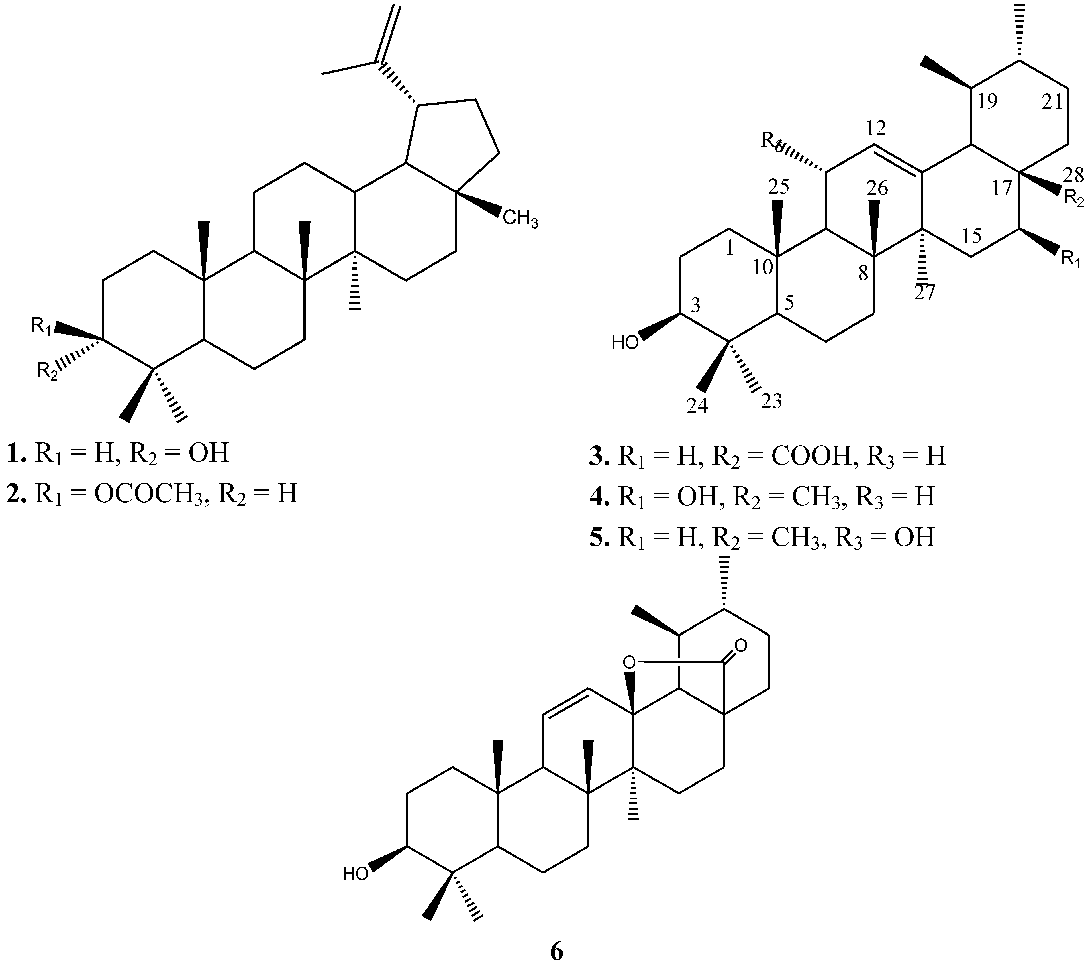
Petroleum ether and chloroform extracts of K. odora exhibited potent activity against T. brucei with IC50 values of 0.5 µg/mL (Table 2). The chloroform extract gave slightly higher selectivity (SI = 63) compared to the petroleum ether extract (SI = 39). The chloroform extract also displayed moderate activity against P. falciparum schizonts and intracellular amastigotes of L. infantum (IC50 8 µg/mL). In comparison, the petroleum ether extract showed similar activity against P. falciparum, L. infantum and T. cruzi (IC50 8.6, 6.8 and 5.7 µg/mL) but with lower selectivity. This motivated us to investigate the antiprotozoal activity of the isolated and identified triterpenoids from the chloroform extract.
Brein (4) and 3β, 11α-dihydroxy urs-12-ene (5) were found to be active and selective against more than one of the investigated protozoa. Brein (4) showed selective and potent activity against T. brucei (IC50 2.3 µM, SI >27.8), with moderate side-activity against P. falciparum, L. infantum and T. cruzi (IC50 9.3–9.9 µM) and acceptable selectivity (SI > 6.5). Compound 5 showed potent and selective activity against L. infantum (IC50 3.2 µM, SI > 20) with some side-activity against T. cruzi and T. brucei (IC50 8.1 and 7.9 µM, respectively, SI > 7.9).
Figure 1. Structures of the triterpenoid compounds 16.
Molecules 18 09207 g001
Although ursolic acid (3) showed efficacy against T. brucei (IC50 2.2 µM) comparable to that of Brein (4), its effect against T. cruzi and L. infantum (IC50 8.8 and 7.4 µM) was associated with low selectivity. Meanwhile, ursolic acid lactone 6 was devoid of any antiprotozoal activity at the highest concentration tested (64 µg/mL). The antitrypanosomal and antileishmanial activity of urosolic acid obtained herein is in consistent with the data found in literature. It has been shown to be active against T. brucei at IC50 1.0 ± 0.2 µg/mL (2.2 µM) [24,25]. In addition, it was also claimed active against T. cruzi with IC50 21 µM [26] and L. donovani (IC50 = 3.5 µg/mL) (7.6 µM) [27]. It is worthy of pointing out that in our investigation, the antiplasmodial activity of this compound (IC50 29.7 µM) is not in agreement with previously reported data which lists IC50 values of 3.1 µg/mL (6.8 µM) [28] and 4.9 µg/mL (10.7 µM) [29].
Table 1. 1H- and 13C-NMR data for compounds 16.
#Compound 1 in CDCl3Compound 2 in CDCl3Compound 3 in DMSOCompound 4 in MeODCompound 5 in CDCl3Compound 6 in MeOD
δCδHδCδHδCδHδCδHδCδHδCδH
133.2 38.4 38.2 40.1 39.7 39.4
225.1 21.3 28.2 27.9 27.4 27.7
376.33.39 (br s)81.04.47 (dd, 5.5, 10.5 Hz)76.8 79.73.18 (d, 7.5 Hz)78.83.23 (br s)79.53.2 (dd, 6.5, 10 Hz)
437.5 38.0 38.8 39.9 39.1 40.0
549.0 55.4 54.8 56.7 55.3 56.1
618.3 18.2 18.0 19.5 18.4 18.8
734.1 34.2 32.7 31.8 33.5 32.3
841.0 40.8 40.0 41.0 43.3 41.5
950.1 50.3 47.1 48.5 55.82.2 (d, 10 Hz)54.0
1037.2 37.8 36.7 36.8 38.1 37.5
1120.8 20.9 22.9 24.2 68.34.26129.95.61 (d, 8.5 Hz)
1225.3 23.7 124.55.16 (br s)126.35.23 (br s)128.65.18 (d, 2 Hz)135.06.07 (d, 10 Hz)
1338.0 37.1 138.2 139.7 143.0 91.9
1442.9 42.8 41.6 45.1 42.1 42.0
1527.4 25.1 27.2 36.4 27.9 26.6
1635.6 35.6 25.6 67.44.19 (d, 6.5 Hz)27.4 23.9
1743.0 43.0 46.7 39.5 33.6 46.6
1848.3 48.3 52.42.11 (d, 11.5 Hz)62.4 58.0 61.8
1948.02.38 (td, 11, 10, 5.5 Hz)48.02.38 (td, 11, 10, 5.5 Hz)38.4 40.9 39.3 41.5
20150.9 151.0 38.1 40.0 39.4 39.4
2129.8 29.8 30.4 31.8 31.1 32.3
2240.0 40.0 36.5 36.8 41.3 32.4
2328.30.82 (s)27.40.85 (s)21.10.90 (s)28.80.82 (s)28.7 28.30.96 (s)
2422.20.93 (s)16.50.84 (s)16.90.68 (s)16.31.02 (s)23.11.29 (s)15.60.80 (s)
2516.00.84 (s)16.21.05 (s)15.10.87 (s)16.41.02 (s)16.80.90 (s)19.41.05 (s)
2615.91.03 (s)16.00.83 (s)16.90.76 (s)18.31.10 (s)18.01.26 (s)18.40.98 (s)
2714.60.96 (s)14.50.79 (s)23.31.10 (s)25.11.20 (s)23.11.30 (s)16.61.25 (s)
2818.00.78 (s)18.00.94 (s)178.6 22.10.80 (s)28.21.10 (s)182.6-
29109.34.69 (br s), 4.57 (br s)109.34.69 (br s), 4.57 (br s)16.90.82, (d, 6.5 Hz)17.00.84 (d, 6.5 Hz)17.60.90 (d)18.31.05 (d, 7.5 Hz)
3019.31.6419.31.68 (s)21.10.92, (d, 7.0 Hz)21.10.97 (d)21.40.95 (d)19.60.98 (d, 6.0 Hz)
1’ 171.0
2’ 27.92.04 (s)
Table 2. Antiprotozoal activity and cytotoxicity of triterpenoid constituents isolated from the plant K. odora.
SampleP. falciparum L. infantum T. cruzi T. brucei MRC-5
IC50SI IC50SI IC50SI IC50SI IC50
Petroleum ether extract8.6 ± 2.12.3 6.8 ± 1.63 5.7 ± 1.63.4 0.5 ± 0.139 19.4 ± 3.4
Chloroform extract8.2 ± 1.94 8.1 ± 2.34 31.0 ± 4.9- 0.5 ± 0.163 31.3 ± 4.2
Cmpd. 3 29.7 ± 5.9<1 7.4 ±1.91.5 8.8 ± 2.31.3 2.2 ± 0.65.2 11.4 ± 2.1
Cmpd. 4 9.7 ± 3.2>6.6 9.3 ± 2.2>6.9 9.9 ± 2.6>6.5 2.3 ± 0.4>27.8 >64.0
Cmpd. 5 23.9 ± 5.72.7 3.2 ± 0.9>20 8.1 ± 1.8>7.9 7.8 ± 1.8>8.2 >64.0
Cmpd. 6 >64.0->64.0->64.0-40.9 ± 8.1- >64.0
Chloroquine0.3 ± 0.05
Miltefosine 10.4 ± 2.1
Benznidazole 1.9 ± 0.3
Suramine 0.03 ± 0.01
Tamoxifen 11.4 ± 3.2
IC50 μg/mL for extracts; µM for pure compounds.
These divergent results may be attributed to the diversity of parasite strains, parasite load, stages of parasite life cycle or be related to differences in the experimental conditions [30]. Ursolic acid lactone 6 was reported to have activity against L. donovani (IC50 191.52 µM) without cytotoxicity towards peripheral blood mononuclear cells [31]. In our opinion, this result was misinterpreted by the authors since the standard miltefosine under the same experimental conditions showed an IC50 of 9.31 µM [31]; hence, the compound should be considered as inactive. To the best of our knowledge, this is the first report on the antiprotozoal evaluation of compounds 4, 5 and 6 against P. falciparum, L. infantum, T. cruzi, and T. brucei.

3. Experimental

3.1. General

The 1D-NMR and 2D-NMR spectra were recorded on a Bruker AMX-400 spectrometer with tetramethylsilane (TMS) as an internal standard. Thin layer chromatography (TLC) was performed on precoated silicagel F254 plates (E. Merck, Darmstadt, Germany). All chemicals were purchased from Sigma Chemical Company (St. Louis, MO, USA).

3.2. Plant Materials

The plant Kleinia odora was collected from the South of Saudi Arabia in February 2011 and identified at the Pharmacognosy Department, College of Pharmacy, King Saud University. A voucher specimen was deposited at the Pharmacognosy Department, College of Pharmacy, King Saud University (Voucher # P-15129).

3.3. Extraction and Isolation

The air-dried and powdered aerial part of K. odora (1 kg) was extracted by maceration with 70% ethanol (4 × 2 L) at room temperature. The combined obtained ethanolic extract was filtered and evaporated at 40 °C using a rotary evaporator. The dried ethanolic extract (50 g) was subsequently redissolved in water (200 mL) and partitioned successively for several times with petroleum ether (3 × 200 mL), chloroform (3 × 200 mL) and n-butanol (3 × 200 mL) to provide the corresponding extracts. The petroleum ether extract (6 g) was subjected to column chromatography on pre-packed silica gel columns (35 mm i.d. × 350 mm) to give nine fractions. The elution was performed with a gradient of hexane-ethyl acetate (10:1) to pure ethyl acetate. TLC analysis of the fractions with anisaldehyde/sulfuric acid and heating at 100 °C allowed the analysis of the nine fractions. Fraction 1 was further purified by using a chromatotron (Harrison Research, Palo Alto, CA, USA) (silica gel 60 F254, layer thickness 2 mm, Merck, Darmstadt, Germany) to give Compound 2 (27 mg). The elution was performed with a mobile phase composed of hexane–dichloromethane (60:40, v/v). The chloroform extract (4 g) was applied on a silica gel column and eluted with a gradient of dichloromethane-ethyl acetate (9:1) to pure ethyl acetate to give seven fractions. Fraction 1 was rechromatographed on a silica gel column (dichloromethane-acetone, 9:1) and on a RP-18 column (MeOH–H2O, 90:10) to produce Compound 4 (48 mg) and Compound 6 (21 mg). Direct crystallization of fraction 2 eluted by 30% acetone-dichlormethane gave Compound 3 (35 mg). Fraction 5 was purified on a RP-18 column (MeOH–H2O, 90:10) to give Compound 5 (8 mg). Fraction 7 (80 mg) was subjected to RP-18 column chromatography with MeOH–H2O (90:10) as a solvent to produce Compound 1 (20 mg).
Epilupeol (1). Amorphous powder; m.p. 205 °C; NMR (CDCl3): see Table 1.
Lupeol acetate (2). White crystals; m.p. 215–218 °C; NMR (DMSO): see Table 1.
Ursolic acid (3). White powder; m.p. 236 °C; NMR (DMSO): see Table 1.
Urs-12-ene-3β,16β-diol (4). Solid; m.p. 221 °C; NMR (MeOD): see Table 1.
3β,11α-Dihydroxyurs-12-ene (5). Amorphous powder; NMR (CDCl3): see Table 1.
3-Hydroxy-13,28-epoxyurs-11-en-28-one (6, ursolic acid lactone). White powder; m.p. 268 °C; NMR (MeOD): see Table 1.

3.4. Antiprotozoal Assay

3.4.1. Standard Drugs

For the different tests, appropriate reference drugs were used as positive control: tamoxifen for MRC-5, chloroquine for P. falciparum, miltefosine for L. infantum, benznidazole for T. cruzi and suramin for T. brucei. All reference drugs were either obtained from the fine chemical supplier Sigma-Aldrich, Taufkirchen, Germany (tamoxifen, suramin) or from WHO-TDR, Geneva, Switzerland (chloroquine, miltefosine, benznidazole).

3.4.2. Biological Assays

The integrated panel of microbial screens and standard screening methodologies were adopted as previously described [32]. All assays were performed in triplicate at the Laboratory of Microbiology, Parasitology and Hygiene at the University of Antwerp (Belgium). Plant extracts were tested at 5 concentrations (64, 16, 4, 1 and 0.25 μg/mL) to establish a full dose-titration and determination of the IC50 (inhibitory concentration 50%). The final in-test concentration of DMSO did not exceed 0.5%, which is known not to interfere with the different assays [32]. Selectivity of activity was assessed by simultaneous evaluation of cytotoxicity on a fibroblast (MRC-5) cell line. The criterion for activity was an IC50 <10 μg/mL and a selectivity index (SI) of > 4.

3.4.3. Antiplasmodial Activity

Chloroquine-resistant P. falciparum K 1-strain was cultured in human erythrocytes O+ at 37 °C under a low oxygen atmosphere (3% O2, 4% CO2, and 93% N2) in RPMI-1640, supplemented with 10% human serum. Infected human red blood cells (200 μL, 1% parasitaemia, 2% haematocrit) were added to each well and incubated for 72 h. After incubation, test plates were frozen at −20 °C. Parasite multiplication was measured using the Malstat assay, a colorimetric method based on the reduction of 3-acetyl pyridine adenine dinucleotide (APAD) by parasite-specific lactate-dehydrogenase (pLDH) [32,33].

3.4.4. Antileishmanial Activity

L. infantum MHOM/MA(BE)/67 amastigotes were collected from the spleen of an infected donor hamster and used to infect primary peritoneal mouse macrophages. To determine in vitro antileishmanial activity, 3 × 10⁴ macrophages were seeded in each well of a 96-well plate. After 2 days outgrowth, 5 × 10⁵ amastigotes/well, were added and incubated for 2 h at 37 °C. Pre-diluted plant extracts were subsequently added and the plates were further incubated for 5 days at 37 °C and 5% CO2. Parasite burdens (mean number of amastigotes/macrophage) were microscopically assessed on 500 cells after Giemsa staining of the testplates, and expressed as a percentage of the blank controls without plant extract.

3.4.5. Antitrypanosomal Activity

Trypanosoma brucei Squib-427 strain (suramin-sensitive) was cultured at 37 °C and 5% CO2 in Hirumi-9 medium [34], supplemented with 10% fetal calf serum (FCS). About 1.5 × 10⁴ trypomastigotes/well were added to each well and parasite growth was assessed after 72 h at 37 °C by adding resazurin [35]. For Chagas disease, T. cruzi Tulahuen CL2 (benznidazole-sensitive) was maintained on MRC-5 cells in minimal essential medium (MEM) supplemented with 20 mM l-glutamine, 16.5 mM sodium hydrogen carbonate and 5% FCS. In the assay, 4 × 10³ MRC-5 cells and 4 × 10⁴ parasites were added to each well and after incubation at 37 °C for 7 days, Parasite growth was assessed by adding the alpha-galactosidase substrate chlorophenol red alpha-D-galactopyranoside [36]. The color reaction was read at 540 nm after 4 h and absorbance values were expressed as a percentage of the blank controls.

3.4.6. Cytotoxicity against MRC-5 Cells

MRC-5 SV2 cells were cultivated in MEM, supplemented with l-glutamine (20 mM), 16.5 mM sodium hydrogen carbonate and 5% FCS. For the assay, 104 MRC-5 cells/well were seeded onto the test plates containing the pre-diluted sample and incubated at 37 °C and 5% CO2 for 72 h. Cell viability was assessed fluorimetrically after 4 h of addition of resazurin. Fluorescence was measured (excitation 550 nm, emission 590 nm) and the results were expressed as % reduction in cell viability compared to control.

4. Conclusions

Two lupane and four ursane triterpenes were from isolated aerial parts of K. odora and identified. Ursane type triterpenes from the biologically active chloroform extract were investigated for their antiprotozoal potential. All were found to have activity against more than one type of the tested parasites, with exception of compound 6.

Acknowledgments

The authors extend their appreciation to the NPST program by King Saud University for funding the work through the project number (10-MED1288-02). The authors gratefully acknowledge that financial support. An Matheeussen and Margot Desmet are acknowledged for performing all the in vitro assays.

Conflicts of Interest

The authors declare no conflict of interest.

References

  1. Kondrashin, A.V.; Baranova, A.M.; Morozova, L.F.; Stepanova, E.V. Urgent tasks of malaria elimination programs. Med. Parazitol. (Mosk) 2011, 3, 3–9. [Google Scholar]
  2. Tengku, S.A.; Norhayati, M. Public health and clinical importance of amoebiasis in Malaysia: A review. Trop. Biomed. 2011, 28, 194–222. [Google Scholar]
  3. Kondrashin, A.V.; Baranova, A.M.; Morozova, L.F.; Stepanova, E.V. Global trends in malaria control. Progress and topical tasks in malaria control programs. Med. Parazitol. (Mosk) 2011, 4, 3–8. [Google Scholar]
  4. Ferrari, B.C.; Cheung-Kwok-Sang, C.; Beggs, P.J.; Stephens, N.; Power, M.L.; Waldron, L.S. Molecular epidemiology and spatial distribution of a waterborne cryptosporidiosis outbreak in Australia. Appl. Environ. Microbiol. 2011, 77, 7766–7771. [Google Scholar] [CrossRef]
  5. Nwaka, S.; Ridley, R.G. Virtual drug discovery and development for neglected diseases through public private partnerships. Nat. Rev. Drug Discov. 2003, 2, 919–928. [Google Scholar] [CrossRef]
  6. Nour, A.M.M.; Khalid, S.A.; Kaiser, M.; Brun, R.; Abdalla, W.E.; Schmidt, T.J. The antiprotozoal activity of methylated flavonoids from Ageratum conyzoides L. J. Ethnopharmacol. 2010, 129, 127–130. [Google Scholar] [CrossRef]
  7. Halliday, P. Kleinia saginata Compositae. Curtis's Bot. Mag. 1989, 6, 151–156. [Google Scholar] [CrossRef]
  8. Hulin, M. New species and combinations in Kleinia (Asteraceae) from the Horn of Africa. Nord. J. Bot. 2002, 22, 419–426. [Google Scholar]
  9. Halliday, P. The genus Klenia (Compositae) in Arabia. Kew Bull. 1983, 39, 817–827. [Google Scholar] [CrossRef]
  10. Collenette, S. An Illustrated Guide to the Flowers of Saudi Arabia; Scorpion Publishing Ltd.: London, UK, 1985. [Google Scholar]
  11. Migahid, A.M. Flora of Saudi Arabia, 3rd ed.; King Saud University Press: Riyadh, Saudi Arabia, 1996; Volume 2, p. 256. [Google Scholar]
  12. Bohlmann, F.; Knoll, K.-H.K. Zwei neue acylpyrrole aus Kleinia kleinioides. Phytochemistry 1978, 17, 599–601. [Google Scholar] [CrossRef]
  13. Bohlmann, F.; Suding, H. Weitere abrotanifolon-derivate aus Kleinia tomentosa. Phytochemistry 1980, 19, 687–688. [Google Scholar] [CrossRef]
  14. Bohlmann, F.; Ahmed, M.; Jakupovic, J.; Jeffrey, C. Sesquiterpenes from Kleinia species. Phytochemistry 1981, 20, 201–251. [Google Scholar]
  15. Dominguez-Carmona, D.B.; Escalante-Erosa, F.; Garcia-Sosa, K.; Ruiz-Pinell, G.; Gutierrez-Yapu, D. Antiprotozoal activity of betulinic acid derivatives. Phytomedicine 2010, 17, 379–382. [Google Scholar] [CrossRef]
  16. Fukuda, Y.; Yamada, T.; Wada, S.I.; Sakai, K.; Matsunaga, S.; Tanaka, R. Lupane and oleanane triterpenoids from the cones of Liquidamber styraciflua. J. Nat. Prod. 2006, 69, 142–144. [Google Scholar] [CrossRef]
  17. Thu, V.K.; Kiem, P.V.; Minh, C.V.; Yen, P.H.; Cuong, N.X.; Huong, H.T. A new flavan glucoside from Glochidion eriocarpum. J. Chem. 2010, 48, 125–131. [Google Scholar]
  18. Waterman, P.G.; Ampofo, S. Dammarane triterpenes from the stem bark of Commiphora dazielii. Phytochemistry 1985, 24, 2925–2928. [Google Scholar] [CrossRef]
  19. Jamal, A.K.; Yaacob, W.A.; Din, L.B. A Chemical Study on Phyllanthus reticulates. J. Phys. Sci. 2008, 19, 45–50. [Google Scholar]
  20. Jang, D.S.; Kim, J.M.; Lee, G.Y.; Kim, J.H.; Kim, J.S. Ursane-type triterpenoids from the Aerial parts of Potentilla discolour. Agric. Chem. Biotechnol. 2006, 49, 48–50. [Google Scholar]
  21. Lima, M. da P.; Braga, P.A.; Macedo, M. L.; Silva, M. F.; Ferreira, G.; Fernandes, J.B.; Vieira, P.C. Phytochemistry of Trattinnickia burserifolia, T. rhoifolia and Dacryodes hopkinsii: Chemosystematic Implications. J. Braz. Chem. Soc. 2004, 15, 385–394. [Google Scholar] [CrossRef]
  22. Bohlmann, F.; Zdero, C.; King, R.M.; Robinson, H. A hydroxygermacrene and other constituents from Pseudobrickellia brasiliensis. Phytochemistry 1984, 23, 1798–1799. [Google Scholar] [CrossRef]
  23. Wang, H.; Fujimoto, Y. Triterpene esters from Eucalyptus tereticornis. Phytochemistry 1993, 33, 151–153. [Google Scholar]
  24. Bero, J.; Hannaert, V.; Chataigné, G.; Hérent, M.F.; Quetin-Leclercq, J. In vitro antitrypanosomal and antileishmanial activity of plants used in Benin in traditional medicine and bio-guided fractionation of the most active extract. J. Ethnopharmacol. 2011, 137, 998–1002. [Google Scholar] [CrossRef]
  25. Hoet, S.; Pieters, L.; Muccioli, G.G.; Habib-Jiwan, J.L.; Opperdoes, F.R.; Quetin-Leclercq, J. Antitrypanosomal activity of triterpenoids and sterols from the leaves of Strychnos spinosa and related compounds. J. Nat. Prod. 2007, 70, 1360–1363. [Google Scholar] [CrossRef]
  26. Cunha, W.R.; Martins, C.; Ferreira, D.D.; Crotti, A.F.M.; Lopes, N.P.; Albuquerque, S. In vitro trypanocidal activity of Triterpenes from Miconia species. Planta Med. 2003, 69, 470–472. [Google Scholar] [CrossRef]
  27. Moulisha, B.; Kumar, G.A.; Kanti, H.P. Anti-leishmanial and anti-cancer activities of a pentacyclic triterpenoid isolated from the leaves of Terminalia arjuna Combretaceae. Trop. J. Pharm. Res. 2010, 9, 135–140. [Google Scholar]
  28. Cimanga, R.K.; Tona, G.L.; Mesia, G.K.; Kambu, O.K.; Bakana, D.P.; Kalenda, P.D.T.; Penge, A.O.; Muyembe, J.J.T.; Totte, J.; Pieters, L.; et al. Bioassayguided isolation of antimalarial triterpenoid acids from the leaves of Morinda lucida. Pharmaceut. Biol. 2006, 44, 677–681. [Google Scholar] [CrossRef]
  29. van Baren, C.; Anao, I.; Lira, P.D.; Debenedetti, S.; Houghton, P.; Croft, S.; Martino, V. Triterpenic acids and flavonoids from Satureja parvifolia. Evaluation of their antiprotozoal activity. Z. Naturforsch C. 2006, 61, 189–192. [Google Scholar]
  30. Peixoto, J.A; Silva, M.L.A.; Crotti, A.E.M.; Veneziani, R.C.S.; Gimenez, V.M.M.; Januário, A.H.; Groppo, M.; Magalhães, L.G.; Santos, F.F.; Albuquerque, S.; et al. Antileishmanial activity of the hydroalcoholic extract of Miconia langsdorffii, Isolated compounds, and semi-synthetic Derivatives. Molecules 2011, 16, 1825–1833. [Google Scholar] [CrossRef]
  31. Sidana, J.; Singh, S.; Arora, S.K.; Foley, W.J.; Singh, I.P. Terpenoidal constituents of Eucalyptus loxophleba ssp. Lissophloia. Pharmaceut. Biol. 2012, 50, 823–827. [Google Scholar] [CrossRef]
  32. Cos, P.; Vlietinck, A.J.; Berghe, D.V.; Maes, L. Anti-infective potential of natural products: How to develop a stronger in vitro proof-of-concept. J. Ethnopharmacol. 2006, 106, 290–302. [Google Scholar] [CrossRef]
  33. Makler, M.T.; Ries, J.M.; Williams, J.A.; Bancroft, J.E.; Piper, R.C.; Hinrichs, D.J. Parasite lactate dehydrogenase as an assay for Plasmodium falciparum drug sensitivity. Am. J. Trop. Med. Hyg. 1993, 48, 739–741. [Google Scholar]
  34. Hirumi, H.; Hirumi, K. Continuous cultivation of Trypanosoma brucei blood stream forms in a medium containing a low concentration of serum protein without feeder cell layers. J. Parasitol. 1989, 75, 985–989. [Google Scholar] [CrossRef]
  35. Raz, B.; Iten, M.; Grether-Buhler, Y.; Kaminsky, R.; Brun, R. The Alamar Blue asssay to determine drug sensitivity of African trypanosomes (T. b. rhodesiense, T. b. gambiense) in vitro. Acta Trop 1997, 68, 139–147. [Google Scholar] [CrossRef]
  36. Buckner, F.S.; Verlinde, C.L.; la Flamme, A.C.; van Voorhis, W.C. Efficient technique for screening drugs for activity against Trypanosoma cruzi using parasites expressing beta-galactosidase. Antimicrob. Agents Chemother. (Bethesda) 1996, 40, 2592–2597. [Google Scholar]
  • Sample Availability: Samples of the compounds are available from the authors.

