Letter

A New Lignan Glucoside from the Whole Plants of Salvia Scapiformis

1
Hubei Key Laboratory of Natural Medicinal Chemistry and Resource Evaluation, School of Pharmacy, Tongji Medical College, Huazhong University of Science and Technology, Wuhan 430030, Hubei, China
2
Tongji Hospital Affiliated to Tongji Medical College, Huazhong University of Science and Technology, Wuhan 430030, Hubei, China
*
Authors to whom correspondence should be addressed.
These authors contributed equally to this work.
Molecules 2013, 18(9), 11377-11383; https://doi.org/10.3390/molecules180911377
Submission received: 30 August 2013 / Revised: 6 September 2013 / Accepted: 9 September 2013 / Published: 13 September 2013

Abstract

:
Phytochemical investigation of the whole plants of Salvia scapiformis Hance afforded eight compounds, including one new lignan, (+)-8α-hydroxypinoresinol-8-O-[6′′-O-(4′′′-hydroxybenzoyl)]-β-d-glucopyranoside (1), four known lignans, (+)-8α-hydroxy-pinoresinol-8-O-β-d-glucopyranoside (2), (+)-8α-hydroxypinoresinol (3), (+)-pinoresinol (4), and (+)-medioresinol (5), and three known triterpenoids, ursolic acid (6), 4-epi-niga-ichigoside F1 (7), and niga-ichigoside F1 (8). Their structures were determined on the basis of extensive spectroscopic analyses and comparison with spectroscopic data in the literature. The absolute configuration of the new compound 1 was determined by chemical transformation and GC analysis.

1. Introduction

Salvia is an important genus in the family Lamiaceae, 84 species of which are widely distributed all over the country in China [1]. Many species of this genus have been cultivated worldwide for ornamental and culinary purposes, and some species are used as folk medicine. Salvia scapiformis Hance, known as “Bai-Bu-Yao”, is used as a folk medicine in China to treat cough, hemoptysis, traumatic injuries, traumatic hemorrhage, dysentery, and furunculosis [2]. As part of our ongoing search for novel natural compounds from Chinese medicinal plants, phytochemical study on the Me2CO extracts of S. scapiformis resulted in eight compounds. In this paper, we describe the isolation and structure elucidation of one new lignan glucoside (1) and seven known compounds 28 (Figure 1).
Figure 1. Chemical structures of compounds 18 from the whole plants of Salvia scapiformis.
Molecules 18 11377 g001

2. Results and Discussion

The acetone extract of the whole plants of S. scapiformis was subjected to silica gel column chromatography (CC) to give seven fractions A–G. Further separation of fractions D and G provided compounds 18 (Figure 1). The known compounds 28 were identified as (+)-8α-hydroxypinoresinol-8-O-β-d-glucopyranoside (2) [3,4], (+)-8α-hydroxypinoresinol (3) [3], (+)-pinoresinol (4) [5], (+)-medioresinol (5) [6], ursolic acid (6) [7], 4-epi-niga-ichigoside F1 (7) [8], and niga-ichigoside F1 (8) [9], respectively, by comparison of their spectroscopic data with those reported in the corresponding literature.
Compound 1 was obtained as a colorless gum with the molecular formula C33H36O14, as deduced from HRESIMS at m/z 679.1985 [M + Na]+ (calcd for C33H36O14Na, 679.2003). The IR spectrum of 1 showed absorptions of hydroxy (3,359 cm−1), carbonyl (1,700 cm−1), and aromatic (1,607 and 1,517 cm−1) functionalities. In the 1H-NMR spectrum of 1, two sets of 1,3,4-trisubstituted and one set of typical AA′BB′ coupling aromatic ring signals at δH 6.72 (1H, d, J = 8.1 Hz), 6.77 (1H, d, J = 8.1Hz), 6.81−6.85 (4H, overlapped), 7.01 (1H, d, J = 1.7 Hz), 7.03 (1H, d, J = 1.7 Hz), and 7.88 (2H, d, J = 8.8 Hz), two pairs of oxygen-bearing methylene protons at δH 4.46 (1H, t, J = 8.7), 4.38 (1H, d, J = 10.4), 3.85 (1H, d, J = 10.4), and 3.76 (1H, dd, J = 9.2 and 5.7 Hz), and two oxygen-bearing methyl singlets at 3.84 and 3.82 were observed. Its 13C-NMR spectrum showed two oxygenated methyl, three methylene, eighteen methine (ten aromatic and seven oxygenated), eight aromatic quaternary carbons, one oxygenated quaternary carbon, and one ester carbonyl. In addition, a series of signals in the range of about the protons at δH 3.0–4.5 and their corresponding carbons at δC 65.2, 72.0, 74.9, 75.7, 78.3, and 100.2 indicated a hexosyl sugar moiety [4]. In the HMBC spectrum, the correlations of H-2′′′/H-6′′′ (δH 7.88) and H-3′′′/H-5′′′ (δH 6.83) with C-1′′′ (δC 122.3), C-4′′′ (δC 163.8), and C-7′′′(δC 167.9) showed a p-hydroxybenzoyl unit (Figure 2). On comparison of its spectroscopic data with those of know lignans [2,3,4,5,6], compound 1 appeared to be a lignan derivant with a furofuran skelon. A long-range correlation from the anomeric proton H-1′′ (δH 4.38) to C-8 (δC 99.3) in the HMBC spectrum implied that the sugar unit was located at C-8 of aglycone. Moreover, the HMBC correlations between H-6′′ (δH 4.20 and 4.52) and C-7′′′ implied that the O-(p-hydroxybenzoyl) group was attached to C-6′′.
Figure 2. Key 1H-1H COSY and HMBC correlations of compound 1.
Molecules 18 11377 g002
The relative configuration of the furofuran skelon was determined by NOESY experiment (Figure 3). Assuming that H-8′ was α-oriented due to the known lignans (compounds 25) from this plant [2,3,4,5,6], the α-orientations of H-9α (δH 4.38) and H-9′α (δH 4.46) were established from the NOESY correlations from H-8′ (δH 3.40) to H-9α and H-9′α. The NOESY correlations of H-9β (δH 3.85) and H-9′β (δH 3.85) with H-7 (δH 4.66) and H-7′ (δH 4.77) implied that H-7 and H-7′ should be positioned on the β face of the molecule.
Moreover, the structure and absolute configuration of compound 1 was further determined by chemical transformation. Alkaline hydrolysis of compound 1 gave p-hydroxybenzoic acid and compound 2, and the later was hydrolyzed by cellulase (EC 3.2.1.4) to obtain compound 3 and sugar moiety. In addition, the sugar moiety was unambiguously determined to be β-d-glucose according to the coupling constant of its anomeric proton (J = 7.7 Hz) and GC analysis. Finally, compound 1 was elucidated as (+)-8α-hydroxypinoresinol-8-O-[6′′-O-(4′′′-hydroxybenzoyl)]-β-d-glucopyranoside.
Figure 3. Key NOESY correlations of the furofuran skeleton of compound 1.
Molecules 18 11377 g003

3. Experimental

3.1. General

Optical rotations were measured on a Perkin-Elmer PE-341LC polarimeter. UV spectra were recorded using a Varian Cary 50 spectrophotometer. IR spectra were recorded on a Bruker Vertex 70 FT-IR spectrophotometer. NMR spectra were recorded on a Bruker AM-400 spectrometer, using the residual CD3OD (δH 3.31/δC 49.0) signals as references. HRESIMS data were acquired using an API QSTAR Pulsar spectrometer. Column chromatography was performed using silica gel (100−200 mesh and 200−300 mesh, Qingdao Marine Chemical Inc., Qingdao, China), Amberchrom CG161M (75 μm, Rohm and Haas, Philadelphia, PA, USA), ODS (50 μm, YMC, Kyoto, Japan), and Sephadex LH-20 (Pharmacia Biotech AB, Uppsala, Sweden). Semipreparative HPLC was performed on an Agilent 1100 liquid chromatography with an YMC (10 × 250 mm, 5 μm) column. GC analysis was performed with an Agilent Technologies 6890N gas chromatography system. Solvents were distilled prior to use, and spectroscopic grade solvents were used. TLC was carried out on precoated silica gel GF254 plates.

3.2. Plant Material

Whole plants of S. scapiformis Hance were collected at Enshi of Hubei Province, China, in September 2010, and authenticated by Dr. Jianping Wang of Huazhong University of Technology and Science. A voucher specimen (No. 2010-0902) was deposited in the herbarium of Hubei Key Laboratory of Natural Medicinal Chemistry and Resource Evaluation, Tongji Medical College, Huazhong University of Technology and Science.

3.3. Extraction and Isolation

The air-dried and powdered whole plants of S. scapiformis (27 Kg) was extracted with Me2CO (3 × 150 L) at room temperature for seven days. The extract was concentrated under the reduced pressure. The residue (1,300 g) was subjected to silica gel (100−200 mesh) column chromatography (CC) eluting with a gradient of petroleum ether–Me2CO–MeOH (1:0:0→0:1:0→0:0:1) to give seven fractions (A−G). Fractions D and G were decolorized on Amberchrom GC161M CC (eluted with 70%→100% EtOH−H2O) and subjected to ODS eluting with a gradient of MeOH–H2O (4:6→1:0) to obtain non-triterpene subfractions D′ and E′ and triterpene subfractions D′′ and E′′, respectively. Subfraction D′ (8.5 g) was further isolated over silica gel (125g) CC eluting with CHCl3–Me2CO (1:0→10:1) to provide four subfractions (D′1−D′4). Subfraction D′2 was purified over Sephadex LH-20 eluted with MeOH to give fractions D′2.1, D′2.2, D′2.3, and D′2.4. Subfraction D′2.3 was subjected to silica gel CC again, and compounds 4 (6 mg, tR 28 min) and 5 (8 mg, tR 32 min) were purified by semi-preparative HPLC eluted with MeOH–H2O (46:54, flow rate: 2 mL/min). Subfraction D′2.4 was fractionated successively by silica gel CC, Sephadex LH-20 eluted with MeOH, silica gel CC, and semi-preparative HPLC eluted with MeOH–H2O (35:65, flow rate: 2 mL/min) to give compound 3 (9 mg, tR 25 min). One main compound was observed in subfraction D′′ by TLC, and compound 6 (40 g) was obtained by recrystallization in MeOH. Fraction G′ was subjected to repeated chromatography and purified by semipreparative HPLC to afford 1 (15 mg, tR 26 min, MeOH–H2O = 45:55, flow rate: 2 mL/min) and 2 (8 mg, tR 23 min, MeOH–H2O = 4:6, flow rate: 1.8 mL/min). Compound 7 (5 mg, tR 26 min) was separated by semipreparative HPLC eluted with MeOH–H2O (6:4, flow rate: 1.5 mL/min) from subfraction E′′, and compound 8 (40 mg) was recrystallized from the same part.
(+)-8α-Hydroxypinoresinol-8-O-[6′′-O-(4′′′-hydroxybenzoyl)]-β-D-glucopyranoside (1). Colorless gum; Molecules 18 11377 i001 
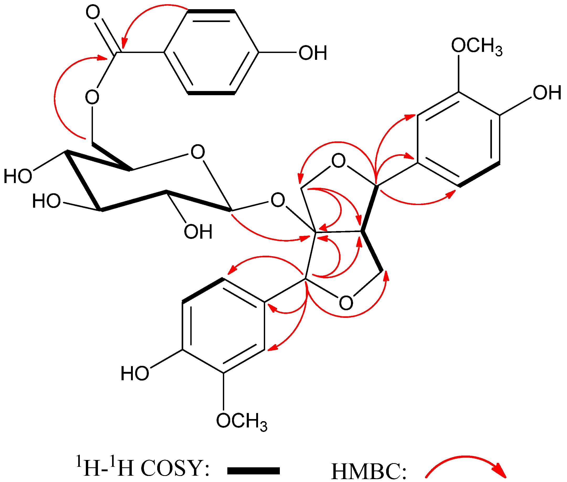
+4.35 (c 0.16, CH3OH); UV (CH3OH) λmax (lg ε) 207 (4.82), 234 (4.26), 259 (4.21) nm; IR (film) vmax 3359, 2943, 1700, 1607, 1517, 1453,1369, 1277 cm−1; 1H and 13C-NMR data, see Table 1; positive HRESIMS m/z 679.1985 [M + Na]+ (calcd for C33H36O14Na, 679.2003).
Table 1. 1H- (400 MHz) and 13C-NMR (100 MHz) data of compound 1 in CD3OD (δ in ppm).
PositionδH (J in Hz)δC PositionδH (J in Hz)δC
1 128.9, s 8′α3.40, ddd (8.0, 6.0, 5.1)60.4, d
27.03, d (1.7)113.6, d9′α4.46, dd (9.2, 8.0)72.4, t
3 149.4, s9′β3.76, dd (9.2, 6.0)
4 147.5, s-OCH33.84, s56.7, q
56.72, d (8.1)115.5, d 3.82, s56.6, q
66.83 (overlapped)122.1, dG1′′4.38, d (7.7)100.2, d
7β4.66, s89.5, dG2′′3.04, m74.9, d
8 99.3, sG3′′3.16, m78.3, d
9α4.38, d (10.4)73.6, tG4′′3.16, m72.0, d
9β3.85, d (10.4)G5′′3.23, dd (15.2, 7.6)75.7, d
1′ 133.2, sG6′′a4.52, dd (11.8,1.8)65.1, t
2′7.01, d (1.7)111.0, dG6′′b4.20, dd (11.8, 7.5)
3′ 148.5, s1′′′ 122.2, s
4′ 147.4, s2′′′, 6′′′7.88, d (8.8)133.0, d
5′6.77, d (8.1)116.3, d3′′′, 5′′′6.83, d (8.8)116.4, d
6′6.83 (overlapped)120.2, d4′′′ 163.8, s
7′β4.77, d (5.1)87.2, d7′′′ 167.9, s

3.4. Hydrolysis of Compound 1 and Determination of the Absolute Configuration of the Sugar Moiety

Compound 1 (5 mg) was dissolved in MeOH (1 mL) and saturated aqueous Na2CO3 solution (4 mL) at room temperature for 4 h. The reaction mixture was concentrated under reduced pressure to provide crude product, which was purified by semi-preparative HPLC to obtain p-hydroxybenzoic acid and compound 2 (3.2 mg). The later and cellulase (10 mg) were stirred in water (5 mL) at 37 °C for 3 days and extracted with EtOAc (3 mL × 3) [10]. The aglycone (1.4 mg), namely compound 3, was purified from the organic phase. Part of the sugar residue, obtained from water layer, was analyzed by co-TLC with an authentic sample (CHCl3–MeOH–H2O, 6:4:1). The other sugar (0.8 mg) and L-cysteine methyl ester hydrochloride (2.0 mg) were dissolved in dry pyridine (1 mL) and then kept at 60 °C for 3 h. The reaction mixture was concentrated to dryness and then trimethylsilylimidazole was added to the residue, followed by stirring at 60 °C for 1 h [11]. Finally, the resultant solution was partitioned with n-hexane and water, and then the organic phase was subjected to GC analysis by using a HP-5MS capillary column (30 m × 0.25 mm × 0.25 µm, Agilent); column temperature, 200 °C; injection temp, 250 °C; detector FID, detector temp, 250 °C. A peak was found at the retention time of 18.10 min for compound 1. When the standard solutions were prepared by the same reaction from corresponding standard glucose, the retention times of presilylated d-glucose and l-glucose were 18.12 and 18.72 min, respectively. Therefore, the sugar in compound 1 was determined to be d-glucose.

4. Conclusions

Phytochemical investigation on the whole plants of Salvia scapiformis Hance resulted in the isolation of one new compound 1 and seven known compounds 28. Their structures were identified to be (+)-8-hydroxypinoresinol-8α-O-[6′′-O-(4′′′-hydroxybenzoyl)]-β-d-glucopyranoside (1), (+)-8α-hydroxypinoresinol-8-O-β-d-glucopyranoside (2), (+)-8α-hydroxypinoresinol (3), (+)-pinoresinol (4), (+)-medioresinol (5), ursolic acid (6), 4-epi-niga-ichigoside F1 (7), and niga-ichigoside F1 (8), respectively, on the basis of extensive spectroscopic analyses, chemical method, and comparison with spectroscopic data in the literature.

Supplementary Materials

Supplementary materials can be accessed at: https://www.mdpi.com/1420-3049/18/9/11377/s1.

Acknowledgments

We are grateful to J. Wang at Huazhong University of Science and Technology for the authentification of this plant material. This work was financially supported by the Hubei Key Laboratory Foundation of Natural Medicinal Chemistry and Resource Evaluation, Huazhong University of Science and Technology (to G. Yao), the Fundamental Research Funds for the Central Universities (HUST: 2012QN003, to G. Yao), Scientific Research Foundation for the Returned Oversea Chinese Scholars, State Education Ministry of China (to G. Yao), and Program for New Century Excellent Talents in University, State Education Ministry of China (NCET-2008-0224, to Y. Zhang).

Conflicts of Interest

The authors declare no conflict of interest.

References

  1. Li, X.W.; Ian, C.H. Lamiaceae. In Flora of China; Wu, Z.Y., Raven, P.H., Hong, D.Y., Eds.; Science Press: Beijing, China; Missouri Botanical Garden Press: St. Louis, MO, USA, 1994; Volume 17, pp. 50–299. [Google Scholar]
  2. Fang, Z.X.; Liao, C.L. Medicinal Flora of Enshi; Hubei Science and Technology Press: Wuhan, China, 2006; Volume 2, pp. 292–293. [Google Scholar]
  3. Choi, S.U.; Yang, M.C.; Lee, K.H.; Kim, K.H.; Lee, K.R. Lignan and terpene constituents from the aerial parts of Saussurea pulchella. Arch. Pharm. Res. 2007, 30, 1067–1074. [Google Scholar] [CrossRef]
  4. Wang, H.B.; Mayer, R.; Rücker, G.; Neugebauer, M. Bisepoxylignan glycosides from Stauntonia hexaphylla. Phytochemistry 1993, 34, 1621–1624. [Google Scholar] [CrossRef]
  5. Cowan, S.; Stewart, M.; Abbiw, D.K.; Latif, Z.; Sarker, S.D.; Nash, R.J. Lignans from Strophanthus gratus. Fitoterapia 2001, 72, 80–82. [Google Scholar] [CrossRef]
  6. Khan, K.A.; Shoeb, A. A lignan from Lonicera hypoleuca. Phytochemistry 1985, 24, 628–630. [Google Scholar] [CrossRef]
  7. Jang, D.S.; Kim, J.M.; Lee, G.Y.; Kim, J.; Kim, J.S. Ursane-type triterpenoids from the aerial parts of Potentilla discolor. Agric. Chem. Biotechnol. 2006, 49, 48–50. [Google Scholar]
  8. Zhou, X.H.; Kasai, R.; Ohtani, K.; Tanaka, O.; Nie, R.L.; Yang, C.H.; Zhou, J.; Yamasaki, K. Oleanane and ursane glucosides from Rubus species. Phytochemistry 1992, 31, 3642–3644. [Google Scholar] [CrossRef]
  9. Reher, G.; Buděšínský, M. Triterpenoids from plants of the Sanguisorbeae. Phytochemistry 1992, 31, 3909–3914. [Google Scholar] [CrossRef]
  10. Liu, S.S.; Zhu, H.L.; Zhang, S.W.; Zhang, X.H.; Yu, Q.; Xuan, L.J. Abietane diterpenoids from Clerodendrum bungei. J. Nat. Prod. 2008, 71, 755–759. [Google Scholar] [CrossRef]
  11. Nhiem, N.X.; Kiem, P.V.; Minh, C.V.; Kim, N.; Park, S.; Lee, H.Y.; Kim, E.S.; Kim, Y.H.; Kim, S.; Koh, Y. Diarylheptanoids and flavonoids from Viscum album inhibit LPS-Stimulated production of pro-inflammatory cytokines in bone marrow-derived dendritic cells. J. Nat. Prod. 2013, 76, 495–502. [Google Scholar] [CrossRef]
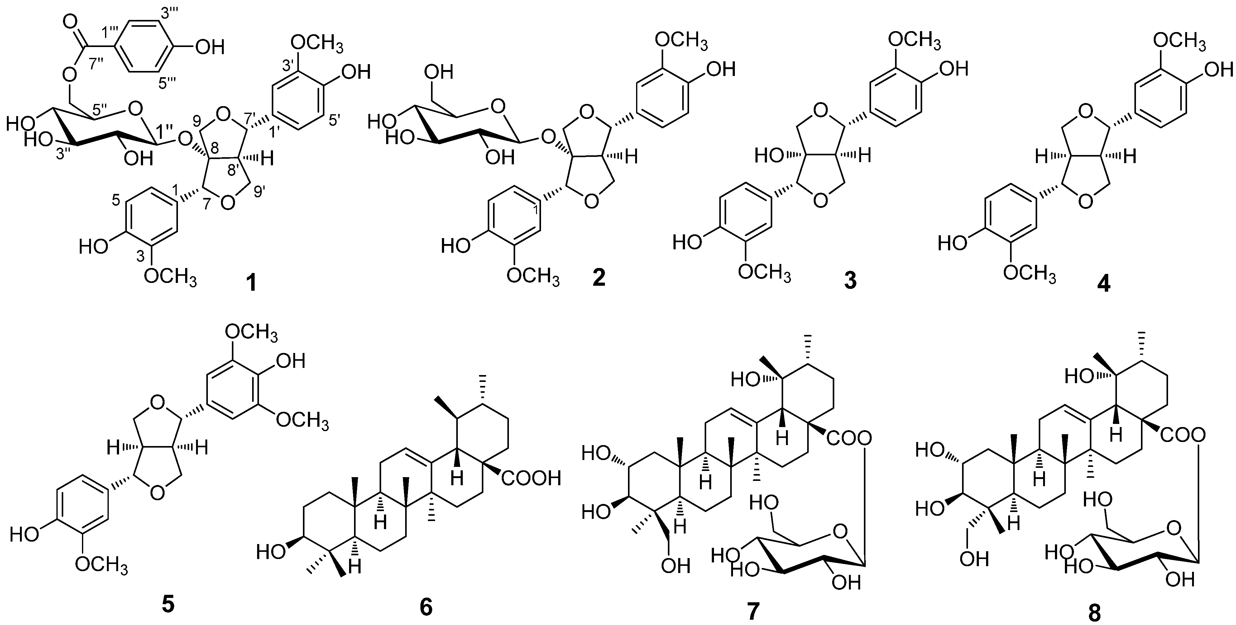
  • Sample Availability: Samples of the compounds 18 are available from the authors.

