Article

The Anthraquinone Derivatives from the Fungus Alternaria sp. XZSBG-1 from the Saline Lake in Bange, Tibet, China

1
School of Chemistry and Chemical Engineering, Sun Yat-sen University, Guangzhou 510275, China
2
Tibet Plateau Institute of Biology, Lhasa 850001, China
3
Zhongshan School of Medicine, Sun Yat-sen University, Guangzhou 510275, China
*
Authors to whom correspondence should be addressed.
Molecules 2014, 19(10), 16529-16542; https://doi.org/10.3390/molecules191016529
Submission received: 7 August 2014 / Revised: 17 September 2014 / Accepted: 19 September 2014 / Published: 14 October 2014
(This article belongs to the Collection Bioactive Compounds)

Abstract

:
Four new anthraquinone derivatives 14 were obtained along with seven known compounds 511 from the extracts of the fungal strain Alternaria sp. XZSBG-1 which was isolated from the sediments of the carbonate saline lake in Bange, Tibet, China. Their structures were determined by spectroscopic methods, mainly by 2D NMR spectra. Compound 1 is a novel tetrahydroanthraquinone with an epoxy ether bond between C-4a and C-9a. In the primary bioassays, compound 3 (alterporriol T) exhibited inhibition of a-glucosidase with a IC50 value 7.2 μM, and compound 9 showed good inhibitory activity against the HCT-116 and HeLa cell lines, with IC50 values of 3.03 and 8.09 μM, respectively.

1. Introduction

In 2002, we reported research on the metabolites of a fungal strain from a saline lake locates in the Bahamas [1]. Since that time, it seems that that little research on fungal metabolites from saline lakes has been published. Recently, we have been interested in the microorganisms from Tibetan saline lakes, which live under the special plateau habitat conditions, including low temperatures and high salt levels that are similar to those of the ocean, and we have thus obtained some unique and significant compounds.
The fungus Alternaria sp. XZSBG-1 collected from the sediment of the salt lake in Bange, Tibet, China was studied. We found that this fungus contained abundant anthraquinone compounds. Four new anthraquinone and tetrahydroanthraquinone derivatives 14, were isolated from this fungus along with seven known compounds (511) (Figure 1). Compound 1, a novel tetrahydroanthraquinone with an epoxy ether bond between C-4a and C-9a, and compound 2, a tetrahydroanthraquinone dimer with a C-4-C-4' linkage, are rare.
Figure 1. Structures of compounds 111 isolated from Alternaria sp. XZSBG-1.
Molecules 19 16529 g001
Anthraquinones and tetrahydroanthraquinones are widely distributed as secondary metabolites in natural biosources, and show important biological activities [2,3,4,5]. So far, 18 compounds of alterporriol family [6,7,8,9,10,11,12] and 14 compounds of altersolanol family [9,13,14,15,16] have been reported. Herein, we report that the isolation, elucidation and biological activities of the anthraquinone and tetrahydroanthraquinone derivatives from Alternaria sp. XZSBG-1.

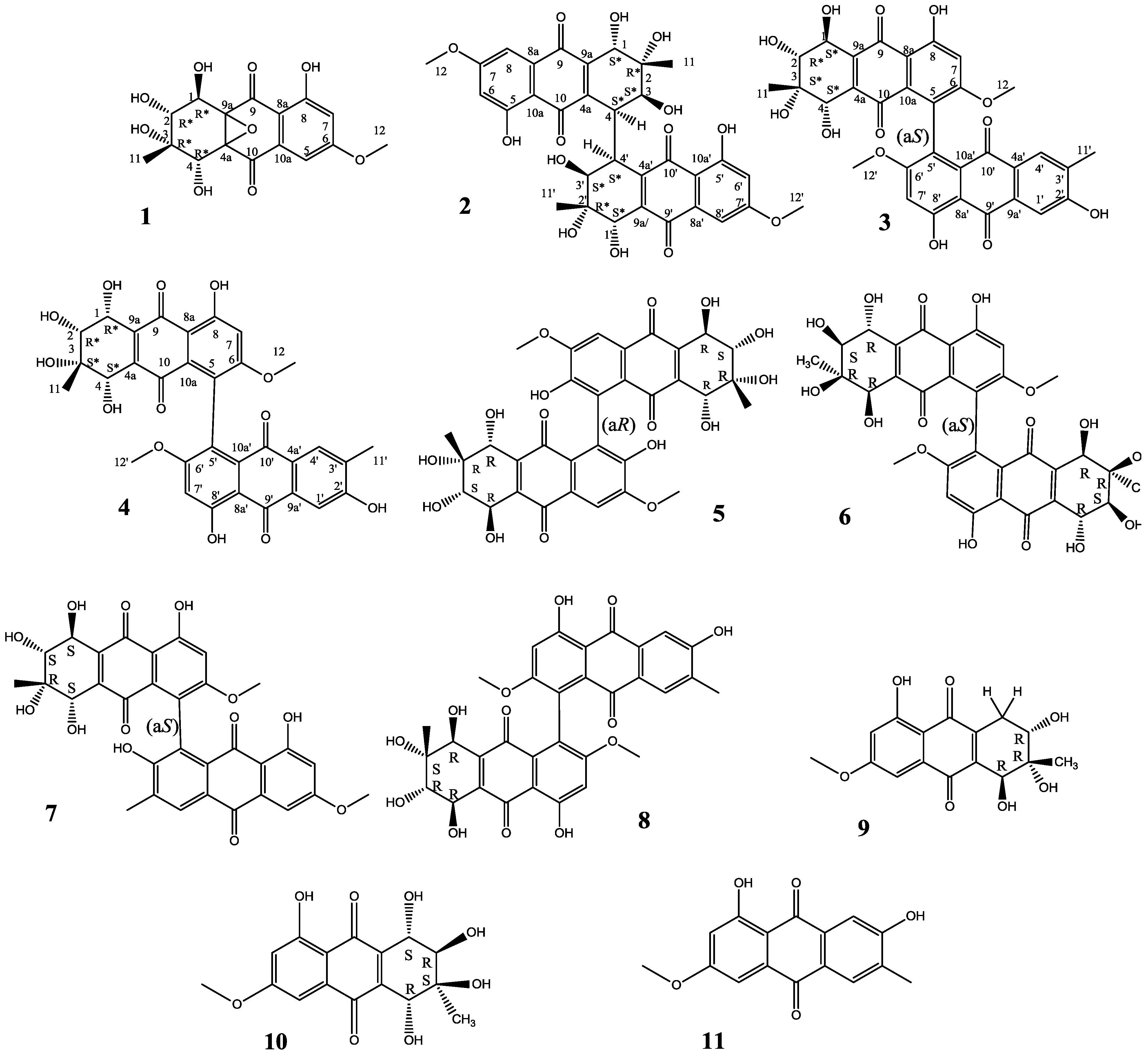
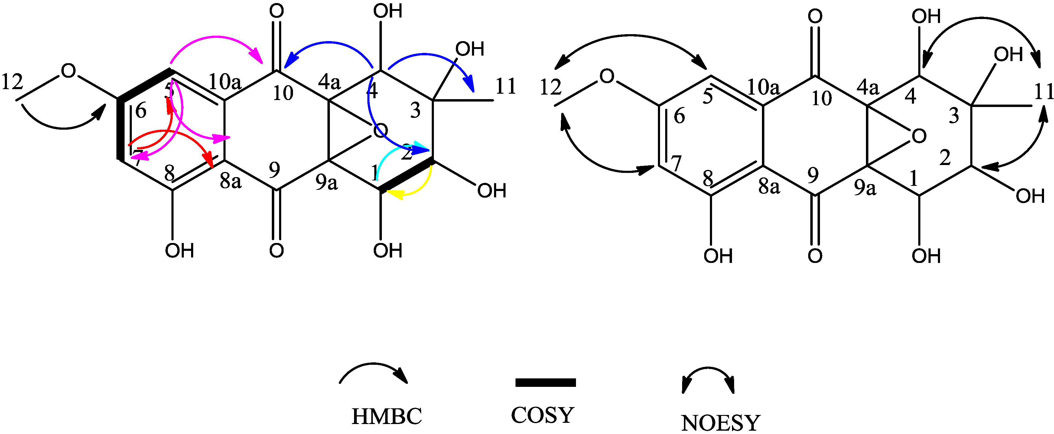
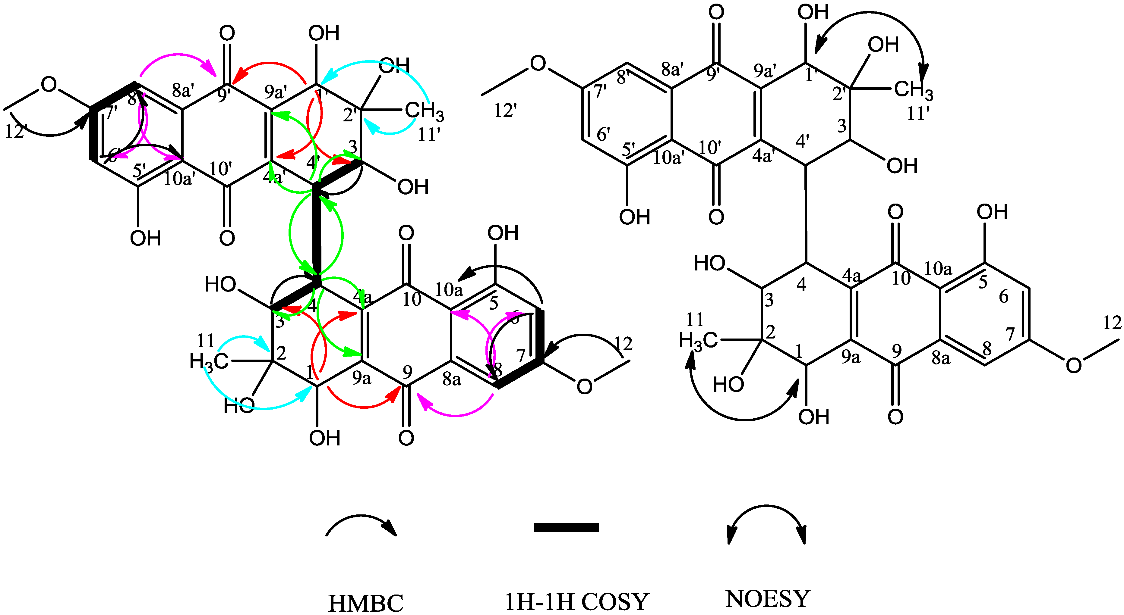
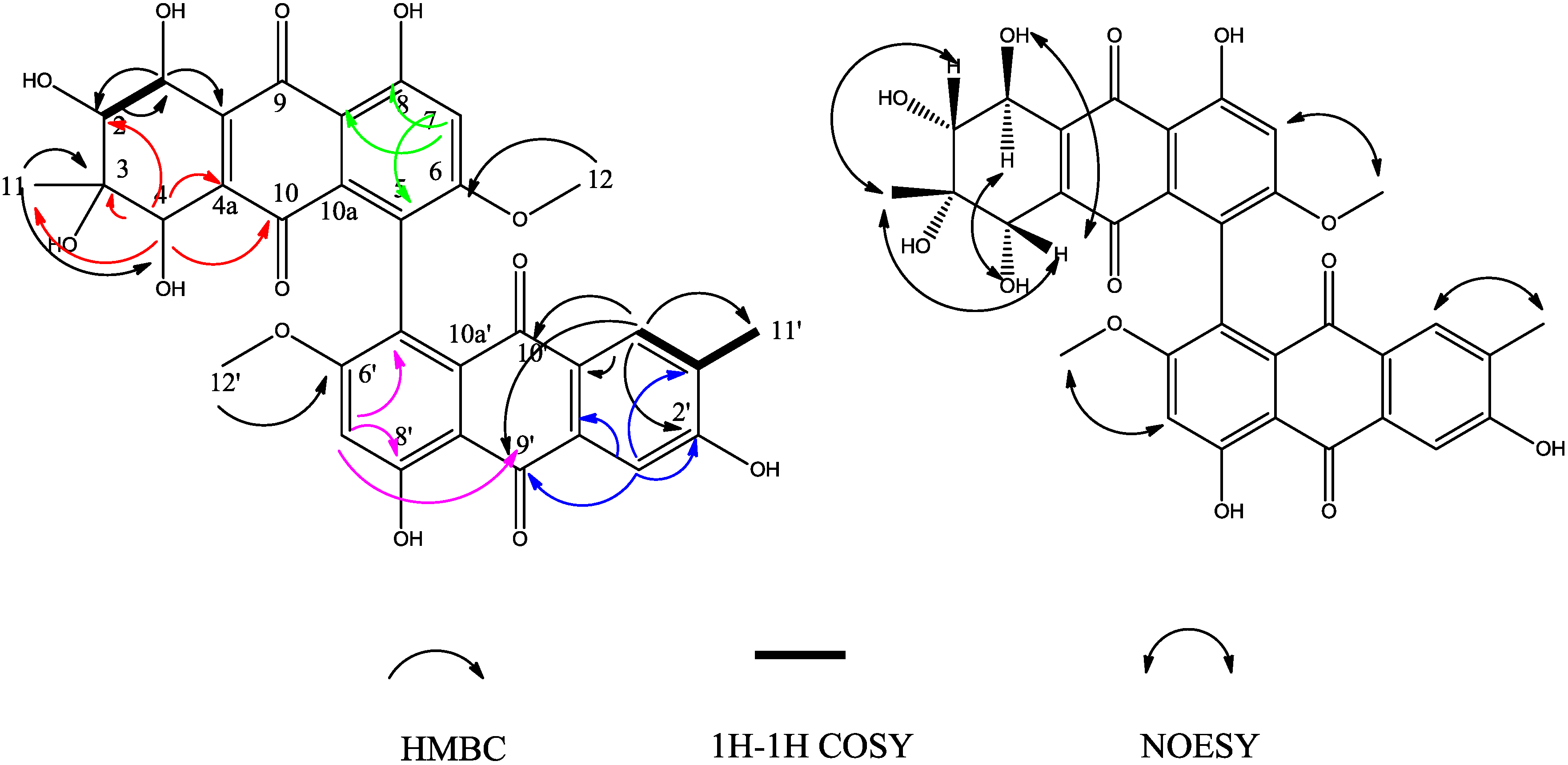
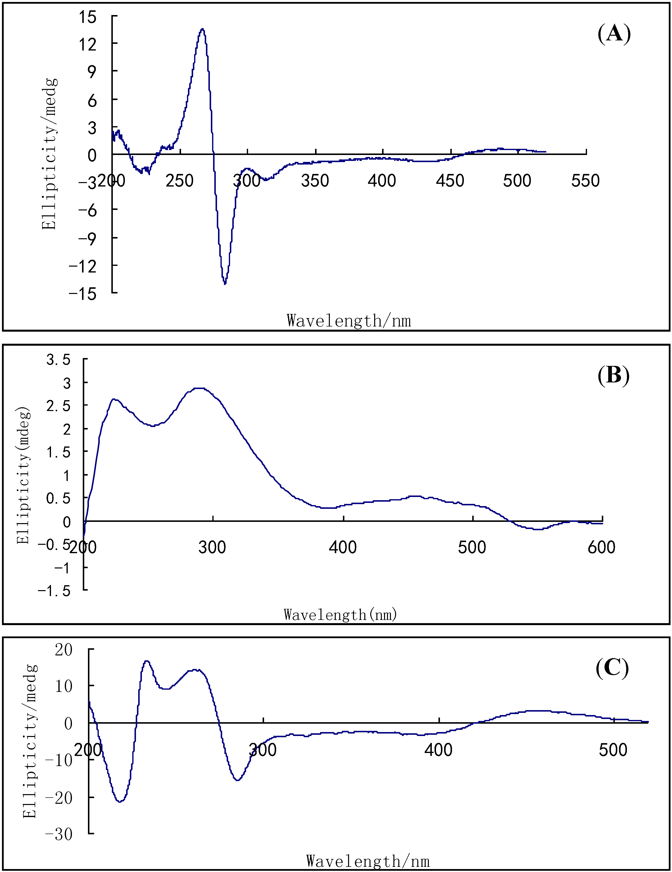
2. Results and Discussion
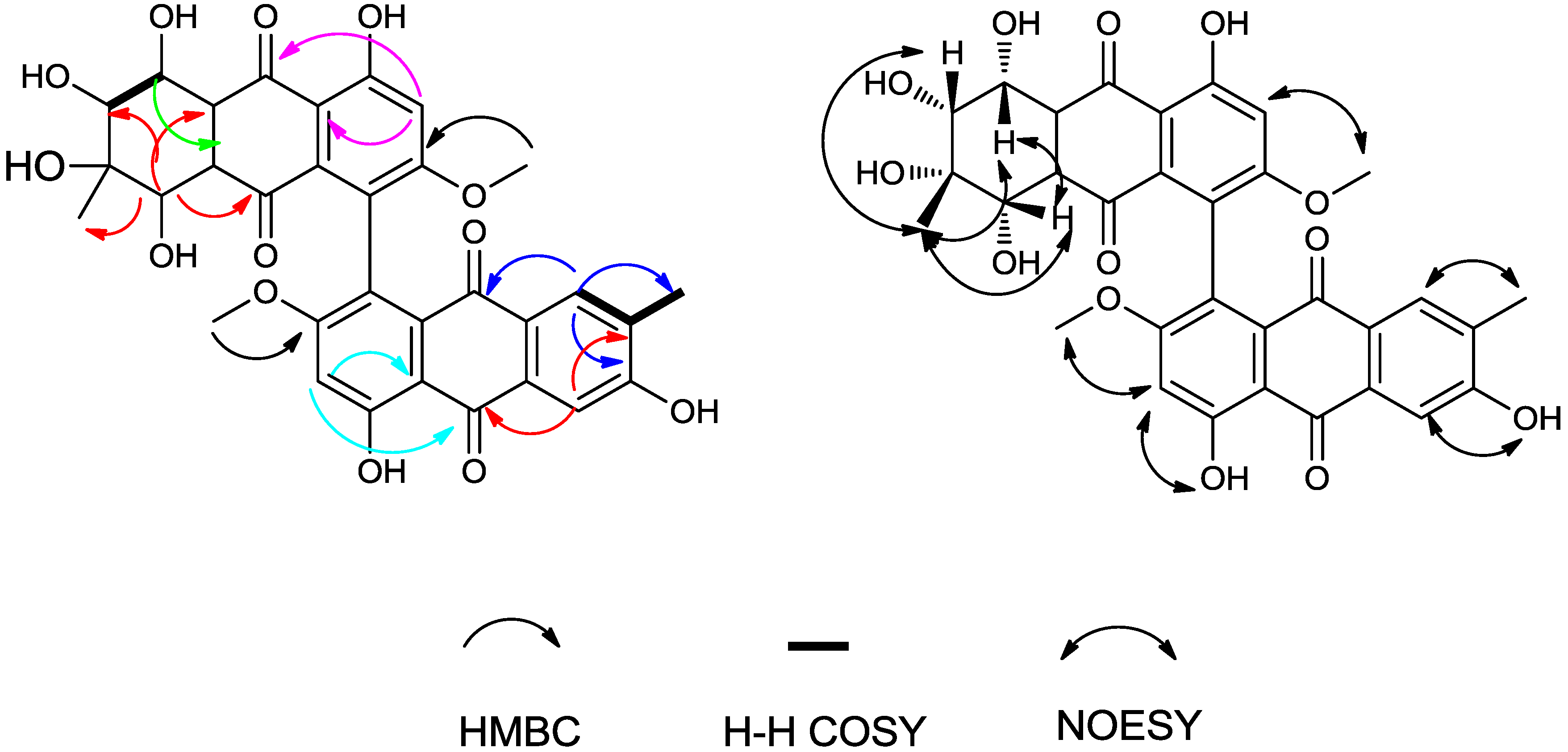
Compound 1 was yellow, amorphous powder. A HR-ESI-TOF-MS peak at m/z = 375.0688 (calcd. for C16H15NaO9, 375.0687) indicated the molecular formula C16H16O9. The 1H-NMR spectrum (Table 1) showed two aromatic protons (δH = 6.91 ppm and δH = 6.81 ppm), one methoxyl (δH = 3.86 ppm), one methyl group singlet (δH = 1.12 ppm), three oxygenated methine groups (δH = 4.43, 4.42 and 3.27 ppm) and a chelated hydroxyl (δH = 11.16 ppm) resonances. The 13C-NMR spectrum (Table 1) displayed two carbonyl signals (δC = 193.61, 191.12 ppm), six aromatic carbon signals, including three quaternary carbons (δC = 74.14, 68.37 and 67.16 ppm) and three methines (δC = 71.84, 67.64 and 67.28 ppm), one methoxyl group (δC = 56.43 ppm) and one methyl group (δC = 21.78 ppm). These data implied that compound 1 possessed a tetrahydroanthraquinone skeletone (Figure 2). In compound 1, the two aromatic protons (δC = 106.19 ppm; δH = 6.91 ppm, d, J = 2.48 Hz) and (δC = 106.98 ppm; δH = 6.81 ppm, d, J = 2.48 Hz) were at meta positions from each other on the aromatic ring, based on their 2.48 Hz coupling constant; The HMBC correlations from H-5 to C-6, C-7, C-8a and C-10, combined with from H-7 to C-5, C-6, C-8 and C-8a, and from H-12 (methoxy) to C-6, in addition, the NOE correlations of H-12(OCH3) to H-5 and H-6 (that supported the deduction that OCH3-12 is attached to C-6), established the substitution pattern of the aromatic ring (Figure 2). The protons in the cyclohexane ring, including three oxygenated methines H-1 (δH = 4.42 ppm, d, J = 8.1 Hz), H-2 (δH = 3.27 ppm, d, J = 8.1 Hz) and H-4 (δH = 4.43 ppm, s) were also observed. The coupled signals from H-1 to H-2 in the 1H-1H COSY spectrum combined with the HMBC correlations from H-1 to C-2, and from H-4 to C-2, C-3, C-4a, C-10 and C-11, established the substructure of the cyclohexane ring (Figure 2).
The HMBC correlations from H-11 to C-2, C-3 and C-4 indicated the methyl group was linked to C-3. One methoxy group, two carbonyl groups and five hydroxyl groups all together occupied eight oxygen atoms, so the remaining two carbons C-4a (δC = 68.37 ppm) and C-9a (δC = 67.16 ppm) must combine with the remaining oxygen to form an epoxy ether bond.
The relative configuration of the chiral centers of C-1, C-2, C-3 and C-4 were deduced by 2D 1H-1H NOESY experiments (Figure 2). A NOESY correlation among CH3-11 (δH = 1.12, singlet), H-2 and H-4 suggested that they are on the same side of the cyclohexane ring, and this was supported by the correlation between H-4 and H-2 (Figure 2). The coupling constant 8.1 Hz between H-1 and H-2 implied that H-1 and H-2 were positioned in a pseudoaxial orientation from each other. We could not deduce the relative configuration of the chiral centers of C-4a and C-9a by NOESY experiments or other spectroscopic methods other than X-ray single crystal diffraction. Therefore, compound 1 was determined as (1R*,2R*,3R*,4R*)-1,2,3,4,8-pentahydroxy-6-methoxy-3-methyl-1,2,3,4-tetrahydro-4a,9a-epoxyanthracene-9,10-dione. We propose for this compound the trivial name altersolanol O.
Table 1. NMR data of compounds 1 and 2 (DMSO-d6), measured at 400 MHz (1H) and 100 MHz (13C).
PositionCompound 1PositionCompound 2
δC, (ppm)δH (ppm)
(mult., J in Hz)
HMBCδC, (ppm)δH (ppm)
(mult., J in Hz)
HMBC
167.284.42 d (8.1)C-21,1'67.784.38 d (4.32)C-2, 3, 4a, 9, 9a, 11
271.843.27 d (8.1)C-12,2'71.69
374.14 3,3'69.593.77 br. d (7.8,4.8 overlap)C-4
467.644.43 sC-2, 3, 10, 114,4'42.73.82 dd (1.3, 4.8)C-3, 4', 4a, 9a
4a68.37 4a,4a'149.43
5106.196.91 d (2.48)C-6, 7, 8a, 105,5'163.04
6165.49 6,6'105.496.80 d (2.5)C-5, 7, 8, 10a
7106.986.81 d (2.48)C-5, 6, 8, 8a7,7'164.89
8162.11 8,8'105.637.05 d (2.5)C-6, 7, 9, 10a
8a110.0 8a,8a'133.69
9193.61 9,9'183.08
9a67.16 9a,9a'140.62
10191.12 10,10'188.59
10a134.46 10a,10a'109.91
1121.781.12 sC-2, 3, 411,11'22.221.13 sC-1, 2, 3
1256.433.86 sC-612,12'56.083.91 sC-7
1-OH 1,1'-OH 5.12 d (4.32)C-1, 2, 9a
2-OH 2,2'-OH 4.14 sC-1
3-OH 3,3'-OH 4.09 d (7.8)C-3, 4
4-OH 5,5'-OH 12.26 sC-5, 6, 10a
8-OH 11.16 s
Figure 2. HMBC correlations and selected NOESY correlations of compound 1.
Molecules 19 16529 g002
Compound 2 was obtained as a red, amorphous powder. The HR-ESI-TOF-MS peak at m/z = 637.1559 [M−H] (calcd. For C32H29O14, 637.1563) indicated the molecular formula C32H30O14. According to the 1H and 13C-NMR spectra (Table 1), we concluded that compound 2 was a symmetrical tetrahydroanthraquinone dimer. Compared to the reported compound alterporriol O [12], their 1H and 13C-NMR spectra were almost identical, but there were notable differences between the corresponding basic UV and optical rotation spectra, whereby the specific rotation value of compound 2 was large (−1200 in acetone) while that of the alterporriol O was −39.0 (in acetone). The NOESY correlation (Figure 3) between CH3-11 (δH = 1.13 ppm, s) and H-1 implied that they were also on the same side of the cyclohexene ring, unlike in alterporriol O. Therefore, compound 2 was identified as (1S*,1'S*,2R*,2'R*,3S*,3'S*,4S*,4'S*)-1,1',2,2',3,3',5,5'-octahydroxy-7,7'-dimethoxy-2,2'-dimethyl-1,1',2,2',3,3',4,4'-octahydro-[4,4'-bianthracene]-9,9',10,10'-tetraone. We propose for this new compound the trivial name alterporriol S.
Figure 3. HMBC correlations and selected NOESY correlations of compound 2.
Molecules 19 16529 g003
Compound 3 was obtained as a red, amorphous powder. The HR-ESI-TOF-MS peak at m/z = 617.1279 [M−H] (calcd. for C32H25O13, 617.1301) indicated the molecular formula C32H26O13. The 1H and 13C-NMR spectra (Table 2) were reminiscent of those obtained for the known monomeric compounds 10 and 11, which suggested that compound 3 is a modified bisanthraquinone dimer. We could readily deduce that compound 3 included one tetrahydroanthraquinone unit and one anthraquinone unit. On the anthraquinone unit, two aromatic proton singlet signals for H-1' (δH = 7.70 ppm, s) and H-4' (δH = 7.56 ppm, s) implied that H-1' and H-4' were in the para position from each other; a chelated hydroxyl signal (δH = 13.60 ppm, s) was observed and HMBC correlations from OH-8' (δH = 13.60 ppm) to C-8a', C-8', C-7' and from OCH3-12' (δH = 3.68 ppm) to C-6' all these indicated the anthraquinone unit to be a macrosporin unit. Furthermore, the HMBC correlations and NOESY correlations (Figure 4) of this unit might indicated that the unit was macrosporin.
In the tetrahydroanthraquinone unit, the HMBC correlations from H-1 to C-2 and C-9a, and from H-4 to C-2, C-3, C-4a, C-10 and C-11, revealed the cyclohexene ring substructure (Figure 4). The chelated hydroxyl signal (δH = 13.04 ppm, s) was observed with its HMBC correlations to C-8a, C-8, C-7, correlations from H-7 to C-5, C-8, C-8a and from OCH3-12 (δH = 3.70 ppm, s) to C-6, and combining with NOESY data, these established the substructure of the aromatic ring (Figure 4).
Table 2. NMR data of compound 3 and 4 (DMSO-d6), measured at 400 MHz (1H) and 100 MHz (13C).
PositionCompound 3Compound 4
δC, (ppm)δH (ppm)
(mult., J in Hz)
HMBCNOEδC, (ppm)δH (ppm)
(mult., J in Hz)
HMBCNOE
168.394.47 dd (5.78, 7.06)C-2, 9aH-4-OH68.454.48 dd (5.60, 6.87)C-2, 4a, 9a, 9H-4
273.783.57 dd (7.06, 7.06)C-1H-1173.743.55 dd (6.87, 6.87)C-1H-11
372.83 72.87
468.224.03 d (6.93)C-2, 3, 4a, 10, 11H-11,1-OH68.274.05 d (6.77)C-2, 9a, 10, 4a, 11H-1, 11
4a143.31 143.42
5122.86 122.59
6164.28 164.33
7103.776.93 sC-5, 8, 8a 103.776.92 sC-6, 8, 8a, 9H-12'
8163.63 163.73
8a109.27 109.3
9188.77 188.79
9a142.78 142.61
10184.09 183.88
10a128.86 129.02
1122.181.13 sC-3, 4H-2,4,1-OH22.221.13 sC-3, 4H-2, 4
1256.743.70 sC-6H-756.713.69 sC-6H-7
1-OH 5.64 d (7.06)C-1, 3, 9aH-4 4.98 d (5.60)C-1, 2, 9aH-4-OH
2-OH 4.36 sC-2, 4 4.81 d (6.87)C-1, 2, 3H-4-OH
3-OH 4.85 d (6.93)C-1 4.38 sC-2, 4, 11H-1-OH
4-OH 5.04 d (5.78)C-2, 4, 4aH-1 5.44 d (6.77)C-3, 4, 4aH- 2-OH
8-OH 13.04 sC-7, 8, 8a 13.04 sC-7, 8, 8a
1'110.447.56 sC- 2', 3', 9', 9a' 110.427.55 sC-2', 3', 9', 9a', 10'H-2'-OH
2'161.19 161.24
3'125.25 125.27
4'130.187.70 d (0.56)C-3', 4a', 10', 11'H-11'130.337.70 d (0.69)C-2', 4a', 9', 10', 11'H-11'
4a'132.39 132.38
5'121.81 123.12
6'164.18 165.25
7'104.016.94 sC- 5', 8', 8a', 9' 103.546.94 sC- 6, 8', 8a', 9'H-8'-OH
8'164.85 165.23
8a'109.98 109.92
9'186.7 186.61
9a'132.2 132.2
10'181.1 180.97
10a'131.48 130.43
11'16.032.20 sC-2',4'H-4'16.092.19 sC-2', 3', 4'H-4'
12'56.653.68 sC-6'H-7'56.833.72 sC-6’H-7'
2'-OH 11.07 br. s 11.03 br. s H-1'
8'-OH 13.60 sC-7', 8', 8a' 13.66 sC-7', 8', 8a'H-7'
Figure 4. HMBC correlations and selected NOESY correlations of compound 3.
Molecules 19 16529 g004
The HMBC correlation between H-12(OCH3) and C-6 indicated that the OCH3 attached at C-6. Further, the HMBC correlations from H-7 to C-5, C-8, C-8a, especially the NOE correlation between OCH3-12 and H-7, established that the junction between the two moieties of 3 was between C-5 and C-5' Besides, based on the HMBC correlations from H-7 to C-12, from H-4' to C-11', from H-7' to C-12' located the positions of three methyl groups.
The relative configuration of the chiral centers of C-1, C-2, C-3 and C-4 were deduced by 2D 1H-1H NOESY experiments (Figure 4). The NOESY correlations from CH3-11 (δH = 1.13 ppm, s) to H-2 and H-4, from H-1 to OH-4, from H-4 to OH-1, suggested that H-2, H-4, OH-1 and CH3-11 were on the same side of the cyclohexene ring, the rest (H-1, OH-2, OH-3 and OH-4) were on the other side of cyclohexene ring (H-1, OH-2, OH-3 and OH-4); finally compound 3 was determined as (1S*,2R*,3S*,4S*)-1,2,2',3,4,8,8'-heptahydroxy-6,6'-dimethoxy-3,3'-dimethyl-1,2,3,4-tetrahydro-[5,5'-bianthracene]-9,9',10,10'-tetraone. We propose for this compound the trivial name alterporriol T.
Compound 4 was also obtained as a red, amorphous powder. The HR-ESI-TOF-MS peak at m/z = 617.1282 (calcd. for C32H25O13, 617.1301) indicated the molecular formula C32H26O13. The 1H- and 13C-NMR spectra of compounds 3 and 4 were very similar. In the 13C-NMR spectrum, only three carbons’ chemical shifts were slightly different, they were C-5', C-6', C-8' (Table 2). The HMBC correlations indicated that compounds 3 and 4 had a same planar configuration. However, the specific rotation value of the two compounds were very different (compound 3, −39.95; compound 4, −337; in ethanol), and their CD spectra were also very different (Figure 5). All these suggested that they were isomers and the relative configuration of compound 4 was different from that of compound 3. In the NOESY correlation spectra, the correlations from CH3-11 (δH = 1.13, s) to H-1, H-2 and H-4 suggested that H-1, H-2, H-4 and CH3-11 were on the same side of the cyclohexene ring.
Figure 5. HMBC correlations and selected NOESY correlations of compound 4.
Molecules 19 16529 g005
NOESY correlations of the anthraquinone unit (from H-1' to OH-2', from H-4' to CH3-11', from H-7' to OCH3-6', and from H-7' to OH-8') were also observed. Finally, we could elucidate the structure of compound 4 as (1R*,2R*,3S*,4S*)-1,2,2',3,4,8,8'-heptahydroxy-6,6'-dimethoxy-3,3' dimethyl-1,2,3,4-tetrahydro-5,5'-bianthracene-9,9',10,10'-tetraone. We propose for this compound the trivial name alterporriol V.
Compounds 3 and 4 also contain a chiral axis. Comparing the CD spectra (Figure 6) with that of the the known compound alterporriol N, because the CD spectra were less sensitive to the configuration of the four chiral centres [10,16], according to the trends of CD between compound 3 and alternporriol N, we suggest that the absolute configuration of the chirality axis for compound 3 can be assigned as aS.
The compounds 511 were identified by comparing their spectroscopic data with those of the corresponding known compounds.
Compounds 111 were evaluated for cytotoxic activity against several human cancer cell lines by MTT assay [17,18]. The results showed that compound 9 showed good inhibitory activity against HCT-116 and HeLa cell lines, the IC50 values are 3.03 and 8.09 μM, respectively (Table 3). The other compounds show no notable inhibitory activity against any tested cancer cell lines.
Figure 6. CD spectra of compounds 3 (A), 4 (B) and 7 (C, alterporriol N) in acetonitrile solution.
Molecules 19 16529 g006
Table 3. Cytotoxicity test with anti-tumor, Epirubicin as a positive control.
CompoundIC50 (μM)
MCF-7/ADRHCT-116HeLa
118.4823.24 ± 2.8543.74 ± 4.02
2>100>50>50
3ND32.38 ± 1.57>50
4>50>50>100
5>100>100>100
6>100>100>100
7>100>100>100
844.99>50>50
9>1003.03 ± 0.058.09 ± 0.89
10>100>100>100
11>100>100>100
EPI2.360.96 ± 0.020.48 ± 0.03
Note: ND—No detected.
Compounds 111 were also tested for their ability to inhibit α-glucosidase by a described method [19]. Compound 3 (Alterporriol T) shows good inhibitory activity on α-glucosidase, with an IC50 value 7.2 μM (Table 4). The other compounds show no inhibitory activity against α-glucosidase.
Table 4. The inhibition activities on α-glucosidase for selected compounds.
Comp.1234567891011Genistein *
IC50 (μM)>10072.07.2>100>400>100>100>100>100>10083.513.6
Note: * Genistein—positive control.

3. Experimental Section

3.1. General Procedures

Column chromatography (CC) was performed using silica gel (200–300 mesh, Qingdao Marine Chemical, Qingdao, China). The HPLC system consisted of a Waters 2010 series (Waters, Milford, MA, USA). A mini ODS column (250 × 10 mm, 10 μm particle size) was used. Melting points were determined on an X-4 micro-melting point apparatus and were uncorrected. Circular dichroism was measured on a Schmidt Haensch Polartronic HH W5 polarimeter (Schmidt, Germany) and was uncorrected. UV spectra were measured on a Shimadzu UV-3501 PC spectrophotometer (Shimadzu, Japan). 1H (400 MHz) and 13C-NMR (100 MHz) data were recorded on a Bruker AVANCE 400 spectrometer (Bruker, Switzerland) with TMS as internal standard. LC/MS data were acquired using an Applied Biosystems/MDS Sciex (Applied Biosystems, Grand Island, NY, USA) and ana ESI source. HR-ESIMS were measured on a Shimadzu LCMS-IT-TOF.

3.2. Fungal Material

The fungal strain Alternaria sp. XZSBG-1 was isolated from a piece of sediment involving rotten branches and leaves, which was collected from the saline carbonate lake of Bamucuo, in Bange county, Tibetan Autonomous Region, China in July 2007. The strain was deposited in the Guangdong Province Key Laboratory of Functional Molecules in Oceanic Microorganism, School of Chemistry and Chemical Engineering, Sun Yat-sen University of China, Guangzhou, China.

3.3. Identification of Fungal Cultures

The fungal strain was identified as an Alternaria sp. according to morphologic traits and a molecular biological protocol by DNA amplification and sequencing as described [12,20]. The sequence data have been submitted to GenBank, accession number HM622756, and the strain was identified as Alternaria sp. Its 591 base pair ITS sequence had 99% sequence identity to that of Alternaria sp. IA249 (AY154699).

3.4. Fermentation, Extraction, and Isolation of Alternaria sp. XZSBG-1

The fungal strain was cultivated in potato glucose liquid medium (15 g of glucose and 3 g of crude sea salt in 1 L of potato infusion) in 1 L Erlenmeyer flasks each containing 400 mL of culture broth at 25 °C without shaking for 4 weeks. The fermentation broth (80 L) was filtered. The culture broth was extracted three times with an equal volume of EtOAc. The combined EtOAc layers were evaporated to dryness under reduced pressure to give an EtOAc extract (25.2 g). The mycelia was extracted with MeOH three times. The MeOH layer was evaporated under vacuum; then the combined residue was suspended in H2O and partitioned with EtOAc to provide the EtOAc extract (28.0 g), all the crude extract combined together (53.2 g) was subjected to silica gel column chromatography (CC, petroleum ether, EtOAc v/v, gradient) to generate eight fractions (Fraction 1−8). Fraction 4 was isolated by CC on silica gel eluted with petroleum ether−EtOAc (v/v, 10:1, 8:1, 6:1, 4:1, 2:1) and then subjected to Sephadex LH-20 CC eluting with mixtures of petroleum ether−CHCl3−MeOH (2:1:1) to obtain macrosporin (11, 3.0 g). Fraction 5 was subjected to repeated silica gel CC (CHCl3−MeOH, v/v, 50:1, 40:1, 30:1, 20:1, 15:1, 10:1, 9:1, 8:1, 7:1, 6:1, 5:1) to obtain altersolanol A (10, 8.0 mg), altersolanol C (9, 3.0 mg), altersolanol O (1, 2.4 mg), alterporriol A (8, 23.0 mg), alterporriol N (7, 20.6 mg), alterporriol S (2, 2.0 mg), fraction 9, and fraction 10, respectively. Fr. 8 was subjected to repeated Sephadex LH-20 CC (MeOH) and further purified on HPLC (40% MeOH−H2O) to afford alterporriol E (5, 18.0 mg) and alterporriol D (6, 11.0 mg). Fr.9 was subjected to repeated silica gel CC (CHCl3−MeOH, v/v, 100:1, 80:1, 60:1, 40:1, 20:1, 15:1, 10:1) to obtain alterporriol U (3, 22 mg); fraction 10 was purified on HPLC (55% MeOH−H2O) to afford alterporriol V (4, 16 mg).
Altersolanol O (1): yellow, amorphous powder; [ α ] D 18 = −29.46 (c = 0.34, MeOH); UV (MeOH) λmax (log ε) = 289 (1.03), 375 (0.40) nm; 1H-NMR (DMSO-d6), see Table 3; 13C-NMR (DMSO-d6), see Table 3; ESIMS m/z 351.0 [M−H]; HRESIMS m/z 375.0688 (calcd. for C16H15NaO9, 375.0687).
Alterporriol S (2): red, amorphous powder; [ α ] D 27 = −1200 (c = 0.2, MeOH); UV (ethanol) λmax (log ε) = 258 (3.99), 396 (0.597) nm; 1H-NMR (DMSO-d6), see Table 3; 13C-NMR (DMSO-d6), see Table 3; ESIMS m/z 637.0 [M−H]; HRESIMS m/z 637.1559 (calcd. for C32H29O14, 637.1563).
Alterporriol T (3): red, amorphous powder; [ α ] D 18 = −39.95 (c = 2.5, MeOH); UV (ethanol) λmax (log ε) = 248 (4.0), 437 (0.542); 1H-NMR (DMSO-d6), see Table 2; 13C-NMR (DMSO-d6), see Table 2; ESIMS m/z 617.0 [M−H]; HRESIMS m/z 617.1279 (calcd. for C32H25O13, 617.1301).
Alterporriol U (4): orange, amorphous powder; [ α ] D 18 = −337 (c = 1.99, MeOH); UV (ethanol) λmax (log ε) = 254 (3.90), 436 (0.722) nm; 1H-NMR (DMSO-d6), see Table 3; 13C-NMR (DMSO-d6), see Table 3; ESIMS m/z 617.0 [M−H]; HRESIMS m/z 617.1282 (calcd. for C32H25O13, 617.1301).

3.5. Biological Assays

3.5.1. Antitumor Activity in Vitro

Cell Culture

MCF-7/ADR, HeLa, HCT-116 cell lines were cultured in Dulbeccos’ modification Eagle’s medium (DMEM, Invitrogen, Carlsbad, CA, USA) supplemented with 10% fetal bovine serum (FBS, Hyclone, Logan, UT, USA), 2 mM L-glutamine, 100 μg/mL streptomycin and 100 U/mL penicillin (Invitrogen). The cells were incubated at 37 °C in a humidified atmosphere with 5% CO2.

Assessment of Antitumor Activity by MTT Assay

Cells were harvested during logarithmic growth phase and seeded in 96-well plates at a density of 1 × 10⁴ cells/mL, and cultured at 37 °C in a humidified incubator (5% CO2) for 24 h, followed by exposure to various concentrations of compounds tested for 48 h. Subsequently 20 μL of MTT (Genview, Houston, TX, USA) solution (5 mg/mL) was added to each well and mixed, the cells were then incubated for an additional 4 h. Culture supernatant was moved, 150 μL of DMSO (Sangon Biotech, Shanghai, China) was added to each well to fully dissolve the MTT-formazan crystals. Cell growth inhibition was determined by measuring the absorbance (Abs) at λ = 570 nm using a microplate reader and calculated according to the following equation:
Growth inhibition = (1 − OD of treated cells/OD of control cells) × 100%
The half maximal inhibitory concentrations (IC50) were obtained from liner regression analysis of the concentration-response curves plotted for each tested compound [17,18].

3.5.2. Enzyme Assays

Alpha-glucosidase activity was assayed using 50 mM phosphate buffer at pH 7.0, and the appropriate PNP glycoside (at 1 mM) were used as substrates. The concentration of the enzyme was specified in each experiment. Curcuminoids at the designated concentration was added to the enzyme solution and incubated at 37 °C for 30 min, and the substrate was then added to initiate the enzyme reaction. The enzyme reaction was carried out at 37 °C for 30 min. Product (PNP) was monitored spectrophotometrically by measuring the absorbance (λ = 400 nm) [19].

4. Conclusions

Alternaria sp. XZSBG-1 is a prolific producer of anthraquinones. Eleven more compounds have been isolated from this strain, including one new altersoanol and three new alterporriols. Compound 1 is a novel tetrahydroanthraquinone with an epoxy ether bond between C-4a and C-9a. In the primary bioassays, compound 9 showed good inhibitory activity against HCT-116 and HeLa cells and compound 3 showed good inhibitory activity on α-glucosidase. Although these new compounds showed no real activity in our primary bioassay, except 3, in view of the structural features of these compounds, it is valuable further to study their other biological activities, especially for compound 1.

Supplementary Materials

Supplementary materials can be accessed at: https://www.mdpi.com/1420-3049/19/10/16529/s1.

Supplementary Files

Supplementary File 1

Acknowledgments

This work was supported by the 863 Foundation of China (2006AA09Z422, 2007AA), Supported by grant from China’s Marine Commonweal Research Project (201005022-2), and Supported by the National Natural Science Foundation of China (20772162, 21272286, 2126203), and supported by grant from The Key Project in Tibet Autonomous Region (2011).

Author Contributions

YC Lin designed research; B Chen, Q Shen and X Zhu performed research and analyzed the data; B Chen and YC Lin wrote the paper. All authors read and approved the final manuscript.

Conflicts of Interest

The authors declare no conflict of interest.

References

  1. Lin, Y.C.; Wu, X.Y.; Deng, Z.J.; Wang, J.; Zhou, S.N.; Vrijmoed, L.L.P.; Jones, E.B.G. The metabolites of the mangrove fungus Verruculina enalia No. 2606 from a salt lake in the Bahamas. Phytochemistry 2002, 59, 469–471. [Google Scholar]
  2. Chen, W.Y.; Wyk, V.B.-E.; Vermaak, I.; Viljoen, M.A. Cape aloes—A review of the phytochemistry, pharmacology and commercialsation of Aloe ferox. Phytochem. Lett. 2012, 5, 1–12. [Google Scholar] [CrossRef]
  3. Shukla, V.; Joshi, G.P.; Rawat, M.S.M. Lichens as a potential natural source of bioactive compounds: A review. Pytochem. Rev. 2010, 9, 303–314. [Google Scholar] [CrossRef]
  4. Mishra, B.B.; Singh, D.D.; Kishore, N.; Tiwari, V.K.; Tripathi, V. Antifungal constituents isolated from the seeds of Aegle marmelos. Phytochemistry 2010, 71, 230–234. [Google Scholar] [CrossRef]
  5. Marcello, L. Anthraquinones: Analytical techniques as a novel tool to investigate on the triggering of biological targets. Curr. Drug Targets 2011, 12, 366–380. [Google Scholar] [CrossRef]
  6. Suemitsu, R.; Horiuchi, K.; Kubota, M.; Okamatsu, T. Production of alterporriols, altersolanols and macrosporin by Alternaria porri and A. Solani. Phytochemistry 1990, 29, 1509–1511. [Google Scholar] [CrossRef]
  7. Ohnishi, K.; Suemitsu, R.; Kubota, M.; Matano, H.; Yamada, Y. Biosyntheses of alterporriol D and E by Alternaria porri. Phytochemistry 1991, 30, 2593–2595. [Google Scholar] [CrossRef]
  8. Phuwapraisirisa, P.; Rangsan, J.; Siripong, P.; Tip-pyang, S. New antitumour fungal metabolites from Alternaria porri. Nat. Prod. Res. 2009, 23, 1063–1071. [Google Scholar] [CrossRef]
  9. Debbab, A.; Aly, A.H.; Edrada-Ebel, R.A.; Wray, V.; Müller, W.E.G.; Totzke, F.; Zirrgiebel, U.; Schächtele, C.; Kubbutat, M.H.G.; Lin, W.H.; et al. Bioactive metabolites from the endophytic fungus Stemphylium globuliferum isolated from Mentha pulegium. J. Nat. Prod. 2009, 72, 626–631. [Google Scholar] [CrossRef]
  10. Debbab, A.; Aly, A.H.; Edrada-Ebel, R.A.; Wray, V.; Pretsch, A.; Pescitelli, G.; Kurtan, T.; Proksch, P. New anthracene derivatives- structure elucidation and antimicrobial activity. Eur. J. Org. Chem. 2012, 2012, 1351–1359. [Google Scholar] [CrossRef]
  11. Huang, C.H.; Pan, J.H.; Chen, B.; Yu, M.; Huang, H.B.; Zhu, X.; Lu, Y.J.; She, Z.G.; Lin, Y.C. Three bianthraquinone derivatives from the mangrove endophytic fungus Alternaria sp. ZJ9–6B from the South China Sea. Mar. Drugs 2011, 9, 832–843. [Google Scholar] [CrossRef]
  12. Zheng, C.J.; Shao, C.L.; Guo, Z.Y.; Chen, J.F.; Deng, D.S.; Yang, K.L.; Chen, Y.Y.; Fu, X.M.; She, Z.G.; Lin, Y.C.; et al. Bioactive hydroanthraquinones and anthraquinones dimmers from a soft coral-derived Alternaria sp. fungus. J. Nat. Prod. 2012, 75, 189–197. [Google Scholar] [CrossRef]
  13. Okamura, N.; Haraguchi, H.; Hashimoto, K.; Yagi, A. Altersolanol-related antimicrobial compounds from a strain of Alternaria solani. Phytochemistry 1993, 34, 1005–1009. [Google Scholar] [CrossRef]
  14. Okamura, N.; Yagi, A.; Haraguchi, H.; Hashimoto, K. Simultaneous high-performance liquid chromatographic determination of altersolanol A, B, C, D, E and F. J. Chromatogr. A 1993, 630, 418–422. [Google Scholar] [CrossRef]
  15. Höller, U.; Gloer, J.B.; Wicklow, D.T. Biologically active polyketide metabolites from an undetermined fungicolous Hyphomycete resembling Clasdosporium. J. Nat. Prod. 2002, 65, 876–882. [Google Scholar] [CrossRef]
  16. Kanamaru, S.; Honma, M.; Murakami, T.; Tsushima, T.; Kudo, S.; Tanaka, K.; Nihei, K.; Nehira, T.; Hashimoto, M. Absolute stereochemistry of altersolanol A and alterporriols. Chirality 2012, 24, 137–146. [Google Scholar] [CrossRef]
  17. Huang, C.H.; Jin, H.; Song, B.; Zhu, X.; Zhao, H.X.; Cai, J.J.; Lu, Y.J.; Chen, B.; Lin, Y.C. The cytotoxicity and anticancer mechanisms of alterporriol L, a marine bianthraquinone, agaist MCF-7 human breast cancer cells. Appl. Microbiol. Biotechnol. 2012, 93, 777–785. [Google Scholar] [CrossRef]
  18. Chen, H.; Zhong, L.L.; Long, Y.H.; Li, J.; Wu, J.H.; Liu, L.; Chen, S.P.; Lin, Y.C.; Li, M.F.; Zhu, X.; et al. Studies on the synthesis of derivatives of marine-derived bostrycin and their structure-activity relationship against tumor cells. Mar. Drugs 2012, 10, 932–952. [Google Scholar] [CrossRef]
  19. Du, Z.Y.; Liu, R.R.; Shao, W.Y.; Mao, X.P.; Ma, L.; Gu, L.Q.; Huang, Z.S.; Chan, A.S.C. Alpha-glucosidase inhibition of natural curcuminoids and curcumin analogs. Eur. J. Med. Chem. 2006, 41, 213–218. [Google Scholar] [CrossRef]
  20. Stewart, C.N., Jr.; Via, L.E. A rapid CTAB DNA isolation technique useful for RAPD fingerprinting and other PCR applications. Biotechniques 1993, 14, 748–749. [Google Scholar]
  • Sample Availability: Sample of the compound 4 is available from the authors.

