Article

Carapanolides J–L from the Seeds of Carapa guianensis (Andiroba) and Their Effects on LPS-Activated NO Production

1
Laboratory of Medicinal Chemistry, Osaka University of Pharmaceutical Sciences, 4-20-1 Nasahara, Takatsuki, Osaka 569-1094, Japan
2
Faculty of Pharmaceutical Sciences, Kinki University, 3-4-1 Kowakae, Higashiosaka, Osaka 577-8502, Japan
*
Author to whom correspondence should be addressed.
Molecules 2014, 19(11), 17130-17140; https://doi.org/10.3390/molecules191117130
Submission received: 5 September 2014 / Revised: 17 October 2014 / Accepted: 20 October 2014 / Published: 24 October 2014
(This article belongs to the Collection Bioactive Compounds)

Abstract

:
A novel gedunin and two novel phragmalin-type limonoids, named carapanolides J–L (compounds 13) as well as a known gedunin-type limonoid 4 were isolated from the seeds of Carapa guianensis (andiroba). Their structures were determined on the basis of 1D and 2D NMR spectroscopy and HRFABMS. Compounds 14 were evaluated for their effects on the production of NO in LPS-activated mouse peritoneal macrophages.

1. Introduction

Carapa guianensis Aublet (Meliaceae), known locally as andiroba, is widely distributed in the Amazonas State of Brazil and its wood is extensively used as commercial timber [1]. Andiroba is a tall rainforest tree that grows up to 40 m in height. The indigenous people in the Amazon have used andiroba in many ways for centuries, and virtually all parts of the tree, as well as the seed oil, are utilized. It can be found growing wild throughout the Amazon rainforest, typically on rich soils, in swamps, and also in the alluvial flats, marshes, and uplands of the Amazon Basin. This tree can also be found wild or under cultivation in Brazil in the Islands region, Tocantins, Rio Solimoes, and near the seaside. It is one of the large-leafed trees of the rainforest and can be identified by its large and distinctively textured leaves. The andiroba tree produces a brown, woody, four-cornered nut with a diameter of 3–4 inches that resembles a chestnut. Andiroba oil is a rich source of essential fatty acids including oleic, palmitic, stearic, and linoleic acids. It yields up to 65% unsaturated fatty acids and can contain approximatoly 9% linoleic acid. Andiroba oil extracts yield up to 65% unsaturated fatty acids and can contain approximately 9% linoleic acid. Extracts from its bark, flowers, and seeds have been used for centuries by the Amazonian people and exhibit various repellent [2], analgesic [3], anti-malarial [4], anti-inflammatory [5], anti-allergic [6], and antiplasmoidal [7] activities, as well as acute and subacute toxicities [8]. Our recent study on the components of the seed oil of Carapa guianasis revealed the structures of two new unusual 9,10-seco-mexicanolide-type limonoids, named carapanolides A and B [9], two novel carbon skeletal limonoids, named guianolides A and B [10], and carapanolides C–I [11]. We herein describe the isolation and structural determination of three novel limonoids 13, named carapanolides J–L, and the effects of 13 and epoxyazadiradione (4) on the production of NO in LPS-activated mouse peritoneal macrophages. The structures of 13 were determined on the basis of NMR spectroscopy, including 1D and 2D (1H, 1H-COSY, NOESY, HSQC, HMBC) NMR, and FABMS.

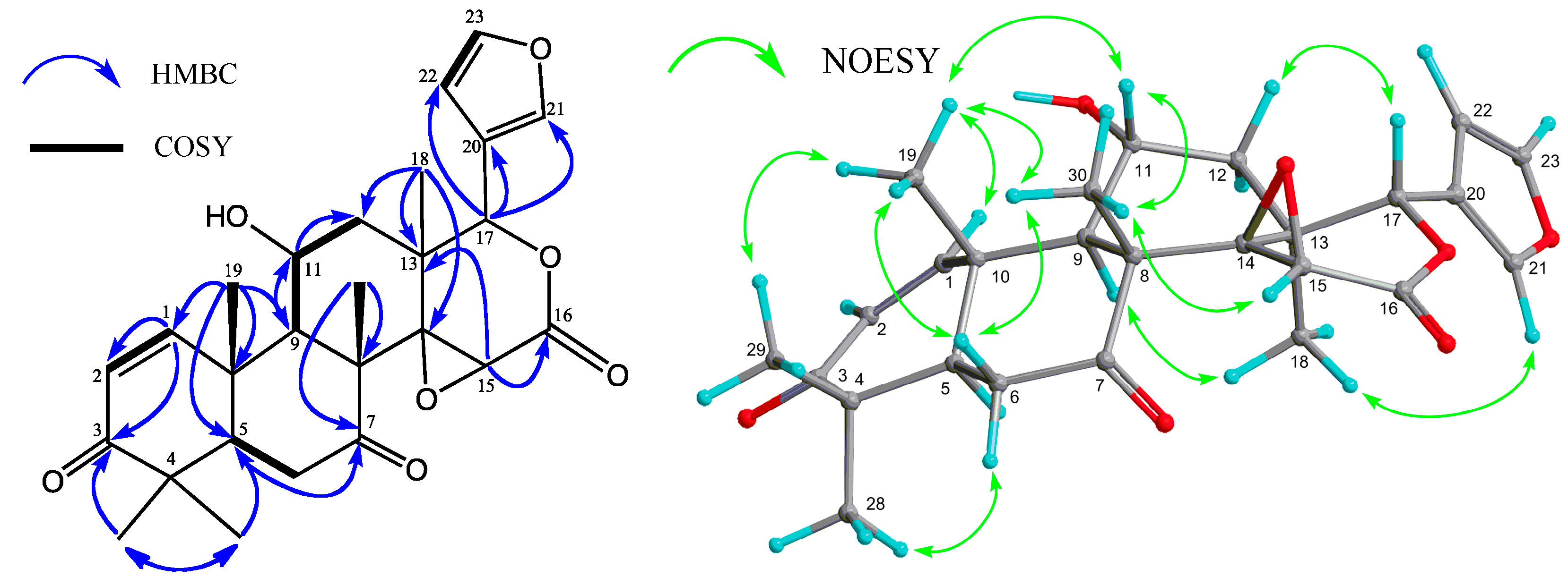
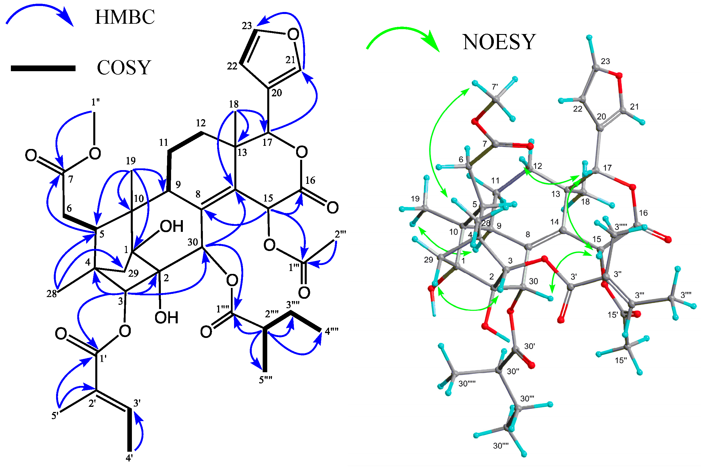
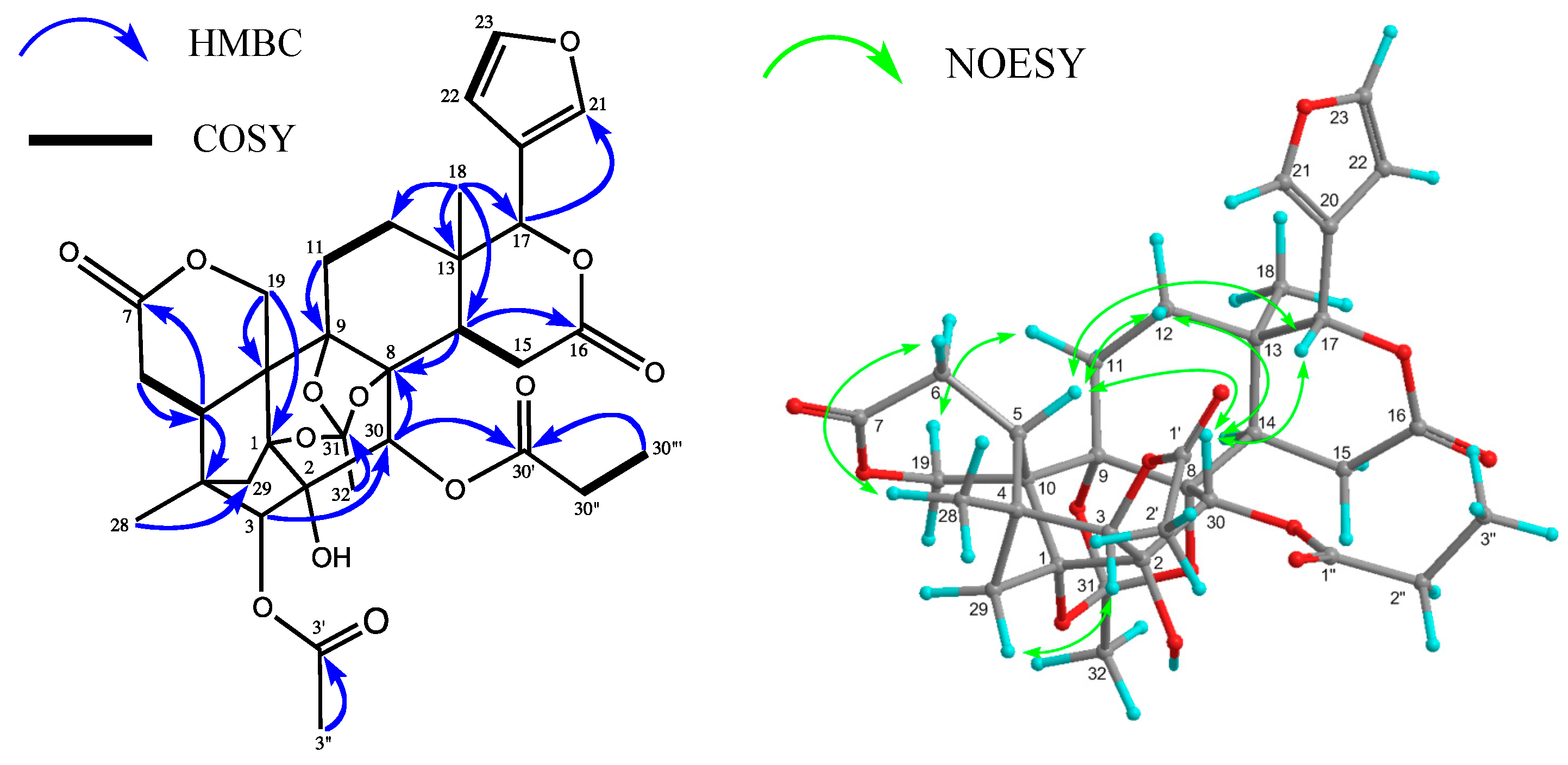
2. Results and Discussion

The seed oil of Carapa guianensis (2.03 kg) was separated by silica gel column chromatography, medium-pressure liquid chromatography (MPLC), and reverse-phase HPLC to obtain three new limonoids 13 and a known limonoid 4, which was identified as epoxyazadiradione (Figure 1) [12].
The molecular formula of carapanolide J (1) was determined as C26H30O7 ([M + H]+ m/z 455.2075) based on HRFABMS. The IR and UV spectra showed bands assignable to a hydroxy group (υmax 3503 cm−1), a six-membered ring ketone (υmax 1727 cm−1), and an α,β-unsaturated six-membered ring ketone [υmax 1671 cm−1; λmax 230 nm (log ε 3.85)]. The 1H and 13C NMR spectra (Table 1) exhibited signals assignable to five tertiary methyls [δH 1.16, 1.17, 1.21, 1.28, and 1.56]; two CH2 groups; five sp3 methine groups, including three oxymethine [δH 3.88 (s), 4.47 (ddd), and 5.49 (s)]; five sp3 quaternary carbons, including an oxycarbon [δC 65.4 (s)]; an α,β-unsaturated six-membered ring ketone [δH 5.84, 8.24 (each 1H, d); δC 203.0 (s)]; a saturated ketone [δC 207.7 (s)]; δ-lactone [δH 5.49 (s); δC 166.4 (s)]; and furan ring [δH 6.39 (dd), 7.42 (t), 7.44 (m)]. In the HMBC spectrum, long-range correlations were observed between Me-18 (δH 1.21) and C-12, C-13, C-14 [δC 65.4 (s)], and C-17 [δC 77.6 (d)]; between Me-19 (δH 1.56) and C-1 (δC 160.2), C-5, C-9, and C-10; between Me-28 (δH 1.17) and C-3 (δC 203.0), C-4, C-5, and C-29; between Me-29 (δH 1.16) and C-3, C-4, C-5, and C-28; between Me-30 (δH 1.28) and C-7 (δC 207.7), C-8, C-9, and C-14; between H-11 (δH 4.47) and C-8, C-9, C-10, C-12, and C-13; between H-15 (δH 3.88) and C-8, C-13, C-14 and C-16 (δC 166.4); and between H-17 (δH 5.49) and C-12, C-13, C-14, C-16, C-18, C-20 [δC 120.0 (s)], C-21 [δC 141.1 (d)], and C-22 [δC 109.7 (s)] (Figure 2). An analysis of the 1H-1H COSY spectrum (H-1–H-2; H-5–H2-6; H-9–H-11–H2-12; and H-22–H-23) revealed the partial structure shown in Figure 2. The HMBC and 1H-1H COSY spectra revealed that 1 was a 11-hydroxy-7-deacetoxy-7-oxogedunin [13]. Selected NOESY correlations were shown in Figure 2. The secondary hydroxyl group at C-11 [δH 4.47 (ddd)] was determined to have an α (equatorial) orientation because significant NOEs were observed between H-11 and Me-19, and Me-30, while coupling constants were observed between H-11β and H-9α (J11β,9α = 10.2 Hz); H-11β and H-12α (J11β,12α = 13.5 Hz); H-11β and H-12β (J11β,12β = 7.9 Hz). Therefore, compound 1 was determined to be 11α-hydroxy-7-deacetoxy-7-oxogedunin, which has been thus isolated in Nature for the first time.
Figure 1. Chemical structures for compounds 14.
Molecules 19 17130 g001
Figure 2. Key HMBC, COSY, and NOESY correlations for carapanolide J (1).
Molecules 19 17130 g002
Table 1. 1H (600 MHz) and 13C (150 MHz) NMR spectroscopic data of compound 1.
Position1Position1
1H a (J, Hz)13C b1H a (J, Hz)13C b
18.24d 10.3 (2)160.21465.4
25.84d 10.3 (1)124.9153.88s54.1
3203.016166.4
445.6175.49s77.6
52.21dd 3.2 (6α), 14.6 (6β)53.9181.21s20.6
6α2.38dd 3.2 (5), 13.8 (6β)36.3191.56s20.9
β2.93dd 13.8 (6α), 14.6 (5) 20120.0
7207.7217.44m141.1
853.4226.39dd 0.6 (21), 1.7(23)109.7
92.45d 10.2 (11)51.3237.42t 1.7 (21, 22)143.3
1040.9281.17s20.7
11β4.47ddd 7.9 (12β), 10.2 (9), 13.5 (12α)67.3291.16s27.4
12α1.46dd 13.5 (11), 13.8 (12β)44.6301.28s18.2
β2.21dd 7.9 (11), 13.8 (12α) 11-OH1.83s
1338.0
a Measured at 600 MHz in CDCl3; b Measured at 150 MHz in CDCl3. Assignments are based on HMBC spectrum.
Carapanolide K (2), which was isolated as a colorless amorphous solid, had the molecular formula C39H50O13 ([M + Na]+; m/z 749.3152, calcd. for 749.3155) as determined by HRFABMS. The IR spectrum showed the presence of a hydroxyl at υmax 3446 cm−1, and ester groups at υmax 1766, 1735, and 1698 cm−1. The 1H -and 13C-NMR spectra (Table 2) indicated the presence of three methyls [δH 0.83, 1.09, 1.14 (each 3H, s)], an acetyl group [δH 2.07 (3H, s), δC 21.1 (q), 169.9 (s)], 2-methylbutanoyl group [δH 0.90 (3H, t), 1.13 (3H, d), 1.48 and 1.64 (each 1H, m), 2.38 (1H, m), δC 176.3 (s)], tigloyl group [δH 1.77 (3H, dd), 1.98 (3H, t), 7.14 (1H, qq); δC 168.5 (s)], methoxycarbonyl group [δH 3.72 (3H, s), δC 52.0 (q), 174.2 (s)], δ-lactone [δH 5.36 (1H, s), δC 80.3 (d), 167.8 (s)], two tertiary hydroxyl groups [δC 77.0 (s), 83.6 (s)], a tetrasubstituted double bond [δC 134.9 (s), 135.4 (s)], and furan ring [δH 6.47 (dd), 7.41 (t), 7.58 (t)]. In the HMBC spectrum, cross-peaks were observed between Me-18 [δH 1.09 (s)] and C-12, C-13, C-14 [δC 135.4 (s)], and C-17 [δC 80.3 (d)]; between Me-19 [δH 1.14 (s)] and C-1 [δC 83.6 (s)], C-5, C-9, and C-10; between Me-28 [δH 0.83 (s)] and C-3 [δC 88.2 (d)], C-4, C-5, and C-29; between H-3 [δH 4.73 (s)] and C-1, C-2 [δC 77.0 (d)], C-4, C-5, C-28, C-29, C-30 [δC 69.7 (d)], and C-3' [δC 168.5 (s)]; between H-5 [δH 2.88 (dd)] and C-1, C-3, C-4, C-6, C-7 [δC 174.2 (s)], C-10, C-19, C-28, and C-29; between H-30 [δH 5.41 (s)] and C-1, C-2, C-3, C-8 [δC 134.9 (s)], C-9, and C-30' [δC 176.3 (s)]. The positions of the hydroxyl, 2-methylbutanoyl, methoxycarbonyl, and tigloyl groups were identified by detailed 1H-1H COSY and HMBC correlations (Figure 3). In addition, the cross peaks between H-9 and H-30, and C-8 [δC 134.9 (s)]; between H-30, H-15, and C-14 [δC 135.4 (s)] revealed that compound 2 was a phragmalin-8(14)-ene derivative [14]. In the NOESY spectrum, significant NOEs (Figure 3) were observed between H-3 [δH 4.73 (s)] and H-29 pro-S, H-30, and Me-28; between H-5 [δH 2.88 (dd)] and Me-28 and H-30; between Me-18 and H-11α and H-12α; between Me-19 and H-11α, between H-15 and H-17β, H-30, H-3', H-5', and H-2'''; therefore, the 2-methylbutanoyl group at C-30 and acetoxy group at C-15 were all α while the tigloyl group at C-3 had a β orientation. The configuration of the 2-methylbutanoyl group at C-30 was deduced to be R because the chemical shift value and NOESY correlation were very similar to that of carapanolide F [11], which was determined as 2R by single-crystal X-ray diffraction analysis.
Carapanolide L (3) was obtained as a colorless amorphous solid, and its molecular formula was established as C33H38O13 ([M + H]+; m/z 643.2391, calcd. for 643.2391) by HRFABMS, implying 15 degrees of unsaturation. The IR spectrum showed the presence of a hydroxyl at υmax 3352 cm−1, and ester groups at υmax 1742 cm−1. The 1H- and 13C-NMR data indicated that eight of the 15 units of unsaturation came from two carbon–carbon double bonds and four ester carbonyls, including two lactone carbonyls. Therefore, the remaining degrees of unsaturation required 3 to be nonacyclic. The 1H- and 13C-NMR spectra of 3 (Table 2) indicated the presence of two tertiary methyls [δH 1.00, 1.13 (each s)], an acetyl [δH 2.19 (s); δC 21.6 (q), 170.4 (s)], propanoyl [δH 1.09 (3H, t), 2.36 (1H, dq), 2.39 (1H, dq); δC 8.6 (q), 27.8 (t), 172.8 (s)], and orthoacetyl group [δH 1.70 (s); δC 21.0 (q), 119.6 (s)], four methylenes, including an oxymethylene [δH 4.38 and 4.77 (each 1H, d), five sp3 methines, including three oxymethines [δH 4.66 (s), 5.35 (s), and 5.71 (s)], a furan ring [δH 6.41 (dd), 7.44 (t), and 7.48 (m)], seven sp3 quaternary carbons, including four oxycarbon [δC 79.5 (s), 85.4 (s), 86.3 (s), and 86.4 (s)], two ester carbonyls [δC 170.4, and 172.8 (each s)], and two lactone carbonyl [δC 169.8, and 171.1 (s)]. An analysis of the 1H-1H COSY spectrum of 3 revealed the partial structures shown in bold face in Figure 4. In the HMBC spectrum (Figure 4), cross-peaks were observed from Me-18 [δH 1.13 (s)] to C-12, C-13, C-14, and C-17 (δC 78.4); from Me-28 [δH 1.00 (s)] to C-3, C-4, C-5, and C-29; from H-30 [δH 5.71 (s)] to C-1 [δC 85.4 (s)], C-2 [δC 79.5 (s)], C-3 [δC 83.9 (d)], C-8 [δC 86.4 (s)], and C-9 [δC 86.3 (s)] from H-14 [δH 1.00 (s)] to C-8, C-9, C-12, C-13, C-15, and C-16 [δC 169.8 (s)]. Therefore, the planar structure of 3 was established as phragmalin-1,8,9-orthoacetate [13], and the positions of the hydroxyl, acetyl, and n-propyl groups were located at C-2, C-3, and C-30 by detailed 1H-1H COSY and HMBC correlations (Figure 3). In the NOESY spectrum, significant NOEs (Figure 3) were observed between H-3 [δH 4.73 (s)] and H-29 pro-S, H-30, and Me-28; between H-5β [δH 2.68 (dd)] and H-12β, Me-28, and H-30; between H-15β [δH 3.19 (dd)] and H-30; between H-17β [δH 5.35 (s)] and H-12β, H-15β, H-22, and H-30β, between Me-18 [δH 1.13 (s)] and H-11α, H-12α and Me-32. Therefore, the relative structure of 3 was established as shown in Figure 1.
Figure 3. Selected 1H-1H COSY, HMBC and NOESYcorrelations for carapanolide K (2).
Molecules 19 17130 g003
Table 2. 1H-NMR and 13C-NMR data for compounds 2 and 3.
Position23
1H a (J, Hz)13C b1H a (J, Hz)13C b
183.685.4
277.079.5
34.73s88.24.66s83.9
443.145.2
52.88dd 1.2 (6B), 5.3 (6A)37.52.68dd 3.5 (6B), 5.5 (6A)33.8
6A2.32d 5.3 (5)33.72.46dd 5.5 (5), 17.6 (6B)31.0
B2.33d 1.2 (5)2.66dd 3.5 (5), 17.6 (6A)
7174.2171.1
8134.986.4
92.73d 7.735.986.3
1047.544.7
11Α1.70m18.31.85dt 2.9 (11α), 14.7 (12α,β)25.7
Β1.89m2.27m
12α1.05m28.51.48m29.4
β1.4dt 3.2 (12α), 14.1 (11β)1.38m
1338.934.5
14135.42.02dd 2.0 (15β), 10.5 (15α)42.8
15α2.70dd 10.5 (14), 20.0 (15β)26.4
β6.28d 2.464.23.19dd 2.0 (14), 20.0 (15α)
16167.8169.8
175.36s80.35.35s78.4
181.09s16.71.13s20.0
19α1.143H, s17.34.77d 13.8 (19β)68.8
β4.38d 13.8 (19α)
20120.5120.8
217.58t 0.8 (22)142.07.48t 0.8 (22)140.8
226.47dd 0.8 (21), 1.6 (23)109.96.41dd 0.8 (21), 1.8 (23)109.6
237.41t 1.6 (22)1437.44t 1.8 (22)143.4
280.83s14.81.00s13.6
29pro-R1.58d 11.0 (29 pro-S)39.81.80d 11.1 (29 pro-S)38.3
pro-S1.86d 11.0 (29 pro-R)2.25d 11.1 (29 pro-R)
305.41s69.75.71s70.0
31119.6
321.70s21.0
1'168.5170.4
2'130.02.19s21.6
3'7.14qq 7.0 (4'), 1.1 (5')12.2
4'1.77dd 1.1 (5'), 7.0 (3')139.2
5'1.98t 1.1 (3', 4')14.5
1''3.72s52.0172.8
2''A2.36dq 7.5 (3''), 9.7 (2''B)27.8
B2.39dq 7.5 (3''), 9.7 (2''A)
3''1.093H, t 7.5 (2''A, 2"B)8.6
1'''169.9
2'''2.07s21.1
1''''176.3
2''''A2.38m40.9
B
3''''A1.48m26.5
B1.64m
4''''0.90t 7.3 (3''''A, 3''''B)16.4
5''''1.13d 7.0 (2'''')11.3
a Measured at 600 MHz in CDCl3; b Measured at 150 MHz in CDCl3. Assignments are based on HMBC spectrum.
Figure 4. Key HMBC, 1H—1H COSY, and NOESY correlations of carapanolide L (3).
Molecules 19 17130 g004
Physiological nitric oxide (NO) is involved in blood pressure regulation and blood flow distribution, whereas its overexpression may induce tissue injury, multiple organ dysfunction, and death, as well as systemic inflammatory responses in sepsis, such as hypotension, cardiodepression, and vascular hyporeactivity [15]. In the present study, four limonoids and l-NMMA, an inducible nitric oxide synthase (iNOS) inhibitor, were evaluated for their inhibitory effects on NO production in LPS-stimulated RAW264.7 cells (Table 3). To determine safe concentrations, the cytotoxicities of these limonoids against RAW 264.7 were assessed by the MTT assay. Compounds 1 and 3 showed non-toxicities at 3–100 μM, whereas 4 and 2 exhibited moderate cytotoxicities (IC50 4: 21.3 μM; 2: 15.2 μM). In the inhibitory assay of NO production, compound 1 showed similar inhibitory activities (produced NO 83.4% at 10 μM; 61.8% at 30 μM; 16.8% at 100 μM) to the positive control, l-NMMA (produced NO 79.3% at 10 μM; 58.2% at 30 μM; 39.9% at 100 μM), with no cytotoxicities. Compound 4 exhibited superior inhibitory activities on NO production at non-toxic concentrations (produced NO 74.0% at 3 μM; 30.0% at 10 μM) to those of l-NMMA. These results suggested that compound 1 may be valuable as potential inhibitors of NO production.
Table 3. Inhobitory effects of NO production by limonoids from the seeds of Carapa guianensis.
CompoundConcentration (μM)
31030100IC50 (μM)
1Produced NO (%) a92.1 ± 1.583.4 ± 3.161.8 ± 1.816.8 ± 0.037.4
Cell viability (%) a102.4 ± 0.8101.0 ± 1.7102.8 ± 0.6103.4 ± 1.8>100
2Produced NO (%)78.6 ± 1.958.3 ± 2.825.8 ± 7.07.1 ± 1.212.0
Cell viability (%)81.4 ± 0.865.6 ± 0.233.6 ± 6.30.4 ± 0.415.2
3Produced NO (%)95.6 ± 2.595.4 ± 1.295.4 ± 2.978.4 ± 2.3>100
Cell viability (%)97.6 ± 0.697.3 ± 1.3100.5 ± 0.494.4 ± 1.0>100
4Produced NO (%)74.0 ± 5.030.0 ± 2.37.5 ± 1.03.9 ± 1.85.9
Cell viability (%)93.6 ± 1.499.7 ± 0.86.8 ± 0.33.3 ± 0.321.3
L-NMMA bProduced NO (%)93.0 ± 3.379.3 ± 0.858.2 ± 2.439.9 ± 1.753.7
Cell viability (%)103.5 ± 0.5102.0 ± 1.594.1 ± 1.496.5 ± 2.5>100
a Produced NO (%) and cell viability (%) were determined based on the absorbance at 570 nm, respectively, by comparison with values for DMSO (100%). Each value represents the mean ± standard error (S.E.) of three determinations. The concentration of DMSO in the sample solution was 2 μL/mL; b Positive control.

3. Experimental Section

3.1. General Procedures

Melting points were determined on a Yanagimoto micro-melting point apparatus and were uncorrected. Optical rotations were measured using a JASCO DIP-1000 digital polarimeter. IR spectra were recorded using a Perkin-Elmer 1720X FTIR spectrophotometer. 1H- and 13C-NMR spectra were obtained on an Agilent vnmrs 600 spectrometer with standard pulse sequences, operating at 600 and 150 MHz, respectively. CDCl3 was used as the solvent and TMS, as the internal standard. FABMS were recorded on a JEOL-7000 mass spectrometer. Column chromatography was carried out over silica gel (70–230 mesh, Merck, Darmstadt, Germany) and MPLC was carried out with silica gel (230–400 mesh, Merck). HPLC was run on a JASCO PU-1586 instrument equipped with a differential refractometer (RI 1531). Fractions obtained from column chromatography were monitored by TLC (silica gel 60 F254, Merck).

3.2. Plant Material

The oil of (2.03 kg) Carapa guianensis AUBLET (Meliaceae) was collected in the Amazon, Brazil, in March 2013. Kindly provided by Mr. Akira Yoshino (who is a representative of the NGO “Green Heart Love Amazon Project”). A voucher specimen (CGS-01-2) was deposited in the Herbarium of the Laboratory of Medicinal Chemistry, Osaka University of Pharmaceutical Sciences.

3.3. Isolation of Compounds 14

The seed oil of Carapa guianensis AUBLET (Meliaceae) (2.03 kg) was dissolved in CHCl3 (1 L) and the CHCl3 solution was subjected to CC (silica gel 14 kg), to afford seven fractions: Fraction A (Fr. No. 1–85, 1.512 kg) was eluted with n-hexane-CHCl3 = 1:1, B (Fr. No. 86–179, 229.1 g) was eluted with CHCl3, C (Fr. No. 180–220, 29.3 g) was eluted with CHCl3-EtOAc = 5:1, D (Fr. No. 221–225, 13.2 g) was eluted with CHCl3-EtOAc = 2:1, E (Fr. No. 226–265, 84.5 g) was eluted with CHCl3-EtOAc = 2:1, F (Fr. No. 266–290, 25.3 g) was eluted with EtOAc, G (Fr. No. 291–315, 72.8 g) was eluted with EtOAc:MeOH = 5:1, and H (Fr. No. 316–333, 45.4 g) was eluted with MeOH. Residue D was rechromatographed over a silica gel column (CC) (230–400 mesh, 300 g) eluted with n-hexane- EtOAc (1:1) to give 13 fractions: D1 (Fr. No. 1–35, 1.52 g), D2 (Fr. No. 36–49, 0.81 g), D(3) (Fr. No. 50–88, 0.70 g), D(4) (Fr. No. 89–115, 0.53 g), D(5) (Fr. No. 116–130, 0.60 g), D(6) (Fr. No. 131–140, 0.52 g), D(7) (Fr. No. 141–205, 0.47 g), D(8) (Fr. No. 206–215, 0.51 g), D(9) (Fr. No. 216–220, 0.42 g), D(10) (Fr. No. 221–240, 0.40 g), D(11) (Fr. No. 241–250, 1.11 g), and D(12) (Fr. No. 251–313, 1.36 g). Fraction D(6) was subjected to CC (230–400 mesh, 40 g) eluted with n-hexane–EtOAc (3:1) to give an amorphous solid (24.1 mg) that was separated by HPLC (ODS, 75% MeOH, at 25 °C, flow rate 4.0 mL·min−1, UV = 220 nm, column 250 × 20 mm i.d., 5 μm) to give compounds 2 (6.2 mg) and 3 (1.79 mg). Fraction D(8) was subjected to CC (230–400 mesh, 40 g) eluted with n-hexane–EtOAc (3:1) to give an amorphous solid (34.0 mg) that was subjected to CC (230–400 mesh, 40 g) eluted with n-hexane–EtOAc (3:1) to give an amorphous solid that was purified by HPLC (ODS, 75% MeOH, at 25 °C, flow rate 4.0 mL·min−1, UV = 220 nm, column 250 × 20 mm i.d., 5 μm) to give compounds 1 (7.5 mg) and 4 (3.8 mg). Fraction D(9) was subjected to CC (230–400 mesh, 30 g) eluted with n-hexane–EtOAc (3:1) to give an amorphous solid (25.5 mg) that was separated by HPLC (ODS, 70% MeOH, at 25 °C, flow rate 4.0 mL·min−1, UV = 220 nm, column 250 × 20 mm i.d., 5 μm) to give compound 3 (6.2 mg).

3.4. Analytical Data

Compound 1. Colorless crystals; mp 172–174 °C (from MeOH-CHCl3); [ α ] D 26 −18.7° (c 0.1, CHCl3); HRFABMS m/z: 455.2075 [M+H]+ (C26H31O7, calcd for 455.2080); UV (EtOH) λmax nm (log ε): 230 (3.85), 237 (3.80), 248 (3.63); IR (KBr) υmax cm−1; 3503 (OH), 2926, 1727 (O-C=O), 1671 (C=C-C=O); 1H- and 13C-NMR, see Table 1. FABMS m/z (rel. int.): 477 ([M+Na]+, 15), 455 ([M+H]+, 71), 83 (100).
Compound 2. Colorless amorphous solids; [ α ] D 26 −72.2° (c 0.1, CHCl3); HRFABMS m/z: 749.3152 [M+Na]+ (C39H50O13Na, calcd for 749.3155); UV λmax (EtOH) nm (log ε): 227 (4.19), 304 (3.98), 315 (4.00), 334 (3.72); IR (KBr) υmax cm−1: 3446 (OH), 2967, 1766 and 1735, 1698; 1H- and 13C-NMR, see Table 2. FABMS m/z (rel. int.): 749 (33) ([M+Na]+, 3), 727 ([M+H]+, 100).
Compound 3. Colorless amorphous solids; [ α ] D 26 −46.8° (c 0.1, CHCl3); HRFABMS m/z: 643.2391 [M+H]+ (C33H38O13, calcd for 643.2391); UV λmax (EtOH) nm (log ε): 208 (1.26), IR (KBr) υmax cm−1: 3352 (OH), 1742 (O-C=O); 1H- and 13C-NMR, see Table 2. FABMS m/z (rel. int.): 665 (33) ([M+Na]+, 12), 643 ([M+H]+, 100).

3.5. Determination of RAW264.7 Cell Proliferation

RAW264.7 cell proliferation was examined according to a method reported previously [16] with some modifications. Briefly, RAW264.7 cells (5 × 10⁴ cells in 100 μL) were seeded onto 96-well microplates, and incubated for 24 h. D-MEM (100 μL) containing test samples (final concentration of 100, 30, 10, or 3 μM) dissolved in DMSO (final concentration 0.2%) was added. After the cells had been treated for 24 h, the MTT solution was added. After 3 h of incubation, 20% sodium dodecyl sulfate (SDS) in 0.1 M HCl was added to dissolve the formazan produced by the cells. The absorbance of each well was read at 570 nm using a microplate reader. The optical density of vehicle control cells was assumed to be 100%.

3.6. Inhibitory Assay of NO Production

An inhibitory assay of nitric oxide production was performed according to a method reported previously [17] with slight modifications. Briefly, RAW264.7 cells (5 × 10⁴ cells in 100 μL) were seeded onto 96-well microplates, and incubated for 24h. D-MEM (100 μL) containing test samples (final concentration of 100, 30, 10, or 3 μM) dissolved in DMSO (final concentration 0.2%) and LPS (final concentration of 5 μg/mL) were added. After cells had been treated for 24 h, 50 μL of 0.1% N-(1-naphtyl)ethylenediamine in H2O and 50 μL of 1% sulfanylamide in 5% phosphoric acid were added. After being incubated for 30 min, the absorbance of each well was read at 570 nm using a microplate reader. The optical density of vehicle control cells was assumed to be 100%.

4. Conclusions

A novel gedunin and two novel phragmalin-type limonoids, named carapanolides J–L (compounds 13), as well as a known gedunin-type limonoid 4 were isolated from the seeds of Carapa guianensis (andiroba). Their structures were determined by spectroscopic analyses. Compound 1 showed similar inhibitory activities (produced NO 83.4% at 10 μM; 61.8% at 30 μM; 16.8% at 100 μM) to positive control, l-NMMA (produced NO 79.3% at 10 μM; 58.2% at 30 μM; 39.9% at 100 μM), with no cytotoxicity. Known compound 4 exhibited superior inhibitory NO production activities at non-toxic concentrations (produced NO 74.0% at 3 μM; 30.0% at 10 μM) to those of l-NMMA. These results suggest that compound 1 may be a valuable potential inhibitor of NO production.

Acknowledgments

We thank Katsuhiko Minoura and Mihoyo Fujitake (this university) for the NMR and MS measurements.

Author Contributions

Y. Matsui and T. Inoue performed the isolation and structure elucidation. T. Kikuchi contributed to evaluation of bioactivities. T. Yamada, O. Muraoka and R. Tanaka prepared the manuscript and supervised whole research project.

Conflicts of Interest

The authors declare no conflict of interest.

References

  1. Tan, Q.G.; Luo, X.D. Meliaceous limonoids: chemistry and biological activities. Chem. Rev. 2011, 111, 7437–7522. [Google Scholar] [PubMed]
  2. Prophiro, J.S.; da Silva Mario, A.N.; Kanis, L.A.; da Rocha, L.C.B.P.; Duque-Luna, J.E.; da Silva, O.S. First report on susceptibility of wild Aedes aegypty (Diptera: Culicidae) using Carapa guianensis (Meliaceae) and Copaifera sp. (Leguminosae). Parasitol. Res. 2012, 110, 7699–7705. [Google Scholar]
  3. Penido, C.; Costa, K.A.; Pennaforte, R.J.; Costa, M.F.S.; Pereira, J.F.G.; Siani, A.C.; Henriques, M.G.M.O. Anti-allergic effects of natural tetranortriterpenoids isolated from Carapa guianensis Aublet on allergen-induced vascular permeability and hyperalgesia. Inflamm. Res. 2005, 54, 295–303. [Google Scholar] [PubMed]
  4. Bickii, J.; Njifutie, N.; Foyere, J.A.; Basco, L.K.; Ringwald, P.J. In vitro antimalarial activity of limonoids from Khaya grandifoliola C.D.C. (Meliaceae). J. Ethnopharmacol. 2000, 69, 27–33. [Google Scholar] [CrossRef]
  5. Penido, C.; Conte, F.P.; Chagas, M.S.S.; Rodrigue, C.A.B.; Pereira, J.F.G.; Henriques, M.G.M.O. Antiinflammatory effects of natural tetranortriterpenoids isolated from Carapa guianensis Aublet on zymosan-induced arthritis in mice. Inflamm. Res. 2006, 55, 457–464. [Google Scholar] [CrossRef] [PubMed]
  6. Ferraris Fausto, K.; Rodrigues, R.; da Silva, V.P.; Figueiredo, R.; Penido, C.; Henriques, M.G.M.O. Modulation of T lymphocyte and eosinophil functions in vitro by natural tetranortriterpenoids isolated from Carapa guianensis Aublet. Int. Immunopharmacol. 2011, 11, 1–11. [Google Scholar]
  7. Miranda Junior, R.N.C.; Dolabela, M.F.; da Silva, M.N.; Povoa, M.M.; Maia, J.G.S. Antiplasmoidal activity of the andiroba (Carapa guianensis Aublet., Meliaceae) oil and its limonoid-rich fraction. J. Ethnopharmacol. 2012, 142, 679–683. [Google Scholar]
  8. Costa-Silva, H.; Lima, C.R.; Silva, E.J.R.; Araujo, A.V.; Fraga, M.C.C.R.; Ribeiro, E.; Ribwiro, A.; Arruda, A.C.; Lafayette, S.S.L.; Wanderley, J. Acute and subacute toxicity of the Carapa guianensis Aublet (Meliaceae) seed oil. J. Ethnopharmacol. 2008, 116, 495–500. [Google Scholar] [CrossRef]
  9. Inoue, T.; Nagai, Y.; Mitooka, A.; Ujike, R.; Muraoka, O.; Yamada, T.; Tanaka, R. Carapanolides A and B: unusual 9,10-seco-mexicanolides having a 2R,9S-oxygen bridge from the seeds of Carapa guianensis. Tetrahedron Lett. 2012, 53, 6685–6688. [Google Scholar] [CrossRef]
  10. Inoue, T.; Matsui, Y.; Kikuchi, T.; In, Y.; Yamada, T.; Muraoka, O.; Matsunaga, S.; Tanaka, R. Guianolides A and B, New Carbon Skeletal Limonoids from the seeds of Carapa guianensis. Org. Lett. 2013, 15, 3018–3021. [Google Scholar] [CrossRef] [PubMed]
  11. Inoue, T.; Matsui, Y.; Kikuchi, T.; In, Y.; Muraoka, O.; Yamada, T.; Tanaka, R. Carapanolides C–I from the seeds of andiroba (Carapa guianensis, Meliaceae). Fitoterapia 2014, 96, 56–64. [Google Scholar] [CrossRef] [PubMed]
  12. Malathi, R.; Rajan, S.S.; Mohan Kumar, R.; Narasimhan, S.; Ravikumar, K. Epoxyazadiradione. Acta Crystallogr. 2007, E63, 2483–2485. [Google Scholar]
  13. Tanaka, Y.; Yamada, T.; In, Y.; Muraoka, O.; Kajimito, T.; Tanaka, R. Absolute stereostructure of Andirolides A-G from the flower of Carapa guianensis (Meliaceae). Tetrahedron 2011, 67, 782–792. [Google Scholar] [CrossRef]
  14. Ravangpai, W.; Sommit, D.; Teerawatananond, T.; Sinpranee, N.; Palaga, T.; Pengpreecha, S.; Muangsin, N.; Pudhom, K. Limonoids from seeds of Thai Xylocarpus moluccensis. Bioorg. Med. Chem. Lett. 2011, 21, 4485–4489. [Google Scholar] [CrossRef] [PubMed]
  15. Kirkeboen, K.A.; Strand, O.A. The role of nitric oxide in sepsis–an overview. Acta Anaesthesiol. Scand. 1999, 43, 275–288. [Google Scholar] [CrossRef] [PubMed]
  16. Yamada, T.; Muroga, Y.; Jinno, M.; Kajimoto, T.; Usami, Y.; Numata, A.; Tanaka, R. New class azaphilone produced by a marine fish-derived Chaetomium globosum. The stereochemistry and biological activities. Bioorg. Med. Chem. 2011, 19, 4106–4113. [Google Scholar] [CrossRef]
  17. Yamasaki, F.; Machida, S.; Nakata, A.; Nugroho, A.E.; Hirasawa, Y.; Kaneda, T.; Morita, H. Haworforbins A-C, new phenolics from Haworthia cymbiformis. J. Nat. Med. 2013, 67, 212–216. [Google Scholar] [CrossRef] [PubMed]
  • Sample Availability: Not available.

