Antibacterial and EGFR-Tyrosine Kinase Inhibitory Activities of Polyhydroxylated Xanthones from Garcinia succifolia


 ,
, 
Abstract
:
1. Introduction

2. Results and Discussion

| S. aureus ATCC 25923 | B. subtilis ATCC 6633 | P. aeruginosa ATCC 27853 | E. coli ATCC 25922 | S. aureus B1 (MRSA) | E. faecalis W1 (VRE) | C. albicans ATCC 10231 | ||||||||
|---|---|---|---|---|---|---|---|---|---|---|---|---|---|---|
| Xanthones | MIC | MBC | MIC | MBC | MIC | MBC | MIC | MBC | MIC | MBC | MIC | MBC | MIC | MFC |
| 1 | ˃256 | - | ˃256 | - | ˃256 | - | ˃256 | - | ˃256 | - | ˃256 | - | ˃256 | - |
| 2 | ˃256 | - | ˃256 | - | ˃256 | - | ˃256 | - | ˃256 | - | ˃256 | - | ˃256 | - |
| 3 | ˃256 | - | ˃256 | - | ˃256 | - | ˃256 | - | ˃256 | - | ˃256 | - | ˃256 | - |
| 4 | 64 | ˃256 | 64 | ˃256 | ˃256 | - | ˃256 | - | 64 | ˃256 | ˃256 | - | 256 | 256 |
| 5 | 64 | ˃256 | 128 | ˃256 | ˃256 | - | ˃256 | - | 64 | ˃256 | ˃256 | - | 256 | ˃256 |
| 6 | 256 | ˃256 | 256 | ˃256 | ˃256 | - | ˃256 | - | ˃256 | - | ˃256 | - | ˃256 | - |
| E. coli G1 | S. aureus B1 (MRSA) | E. faecalis W1 (VRE) | |||||||||||
|---|---|---|---|---|---|---|---|---|---|---|---|---|---|
| Antibiotics | |||||||||||||
| Xanthones | Na | CIP | AMP | CTX | S | Na | OX | AMP | CIP | Na | VA | AMP | E |
| 1 | 8 | 8 | 8 | 12 | 8 | - | - | - | - | - | - | 21 | 9 |
| 2 | 9 | 9 | 9 | 12 | 8 | 9 | 12 | 11 | 9 | 8 | 12 | 21 | 9 |
| 3 | 7.5 | 7.5 | 7.5 | 12 | 7.5 | 8 | 8 | 8 | 8 | - | 9 | 21 | 11 |
| 4 | 7.5 | 7.5 | 7.5 | 12 | 7.5 | - | - | - | - | - | 10 | 21 | 12 |
| 5 | 7.5 | 7.5 | 7.5 | 12 | 7.5 | - | - | 7.5 | - | - | 9 | 21 | 11 |
| 6 | 7.5 | 7.5 | 7.5 | 12 | 7.5 | - | - | 7.5 | - | 9 | 21 | 11 | |
| Control | - | - | 12 | - | - | - | - | - | - | 9 | 21 | 11 | |
| MIC (µg/mL) | |||||
|---|---|---|---|---|---|
| 2 alone | OX alone | 2 with OX | OX with 2 | ΣFIC | |
| S. aureus B1 | ˃256 | 128 | 16 | 32 | ˂0.312 * |
| 2 alone | AMP alone | 2 with AMP | AMP with 2 | ΣFIC | |
| S. aureus B1 | ˃256 | 128 | 128 | 64 | 0.5-1 |
| 2 alone | VA alone | 2 with VA | VA with 2 | ΣFIC | |
| E. faecalis W1 | ˃256 | 256 | ˃256 | 256 | >1 |
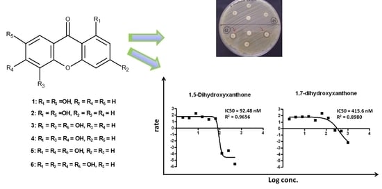
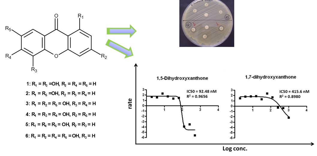
| Compound | % Relative Activity * | IC50 (nM) |
|---|---|---|
| 1 | 81.99 ± 3.30 | 90.34 ± 2.969 |
| 2 | 48.69 ± 9.34 | 223 ± 1.235 |
| 3 | 10.96 ± 9.34 | - |
| 4 | no inhibition | - |
| 5 | 13.71 ± 21.51 | - |
| 6 | no inhibition | - |
| Gefitinib (standard inhibitor, 1 µM) | 67.35 ± 2817 | 33 |
| Control (no inhibitor) | 0 | - |
3. Experimental Section
3.1. General Experimentation Procedures
3.2. Plant Material
3.3. Extraction and Isolation of Xanthones
3.4. Antimicrobial Activity Bioassays
3.4.1. Bacterial Strains
3.4.2. Determination of Minimum Inhibitory and Bactericidal/Fungicidal Concentrations
3.4.3. Synergistic Activity Studies
Screening of Combined Effect between Xanthones and Antibiotics
Synergy Test: Checkerboard Method
3.5. Tyrosine Kinase Inhibition Assay
3.6. Determination of IC50
4. Conclusions
Acknowledgments
Author Contributions
Conflicts of Interest
References
- Kijjoa, A.; Vieira, L.L.M. Triterpenes from the plants of the Family Clusiaceae (Guttiferae): Chemistry and Biological activities. In Natural Products: Chemistry, Biochemistry and Pharmacology; Brahmachari, G., Ed.; Narosa Publishing House Pvt. Ltd.: New Delhi, India, 2009; pp. 326–381. [Google Scholar]
- Ritthiwigrom, T.; Laphookhieo, S.; Pyne, S.G. Chemical constituents and biological activities of Garcinia cowa Roxb. Maejo Int. J. Sci. Technol. 2013, 7, 212–231. [Google Scholar]
- Paveen, M.; Khan, N.U.; Acchari, B.; Dutta, P.K. A triterpene from Garcinia mangostana. Phytochemistry 1991, 30, 361–362. [Google Scholar]
- Rukachaisirikul, V.; Pailee, P.; Hiranat, A.; Tuchinda, P.; Yoosook, C.; Kasisit, J.; Taylor, W.C.; Reutrakul, V. Anti-HIV-1 protostane triterpenes and digeranylbenzophenone from trunk bark and stem of Garcinia speciosa. Planta Medica 2003, 69, 1141–1146. [Google Scholar]
- Mahabusarakam, W.; Chairerk, P.; Taylor, W.C. Xanthones from Garcinia Cowa Roxb. latex. Phytochemistry 2005, 66, 1148–1153. [Google Scholar]
- Panthong, K.; Hutadilok-Towanata, N.; Panthong, A. Cowaxanthone F, a new tetraoxygenated xanthone, and other anti-inflammatory and antioxidant compounds from Garcina cowa. Can. J. Chem. 2009, 87, 1636–1640. [Google Scholar]
- Kijjoa, A.; Gonzalez, M.J.; Pinto, M.; Nascimento, M.S.J.; Campos, N.; Mondranondra, I.O.; Silva, A.M.S.; Eaton, G.; Herz, W. Cytotoxicity of Prenylated Xanthones and Other Constituents from the Wood of Garcinia merguensis. Planta Medica 2008, 8, 864–866. [Google Scholar]
- Iinuma, M.; Tosa, H.; Tanaka, T.; Asai, F.; Kobayashi, Y.; Shimano, R.; Miyauchi, K. Antibacterial activity of xanthones from Guttiferous plants against methicillin-resistant Staphylococcus aureus. J. Pharm. Pharmacol. 1996, 48, 861–865. [Google Scholar]
- Chomnawang, M.T.; Surrasmo, S.; Wongsariya, K.; Bunyapraphatsara, N. Antibacterial activity of Thai medicinal plants against methicillin-resistant Staphylococcus aureus. Fitoterapia 2009, 80, 102–104. [Google Scholar]
- Na, Y. Recent cancer drug development with xanthone structures. J. Pharm. Pharmacol. 2009, 61, 707–712. [Google Scholar]
- Aksoy, D.Y.; Unal, S. New antimicrobial agents for treatment of Gram-positive bacterial infections. Clin. Microbiol. Infect. 2008, 14, 411–420. [Google Scholar]
- Bassetti, M.; Merelli, M.; Temperoni, C.; Astilean, A. New antibiotics for bad bugs: Where are we? Ann. Clin. Microbiol. Antimicrob. 2013, 12. [Google Scholar] [CrossRef]
- Gomes, N.M.; Bessa, L.J.; Buttachon, B.; Costa, P.M.; Buaruang, J.; Dethoup, T.; Silva, A.M.S.; Kijjoa, A. Antibacterial and antibiofilm activities of tryptoquivalines and meroditerpenes isolated from the marine-derived fungi Neosartorya paulistensis, N. laciniosa, N. tsunodae, and the soil fungi N. fischeri and N. siamensis. Mar. Drugs 2014, 12, 822–839. [Google Scholar]
- Zhang, Z.; El Sohly, H.N.; Jacob, M.R.; Pasco, D.S.; Walker, L.A.; Clark, A.M. Natural Products inhibiting Candida albicans secreted aspartic protease from Tovomita krukovii. Planta Medica 2002, 68, 49–54. [Google Scholar]
- Kijjoa, A.; Gonzalez, M.J.; Afonso, C.M.; Pinto, M.M.M.; Anantachoke, C.; Herz, W. Xanthones from Calophyllum teysmannii var. inophylloide. Phytochemistry 2000, 53, 1021–1024l. [Google Scholar]
- Wu, Q.L.; Wang, S.P.; Du, L.J.; Yang, J.S.; Xiao, P.G. Xanthones from Hypericum japonicum and H. henryi. Phytochemistry 1998, 49, 1395–1402. [Google Scholar]
- Jackson, B.; Locksley, H.D.; Scheinmann, F. Extractives from Guttiferae. Part XIII. Isolation and structure of five xanthones from Garcinia eugeniifolia wall. J. Chem. Soc. C 1969, 16, 2201–2203. [Google Scholar]
- Fu, W.M.; Zhang, J.F.; Wang, H.; Tan, H.S.; Wang, W.M.; Chen, S.C.; Zhu, X.; Chan, T.M.; Tse, C.M.; Leung, K.S. Apoptosis induced by 1, 3, 6, 7-tetrahydroxyxanthone in hepatocellular carcinoma and proteomic analysis. Apoptosis 2012, 17, 842–851. [Google Scholar]
- Dharmaratne, H.R.W.; Wijesinghe, W.M.N.M.; Thevanasem, V. Antimicrobial activity of xanthones from Calophyllum species, against methicillin-resistant Staphylococcus aureus (MRSA). J. Ethnopharmacol. 1999, 66, 339–342. [Google Scholar]
- Rukachaisirikul, V.; Kamkaew, M.; Sukavisit, D.; Phongpaichit, S.; Sawangchote, P.; Taylor, C.W. Antibacterial xanthones from the leaves of Garcinia nigrolineata. J. Nat. Prod. 2003, 66, 1531–1535. [Google Scholar]
- Sakagami, Y.; Iinuma, M.; Piyasena, K.G.N.P.; Dharmaratne, H.R.W. Antibacterial activity of α-mangostin against vancomycin resistant Enterococci (VRE) and synergism with antibiotics. Phytomedicine 2005, 12, 203–208. [Google Scholar]
- Woodburn, J.R. The epidermal growth factor receptor and its inhibition in cancer therapy. Pharmacol. Ther. 1999, 82, 241–250. [Google Scholar] [CrossRef]
- Anderson, N.G.; Ahmad, T.; Chan, K.; Dobson, R.; Bundred, N.J. ZD 1839 (Iressa), a novel epidermal growth factor receptor (EGFR) tyrosine kinase inhibitor, potently inhibits the growth of EGFR-positive cancer cell lines with or without erbB2 overexpression. Int. J. Cancer 2001, 94, 774–782. [Google Scholar]
- Hidalgo, M.; Siu, L.L.; Nemunaitis, J.; Rizzo, J.; Hammond, L.A.; Takimoto, C.; Eckhardt, S.G.; Tolcher, A.; Britten, C.D.; Denis, L. Phase I and pharmacology study of OSI-774, an epidermal growth factor receptor tyrosine kinase inhibitor in patients with advanced solid malignancies. J. Clin. Oncol. 2001, 19, 3267–3279. [Google Scholar]
- Harries, M.; Smith, I. The development and clinical use of trastuzumab (Herceptin). Endocr. Relat. Cancer 2002, 9, 75–85. [Google Scholar] [CrossRef]
- Wakeling, A.E.; Guy, S.P.; Woodburn, J.R.; Ashton, S.E.; Curry, B.I.; Barker, A.J.; Gibson, K.H. GEFITINIB (Gefitinib): An orally active inhibitor of epidermal growth factor signaling with potential for cancer chemotherapy. Cancer Res. 2002, 62, 5749–5754. [Google Scholar]
- Clinical and Laboratory Standards Institute (CLSI). Performance Standards for Antimicrobial Susceptibility Testing; Twenty-First Informational Supplement M100-S21; CLSI: Wayne, PA, USA, 2011. [Google Scholar]
- Odds, F.C. Synergy, antagonism, and what the chequerboard puts between them. J. Antimicrob. Chemother. 2003, 52. [Google Scholar] [CrossRef]
- Sample Availability: Samples of the compounds are available from the authors.
© 2014 by the authors. Licensee MDPI, Basel, Switzerland. This article is an open access article distributed under the terms and conditions of the Creative Commons Attribution license ( http://creativecommons.org/licenses/by/4.0/).
Share and Cite
Duangsrisai, S.; Choowongkomon, K.; Bessa, L.J.; Costa, P.M.; Amat, N.; Kijjoa, A. Antibacterial and EGFR-Tyrosine Kinase Inhibitory Activities of Polyhydroxylated Xanthones from Garcinia succifolia. Molecules 2014, 19, 19923-19934. https://doi.org/10.3390/molecules191219923
Duangsrisai S, Choowongkomon K, Bessa LJ, Costa PM, Amat N, Kijjoa A. Antibacterial and EGFR-Tyrosine Kinase Inhibitory Activities of Polyhydroxylated Xanthones from Garcinia succifolia. Molecules. 2014; 19(12):19923-19934. https://doi.org/10.3390/molecules191219923
Chicago/Turabian StyleDuangsrisai, Susawat, Kiattawee Choowongkomon, Lucinda J. Bessa, Paulo M. Costa, Nurmuhammat Amat, and Anake Kijjoa. 2014. "Antibacterial and EGFR-Tyrosine Kinase Inhibitory Activities of Polyhydroxylated Xanthones from Garcinia succifolia" Molecules 19, no. 12: 19923-19934. https://doi.org/10.3390/molecules191219923