Article

Coumarins from Edgeworthia chrysantha

College of Pharmaceutical Science, Zhejiang University of Technology, 18 Chaowang Road, Hangzhou 310014, China
*
Authors to whom correspondence should be addressed.
Molecules 2014, 19(2), 2042-2048; https://doi.org/10.3390/molecules19022042
Submission received: 9 January 2014 / Revised: 23 January 2014 / Accepted: 23 January 2014 / Published: 13 February 2014

Abstract

:
A new coumarin, edgeworic acid (1), was isolated from the flower buds of Edgeworthia chrysantha, together with the five known coumarins umbelliferone (2), 5,7-dimethoxycoumarin (3), daphnoretin (4), edgeworoside C (5), and edgeworoside A (6). Their structures were established on the basis of spectral data, particularly by the use of 1D NMR and several 2D shift-correlated NMR pulse sequences (1H-1H COSY, HSQC and HMBC), in combination with acetylation reactions.

1. Introduction

The genus Edgeworthia (Thymelaeaceae) consists of five species distributed around the World, which are native to China, India, Japan, and southeast of America [1]. E. chrysantha is widely distributed and is endemic to South and East China [1]. The bark of E. chrysantha is used as “Zushima” in some local areas in China for the treatment of traumatic injury and rheumatism [2,3], and the fiber of the stem bark is a raw materials for making high quality paper [1]. The flower buds are often used as the traditional Chinese medicine “Mimenghua” for the treatment of ophthalmalgia and delacrimation [1,4].
Phytochemical studies have revealed that E. chrysantha contains various constituents, such as coumarins [5,6,7,8], flavonoids [9,10,11], terpenes [12,13,14] and lignans [15,16]. Among them coumarins are generally considered as the major anti-inflammatory and analgesia bioactive constituents [17]. In our continuing search for pharmacological and structurally interesting substances from the flower buds of E. chrysantha, a new coumarin, edgeworic acid (1), has been isolated, along with the five known coumarins umbelliferone (2) [18], 5,7-dimethoxycoumarin (3) [19] daphnoretin (4) [20], edgeworoside C (5) [18], and edgeworoside A (6) [18] (Figure 1). The structure of the new compound was elucidated by spectroscopic methods and confirmed by acetylation.
Figure 1. Chemical structures of compounds 16.
Molecules 19 02042 g001

2. Results and Discussion
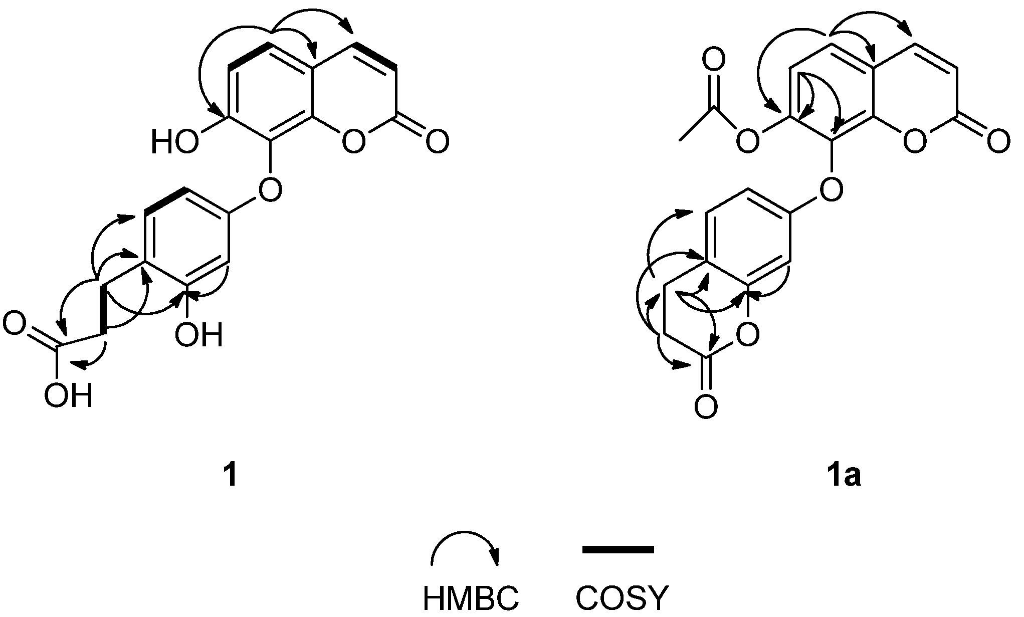
Compound 1 was obtained as a white powder. The molecular formula C18H14O7 was determined by HR-ESI-MS ([M−H] peak at m/z 341.0752), indicating 12 degrees of unsaturation. The 13C-NMR and DEPT spectra resolved 18 carbon signals, which were classified by chemical shifts and HSQC spectrum as two carbonyl groups [δC 174.6 (C-9'), δC 160.2 (C-2)], seven sp2 quaternary carbons [δ C 157.2 (C-1'), δC 156.3 (C-3'), δC 154.8 (C-7), δC 148.8 (C-9), δC 128.6 (C-8), δC 121.0 (C-4'), δC 112.5 (C-10)], seven sp2 methines [δC 145.1 (C-4), δC 130.5 (C-5'), δC 125.7 (C-5), δC 114.1 (C-6), δC 112.1 (C-3), δC 105.7 (C-6'), δC 102.0 (C-2')], two sp3 methylenes [δC 34.3 (C-8'), δC 25.3 (C-7')] (Table 1).
The IR spectrum exhibited vibration bands for free hydroxyl (3334 cm−1), carboxyl (3207, 1732 cm−1), conjugated carbonyl (1692 cm−1), and aromatic (1610, 1519, 1448 cm−1) functionalities. The UV spectrum exhibited a maximum absorption at 322 nm. According to the data mentioned above, it is suggested that compound 1 has a coumarin skeleton. This was further supported by the 1H-NMR signals [δH 8.01 (1H, d, J = 9.2 Hz, H-4); δH 6.25 (1H, d, J = 9.2 Hz, H-3)] (Table 1), and 13C-NMR signals [δC 160.2 (C-2); δC 145.1 (C-4); δC 112.1 (C-3)] [8].
The 1H-NMR spectrum of 1 (Table 1) showed the presence of a set of ortho-coupled aromatic signals [δH 7.46 (1H, d, J = 8.4 Hz, H-5); δH 6.99 (1H, d, J = 8.4 Hz, H-6)]. The 1H-NMR data also showed an ABX-type coupling system [δH 6.97 (1H, d, J = 8.4 Hz, H-5'); δH 6.29 (1H, d, J = 2.4 Hz, H-2'); δH 6.24 (1H, overlapped, H-6')]. The signals at δH 2.68 (2H, t, J = 7.2 Hz, H-7'), δC 25.3 (C-7'), δH 2.43 (2H, t, J = 7.2 Hz, H-8'), δC 34.3 (C-8') showed the existence of a 3-propionic acid group [8], which was confirmed by the HSQC, HMBC, and 1H-1H COSY spectra. (Figure 2) In the HMBC spectrum, the 1H-NMR signal at δH 2.43 (H-8') was correlated to 13C-NMR signal at δC 121.0 (C-4'), and the 1H-NMR signal at δH 2.68 (H-7') showed correlations with 13C-NMR signals at δC 156.3 (C-3'), 121.0 (C-4') and 130.5 (C-5'), indicating that 3-propionic acid group was located at the C-4' position. (Figure 2) The aromatic H-atom at δH 7.46, which correlated with 154.8 (C-7), 148.8 (C-9), 145.1 (C-4) in the HMBC spectrum (Figure 2), could be assigned to H-5. Since it coupled with the H-6, the substitution site at the coumarin skeleton was established at C-7 and C-8.
Table 1. 1H- and 13C-NMR data of 1 and 1a (δ in ppm and J in Hz).
No.1No.1a
δHδCδHδC
2160.2 s2159.3 s
36.25 (d, 9.2)112.1 d36.53 (d, 9.6)116.5 d
48.01 (d, 9.2)145.1 d48.13 (d, 9.6)144.4 d
57.46 (d, 8.4)125.7 d57.71 (d, 8.4)125.6 d
66.99 (d, 8.4)114.1 d67.33 (d, 8.4)120.3 d
7154.8 s7146.1 s
8128.6 s8133.3 s
9148.8 s9147.7 s
10112.5 s10118.9 s
1'157.2 s1'157.0 s
2'6.29 (d, 2.4)102.0 d2'6.65 (d, 2.4)104.0 d
3'156.3 s3'152.7 s
4'121.0 s4'118.1 s
5'6.97 (d, 8.4)130.5 d5'7.24 (d, 8.4)129.6 d
6'6.24 (overlapped)105.7 d6'6.69 (dd, 2.4, 8.4)111.2 d
7'2.68 (t, 7.2)25.3 t7'2.94 (t, 7.2)22.5 t
8'2.43 (t, 7.2)34.3 t8'2.78 (t, 7.2)28.9 t
9'174.6 s9'168.4 s
Ac2.15 (s)20.6 q
168.5 s
Figure 2. Key HMBC and 1H-1H COSY correlations of 1 and 1a.
Molecules 19 02042 g002
According to the molecular formula of 1, there were only two phenolic hydroxyl groups left. Unfortunately, no phenolic hydroxyl groups signals appeared in the 1H-NMR spectrum, so the locations of the phenolic hydroxyl groups were confirmed by acetylation. The acetylated derivative of 1 (compound 1a) was obtained as a white powder. The molecular formula C20H16O7 was determined by ESI-MS ([M+H]+ peak at m/z 367.0), indicating 13 degrees of unsaturation. By comparing the MS data of 1a with those of 1, it was presumed that a six membered lactone ring was formed during the acetylation reaction. The comparison of the 13C-NMR data of 1 with those of 1a (Table 1) revealed that the signals of C-6 and C-8 were shifted downfield in the range of δ 5–6 ppm, the signal of C-7 was shifted upfield by δ 8.7 ppm, and the signal of C-3' was shifted upfield by δ 3.6 ppm. According to the analysis mentioned above, one of the phenolic hydroxyl groups was deduced to be at C-7 [21], and another one was located at C-3', which was confirmed by the HMBC spectrum. (Figure 2).
The five known coumarins were identified as umbelliferone (2) [18], 5,7-dimethoxycoumarin (3) [19] daphnoretin (4) [20], edgeworoside C (5) [18], and edgeworoside A (6) [18], by interpretation of their spectroscopic data and comparison with literature values.

3. Experimental

3.1. General

All chemical solvents used were of analytical grade. Column chromatography (CC): MCI gel (Mitsubishi Chemical Co., Tokyo, Japan); Sephadex LH-20 (Pharmacia Fine Chemical Co. Ltd., Uppsala, Sweden); silica gel (Qingdao Marine Chemical Group Co., Qingdao, China; 200–300 and 400–600 mesh). HPLC: Agilent 1100 series (Agilent Technologies, Palo Alto, CA, USA) equipped with an Agilent DAD spectrophotometer and an Alltima-C18 reversed-phase column (5 µm, 250 × 10 mm) with an Eclipse XDB-C18 guard column. IR spectra: Nicolet-Magna-FT-IR 750 spectrometer (Thermo Scientific, Waltham, MA, USA). UV spectra: Shimadzu UV-2450 spectrophotometer (Shimadzu, Kyoto, Japan). LR- and HR-ESI-MS: Finnigan LCQ-Deca (Thermo Scientific, Waltham, MA, USA) and Waters Micromass Q-TOF-Ultima mass spectrometers (Waters, Milford, MA, USA). 1H- and 13C-NMR spectra were recorded on a Bruker Avance-400 spectrometer (Bruker, Karlsruhe, Germany) (1H- at 400 MHz, 13C- at 100 MHz) in DMSO-d6 at room temperature (22 °C). Chemical shifts are reported in ppm (δ), relative to tetramethylsilane as internal standard, and coupling constants are in Hertz.

3.2. Plant Material

The flower buds of E. chrysantha were collected in a garden of Lishui, Zhejiang Province, China, in February 2008. The plants were authenticated by Dr. Chu Chu, Zhejiang University of Technology, China. A voucher specimen (TCM 2008-026) was deposited in College of Pharmaceutical Science, Zhejiang University of Technology.

3.3. Extraction and Isolation

The air-dried material (10 kg) were extracted at room temperature and for 36 h × 3 with 95% (v/v) EtOH (5 L × 3) to give, after removal of the solvent, 220 g of crude extract which was dissolved in 4 L of H2O to form a suspension and successively partitioned with petroleum ether (60–90 °C) (3,000 mL × 3), ethyl acetate (3,000 mL × 3) and n-butanol (3,000 mL × 3). The ethyl acetate extracts (14 g) were chromatographed on a silica gel column (petroleum ether/ethyl acetate, 4:1–0:1 v/v) to give eight fractions 1–8. Fraction 1 (4:1 v/v; 1.2 g) was separated on a silica gel H column (petroleum ether/ethyl acetate, 6:1 v/v) to afford three fractions 1'–3'. Subfraction 3' (100 mg) was chromatographed on a silica gel H column (petroleum ether/acetone, 6:1 v/v) to afford compound 2 (28 mg) and 3 (27 mg). Fraction 3 (3:1 v/v; 0.2 g) was recrystallized with methanol to give compound 4 (45 mg). Fraction 4 (2.5:1 v/v; 1.0 g) was separated on a silica gel H column (CHCl3/acetone, 6:1 v/v) to afford compound 1 (43 mg). Fraction 7 (1:1 v/v; 3.8 g) was chromatographed on a MCI gel column (MeOH/H2O, 1:9–8:2 v/v) to give three fractions 1'–3'. Subfraction 1' (100 mg) was recrystallized from methanol to afford compound 5 (55 mg). Subfraction 3' (80 mg) was subjected to Sephadex LH-20 column chromatography (3 × 100 cm) eluted with CHCl3/MeOH (1:1 v/v) to remove the pigments and finally purified by semipreparative-HPLC using MeOH/H2O (64:36 v/v, 25 °C, 3.0 ml/min) to afford compound 6 (13 mg, tR = 17.38 min).

3.4. Acetylation of Edgeworic acid (1)

A mixture of compound 1 (20 mg), Ac2O (5 mL), and pyridine (5 mL) was stirred at room temperature overnight. The resulting solution was concentrated under vacuum. The residue was dissolved in ethyl acetate and washed with water (5 mL). The product was purified on a silica gel column (n-hexane/ethyl acetate, 3:2 v/v) to afford 1a (15 mg).
Acetylated derivative of edgeworic acid (1a). White powder; ESI-MS (+) m/z 367.0 [M+H]+; 1H-NMR (DMSO-d6) and 13C-NMR (DMSO-d6) data: see Table 1.

3.5. Spectral Data

Edgeworic acid (1). White powder; UV (MeOH) λmax (log ε): 322 (4.20); IR (KBr) vmax: 3,334, 3,207, 1732, 1692, 1610, 1519, 1448 cm−1; HR-ESI-MS (−) m/z 341.0752 [M−H] (calcd. for C18H13O7, 341.0661); 1H-NMR (DMSO-d6) and 13C-NMR (DMSO-d6) data: see Table 1.
Umbelliferone (2). Colorless needles; m.p.: 225–228 °C; 1H-NMR (DMSO-d6) δ: 10.6 (1H, s, 7-OH), 7.92 (1H, d, J = 9.5 Hz, H-4), 7.52 (1H, d, J = 8.5 Hz, H-5), 6.78 (1H, dd, J = 2.4, 8.5 Hz, H-6), 6.71 (1H, d, J = 2.4 Hz, H-8), 6.19 (1H, d, J = 9.5 Hz, H-3).
5,7-Dimethoxycoumarin (3). Colorless needles; m.p.: 144–145 °C; 1H-NMR (DMSO-d6) δ: 7.96 (1H, d, J = 9.6 Hz, H-4), 6.41 (1H, s, H-8), 6.28 (1H, s, H-6), 6.15 (1H, d, J = 9.6 Hz, H-3), 3.89 (3H, s, OCH3), 3.86 (3H, s, OCH3).
Daphnoretin (4). Yellow needles; m.p.: 223–225 °C; 1H-NMR (DMSO-d6) δ: 10.3 (1H, s, 7-OH), 8.05 (1H, d, J = 9.5 Hz, H-4'), 7.88 (1H, s, H-4), 7.72 (1H, d, J = 8.6 Hz, H-5'), 7.22 (1H, s, H-5), 7.20 (1H, d, J = 2.4 Hz, H-8'), 7.12 (1H, dd, J = 8.6, 2.4 Hz, H-6'), 6.87 (1H, s, H-8), 6.39 (1H, d, J = 9.5 Hz, H-3'), 3.82 (3H, s, 6-OCH3); 13C-NMR (DMSO-d6) δ: 160.4 (C-2), 160.1 (C-2'), 157.4 (C-7'), 155.5 (C-9'), 150.8 (C-7), 147.9 (C-9), 146.1 (C-6), 144.5 (C-4'), 136.2 (C-3), 131.2 (C-4), 130.3 (C-5'), 114.9 (C-10'), 114.3 (C-3'), 113.9 (C-6'), 110.6 (C-10), 110.0 (C-5), 104.5 (C-8'), 103.2 (C-8), 56.5 (7-OCH3).
Edgeworoside C (5). White powder; 1H-NMR (DMSO-d6) δ: 10.6 (1H, br s, 7'-OH), 8.09 (1H, d, J= 9.2 Hz, H-4'), 8.03 (1H, d, J = 9.4 Hz, H-4), 7.78 (1H, d, J = 8.5 Hz, H-5'), 7.64 (1H, d, J = 8.3 Hz, H-5), 7.32 (1H, d, J = 8.5 Hz, H-6'), 7.00 (1H, d, J = 8.3 Hz, H-6), 6.33 (1H, d, J = 9.2 Hz, H-3'), 6.21 (1H, d, J = 9.4 Hz, H-3), 5.48 (1H, s, H-1''), 3.44 (1H, s, H-2''), 3.18 (2H, m, H-4'', 5''), 3.01 (1H, br s, H-3''), 1.06 (3H, d, J = 6.0 Hz, 5''-CH3); 13C NMR (DMSO-d6) δ: 160.7 (C-2), 160.5 (C-2'), 159.7 (C-7), 157.5 (C-7'), 153.6 (C-9), 153.1 (C-9'), 145.4 (C-4'), 145.1 (C-4), 129.8 (C-5, 5'), 113.9 (C-10'), 113.4 (C-3'), 113.1 (C-6), 111.8 (C-6'), 111.6 (C-10), 111.5 (C-3), 110.4 (C-8'), 106.9 (C-8), 99.0 (C-1''), 71.9 (C-4''), 70.6 (C-3''), 70.4 (C-2''), 70.1 (C-5''), 18.3 (C-6'').
Edgeworoside A (6). White powder; 1H-NMR (DMSO-d6) δ: 10.5 (1H, br s, 7-OH), 8.10 (1H, d, J = 9.6 Hz, H-4''), 8.02 (1H, s, H-4), 8.01 (1H, d, J = 9.5 Hz, H-4'), 7.80 (1H, d, J = 8.9 Hz, H-5''), 7.69 (1H, d, J = 8.50 Hz, H-5′), 7.64 (1H, d, J = 8.6 Hz, H-5), 7.32 (1H, d, J = 8.9 Hz, H-6’’), 7.18 (1H, d, J = 2.2 Hz, H-8'), 7.07 (1H, dd, J = 8.5, 2.2 Hz, H-6'), 7.06 (1H, d, J = 8.6 Hz, H-6), 6.37 (1H, d, J = 9.5 Hz, H-3'), 6.34 (1H, d, J = 9.6 Hz, H-3''), 5.50 (1H, br s, H-1'''), 3.54 (1H, m, H-2'''), 3.29 (1H, m, H-5'''), 3.19 (1H, m, H-4'''), 2.94 (1H, m, H-3'''), 1.04 (3H, d, J = 6.2 Hz, 5'''-CH3).

4. Conclusions

A new coumarin, edgeworic acid (1), was isolated from the flower buds of E. chrysantha together with the five known compounds umbelliferone (2), 5,7-dimethoxycoumarin (3), daphnoretin (4), edgeworoside C (5), and edgeworoside A (6). Their structures were determined by spectroscopic analysis 1D-NMR, 2D-NMR and MS experiment combined with an acetylation reaction.

Acknowledgments

The authors gratefully acknowledge financial support from the National Natural Science Foundation of China (No. 31270397) and Science and Technology Department of Zhejiang Province (2012C23112).

Conflicts of Interest

The authors declare no conflict of interest.

References

  1. Editorial Group of Flora Republicae Popularis Sinicae. Thymelaeaceae. In Flora Republicae Popularis Sinicae; Gu, C.Z., Li, Z.Y., Eds.; Science Press: Beijing, China, 1999; Volume 52, pp. 389–391. [Google Scholar]
  2. Li, S.H.; Wu, L.J.; Yin, H.Y. Chemical and pharmacological advances of the study on Zushima. J. Chin. Mater. Med. 2002, 27, 401–403. [Google Scholar]
  3. Jiangsu New Medical College. Dictionary of Chinese Crude Drugs; Shanghai People’ Publishing House: Shanghai, China, 1977; Volume 2, p. 1739. [Google Scholar]
  4. Jiangsu New Medical College. Dictionary of Chinese Crude Drugs; Shanghai People’ Publishing House: Shanghai, China, 1977; Volume 2, p. 2264. [Google Scholar]
  5. Baba, K.; Tabata, Y.; Taniguti, M.; Kozawa, M. Coumarins from Edgeworthia chrysantha. Phytochemistry 1989, 28, 221–225. [Google Scholar]
  6. Baba, K.; Taniguti, M.; Yoneda, Y.; Kozawa, M. Coumarin glycosides from Edgeworthia chrysantha. Phytochemistry 1990, 29, 247–249. [Google Scholar]
  7. Li, S.H.; Wu, L.J.; Gao, H.Y.; Chen, Y.H.; Li, Y. A new dicoumarinoid glycoside from Daphne giraldii. J. Asian Nat. Prod. Res. 2005, 7, 839–842. [Google Scholar] [CrossRef]
  8. Hu, X.J.; Jin, H.Z.; Zhang, W.D.; Zhang, W.; Yan, S.K.; Liu, R.H.; Shen, Y.H.; Xu, W.Z. Two new coumarins from Edgeworthia chrysantha. Nat. Prod. Res. 2009, 23, 1259–1264. [Google Scholar]
  9. Zhang, H.J.; Zhao, Y.Y.; Li, O.Y. Studies on the chemical constituents from the flowers of Edgeworthia chrysantha. Nat. Prod. Res. Dev. 1997, 9, 24–27. [Google Scholar]
  10. Zhou, G.X.; Yang, Y.C.; Shi, J.G.; Hu, W.Y. Study on biflavonoids from stem bark of Daphne giraldii. Chin. Tradit. Herb. Drugs. 2002, 33, 1061–1063. [Google Scholar]
  11. Zhou, T.; Zhang, S.W.; Liu, S.S.; Cong, H.J.; Xuan, L.J. Daphnodorin dimers from Edgeworthia chrysantha with α-glucosidase inhibitory activity. Phytochem. Lett. 2010, 3, 242–247. [Google Scholar] [CrossRef]
  12. Ohigashi, H.; Hirota, M.; Ohtsuka, R.; Koshimizu, K.; Tokuds, H.; Tennen, Y. Plant constituents with Epstein-Barr virus inducing activity: Tigliane and 1-alkyl-daphnane type esters. Symp. Chem. Nat. Prod. 1983, 26, 24–31. [Google Scholar]
  13. Wang, C.R.; An, B.Z.; Li, S.M.; Zhou, B.N. The studies on the bioactive diterpenes from Daphne giraldii. Acta Chim. Sin. 1987, 45, 993–996. [Google Scholar]
  14. Liao, S.G.; Zhang, B.L.; Wu, Y.; Yue, J.M. New phenolic components from Daphne giraldii. Helv. Chim. Acta 2005, 88, 2873–2878. [Google Scholar] [CrossRef]
  15. Zhuang, L.G.; Seligmann, O.; Jurcic, K.; Wagner, H. Constituents of Daphne tangutica. Planta Med. 1982, 45, 172–176. [Google Scholar] [CrossRef]
  16. Zhuang, L.G.; Seligmann, O.; Wagner, H. Daphneticin, a coumarinolignoid from Daphne tangutica. Phytochemistry 1983, 22, 617–619. [Google Scholar] [CrossRef]
  17. Zhang, W.; Su, J.; Hu, X.J.; Liu, R.H.; Zhang, W.D. Chemical constituents and pharmacological activities of three origin plants of Traditional Chinese Medicine Zushima. Chin. J. Pharm. 2007, 38, 233–238. [Google Scholar]
  18. Hu, X.J.; Jin, H.Z.; Su, J.; Zhang, W.; Xu, W.Z.; Yan, S.K.; Liu, R.H.; Li, J.Q.; Zhang, W.D. Coumarins from Daphne retusa. Chin. J. Nat. Med. 2009, 7, 34–36. [Google Scholar] [CrossRef]
  19. Yin, F.; Lou, F.C. Studies on the constituents of Citrus medica L. var. Sarcodactylis. Chin. Pharm. J. 2004, 39, 20–21. [Google Scholar]
  20. Chen, Y.Y.; Duan, J.A.; Tang, Y.P.; Guo, S. Chemical constituents from flower buds of Daphne genkwa. Chin. Tradit. Herb. Drugs 2013, 44, 397–402. [Google Scholar]
  21. Wu, L.J.; Qiu, F.; Kong, L.Y.; Liu, Y.X.; Song, S.J.; Lou, H.X. Spectrometric Identification of Organic Compounds, 3rd ed.; China Medical Science Press: Beijing, China, 2009; pp. 167–168. [Google Scholar]
  • Sample Availability: Not available.

