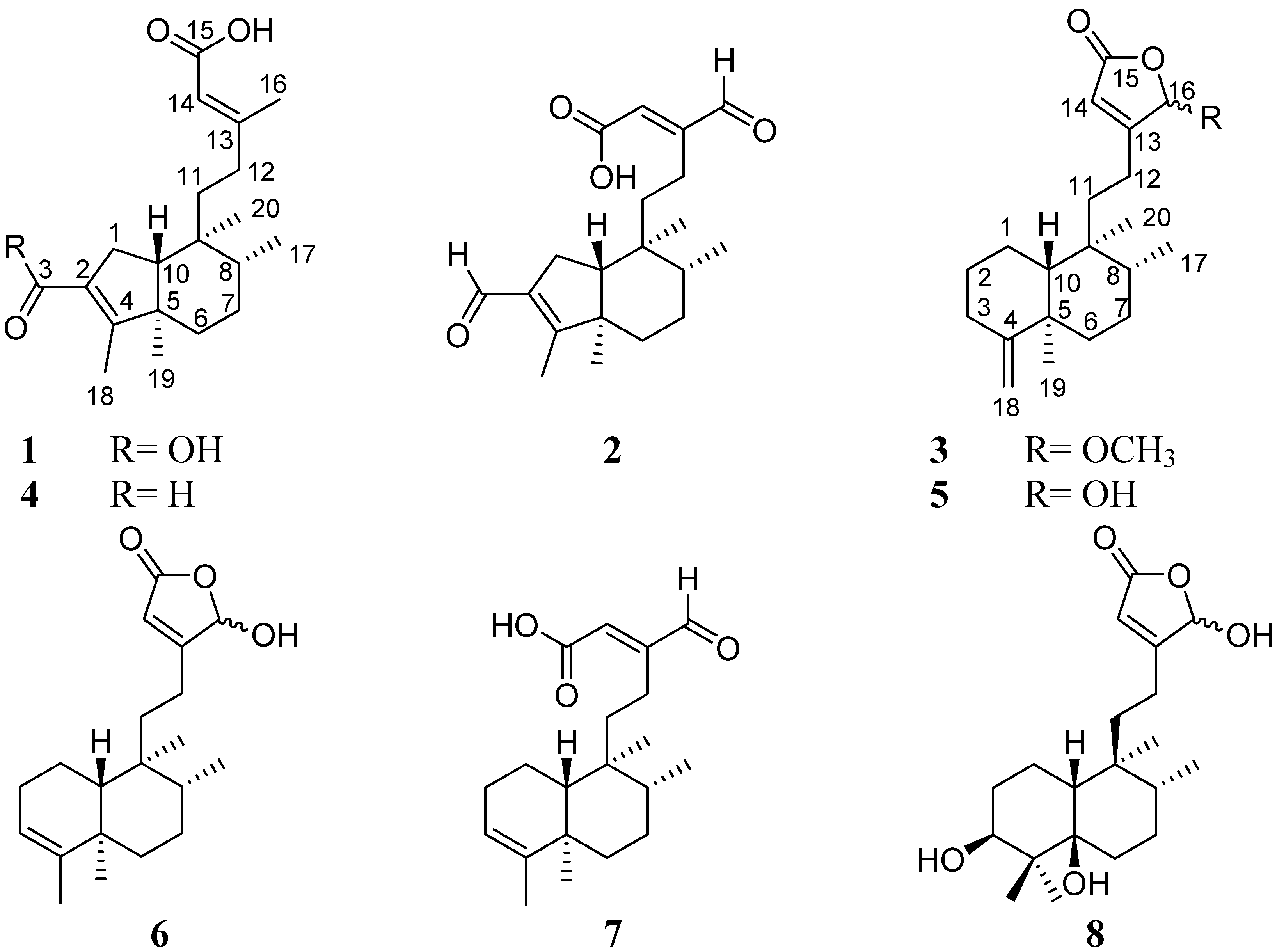
Three New Clerodane Diterpenes from Polyalthia longifolia var. pendula


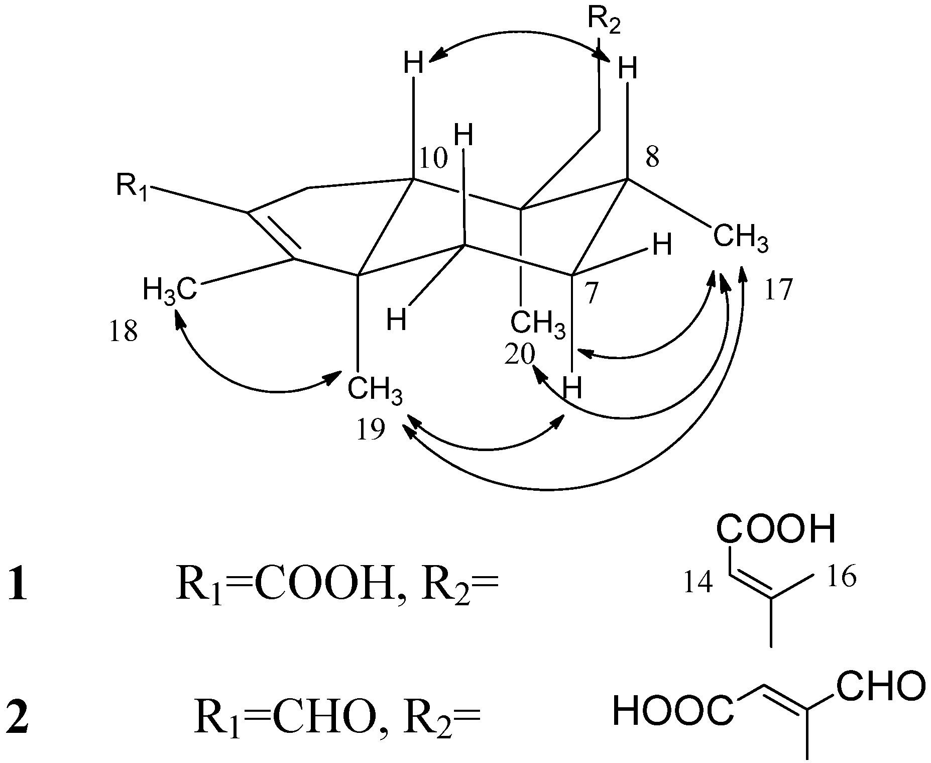
 and
and
Abstract
:1. Introduction

2. Results and Discussion

| No. | 1 a | 2 b | 3 c | ||||
|---|---|---|---|---|---|---|---|
| δC | δH | δC | δH | δC | δH | ||
| 1 | 29.3 | 2.30 dd (14.6, 6.0) | 25.5 | 2.64 m | 21.1 | 1.40–1.55 m | |
| 2.35 dd (14.6, 2.1) | 2.15 m | ||||||
| 2 | 126.7 | - | 137.3 | - | 32.4 | 2.05 m | |
| 2.24 m | |||||||
| 3 | 169.6 * | - | 189.8 | 9.95 s | 28.01/28.03 | 1.20 m | |
| 1.84 m | |||||||
| 4 | 164.9 | - | 174.9 | - | 159.0 | - | |
| 5 | 50.1 | - | 51.1 | - | 40.0 | - | |
| 6 | 34.4 | 1.37 td (11.5, 5.0) | 38.5 | 1.65 m | 36.8 | 1.45–1.58 m | |
| 1.67 m | 1.45 m | ||||||
| 7 | 28.1 | 1.57 m | 28.3 | 1.60 m | 26.9 | 1.40–1.55 m | |
| 1.60 m | |||||||
| 8 | 37.2 | 1.55 m | 37.1 | 1.64 m | 36.1 | 1.38–1.43 m | |
| 9 | 37.7 | - | 38.5 | - | 38.72/38.68 | - | |
| 10 | 54.1 | 1.69 dd (6.0, 2.1) | 53.8 | 1.71 m | 48.1 | 1.03 dt (12.0, 3.0 ) | |
| 11 | 38.1 | 1.47 td (8.5, 4.5) | 34.0 | 1.36 m | 34.00/34.02 | 1.40–1.60 m | |
| 12 | 34.5 | 2.07 m | 19.3 | 2.52 m | 20.7 | 2.02 m | |
| 2.12 m | 2.66 m | 2.23 m | |||||
| 13 | 160.2 | - | 154.7 | - | 168.9 | - | |
| 14 | 115.8 | 5.70 s | 135.3 | 6.49 s | 117.11/117.20 | 6.09 br s | |
| 15 | 169.4 * | - | 167.2 | - | 170.4 | - | |
| 16 | 17.7 | 2.15 s | 194.4 | 9.55 s | 104.0 | 5.91 s | |
| 17 | 14.0 | 0.87 d (6.0) | 14.9 | 0.92 d (6.5) | 15.8 | 0.77 d (6.5) | |
| 0.79 d (6.5) | |||||||
| 18 | 10.2 | 2.00 s | 9.9 | 2.08 s | 103.0 | 4.48 d (3.5) | |
| 19 | 15.9 | 0.95 s | 17.1 | 0.96 s | 20.5 | 1.02 s | |
| 20 | 17.1 | 0.92 s | 18.0 | 0.85 s | 17.7 | 0.74 s | |
| 16-OCH3 | 56.1 | 3.43 s/3.43 s | |||||
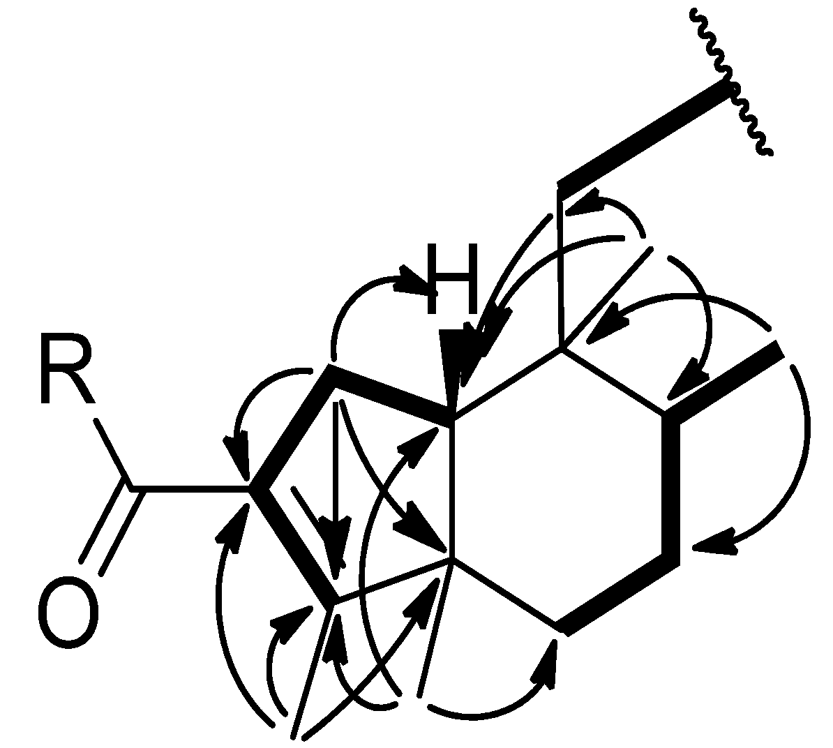
 ) and COSY (
  ) correlations of compounds 1–2.


3. Experimental
3.1. General
3.2. Plant Material
3.3. Extraction and Isolation
3.4. Spectral Data
 +2.0 (c 0.10, CHCl3), UV λmax (log ε) (MeOH) nm: 220 (3.24). IR νmax (KBr) cm−1: 3,361, 2,957, 2,922, 1,684, 1,654, 1,437, 1,208, 1,142. ESIMS (positive mode) m/z: 357.3 [M+Na]+. ESIMS (negative mode) m/z: 333.3 [M-H]−. HR-FAB-MS m/z: 335.2226 [M+H]+ (calcd. for C20H31O4 335.2222). 1H-NMR (CD3OD, 500 MHz) and 13C-NMR (CD3OD, 125 MHz), see Table 1. +13.3 (c 1.0, CHCl3), UV λmax (log ε) (MeOH) nm: 219 (2.78). IR νmax (KBr) cm−1: 2,924, 2,853, 1,687, 1,676, 1,639, 1,564, 1,383, 1,208, 1,183. ESIMS (positive mode) m/z: 355.2 [M+Na]+. ESIMS (negative mode) m/z: 331.3 [M-H]−. HR-FAB-MS m/z: 355.1880 [M+Na]+ (calcd for C20H28O4Na, 355.1885), 333.2060 [M+H]+ (calcd. for C20H29O4+, 333.2066). 1H-NMR (CDCl3, 500 MHz) and 13C-NMR (CDCl3, 125 MHz), see Table 1. −71.0 (c 0.1, MeOH), UV λmax (log ε) (MeOH) nm: 207 (4.32). IR νmax (KBr) cm−1: 2,955, 2,924, 2,853, 1,758, 1,680, 1,639, 1,456, 1,206. ESIMS (positive mode) m/z: 355.24 [M+Na]+. ESIMS (negative mode) m/z: 331.20 [M-H]−. HR-FAB-MS m/z: 355.2251 [M+Na]+ (calcd. for C21H32O3Na, 355.2249), 333.2427 [M+H]+ (calcd. for C21H33O3+, 333.2430). 1H-NMR (DMSO-d6, 500 MHz) and 13C-NMR (DMSO-d6, 125 MHz), see Table 1.
3.5. Cell Culture
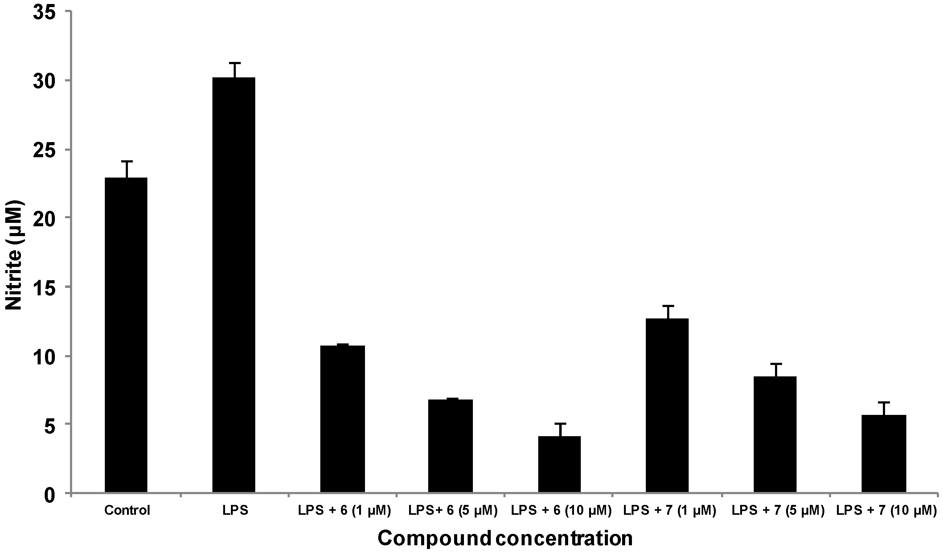
3.6. Detection of Nitric Oxide Expression by Griess Reaction
3.7. Statistical Analysis
4. Conclusions
Supplementary Materials
Acknowledgments
Author Contributions
Conflicts of Interest
References
- Ghosh, A.; Das, B.K.; Chatterjee, K.C.; Chandra, G. Antibacterial potentiality and phytochemical analysis of mature leaves of Polyalthia longifolia (Magnoliales: Annonaceae). South Pac. J. Nat. Sci. 2008, 26, 68–72. [Google Scholar] [CrossRef]
- Saleem, R.; Ahmed, M.; Ahmed, S.I.; Azeem, M.; Khan, R.A.; Rasool, N.; Saleem, H.; Noor, F.; Faizi, S. Hypotensive activity and toxicology of constituents from root bark of Polyalthia longifolia var. pendula. Phytother. Res. 2005, 19, 881–884. [Google Scholar] [CrossRef]
- Zhao, G.X.; Jung, J.H.; Smith, D.L.; Wood, K.V.; McLaughlin, J.L. Cytotoxic clerodane diterpenes from Polyalthia longifolia. Planta Med. 1991, 57, 380–383. [Google Scholar] [CrossRef]
- Chen, C.Y.; Chang, F.R.; Shih, Y.C.; Hsieh, T.J.; Chia, Y.C.; Tseng, H.Y.; Chen, H.C.; Chen, S.J.; Hsu, M.C.; Wu, Y.C. Cytotoxic constituents of Polyalthia longifolia var. pendula. J. Nat. Prod. 2000, 63, 1475–1478. [Google Scholar] [CrossRef]
- Marthanda Murthy, M.; Subramanyam, M.; Hima Bindu, M. Antimicrobial activity of clerodane diterpenoids from Polyalthia longifolia seeds. Fitoterapia 2005, 76, 336–339. [Google Scholar] [CrossRef]
- Sashidhara, K.V.; Singh, S.P.; Shukla, P.K. Antimicrobial evaluation of clerodane diterpenes from Polyalthia longifolia var. pendula. Nat. Prod. Commun. 2009, 4, 327–330. [Google Scholar]
- Chang, F.R.; Hwang, T.L.; Yang, Y.L.; Li, C.E.; Wu, C.C.; Issa, H.H.; Hsieh, W.B.; Wu, Y.C. Anti-inflammatory and cytotoxic diterpenes from formosan Polyalthia longifolia var. pendula. Planta Med. 2006, 72, 1344–1347. [Google Scholar] [CrossRef]
- Misra, R.; Pandey, R.C.; Dev, S. Higher isoprenoids-IX: Diterpenoids from the oleoresin of Hardwickia pinnata Part 2: Kolavic, kolavenic, kolavenolic and kolavonic acids. Tetrahedron 1979, 35, 979–984. [Google Scholar] [CrossRef]
- Hara, N.; Asaki, H.; Fujimoto, Y.; Gupta, Y.K.; Singh, A.K.; Sahai, M. Clerodane and ent-halimane diterpenes from Polyalthia longifolia. Phytochemistry 1995, 38, 189–194. [Google Scholar] [CrossRef]
- Hao, X.J.; Yang, X.S.; Zhang, Z.; Shang, L.J. Clerodane diterpenes from Polyalthia cheliensis. Phytochemistry 1995, 39, 447–448. [Google Scholar] [CrossRef]
- Phadnis, A.P.; Patwardhan, S.A.; Dhaneshwar, N.N.; Tavale, S.S. Clerodane diterpenoids from Polyalthia longifolia. Phytochemistry 1988, 27, 2899–2901. [Google Scholar] [CrossRef]
- Tori, M.; Katto, A.; Sono, M. Nine new clerodane diterpenoids from rhizomes of Solidago altissima. Phytochemistry 1999, 52, 487–493. [Google Scholar] [CrossRef]
- Bomm, M.D.; Zukerman-Schpector, J.; Lopes, L.M.X. Rearranged (4→2)-abeo-clerodane and clerodane diterpenes from Aristolochia chamissonis. Phytochemistry 1999, 50, 455–461. [Google Scholar]
- Bohlmann, F.; Singh, P.; Singh, R.K.; Joshi, K.C.; Jakupovic, J. A diterpene with a new carbon skeleton from Solidago altissima. Phytochemistry 1985, 24, 1114–1115. [Google Scholar] [CrossRef]
- Kijjoa, A.; Pinto, M.M.M.; Pinho, P.M.M.; Tantisewie, B.; Herz, W. Clerodane derivatives from Polyalthia viridis. Phytochemistry 1990, 29, 653–655. [Google Scholar] [CrossRef]
- Manabe, S.; Nishino, C. Stereochemistry of cis-clerodane diterpenes. Tetrahedron 1986, 42, 3461–3470. [Google Scholar] [CrossRef]
- Kijjoa, A.; Pinto, M.M.M.; Pinho, P.M.M.; Herz, W. Clerodanes from Polyalthia viridis. Phytochemistry 1993, 34, 457–460. [Google Scholar] [CrossRef]
- Aktan, F. iNOS-mediated nitric oxide production and its regulation. Life Sci. 2004, 75, 639–653. [Google Scholar] [CrossRef]
- Palmer, R.M.; Ferrige, A.G. Nitric oxide release accounts for the biological activity of endothelium-derived relaxing factor. Nature 1987, 327, 524–526. [Google Scholar] [CrossRef]
- Radomski, M.W.; Palmer, R.M.; Moncada, S. The anti-aggregating properties of vascular endothelium: Interactions between prostacyclin and nitric oxide. Br. J. Pharmacol. 1987, 92, 639–646. [Google Scholar] [CrossRef]
- Garthwaite, J. Glutamate, nitric oxide and cell-cell signalling in the nervous system. Trends Neurosci. 1991, 14, 60–67. [Google Scholar] [CrossRef]
- Stichtenoth, D.O.; Frolich, J.C. Nitric oxide and inflammatory joint diseases. Br. J. Rheumatol. 1998, 37, 246–257. [Google Scholar] [CrossRef]
- Lee, J.Y.; Kim, Y.J.; Kim, H.J.; Kim, Y.S.; Park, W. Immunostimulatory effect of laminarin on RAW 264.7 mouse macrophages. Molecules 2012, 17, 5404–5411. [Google Scholar] [CrossRef]
- Wang, S.G.; Xu, Y.; Chen, J.D.; Yang, C.H.; Chen, X.H. Astragaloside IV stimulates angiogenesis and increases nitric oxide accumulation via JAK2/STAT3 and ERK1/2 pathway. Molecules 2013, 18, 12809–12819. [Google Scholar] [CrossRef]
- Kobayashi, Y. The regulatory role of nitric oxide in proinflammatory cytokine expression during the induction and resolution of inflammation. J. Leukoc. Biol. 2010, 88, 1157–1162. [Google Scholar] [CrossRef]
- Tanna, A.; Nair, R.; Chanda, S. Assessment of anti-inflammatory and hepatoprotective potency of Polyalthia longifolia var. pendula leaf in Wistar albino rats. J. Nat. Med. 2009, 63, 80–85. [Google Scholar] [CrossRef]
- Shih, Y.T.; Hsu, Y.Y.; Chang, F.R.; Wu, Y.C.; Lo, Y.C. 6-Hydroxycleroda-3,13-dien-15,16-olide protects neuronal cells from lipopolysaccharide-induced neurotoxicity through the inhibition of microglia-mediated inflammation. Planta Med. 2010, 76, 120–127. [Google Scholar] [CrossRef]
- Lee, K.C.; Chang, H.H.; Chung, Y.H.; Lee, T.Y. Andrographolide acts as an anti-inflammatory agent in LPS-stimulated RAW264.7 macrophages by inhibiting STAT3-mediated suppression of the NF-κB pathway. J. Ethnopharmacol. 2011, 135, 678–684. [Google Scholar] [CrossRef]
- Sample Availability: Samples of all compounds in the manuscript are available from the authors.
© 2014 by the authors. Licensee MDPI, Basel, Switzerland. This article is an open access article distributed under the terms and conditions of the Creative Commons Attribution license ( http://creativecommons.org/licenses/by/3.0/).
Share and Cite
Wu, T.-H.; Cheng, Y.-Y.; Chen, C.-J.; Ng, L.-T.; Chou, L.-C.; Huang, L.-J.; Chen, Y.-H.; Kuo, S.-C.; El-Shazly, M.; Wu, Y.-C.; et al. Three New Clerodane Diterpenes from Polyalthia longifolia var. pendula. Molecules 2014, 19, 2049-2060. https://doi.org/10.3390/molecules19022049
Wu T-H, Cheng Y-Y, Chen C-J, Ng L-T, Chou L-C, Huang L-J, Chen Y-H, Kuo S-C, El-Shazly M, Wu Y-C, et al. Three New Clerodane Diterpenes from Polyalthia longifolia var. pendula. Molecules. 2014; 19(2):2049-2060. https://doi.org/10.3390/molecules19022049
Chicago/Turabian StyleWu, Tung-Ho, Yung-Yi Cheng, Chao-Jung Chen, Lean-Teik Ng, Li-Chen Chou, Li-Jiau Huang, Yung-Husan Chen, Sheng-Chu Kuo, Mohamed El-Shazly, Yang-Chang Wu, and et al. 2014. "Three New Clerodane Diterpenes from Polyalthia longifolia var. pendula" Molecules 19, no. 2: 2049-2060. https://doi.org/10.3390/molecules19022049