Article

Limonoids from the Fruits of Khaya ivorensis

1
CAS Key Laboratory of Tropical Plant Resource and Sustainable Use, Xishuangbanna Tropical Botanical Garden, Chinese Academy of Sciences, Menglun, Mengla, Yunnan 666303, China
2
University of Chinese Academy of Sciences, Beijing 100049, China
3
Engineering Research Center for the Development and Application of Ethnic Medicines and TCM, School of Pharmacy, Guiyang Medical College, 9 Beijing Road, Guiyang, Guizhou 550004, China
*
Authors to whom correspondence should be addressed.
Molecules 2014, 19(3), 3004-3011; https://doi.org/10.3390/molecules19033004
Submission received: 22 January 2014 / Revised: 27 February 2014 / Accepted: 28 February 2014 / Published: 7 March 2014
(This article belongs to the Section Natural Products Chemistry)

Abstract

:
Two new limonoids, namely 14,15-didehydroruageanin A (1) and 3-O-methyl- butyrylseneganolide A (2), were isolated from the fruits of Khaya ivorensis along with six known limonoids: seneganolide A (3), 1,3-dideacetylkhivorin (4), 7-deacetylkhivorin (5), 3-deacetylkhivorin (6), 1-deacetylkhivorin (7), and 3-deacetyl-7-oxokhivorin (8). All the compounds were evaluated for their cytotoxicity against five tumor cell lines.

Graphical Abstract

1. Introduction

Meliaceous limonoids, the major metabolites of the Meliaceae family, have attracted great interest in the natural products field due to their structural diversity and broad range of bioactivities [1,2]. Khaya A. Juss. (Meliaceae) is a genus of eight species mainly distributed in Africa and Madagascar [3]. Khaya ivorensis A. Chev. is a popular traditional African medicinal plant in this genus that is also cultivated in southern China [4,5,6]. Its ethanol stem bark extract has shown tissue toxicity, antimalarial and antiinflammatory activities [5]. Previous chemical investigation of K. ivorensis indicated that this plant was a good source of limonoids [6,7,8,9,10,11]. As a continuation of our studies of this medicinal plant in search of biologically significant secondary metabolites, the fruits of K. ivorensis were investigated. As a result, the EtOAc-soluble fraction of the ethanolic extract has produced after extensive column chromatography two new limonoids, 14,15-didehydroruageanin A (1), 3-O-methylbutyrylseneganolide A (2), and six known limonoids 38, of which compounds 3, 4, 7, and 8 were isolated in this plant for the first time (Figure 1). Their structures were established by NMR spectroscopic method and by comparison with literature data. The cytotoxic activities of all isolated compounds were also tested. Herein, the extraction, isolation, structure elucidation, and cytotoxic evaluation of these compounds are described.
Figure 1. The chemical structures of compounds 18.
Molecules 19 03004 g001

2. Results and Discussion

Compound 1, was obtained as a white amorphous powder. Its HREIMS revealed a molecular ion peak at m/z 554.2501 (calcd. 554.2516), consistent with a molecular formula of C31H38O9 possessing 13 degrees of unsaturation. The 1H- and 13C-NMR (with DEPT) spectra (Table 1) showed, in addition to resonances for an isobutyryl [δH 1.23 (3H, d, J = 7.1 Hz), 1.26 (3H, d, J = 7.1 Hz), 2.81 (1H, m); δC 19.2 CH3, 19.6 CH3, 34.9 CH, 176.0 C] [12] and a methoxycarbonyl group [δH 3.65 (3H, s); δC 52.31 OCH3, 174.3 C], signals for a ketone (δC 214.8 C), four tertiary methyls (δH 0.79 s, 0.90 s, 1.13 s, 1.22 s; δC 16.1 CH3, 21.60 CH3, 21.7 CH3, 22.9 CH3), a furan ring [δH 6.70 (1H, br s), 7.70 (1H, br s), 7.88 (1H, br s); δC 111.4 CH, 121.1 C, 143.1 CH, 144.2 CH], and an α,β-unsaturated δ-lactone [δH 6.66 (1H, s); δC 119.5 CH, 162.0 C, 164.6 C]. An epoxy group was also identified by the NMR data [δH 4.51 (1H, d, J = 2.0); δC 61.5 C, 62.7 CH]. The above signals accounted for seven out of the 13 degrees of unsaturation and suggested the compound to thus be hexacyclic. These observations indicated that compound 1 was possibly a mexicanolide-type limonoid [12]. 2D-NMR correlation analysis (Figure 2) further confirmed this conclusion and allowed establishment of the full structure for 1. In particular, HMBC correlation from H2-6 to the carboxyl at δC 174.3 located the methoxycarbonyl group at C-6, while that from H-3 to the isobutyryl carbonyl placed the isobutyryloxy group at C-3. The epoxy group was further revealed to be an 8,30-epoxy by 1H-1H COSY correlations between H-30/H-2/H-3 and HMBC correlations H-30/C-8, H-9/C-8, and H-15/C-8 (Figure 2).
Table 1. 1H- and 13C-NMR of 1 and 2.
12
δC, type aδH (J in Hz) bδC, type aδH (J in Hz) b
1214.8, s 214.8, s
249.7, d3.92 dd (9.3, 2.0)49.9, d3.94 ddd (9.0, 6.1, 1.3)
377.6, d5.39 d (9.3)79.1, d5.14 d (9.2)
440.3, s 39.3, s
542.6, d3.67 d (2.0)41.2, d3.57 dd (7.2, 4.7)
633.6, t2.44 m33.5, t2.55 m
7174.3, s 174.5, s
861.5, s 136.9, s
956.3, d2.11 br s54.1, d2.46 m
1049.0, s 52.5, s
1121.59, tα 1.22 m, β 1.65 m22.1, tα 1.81 m, β 1.66 m
1233.2, tα 1.26 m, β 2.11 m32.9, tα 1.19 m, β 1.95 m
1339.7, s 38.0, s
14162.0, s 161.1, s
15119.5, d6.66 s113.3, d6.57 s
16164.6, s 165.3, s
1780.0, d5.57 s80.1, d5.20 s
1821.7, q1.22 s22.2, q1.08 s
1916.1, q1.13 s16.1, q1.30 s
20121.1, s 121.6, s
21143.1, d7.88 br s142.6, d7.81 s
22111.4, d6.70 br s111.3, d6.66 d (1.0)
23144.2, d7.70 br s144.1, d7.67 t (1.6)
2822.9, q0.90 s23.0, q0.93 s
2921.60, q0.79 s21.4, q0.84 s
3062.7, d4.51 d (2.0)129.8, d6.55, dd (6.0, 2.9)
7-OMe52.3, q3.65 s52.3, q3.69 s
3-acyl-1'176.0, s 172.7, s
2'34.9, d2.81 m43.4, t2.35 m
3'19.6, q1.26 d (7.1)26.0, d2.19 dt (13.7, 6.8)
4'19.2, q1.23 d (7.1)22.82, q0.95 d (6.7)
5' 22.85, q0.92 d (6.7)
a Recorded at 150 MHz in pyridine-d5; b Recorded at 600 MHz in pyridine-d5.
The relative configuration of 1 was established by ROESY correlation analysis (Figure 2a). The ROESY correlations of H-9/H3-19 and H-9/H3-18 indicated that H-9 was α-oriented, while those of H-5/H-11β, H-5/H3-28, and H-17/H-11β showed H-5 and H-17 were β-oriented. The ROESY correlation of H3-29/H-3 suggested that H-3 was α-oriented. The close coupling constant of H-3 in 1 (J = 9.3 Hz) and 2'R-cipadesin A (J = 9.5 Hz) [13], whose structure has been confirmed by X-ray crystal analysis [14], indicated that both compounds shared the same stereochemistry for H-2 and H-3 and H-2 was assigned to be α-oriented. The 8,30-epoxy was then determined to be α-oriented on the basis of the small coupling constant of H-30 (J = 2.0 Hz) [13]. Therefore, the relative stereochemistry of 1 was established as shown in Figure 1 and it was named 14,15-didehydroruageanin A.
Figure 2. 1H-1H COSY (bold) and selected HMBC correlations of 1 (a); Selected key ROESY correlations of 1 (b).
Molecules 19 03004 g002
Compound 2, was obtained as a white amorphous powder. Its molecular formula C32H40O8 was established by the HREIMS ion peak at m/z 552.2715 (calcd. 552.2723). Analysis of its 1H- and 13C-NMR data (Table 1) showed that 2 was also a mexicanolide-type compound, very similar to the known compound seneganolide A [15]. The only differences in the NMR data of the two compounds was the upfield-shift of the signals for C-3 (δC from 80.2 to 79.1) and the appearance of additional signals for a 3-methylbutyryl group [δH 0.92 (3H, d, J = 6.7 Hz), 0.95 (3H, d, J = 6.7 Hz), 2.19 (1H, dt, J = 13.7, 6.8 Hz), 2.35 (2H, m); δC 22.82 CH3, 22.85 CH3, 26.0 CH, 43.4 CH2, 172.7 C], which suggested that compound 2 was the 3-O-methylbutyryl derivative of seneganolide A. 1H-1H COSY correlations H-4'/H-3'/H-2' and the HMBC correlation H-3/C-1' (Figure 3a) further confirmed this conclusion. ROESY correlation analysis (Figure 3b) also supported that both compounds shared the same stereochemistry. Thus, the relative structure of 2 was elucidated as shown in Figure 1 and it was named 3-O-methylbutyrylseneganolide A.
Figure 3. 1H-1H COSY (bold) and selected HMBC correlations of 2 (a); Selected key ROESY correlations of 2 (b).
Molecules 19 03004 g003
Six known limonoids were also isolated and identified by spectroscopic methods to be seneganolide A (3) [15], 1,3-dideacetylkhivorin (4) [16], 7-deacetylkhivorin (5) [7], 3-deacetylkhivorin (6) [11], 1-deacetylkhivorin (7) [17], and 3-deacetyl-7-oxokhivorin (8) [18]. Among these known limonoids, compounds 3, 4, 7, and 8 were obtained from K. ivorensis for the first time.
Limonoids 18 were all evaluated for their cytotoxic activities against five human tumor cell lines: myeloid leukemia (HL-60), hepatocellular carcinoma (SMMC-7721), lung cancer (A-549), breast cancer (MCF-7), and colon cancer (SW480). However, only compounds 2, 3, and 4 exhibited cytotoxicity against certain tumor cell lines with the IC50 values in the range of 21.1–39.5 μM (Table 2).
Table 2. The cytotoxicity (IC50 μM) of isolated compounds 18.
CompoundHL-60SMMC-7721A-549MCF-7SW480
1>40>40>40>40>40
2>40>4037.3>40>40
321.221.123.8>4032.6
4>40>4039.526.1>40
5>40>40>40>40>40
6>40>40>40>40>40
7>40>40>40>40>40
8>40>40>40>40>40
Cisplatin a1.14.56.613.111.1
a Positive control.

3. Experimental

3.1. General Information

Optical rotations were obtained with a JASCO P-1020 polarimeter. UV spectra were measured with a Shimadzu UV 2401PC. IR spectra (KBr) were determined on a Bruker Tensor-27 infrared spectrophotometer. 1D- and 2D-NMR spectra were recorded on Bruker AM-400, Bruker DRX-500 and Avance III 600 spectrometers with TMS as an internal standard (Karlsruhe, Germany). ESIMS and HREIMS recorded on a Xevo TQ-S mass spectrometer (Manchester, UK) and a Waters AutoSpec Premier P776 instrument (Milford, CT, USA), respectively. Semi-preparative HPLC was carried out using a Waters system (Milford, CT, USA) consisting of a 600 pump and a 2996 Photodiode Array Detector. Silica gel (200–300 mesh, Qingdao Marine Chemical Factory, Qingdao, China), Sephadex LH-20 gel (40–70 μM, Amersham Pharmacia Biotech AB, Uppsala, Sweden), and MCI gel (CHP20/P120, 75–150 μM, high porous polymer, Mitsubishi Chemical Corporation, Japan) were used for column chromatography (CC).

3.2. Plant Material

The fruits of K. ivorensis were collected from Xishuangbanna Tropical Botanical Garden (XTBG), Chinese Academy of Science (CAS), Mengla Country, Yunnan Province, People’s Republic of China, in July 2013 and were identified by one of the authors (Y.-K. Xu). A voucher specimen (No. 028765) was deposited in the herbarium of XTBG.

3.3. Extraction and Isolation

The air-dried and powdered fruits of K. ivoremsis (4.0 kg) were extracted three times (each for 7 days) with EtOH–H2O (95/5, v/v, 30 L) at room temperature. Removal of solvent from the combined extracts under vacuum afforded a crude residue (200 g). The residue was then suspended in H2O and partitioned with EtOAc (60 g). The EtOAc-soluble fraction was subjected to silica gel CC (petroleum ether-acetone from 1/0 to 0/1, v/v) to produce four fractions (1–4). Fraction 1 (5.0 g) was subjected to MCI gel CC (MeOH–H2O from 1/6 to 1/1, v/v) to give 4 (12 mg), 5 (5 mg), and 6 (7 mg). Fraction 2 (12.5 g) was subjected to silica gel CC to obtain 3 (600 mg) and sub-fraction 2A (10 mg). Fraction 2A was then purified by semi-preparative HPLC (MeOH–H2O from 2/3 to 7/3, v/v) to yield 2 (4 mg). Fraction 3 (2.0 g) was separated by Sephadex LH-20 CC eluted with MeOH–H2O (from 1/4 to 1/1, v/v) and further purified by semi-preparative HPLC to give 1 (3 mg). Fraction 4 (12.0 g) was applied to a silica gel column and eluted with petroleum ether-acetone (from 1/9 to 1/1, v/v) to give 7 (800 mg) and a sub-fraction 4A (50 mg), purification of which by Sephadex LH-20 CC (MeOH–H2O from 3/7 to 3/2, v/v) and semi-preparative HPLC (CH3CN–H2O from 2/3 to 7/3, v/v) yielded 8 (4 mg).

3.4. Spectral Data

14,15-Didehydroruageanin A (1). White amorphous powder; [α]20.5D +9.3 (c 0.045, MeOH); UV (MeOH) λmax nm (log ε) 204.8 (4.23); IR (KBr) νmax (cm−1): 2972, 2931, 1732, 1634, 1460, 1384, 1271, 1197, 1114, 1028; 1H- and 13C-NMR data, see Table 1; positive ESIMS m/z 555 [M+H]+; HREIMS m/z 554.2501 M (calcd for C31H38O9, 554.2516).
3-O-Methylbutyrylseneganolide A (2). White amorphous powder; [α]15.4D +51.0 (c 0.076, MeOH); UV (MeOH) λmax nm (log ε) 203 (4.08), 282.4 (3.89); IR (KBr) νmax (cm−1): 2961, 1630, 1463, 1384, 1295, 1256, 1167, 1119, 1107, 1028, 1001; 1H- and 13C-NMR data, see Table 1; positive ESIMS m/z 553 [M+H]+; HREIMS m/z 552.2715 M (calcd for C32H40O8, 552.2723).

3.5. Cytotoxicity Assay

The MTT method [19] was used for assessing the cytotoxicity of all isolated compounds against the five tumor cell lines (HL-60 human myeloid leukemia, SMMC-7721 hepatocellular carcinoma, A-549 lung cancer, MCF-7 breast cancer, and SW480 colon cancer) with cisplatin as the positive control.

4. Conclusions

In summary, three mexicanolide-type limonoids, 14,15-didehydroruageanin A (1), 3-O-methylbutyrylseneganolide A (2), and seneganolide A (3), along with five known d-seco limonoids, 1,3-dideacetylkhivorin (4), 7-deacetylkhivorin (5), 3-deacetylkhivorin (6), 1-deacetylkhivorin (7) and 3-deacetyl-7-oxokhivorin (8), were isolated from the fruits of Khaya ivorensis. Among these compounds, 1 and 2 were new compounds; and compounds 3, 4, 7, and 8 were obtained from this plant for the first time. The cytotoxicity evaluation showed that only compounds 2, 3, and 4 exhibited cytotoxicity against certain tumor cell lines.

Supplementary Materials

Supplementary materials can be accessed at: https://www.mdpi.com/1420-3049/19/3/3004/s1.

Acknowledgments

NSFC-U1302222, CAS 135 program (XTBG-F02), and Ethnobotanical Investigation of Plants for Industrialization in Southwest China (2012FY110300) are gratefully acknowledged.

Author Contributions

YKX, PZ, and KLJ designed research; KLJ, SGL, ZN, HBH, and XLZ performed research and analyzed the data; KLJ, SGL, and YKX wrote the paper. All authors read and approved the final manuscript.

Conflictts of Interest

The authors declare no conflict of interest.

References

  1. Fang, X.; Di, Y.T.; Hao, X.J. The advances in the limonoid chemistry of the Meliaceae family. Curr. Org. Chem. 2011, 15, 1363–1391. [Google Scholar] [CrossRef]
  2. Tan, Q.G.; Luo, X.D. Meliaceous limonoids: Chemistry and biological activities. Chem. Rev. 2011, 111, 7437–7522. [Google Scholar] [CrossRef]
  3. Chen, S.K.; Li, H.; Chen, B.Y. In Zhongguo Zhiwu Zhi; Science Press: Beijing, China, 1997; pp. 46–47. [Google Scholar]
  4. Zhang, B.; Wang, Y.; Yang, S.P.; Zhou, Y.; Wu, W.B.; Tang, W.; Zuo, J.P.; Li, Y.; Yue, J.M. Ivorenolide A, an unprecedented immunosuppressive macrolide from Khaya ivorensis: Structural elucidation and bioinspired total synthesis. J. Am. Chem. Soc. 2012, 134, 20605–20608. [Google Scholar]
  5. Agbedahunsi, J.; Fakoya, F.; Adesanya, S. Studies on the anti-inflammatory and toxic effects of the stem bark of Khaya ivorensis (Meliaceae) on rats. Phytomedicine 2004, 11, 504–508. [Google Scholar] [CrossRef]
  6. Zhang, B.; Yang, S.P.; Yin, S.; Zhang, C.R.; Wu, Y.; Yue, J.M. Limonoids from Khaya ivorensis. Phytochemistry 2009, 70, 1305–1308. [Google Scholar] [CrossRef]
  7. Adesogan, E.K.; Taylor, D.A.H. Limonoid extractives from Khaya ivorensis. J. Chem. Soc. C 1970, 12, 1710–1714. [Google Scholar] [CrossRef]
  8. Adesida, G.; Adesogan, E.; Okorie, D.; Taylor, D.; Styles, B. The limonoid chemistry of the genus Khaya (Meliaceae). Phytochemistry 1971, 10, 1845–1853. [Google Scholar] [CrossRef]
  9. Taylor, D.A.H. The structure of an extractive from Khaya ivorensis. Phytochemistry 1977, 16, 1847–1849. [Google Scholar] [CrossRef]
  10. Vanucci, C.; Lange, C.; Lhommet, G.; Dupont, B.; Davoust, D.; Vauchot, B.; Clement, J.L.; Brunck, F. An insect antifeedant limonoid from seed of khaya ivorensis. Phytochemistry 1992, 31, 3003–3004. [Google Scholar] [CrossRef]
  11. Abdelgaleil, S.A.M.; Hashinaga, F.; Nakatani, M. Antifungal activity of limonoids from Khaya ivorensis. Pest Manag. Sci. 2005, 61, 186–190. [Google Scholar]
  12. Li, M.Y.; Yang, X.B.; Pan, J.Y.; Feng, G.; Xiao, Q.; Sinkkonen, J.; Satyanandamurty, T.; Wu, J. Granatumins A–G, limonoids from the seeds of a Krishna Mangrove, Xylocarpus granatum. J. Nat. Prod. 2009, 72, 2110–2114. [Google Scholar] [CrossRef]
  13. Gan, L.S.; Wang, X.N.; Wu, Y.; Yue, J.M. Tetranortriterpenoids from Cipadessa baccifera. J. Nat. Prod. 2007, 70, 1344–1347. [Google Scholar] [CrossRef]
  14. Siva, B.; Poornima, B.; Venkanna, A.; Prasad, K.R.; Sridhar, B.; Nayak, V.L.; Ramakrishna, S.; Babu, K.S. Methyl angolensate and mexicanolide-type limonoids from the seeds of Cipadessa baccifera. Phytochemistry 2014, 98, 174–182. [Google Scholar] [CrossRef]
  15. Abdelgaleil, S.A.M.; Iwagawa, T.; Doe, M.; Nakatani, M. Antifungal limonoids from the fruits of Khaya senegalensis. Fitoterapia 2004, 75, 566–572. [Google Scholar] [CrossRef]
  16. Akisanya, A.; Arene, E.; Bevan, C.; Ekong, D.; Nwaji, M.; Okogun, J.; Powell, J.; Taylor, D. West African timbers. Part XII. The inter-relation of gedunin and khivorin. J. Chem. Soc. C 1996, 506–509. [Google Scholar]
  17. Bickii, J.; Njifutie, N.; Foyere, J.A.; Basco, L.K.; Ringwald, P. In vitro antimalarial activity of limonoids from Khaya grandifoliola C.D.C. (Meliaceae). J. Ethnopharmacol. 2000, 69, 27–33. [Google Scholar] [CrossRef]
  18. Tchimene, M.K.; Ngamga, D.; Tane, P.; Sterner, O.; Connolly, J.D. 3,7-Dideacetyl-6 alpha-hydroxykhivorin, a new limonoid from Khaya senegalensis (Meliaceae). Bull. Chem. Soc. Ethiop. 2006, 20, 69–73. [Google Scholar]
  19. Mosmann, T. Rapid colorimetric assay for cellular growth and survival: Application to proliferation and cytotoxicity assays. J. Immunol. Methods 1983, 65, 55–53. [Google Scholar] [CrossRef]
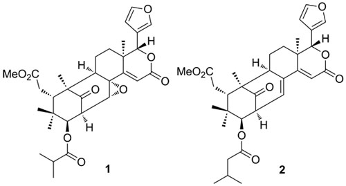
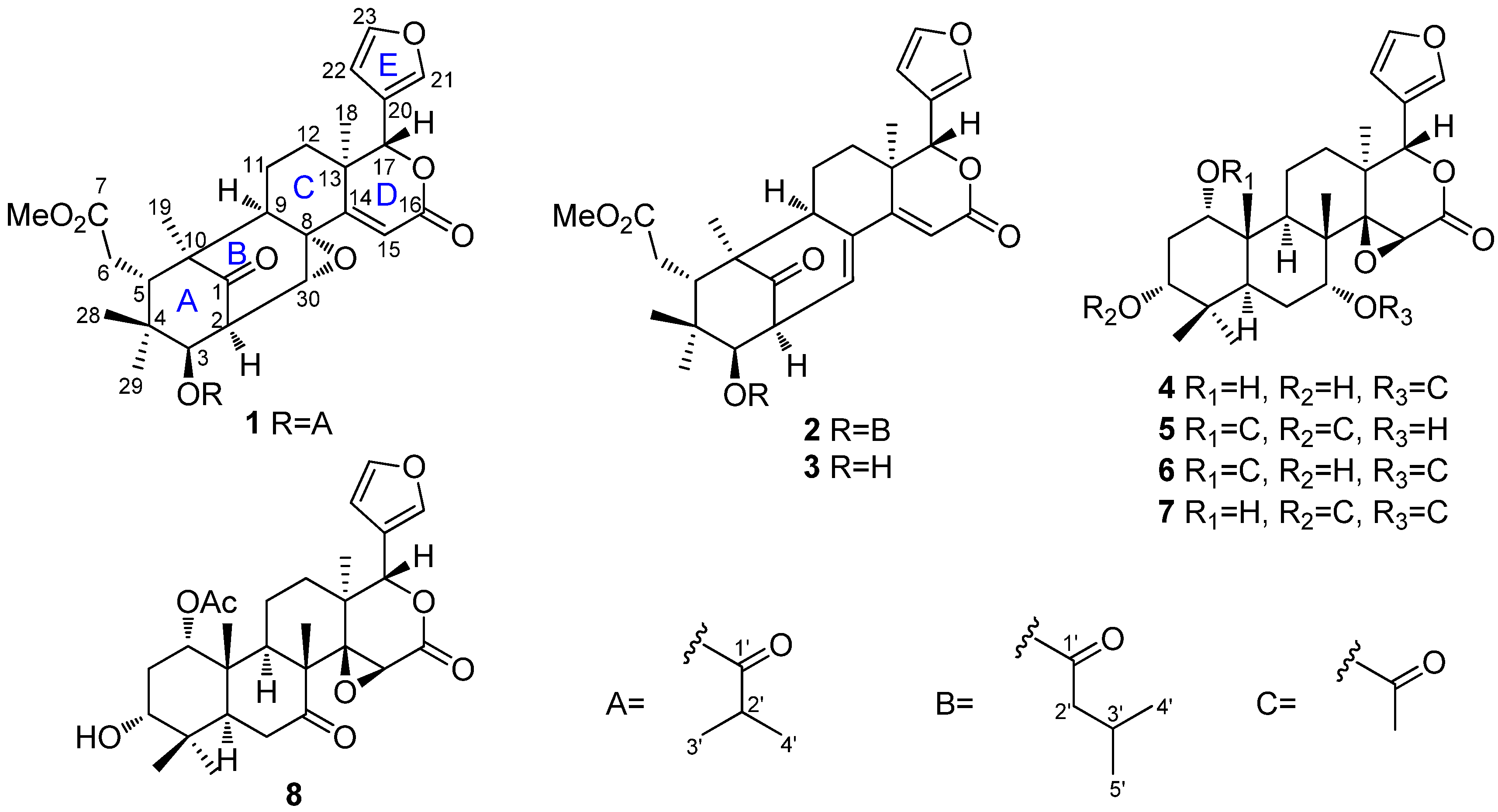
  • Sample Availability: Samples of the compounds 18 are available from the authors.

