Article

Additional New Minor Cucurbitane Glycosides from Siraitia grosvenorii

by
Indra Prakash
1,* and
Venkata Sai Prakash Chaturvedula
2
1
The Coca-Cola Company, Organic Chemistry Department, Global Research and Development, One Coca-Cola Plaza, Atlanta, GA 30313, USA
2
Natural Ingredient Development, Blue California, 30111 Tomas, Rancho Santa Margarita, CA 92688, USA
*
Author to whom correspondence should be addressed.
Molecules 2014, 19(3), 3669-3680; https://doi.org/10.3390/molecules19033669
Submission received: 25 February 2014 / Revised: 14 March 2014 / Accepted: 17 March 2014 / Published: 24 March 2014
(This article belongs to the Section Natural Products Chemistry)

Abstract

:
Continuous phytochemical studies of the crude extract of Luo Han Guo (Siraitia grosvenorii) furnished three additional new cucurbitane triterpene glycosides, namely 11-deoxymogroside V, 11-deoxyisomogroside V, and 11-deoxymogroside VI. The structures of all the isolated compounds were characterized on the basis of extensive NMR and mass spectral data as well as hydrolysis studies. The complete 1H- and 13C-NMR spectral assignments of the three unknown compounds are reported for the first time based on COSY, TOCSY, HSQC, and HMBC spectroscopic data.

1. Introduction

The fruit of Siraitia grosvenorii (Momordica grosvenorii; Cucurbitaceae), known as Luo Han Guo, has been used for centuries in Traditional Chinese medicine for the treatment of dry cough, sore throat, and minor stomach and intestinal troubles [1,2,3,4]. An intriguing characteristic of Luo Han Guo that is well known now throughout the world is its intense sweet taste and it has been used as a non-caloric natural sweetener in some countries [5]. The extract from this fruit has been reported to be about 150 times sweeter than sucrose. The non-caloric sweet taste of S. grosvenorii fruit results primarily from the content of a group of cucurbitane-type triterpene glycosides also known as mogrosides that are present at about 1% in the flesh of the fruit. Early chemical investigation of Luo Han Guo resulted in the isolation of various triterpenoid glycosides and polyphenols [1,2,3,4,5].
In continuation of our study on the isolation of natural sweeteners from S. grosvenorii [6,7,8,9], we have investigated a commercial aqueous alcoholic extract of Luo Han Guo, which resulted in the isolation of three new curcubitane glycosides, 11-deoxymogroside V (1), 11-deoxyisomogroside V (2), and 11-deoxymogroside VI (3) (Figure 1). This paper describes the isolation, purification, and structure elucidation of these three new mogrosides 13 based on the extensive NMR and mass spectroscopic as well as hydrolysis studies. The complete assignments of the 1H- and 13C-NMR values of the isolated compounds were made on the basis of COSY, TOCSY, HSQC, and HMBC spectral data as well as comparison with the spectral data of known compounds 46.
Figure 1. Structures of 11-deoxymogroside V (1), 11-deoxyisomogroside V (2), 11-deoxymogroside VI (3), mogroside V (4), isomogroside V (5), and mogroside VI (6).
Molecules 19 03669 g001

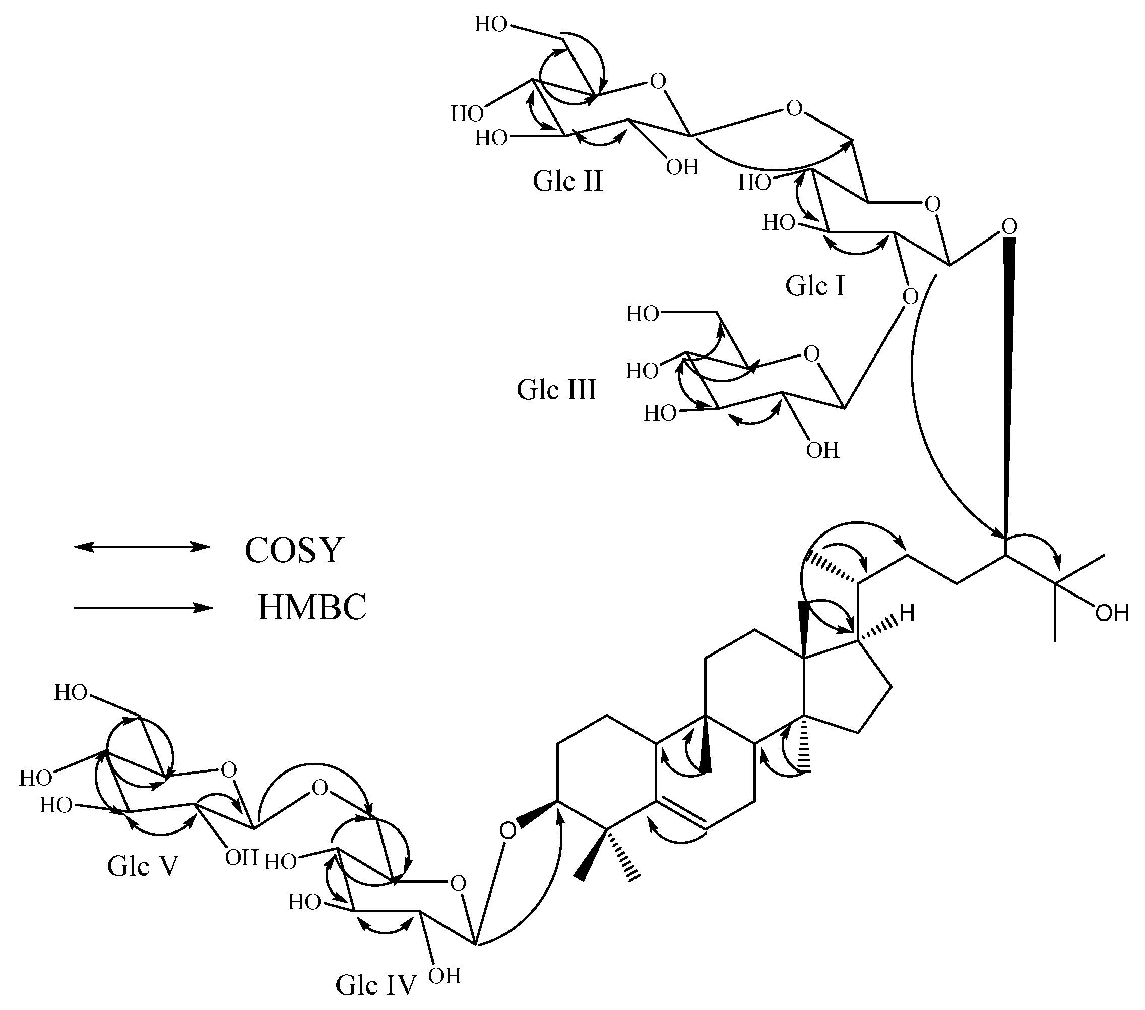
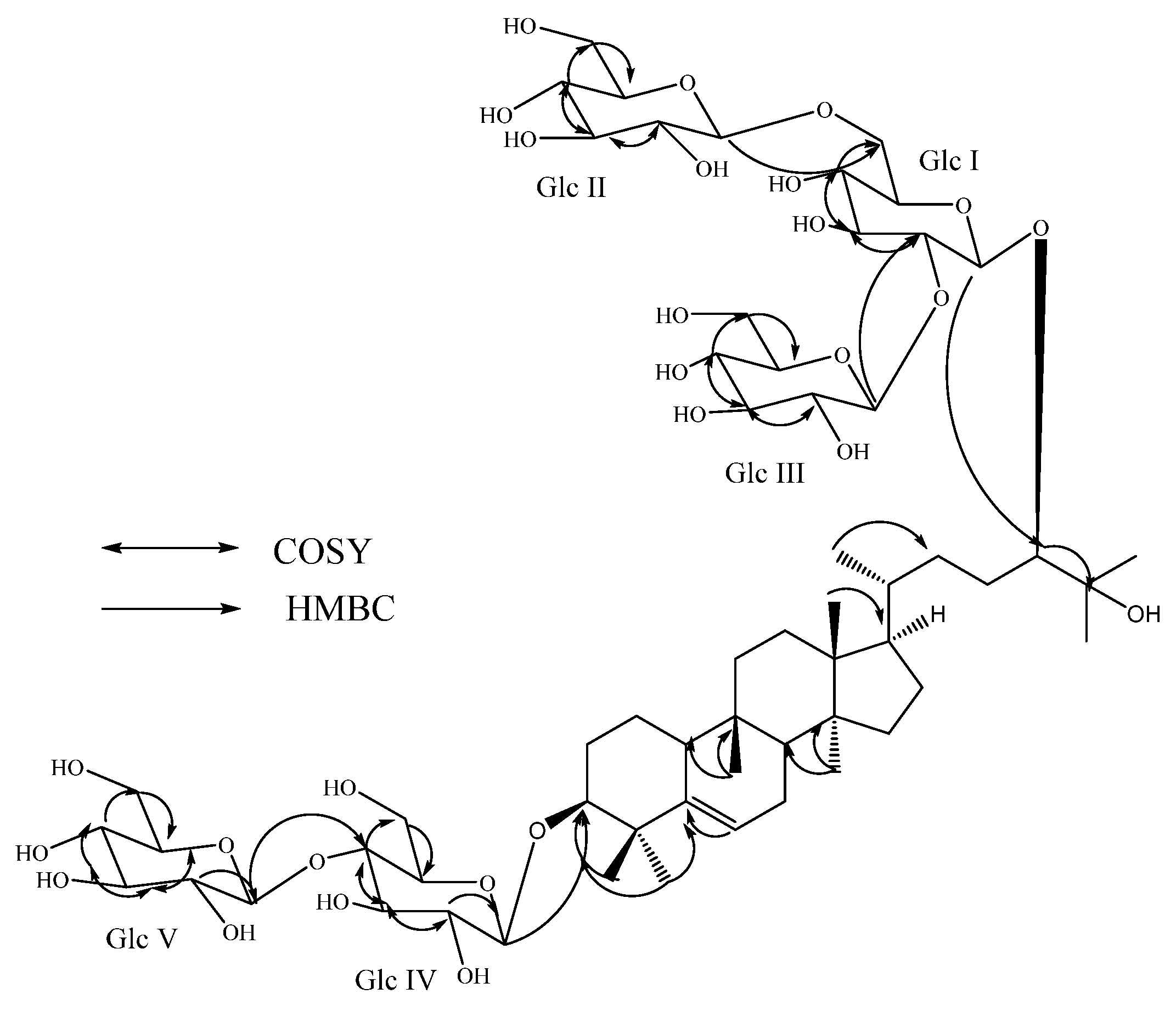
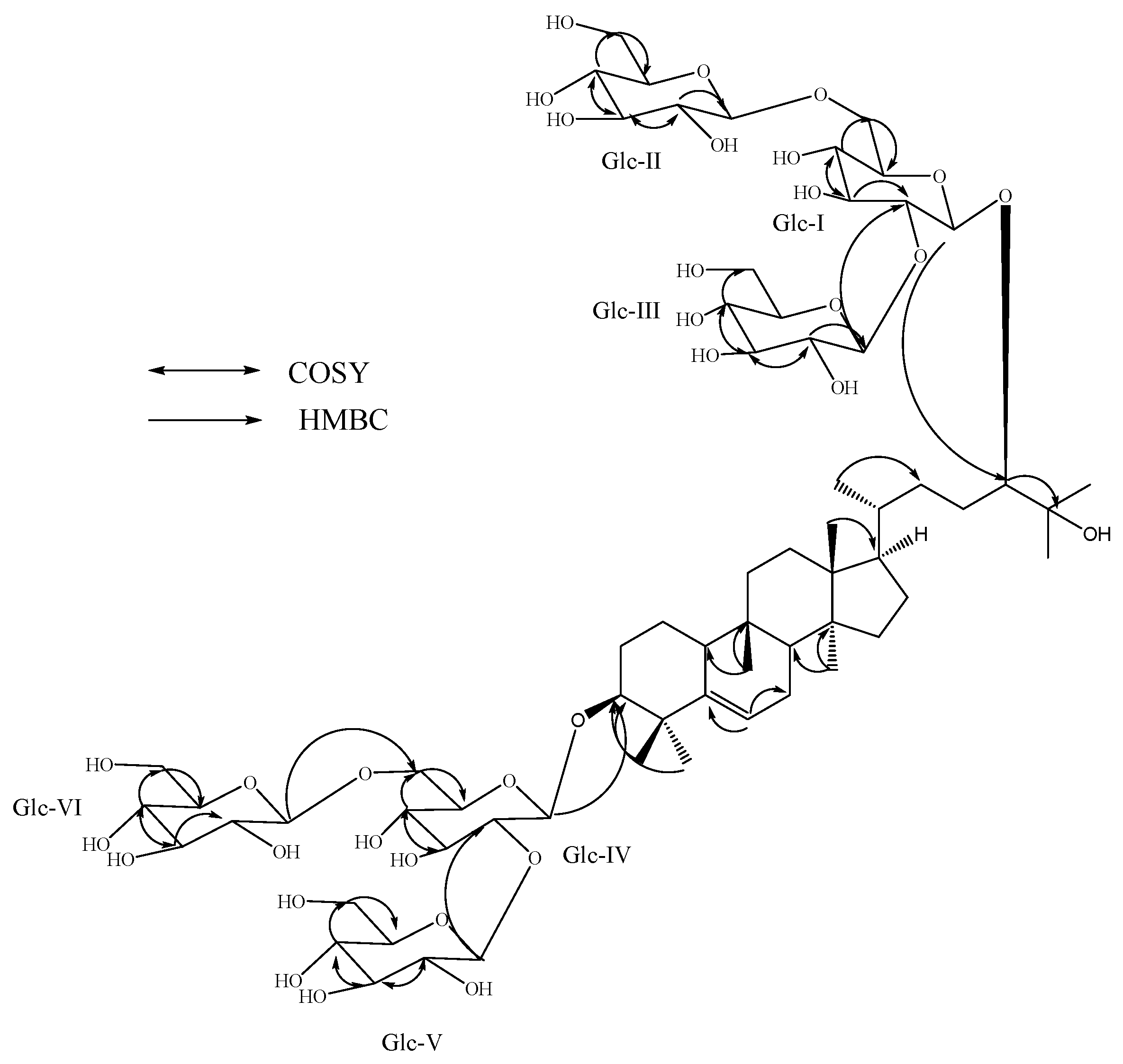
2. Results and Discussion
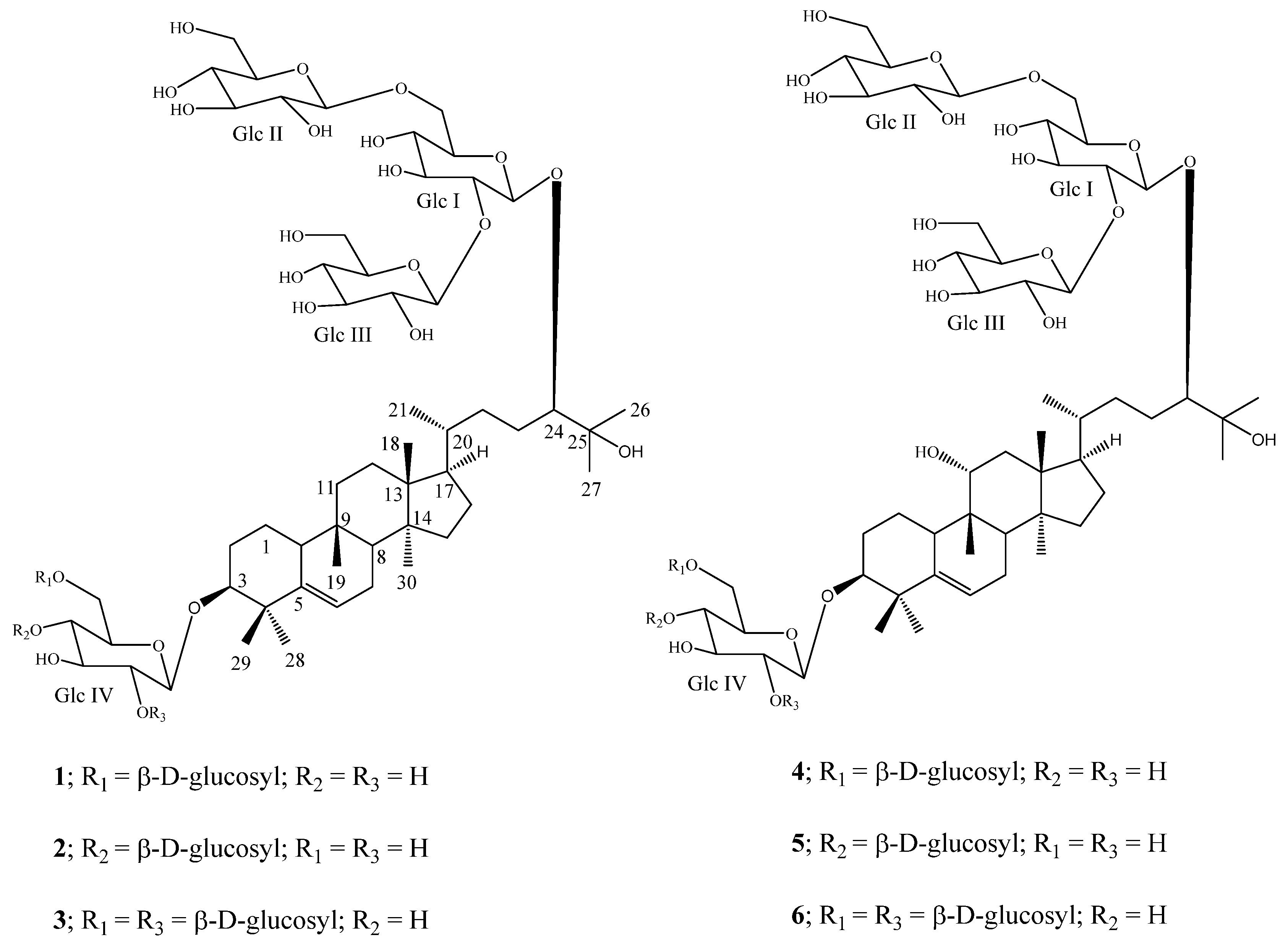
Compound 1 was isolated as a white powder and its molecular formula was deduced as C60H102O28 from the positive ESI Time of Flight (TOF) mass spectrum (MS) studies which showed an [M+H]+ ion at m/z 1,271.6649. The 1H-NMR spectrum of 1 showed the presence of seven methyl singlets at δ 0.84, 0.85, 0.91, 1.09, 1.39, 1.51, and 1.52; a secondary methyl signal as a doublet at 1.11 (J = 6.0 Hz); nine sp3 methylene and five sp3 methine groups between δ 1.09–2.54. The 1H-NMR spectrum of 1 also showed the presence of two oxymethine protons at δ 3.73 and 3.80; an olefinic proton corresponding to a trisubstituted double bond at δ 5.49. The 13C-NMR spectrum of 1 showed a tertiary hydroxy group resonating at δC 72.9. The above spectral data is characteristic to the aglycone moiety of the triterpenoid mogrol, isolated earlier from S. grosvenorii [5,6]. The 1H-NMR spectrum of 1 also showed the presence of five anomeric protons at δ 4.84, 4.85, 4.93, 5.18, and 5.56, indicating the presence of five hexose moieties. This was confirmed by the MS/MS spectrum of 1, selecting the [M-H] ion at m/z 1,269 for fragmentation, indicated the loss of five sugar moieties at m/z 1,107.5968, 945.5433, 783.4903, 621.4369, and 459.3816 having a difference of 162 atomic mass units, indicative of the presence of these five hexose moieties. Acid hydrolysis of 1 with 5% H2SO4 afforded glucose, which was identified by direct comparison with an authentic sample by TLC [10,11,12], suggested the presence of five glucosyl moieties in its structure. The configuration of the sugars present in 1 was identified by preparing the corresponding thiocarbamoyl-thiazolidine carboxylate derivatives and comparison of their retention times with the standard sugars as described in the literature [13]. The placement of the five sugar units in 1 was confirmed as 1→6 linked β-D-glucobiosyl substituent at C-3 and 2,6-branched β-D-glucotriosyl moiety at C-24 were identified on the basis of the key COSY, and HMBC correlations as shown in Figure 2.
Figure 2. Key COSY and HMBC correlations of 11-deoxymogroside V (1).
Molecules 19 03669 g002
The large coupling constants observed for the five D-glucose anomeric protons suggested the β-orientation, as reported for mogrol glycosides. A series of 1D TOCSY experiments selecting each anomeric proton resonating at δ 4.84, 4.85, 4.93, 5.18, and 5.56 together with their HMBC correlations helped in assigning the 1H-NMR spectral data for the sugar units in 1. The 1H- and 13C-NMR values for all the protons and carbons were assigned on the basis of COSY, TOCSY, HSQC and HMBC correlations and are given in Table 1. A close comparison of the NMR and MS spectral data of 1 with mogroside V (4) [6] suggested that the structure of 1 is identical to 4 except for the absence of the 11-hydroxyl group. The stereochemistry of various chiral centers in 1 was considered similar to 4 based on identical 1H- and 13C-NMR values and multiplicities at the corresponding positions [6,14,15,16]. Thus, the structure of 1 was deduced as 11-deoxymogroside V.
Table 1. 1H- and 13C-NMR chemical shift values (δ, ppm) for the compounds 13 in pyridine-d5/D2O (10:1) a–c.
Position123
1H13C1H13C1H13C
11.62 m
1.85 m
22.61.51 m
1.80 m
22.61.61 m
1.92 m
22.5
21.97 m
2.54 br d (12.0)
28.91.91 m
2.38 br d (12.0)
28.91.97 m
2.43 br d (11.0)
28.8
33.73 br s (W1/2 = 7.5)87.53.65 br s (W1/2 = 6.8)87.73.72 br s (W1/2 = 7.1)86.6
4---41.9---42.4---41.5
5---143.4---143.3---142.9
65.49 d (4.2)118.85.50 m118.95.70 br s119.2
71.64 m
2.24 m
24.41.65 m
2.24 m
24.51.67 m
2.43 m
24.7
81.62 m43.61.62 m43.81.59 m43.8
9---35.1---36.6---34.4
102.27 m38.32.28 m38.42.28 m38.2
111.36 m
1.59 m
32.41.37 m
1.58 m
32.61.35 m
1.62 m
32.5
121.45 m
1.62 m
30.61.46 m
1.65 m
30.81.44 m
1.63 m
30.9
13---46.7---46.5---46.1
14---49.4---49.7---49.3
151.09 m
1.19 m
34.91.09 m
1.19 m
35.01.08 m
1.20 m
35.1
161.49 m
2.16 m
28.21.48 m
2.15 m
28.41.50 m
2.16 m
28.5
171.69 m50.81.68 m50.91.69 m50.8
180.85 s15.50.85 s15.70.84 s15.8
190.91 s28.10.88 s28.20.99 s28.3
201.57 m36.21.56 m36.31.57 m36.2
211.11 d (6.0)19.11.10 d (6.3)19.21.10 d (6.1)19.3
221.76 m
2.00 m
33.31.76 m
1.99 m
33.41.75 m
2.00 m
33.3
231.66 m
1.97 m
29.01.65 m
1.93 m
29.11.67 m
1.94 m
29.0
243.80 d (9.0)92.43.80 d (8.9)92.63.80 d (9.4)92.6
25---72.9---73.2---72.8
261.39 s26.71.39 s26.91.39 s26.9
271.52 s24.41.51 s24.51.52 s24.6
281.09 s28.21.11 s28.31.06 s28.2
291.51 s25.61.52 s25.81.50 s25.9
300.84 s18.00.83 s18.20.85 s18.2
Glc-1
14.93 d (6.8)103.74.92 d (6.8)103.84.93 d (7.1)103.8
24.29 m81.14.27 m81.24.29 m81.1
34.30 m78.34.29 m78.44.29 m78.2
43.98 m71.03.97 m71.13.98 m71.2
54.10 m76.14.09 m76.24.09 m76.3
63.98 m
4.89 d (9.7)
69.93.97 m
4.88 d (9.4)
70.03.98 m
4.89 d (9.5)
70.0
Glc-2
14.850 d (7.3)104.54.85 d (7.7)104.64.850 d (7.7)104.5
24.06 m74.94.06 m75.04.06 m75.0
34.27 m77.64.28 m77.64.29 m78.2
44.18 m71.34.18 m71.44.18 m71.5
53.95 m78.03.94 m78.23.93 m78.2
64.30 m
4.50 d (11.0)
62.24.30 m
4.50 m
62.24.30 m
4.50 dd (1.9, 12.0)
62.4
Glc-3
15.56 d (7.9)104.65.18 d (7.7)104.85.56 m104.7
24.11 m75.64.08 m74.74.11 m75.8
34.23 m77.94.27 m77.84.23 m78.0
44.07 m71.94.16 m71.44.07 m72.2
54.00 m78.14.02 m78.34.01 m78.2
64.32 m
4.60 d (12.0)
63.14.23 m
4.53 m
62.24.32 m
4.60 dd (2.3, 12.0)
63.3
Glc-4
14.845 d (7.3)106.64.84 d (7.7)106.74.86 d (7.7)104.5
23.95 m75.13.98 m74.84.28 m81.3
34.23 m77.94.27 m76.54.29 m78.2
44.13 m77.04.27 m81.43.98 m71.2
54.06 m74.93.92 m76.24.07 m76.8
64.35 m
4.83 m
69.94.30 m
4.50 m
62.1 4.32 m
4.78 m
69.8
Glc-5
15.18 d (7.9)104.95.57 d (8.1)104.85.34 d (7.7)104.7
24.06 m74.94.10 m75.84.09 m76.8
34.31 m78.04.23 m77.84.24 m77.7
44.19 m71.34.07 m72.24.30 m71.3
54.00 m78.14.00 m78.33.92 m78.2
64.35 m
4.54 d (12.0)
62.5 4.31 m
4.59 m
63.24.46 m
4.55 m
62.5
Glc-6
1 5.16 d (7.8)105.0
2 4.07 m74.9
3 4.29 m78.2
4 4.19 m71.5
5 4.01 m78.3
6 4.35 m4.57 m62.6
a Assignments were made on the basis of COSY, TOCSY, HSQC and HMBC spectral data; b Coupling constants are in Hz; c Chemical shift values are in δ (ppm); Glc: β-D-glucosyl.
Compound 2, was also isolated as a white amorphous powder and its molecular formula was inferred as C60H102O28 from the ESI TOF MS data, suggesting its molecular weight as 1,270; identical to 1. The 1H-NMR spectrum of 2 showed the presence of seven methyl singlets at δ 0.83, 0.85, 0.88, 1.11, 1.39, 1.51, and 1.52; a secondary methyl signal as doublet at δ 1.10 (J = 6.3 Hz); nine methylene and four methine groups between δ 1.09–2.38; two oxymethine protons at δ 3.65 and 3.80; an olefinic proton corresponding to a trisubstituted double bond at δ 5.50; five anomeric protons at 4.84, 4.85, 4.92, 5.18, and 5.57; a tertiary hydroxy group resonating at δC 73.2. Acid hydrolysis of 2 with 5% H2SO4 also afforded glucose, which was identified by direct comparison with an authentic sample by co-TLC [10,11,12] and its configuration was identified as D, similar to 1. The large coupling constants observed for the five D-glucose anomeric protons suggested the β-orientation as in 1 and other mogrosides [5,6]. From the above spectral and chemical studies, compound 2 was found to be similar to compound 1 in its structure by having identical chemical shifts for the aglycone moiety in its 1H-NMR spectral data and five glucosyl moieties. Based on the key COSY and HMBC correlations of 2 as shown in Figure 3, it was found that three glucosyl units are present at C-24 as 2,6-branched β-D-glucotriosyl moiety similar to 1, whereas the additional two glucosyl units at C-3 were attached as 1→4 linked β-D-glucobiosyl substituent suggesting a structure identical to that of isomogroside V (5) [17], except for the absence of the 11-hydroxyl group. This was supported by the MS data of 2 which showed 16 atomic mass units less for 5. The 1H- and 13C-NMR values for all the protons and carbons were assigned on the basis of COSY, TOCSY, HSQC and HMBC correlations and are given in Table 1. Considering the stereochemistry at all chiral centers similar to isomogroside V (5) [17] by the identical 1H- and 13C-NMR values as well as in comparison with reported values for various cucurbitane glycosides [6,14,15,16], the structure of 2 was assigned as 11-deoxyisomogroside V.
Figure 3. Key COSY and HMBC correlations of 11-deoxyisomogroside V (2).
Molecules 19 03669 g003
Compound 3, was also obtained as a white amorphous powder. The positive ESI TOF mass spectrum of 3 showed an [M+H]+ ion at m/z 1,433.7169 which was in good agreement with the molecular formula C66H112O33. The 1H- and 13C-NMR spectrum of 3 showed seven methyl singlets; a secondary methyl signal as doublet; nine methylene and five methine groups; two oxymethine protons; an olefinic proton corresponding to a trisubstituted double bond; a tertiary hydroxy group similar to the aglycone moiety peaks observed in 1 and 2. The MS/MS spectrum of 3, selecting the [M–H] ion at m/z 1431 for fragmentation, indicated the loss of six sugar moieties at m/z 1269.6481, 1107.5962, 945.5418, 783.4893, 621.4377, and 459.3869, and further the difference of 162 mass units suggests the presence of six hexose moieties. This was also supported by the presence of anomeric protons resonating at δ 4.85, 4.86, 4.93, 5.16, 5.34, and 5.56 in its 1H-NMR spectral data. Acid hydrolysis of 3 that was performed as in 1 and 2 confirmed the sugars as D-glucose and the large coupling constants observed for the six D-glucose anomeric protons suggested their β-orientation. The above spectral and hydrolysis studies indicated that the structure of 3 has six β-D-glucotriosyl units on the aglycone moiety presented in compounds 1 and 2. The presence of a 2,6-branched β-D-glucotriosyl moieties at the C-24 and C-3 positions in 3 was supported on the basis of the key COSY and HMBC correlations shown in Figure 4 and in comparison of its NMR values with 1 and mogroside VI (6) [6]. The 1H- and 13C-NMR values for all the protons and carbons were assigned on the basis of COSY, TOCSY, HSQC and HMBC correlations and are given in Table 1, suggesting the stereochemistry at all chiral centers identical to compounds 1 and 2 [6,14,15,16]. Thus, the structure of 3 was confirmed as 11-deoxy- mogroside VI.
Figure 4. Key COSY and HMBC correlations of 11-deoxymogroside VI (3).
Molecules 19 03669 g004

3. Experimental

3.1. General

NMR spectra were acquired on a Bruker Avance DRX 500 MHz instrument with a 5 mm inverse detection probe using standard pulse sequences. The spectrum was referenced to the residual solvent signal (δH 8.71, δC 149.9 for pyridine-d5), chemical shifts are given in δ (ppm), and coupling constants are reported in Hz, MS and MS/MS data were generated with a Waters Premier Quadrupole Time-of-Flight (Q-Tof) mass spectrometer equipped with an electrospray ionization source operated in the positive-ion mode and ThermoFisher Discovery OrbiTrap in the positive Positive Mode Electrospray. Samples were diluted with water: acetonitrile (1:1) containing 0.1% formic acid and introduced via infusion using the onboard syringe pump. The details of various columns and other parameters used for HPLC purification for methods 1–6 are given below:
Method 1; Column: Phenomenex Gemini NX C18, 250 × 21.2 mm, 5 μm (p/n 00G-4097-P0) with a Phenomenex guard column; UV Detection: 210 nm; Mobile Phase A: water; Mobile Phase B: Acetonitrile; Flow Rate: 20 mL/min.
Methods 2 and 5: Column: Phenomenex Synergi Hydro RP, 250 × 10 mm, 4 μm (p/n 00G-4375-N0) with a Phenomenex guard column; UV Detection: 210 nm; Mobile Phase A: water; Mobile Phase B: Acetonitrile; Flow Rate: 5 mL/min.
Methods 3 and 6: Column: ZIC-HILIC, 250 × 10 mm, 5 μm (p/n HX 129616) with a Phenomenex guard column; UV Detection: 210 nm; Mobile Phase A: water; Mobile Phase B: Acetonitrile; Flow Rate: 5 mL/min.
Method 4: Column: Waters Atlantis Prep T3, 250 × 10 mm, 5 μm (p/n 186003694) with a Phenomenex guard column; UV Detection: 210 nm; Mobile Phase A: water; Mobile Phase B: Acetonitrile; Flow Rate: 5 mL/min.

3.2. Plant Material

The Luo Han Guo commercial extract was purchased from Chengdu Biopurify Phytochemicals, Chengdu, Sichuan, China. A voucher specimen is deposited at The Coca Cola Company, No. VSPC-3166-99.

3.3. Isolation

An aliquot of commercial crude extract of mogrosides (15 g, 90% mogrosides) was taken in a round-bottomed flask and MeOH (60 mL) was added. The mixture was stirred for 15 min and heated to reflux. After refluxing the mixture for 1 h, it was cooled to RT and stirring was continued for an additional 24 h after the addition of acetone (300 mL). The solid formed was filtered out and concentrated the filtrate concentrated to obtain a residue (2.3 g). A series of HPLC purifications were performed on this residue (Table 2) to obtain the three new mogrosides. A first round of HPLC purification was performed using method 1 and fractions eluted between 19 and 21 min were collected, which on evaporation under vacuum furnished Fraction 1. A second round of purification of Fraction 1 was performed using method 2 and fractions collected between 6.5 and 8.5 min, as well as 19 and 23 min were collected, which on concentration yielded Fractions 2 and 3, respectively. Purification of Fraction 2 using HPLC method 3, collection of the elute at 16.4 min and concentration, followed by a final round of purification using HPLC method 4 over multiple runs furnished 3 (tR 16.4 min, 4.2 mg). Similarly, purification of Fraction 3 using HPLC method 5, collection of the elute between 15 and 17 min, and a final round of purification using HPLC 6 method over multiple runs furnished 1 (4.2 mg), and 2 (3.6 mg).
Table 2. RP-HPLC methods for the isolation and purification of cucurbitane glycosides 13.
HPLC MethodTime (min)% of Mobile Phase A% of Mobile Phase B
Method 10.09010
15.07525
20.05446
21.01090
26.01090
26.59010
31.09010
Method 20.07525
15.07525
16.07030
30.07030
31.01090
36.01090
37.07525
45.07525
Method 30.01882
15.01882
20.05050
21.0955
25.0955
26.01882
31.01882
Method 40.07525
15.07525
30.06535
31.03070
35.03070
36.07525
40.07525
Method 50.07525
8.57525
10.07129
16.57030
18.56634
24.56634
26.54852
29.04852
Method 531.03070
37.03070
37.17525
45.07525
Method 60.01585
20.01585
30.0955
31.0955
36.01585
37.01585
11-deoxymogroside V (1). White powder, 1H-NMR [500 MHz, pyridine-d5/D2O (10:1), δ ppm] and 13C-NMR [125 MHz, pyridine-d5/D2O (10:1), δ ppm] data see Table 1; HRMS (M+H)+ m/z 1271.6649 (calcd. for C60H103O28: 1271.6636).
11-deoxyisomogroside V (2). White powder; 1H-NMR [500 MHz, pyridine-d5/D2O (10:1), δ ppm] and 13C-NMR [125 MHz, pyridine-d5/D2O (10:1), δ ppm] data see Table 1; HRMS (M+Na)+ m/z 1293.6447 (calcd. for C60H102O28Na: 1293.6455).
11-deoxymogroside VI (3). White powder; 1H-NMR [500 MHz, pyridine-d5/D2O (10:1), δ ppm] and 13C-NMR [125 MHz, pyridine-d5/D2O (10:1), δ ppm] data see Table 1; HRMS (M+H)+ m/z 1433.7169 (calcd. for C66H113O33: 1433.7164).
Acid Hydrolysis of Compounds 13. To a solution of each compound (2 mg) in MeOH (3 mL) was added 5% H2SO4 (3 mL) and the mixture was refluxed for 8 h. The reaction mixture was then neutralized with saturated sodium carbonate and extracted with ethyl acetate (EtOAc, 2 × 25 mL) to give an aqueous fraction containing sugars and an EtOAc fraction containing the aglycone part. The aqueous phase was concentrated and compared with standard sugars using the TLC systems EtOAc/n-butanol/water (2:7:1) and CH2Cl2/MeOH/water (10:6:1) [10,11,12]. The sugar was identified as glucose for all three compounds 13.
General Procedure for Acid Hydrolysis and Determination of Sugar Configuration in 13
Each compound 13 (1 mg) was hydrolyzed with 0.5 M HCl (0.5 mL) for 1.5 h. After cooling, the mixture was passed through an Amberlite IRA400 column and the eluate was lyophilized. The residue was dissolved in pyridine (0.25 mL) and heated with L-cysteine methyl ester HCl (2.5 mg) at 60 °C for 1.5 h. Then, O-tolyl isothiocyanate (12.5 µL) was added to the mixture and heated at 60 °C for an additional 1.5 h. The reaction mixture was analyzed by HPLC: column Phenomenex Luna C18, 150 × 4.6 mm (5 μm); 25% acetonitrile-0.2% TFA water, 1 mL/min; UV detection at 250 nm. The sugar was identified as D-glucose (tR, 12.42, 12.57 and 12.51 min) in all compounds 13 [authentic samples, D-glucose (tR, 12.44) and L-glucose (tR, 11.24 min)] [13].

4. Conclusions

Three new triterpenoid glycosides 13 were isolated from a commercial extract of S. grosvenorii. The structures of the new compounds were identified as 11-deoxymogroside V, 11-deoxyisomogroside V and 11-deoxymogroside VI, respectively, on the basis of extensive NMR and mass spectroscopic data and chemical studies. To the best of our knowledge this is the first report of the isolation of these three triterpene glycosides from S. grosvenorii in Nature. The discovery of these compounds is an important addition in expanding our understanding of the diversity of the triterpenoid glycosides present in the S. grosvenorii.

Supplementary Materials

Supplementary materials can be accessed at: https://www.mdpi.com/1420-3049/19/3/3669/s1.

Acknowledgments

We wish to thank Chengdu Biopurify Phytochemicals, China for providing the crude mogroside extract of S. grosvenorii.

Conflictts of Interest

The authors declare no conflict of interest.

References

  1. Takemoto, T.; Tsunematsu, A.S.; Nakajima, T.; Okuhira, M. Studies on the constituents of Fructus Momordicae. I. On the sweet principle. Yakugaku Zasshi 1983, 103, 1151–1154. [Google Scholar]
  2. Takemoto, T.; Tsunematsu, A.S.; Nakajima, T.; Okuhira, M. Studies on the constituents of Fructus Momordicae. II. Structure of sapogenin. Yakugaku Zasshi 1983, 103, 1155–1166. [Google Scholar]
  3. Takemoto, T.; Tsunematsu, A.S.; Nakajima, T.; Okuhira, M. Studies on the constituents of Fructus Momordicae. III. Structure of mogrosides. Yakugaku Zasshi 1983, 103, 1167–1173. [Google Scholar]
  4. Yasushi, A.S.; Yuji, M.; Hiroshi, I.; Masaki, S.; Yoshihisa, N. Triterpene Glycosides of Siraitia grosvenori inhibit rat intestinal maltase and suppress the rise in blood glucose level after a single oral administration of maltose in rats. J. Agric. Food Chem. 2005, 53, 2941–2946. [Google Scholar] [CrossRef]
  5. Yan, X.; Mario, R.-H.; Brianna, H.H.; William, D.M. Isolation of the sweet components from Siraitia grosvenorii. Food Chem. 2008, 107, 1022–1028. [Google Scholar] [CrossRef]
  6. Chaturvedula, V.S.P.; Prakash, I. Cucurbitane glycosides from Siraitia grosvenorii. J. Carbohydr. Chem. 2011, 30, 16–26. [Google Scholar] [CrossRef]
  7. Chaturvedula, V.S.P.; Prakash, I. Kaempferol glycosides from Siraitia grosvenorii. J. Chem. Pharm. Res. 2011, 3, 799–804. [Google Scholar]
  8. Chaturvedula, V.S.P.; Prakash, I. Additional cucurbitane glycosides from Siraitia grosvenorii. IOSRPHR 2012, 2, 7–12. [Google Scholar] [CrossRef]
  9. Chaturvedula, V.S.P.; Prakash, I. Isolation and structure elucidation of daidzein and genistein from Siraitia grosvenorii. Asian J. Pharm. Res. Dev. 2013, 1, 67–72. [Google Scholar]
  10. Bedir, E.; Toyang, N.J.; Khan, I.A.; Walker, L.A.; Clark, A.M. A new dammarane type triterpene glycoside from Polyscias fulva. J. Nat. Prod. 2001, 64, 95–97. [Google Scholar] [CrossRef]
  11. Chaturvedula, V.S.P.; Schilling, J.K.; Miller, J.S.; Andriantsiferana, R.; Rasamison, V.E.; Kingston, D.G.I. New cytotoxic oleanane saponis from the infructescences of Polyscias amplifolia from the Madagascar rainforest. Planta Med. 2003, 69, 440–444. [Google Scholar] [CrossRef]
  12. Huan, V.D.; Yamamura, S.; Ohtani, K.; Kasai, R.; Yamasaki, K.; Nham, N.T. Oleanane saponins from Polyscias fructicosa. Phytochemistry 1998, 47, 451–457. [Google Scholar] [CrossRef]
  13. Tanaka, T.; Nakashima, T.; Ueda, T.; Tomii, K.; Kouno, I. Facile discrimination of aldose enantiomers by reversed-phase HPLC. Chem. Pharm. Bull. 2007, 55, 899–901. [Google Scholar] [CrossRef]
  14. Toshihiro, A.; Yosuke, H.; Harukuni, T.; Norihiro, B.; Naoto, S.; Takashi, S.; Yumiko, K. Cucurbitane glycosides from the fruits of Siraitia grosvenorii and their inhibitory effects on epstein-barr virus activation. J. Nat. Prod. 2007, 70, 783–788. [Google Scholar] [CrossRef]
  15. Dianpeng, Li.; Tsuyoshi, I.; Nanae, M.; Toshihiro, N.; Hourui, Z.; Tatsunori, S.; Gen-Ichiro, N. Cucurbitane glycosides from unripe fruits of lo han kuo (Siraitia grosvenori). Chem. Pharm. Bull. 2006, 54, 1425–1428. [Google Scholar] [CrossRef]
  16. Motohiko, U.; Toshihiro, A.; Harukuni, T.; Masakazu, T.; Teruo, M.; Norihiro, B.; Yumiko, K.; Jun-Ichi, H.; Hoyoku, N. Inhibitory effects of cucurbitane glycosides and other triterpenoids from the fruit of Momordica grosvenori on epstein-barr virus early antigen induced by tumor promoter 12-O-tetradecanoylphorbol-13-acetate. J. Agric. Food Chem. 2002, 50, 6710–6715. [Google Scholar] [CrossRef]
  17. Zhonghua, J.; Xiaogen, Y. A minor, sweet cucurbitane glycoside from Siraitia grosvenorii. Nat. Prod. Commun. 2009, 4, 769–772. [Google Scholar]
  • Sample Availability:Samples of the new triterpene glycosides 13, and the known mogrosides 46, are available from the authors.

