Article

New Flavanol and Cycloartane Glucosides from Landoltia punctata

Key Laboratory of Environmental and Applied Microbiology, Chengdu Institute of Biology, Chinese Academy of Sciences, Chengdu 610041, China
*
Authors to whom correspondence should be addressed.
Molecules 2014, 19(5), 6623-6634; https://doi.org/10.3390/molecules19056623
Submission received: 26 February 2014 / Revised: 14 May 2014 / Accepted: 16 May 2014 / Published: 22 May 2014
(This article belongs to the Section Natural Products Chemistry)

Abstract

:
Chemical investigation on the constituents of Landoltia punctata led to the isolation and identification of 17 compounds, four of which were new and identified as (3β,24S)-9,19-cycloartane-3,22,24,25-tetraol 3-O-[β-d-glucopyranosyl-(1→2)]-[β-d-glucopyranosyl-(1→6)]-β-d-glucopyranoside (1), (3β,24S)-9,19-cycloartane-3,24,25-triol 3-O-[β-d-glucopyranosyl-(1→2)]-[β-d-glucopyranosyl-(1→6)]-β-d-glucopyranoside (2), 3,4'-dihydroxy-7,3'-dimethoxyflavan-5-O-β-d-glucopyranoside (3) and 3,4'-dihydroxy-4,7,3'-trimethoxyflavan-5-O-β-d-glucopyranoside (4). Their structures were elucidated by spectroscopic, chemical, and biochemical methods. Thus, cycloartane triterpenoids were discovered in the Lemnaceae family for the first time. Compound 3 showed antioxidant capacity in the positively charged 2,2'-azino-bis-3-ethylbenzothiazoline-6-sulfonic acid radical (ABTS+•) and superoxide anion radical scavenging assays.

Graphical Abstract

1. Introduction

The plant of the Lemnaceae, commonly known as duckweed, is a kind of aquatic monocot angiosperm and the smallest with flowers, which is widely distributed on the surface of still, slowly flowing, and polluted waters around the World. In this family, there are 37 species, representing five genera: Lemna, Landoltia, Spirodela, Wolffia and Wolffiella [1,2]. Duckweeds grow very fast and proliferate quickly by budding, allowing them to colonize freshwater habitats rapidly and produce 13 to 38 metric tons/hectare/year dry weight of plant mass [3,4]. Since duckweeds can absorb pollutants (eg. N, P and heavy metals) from wastewater, they have been commonly used in the treatment of domestic and animal wastewater streams for many years [5,6,7]. Meanwhile, because of the relatively high starch and low lignin content in this plant, it has been proved to be an ideal bioresource for bioethanol production [4,8]. Supported by the Minister of Science and Technology and the Major Projects of Knowledge Innovation Program of Chinese Academy of Sciences, our group have been focused on the exploitation of duckweed in the fields of biofuels and natural products.
In our previous study, Landoltia punctata demonstrated obvious advantages in the biofuel aspect for its higher starch content and easier acquisition among the five genera in Lemnaceae [9]. Large scale cultivation of L. punctata has been carried out by our group, and tons of L. punctata biomass can be obtained in a very short time. It is no doubt that L. punctata has become a new resource for natural products such as proteins, polysaccharides, amino acids, and other small molecules.
There are a few reports about the chemical composition of L. punctata. To date, only eight compounds have been reported. One is an anthocyanin, and the other seven isolated by paper chromatography were determined by Rf values, UV spectral maxima and color reactions to be saponarin, isosaponarin, saponaretin and the glucosides of saponaretin and vitexin [10]. Detailed chemical exploration on this plant is imperative. In this study, we investigated the chemical components of L. punctata by chromatographic and spectroscopic methods. As a result, four new compounds: (3β,24S)-9,19-cycloartane-3,22,24,25-tetraol 3-O-[β-d-glucopyranosyl-(1→2)]-[β-d-glucopyranosyl-(1→6)]-β-d-glucopyranoside (1), (3β,24S)-9,19-cycloartane-3,24,25-triol 3-O-[β-d-glucopyranosyl-(1→2)]-[β-d-glucopyranosyl-(1→6)]-β-d-glucopyranoside (2), 3,4'-dihydroxy-7,3'-dimethoxyflavan-5-O-β-d-glucopyranoside (3) and 3,4'-dihydroxy-4,7,3'-trimethoxyflavan-5-O-β-d-glucopyranoside (4), (Figure 1), together with 13 known ones apigenin (5), luteolin (6), apigenin-7-O-β-glucoside (7), luteolin-7-O-β-glucoside (8) [11], vitexin (9), isovitexin (10), orientin (11), isoorientin (12) [12], 6,8-di-C-β-glucosylapigenin (13) [13], 6-C-β-glucosyl-8-C-β-galactosylapigenin (14) [14], β-sitosterol (15), stigmasterol (16) [15], 5,22-diene-3,6-dicarbonyl-stigmasterol (17) [16] were isolated and identified from the 95% ethanol extract of L. punctata.
Figure 1. Chemical structures of 14.
Molecules 19 06623 g001
This is the first report of cycloartane-type triterpenoids from the Lemnaceae family. Here the isolation and structural elucidation of these compounds as well as bioactivities of compound 3 were described.

2. Results and Discussion

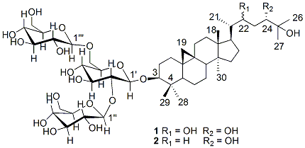
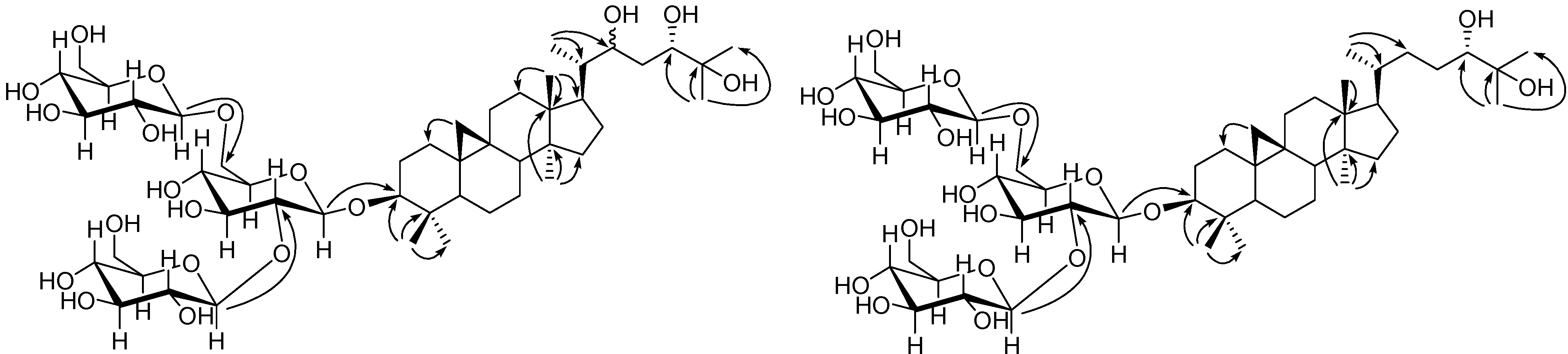
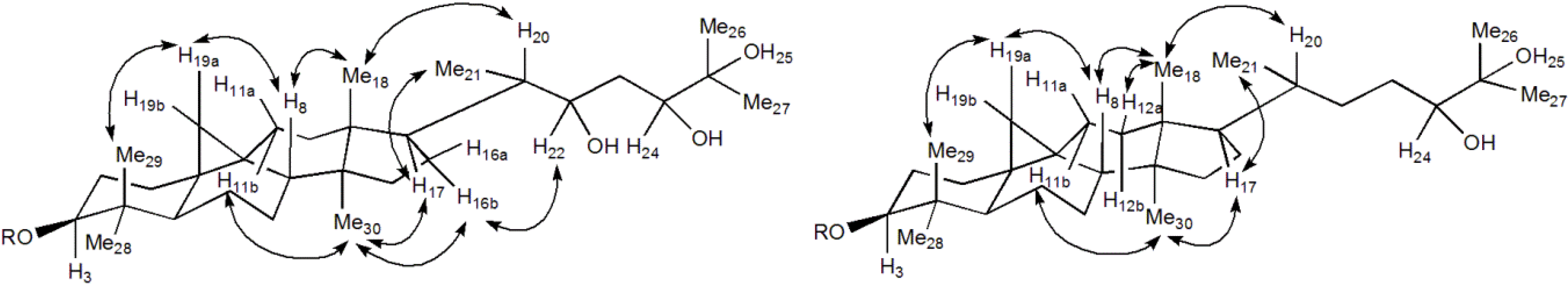
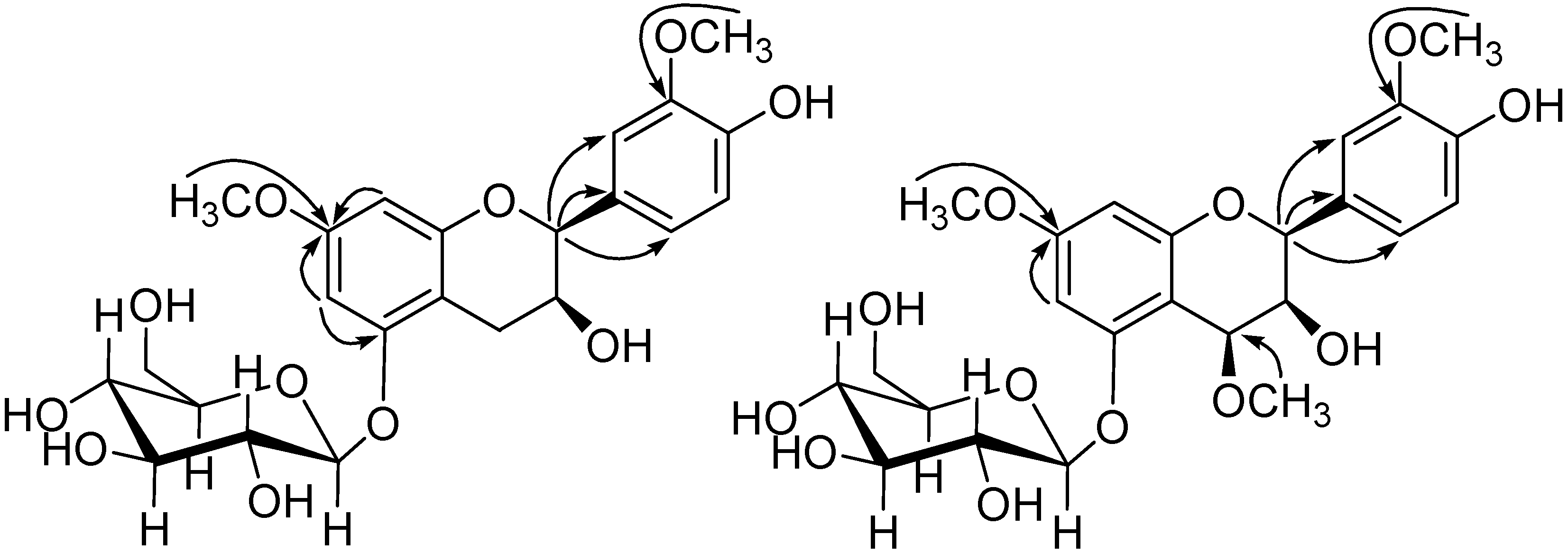
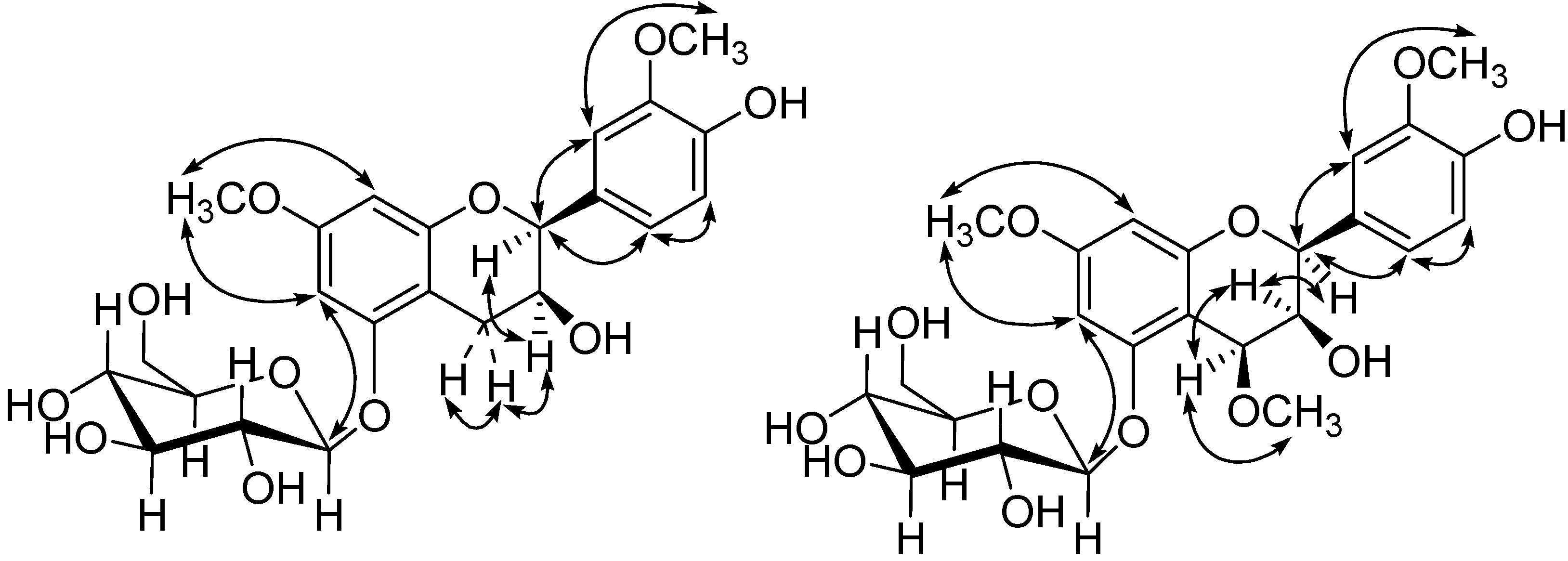
2.1. Structure Elucidation of Compounds 14

Compound 1 was obtained as an amorphous powder. Its HR-ESI-MS quasi-molecular ion peak at m/z 985.5371 ([M+Na]+) corresponded to a molecular formula of C48H82O19, which was supported by the NMR signals. 1H-NMR spectrum of 1 showed characteristic signals of a cyclopropane CH2 at δH 0.64 (d, J = 3.72 Hz, H-19a) and 0.44 (d, J = 3.72 Hz, H-19b), six tertiary Me groups (δH 1.11, 1.24, 1.23, 1.13, 0.95, 1.01; respectively, H3-18, H3-26, H3-27, H3-28, H3-29, H3-30) and a secondary Me group at δH 0.95 (H3-21) [17,18]. Additionally, three anomeric proton signals were observed at δH 4.74 (d, J = 7.68 Hz), 4.52 (d, J = 7.20 Hz) and 4.48 (d, J = 7.74 Hz), indicative of the presence of three β-linked sugar units. The 13C-NMR and DEPT spectra permitted differentiation of the 48 resonances into 6 C, 22 CH, 13 CH2, and 7 CH3 groups, of which 30 were attributed to a triterpene skeleton and 18 to three hexose groups. Acid hydrolysis of 1 gave d-glucoses with optical rotation of Molecules 19 06623 i001+ 51.2, which was determined by TLC analysis and optical rotation measurement [19,20]. Therefore, 1 was considered to be a cycloartane-type triterpene glucoside.
In the HMBC spectrum (Figure 2), correlations of δH 1.13 (H-28) and δH 0.95 (H-29) with δC 90.9 (C-3) suggested that C-3 was substituted by the OH group; 26-CH3, 27-CH3 with C-24 (δC 76.2) and C-25 (δC 74.0), and 21-CH3 with C-22 (δC 71.3) indicated the presence of 24-OH, 25-OH and 22-OH respectively. The glycosidic linkage was determined on the basis of following key spectral signals: HMBC correlations of H-1' with C-3, H-1'' with C-2', and H-1''' with C-6', 1H,1H-COSY cross peaks of H-1'/H-2', H-6'/H-5', and the NOESY cross peaks of H-1'/H-3' and H-5'. Furthermore, the stereochemistry of C-24 was assigned to be S by comparing its spectral data with those reported for analogs [21,22]. Additionally, NOESY correlations between δH 1.62 (H-8) and δH 0.44 (H-19a)/18-CH3, 18-CH3 and δH 1.79 (H-20), δH 0.44 (H-19a) and 29-CH3, δH 2.04 (H-16b) and δH 4.01 (H-22)/30-CH3, 21-CH3 and δH 1.80 (H-17), H-17 and 30-CH3 were observed, revealing their relative configuration (Figure 3).
Figure 2. Key HMBC correlations of 1 and 2.
Molecules 19 06623 g002
Consequently, the structure of 1 was established as (3β,24S)-9,19-cycloartane-3,22,24,25-tetraol 3-O-[β-d-glucopyranosyl-(1→2)]-[β-d-glucopyranosyl-(1→6)]-β-d-glucopyranoside.
The HR-ESI-MS spectrum of 2 (m/z 969.5406 [M + Na]+, calc. for C48H82O18Na) supported a molecular formula of C48H82O18. The NMR spectra of 2 were very similar to those of 1, except for one less oxygenated methine at δC 71.3. Detailed inspection of the HMBC spectrum led to a conclusion that 2 had one less OH on C-22 than 1, on the basis of the key HMBC correlation of 21-CH3H 1.01) with C-22 (δC 23.2). The linkage of three glucoses on the triterpenoid aglycon was determined to be the same way as 1 on the basis of HMBC and 1H,1H-COSY signals. The stereochemistry of 2 was determined on the basis of NOESY cross peaks of H-8 and H-19a/18-CH3, 18-CH3 and H-12a, H-19a and 29-CH3, H-16b and 30-CH3, 21-CH3 and H-17, H-17 and 30-CH3. Acid hydrolysis of 2 gave d-glucoses, which was identified by comparing the optical rotation value with an authentic sample. Finally the structure of 2 was determined to be (3β,24S)-9,19-cycloartane-3,24,25-triol 3-O-[β-d-glucopyranosyl-(1→2)]-[β-d-glucopyranosyl-(1→6)]-β-d-glucopyranoside.
Figure 3. Key NOE correlations of 1 and 2.
Molecules 19 06623 g003
Compound 3 was obtained as colorless needles. Its molecular formula was deduced to be C23H28O11 from the quasi-molecular ion peak at m/z 479.1549 ([MH]) in the HR-ESI-MS spectrum, indicating 10 degrees of unsaturation. The IR spectrum revealed the existence of OH (3434 cm−1) and aromatic ring (1623 cm−1). The 1H-NMR spectrum displayed two meta-coupled protons at δH 6.49 (d, J = 2.16 Hz) and 6.29 (d, J = 2.16 Hz), and three ABX system aromatic protons at δH 7.22 (d, J = 1.06 Hz), 6.99 (dd, J = 8.16, 1.06 Hz), and 6.87 (d, J = 8.16 Hz). In addition, two methoxyl H-atoms at δH 3.82 (s, 3H) and 3.95 (s, 3H), two CH at δH 4.29 (br. s) and 4.98 (br. s), and one CH2 at δH 3.09 (d, J = 17.60 Hz, 1H) and 3.02 (dd, J = 17.60, 4.38 Hz, 1H) were observed. Meanwhile an anomeric H-atom at δH 4.95 (d, J = 7.20 Hz) together with the signals at δH 3.42-3.55 indicated the presence of a β-linked glycosyl group. Enzymatic hydrolysis of 3 with β-d-glucosidase afforded d-glucose, which was identified by direct comparison with the authentic sample by TLC [19]. In view of above evidences, it was concluded that 3 was a flavanol glucoside.
In the combination of the 13C-NMR and HSQC spectra of 3, the 23 carbon resonances could be easily attributed to a flavanol moiety, a glucopyranose unit (δC 62.7, 71.6, 75.1, 78.2, 78.4, 102.8), and two methoxyls (δC 56.0, 56.6). In order to determine the location of substituent groups on the flavanol moiety, HMBC and NOESY experiments were performed. As a result, the NOESY correlations of the methoxyl H-atoms at δH 3.82 with H-6 and H-8, H-6 with H-1', and the other methoxyl H-atoms at δH 3.95 with H-2', together with the HMBC correlations of 7-OCH3 to C-7, 3'-OCH3 to C-3', indicated the glucose and two methoxyls were situated at C-5, C-7, and C-3' respectively (Figure 4 and Figure 5).
The NOESY correlation of δH 4.98 (H-2)/δH 4.29 (H-3) and resonance of H-2 as a broad singlet indicated that the relative configuration of 2,3 was cis [23,24,25,26,27,28]. Therefore, the structure of 3 was elucidated to be cis-3,4'-dihydroxy-7,3'-dimethoxyflavan-5-O-β-d-glucopyranoside.
Compound 4 was isolated as amorphous powder. The molecular formula was C24H30O12, determined by negative-ion at m/z 509.1662 in the HR-ESI-MS. Enzymatic hydrolysis of compound 4 with β-d-glucosidase gave d-glucose. Comparing its NMR spectra with those of 3, it was evident that 4 contained one more methoxyl at δH 3.65/δC 56.6 and one oxygenated CH (δC 73.5) than 3.
The location of one more methoxyl at C-4 was determined by the HMBC correlation of 4-OCH3 at δH 3.65 with C-4 (δC 73.5). The other substitutions of 4 were confirmed by the same way as 3.
Figure 4. Key HMBC correlations of 3 and 4.
Molecules 19 06623 g004
The configuration of 4 was elucidated on the basis of NOESY correlations and coupling constants. Resonances of H-2 as a broad singlet and H-4 as a small doublet (J = 2.28 Hz) were observed, which suggested that the relative 2,3/3,4-stereochemistry were both cis [26]. Additionally the NOESY correlation between δH 5.05 (H-2)/δH 4.62 (H-4) also supported the above assignment. The structure of 4 was identified to be cis-3,4'-dihydroxy-4,7,3'-trimethoxyflavan-5-O-β-d-glucopyranoside.
Figure 5. Key NOE correlations of 3 and 4.
Molecules 19 06623 g005

2.2. Biological Activity Assay

According to previous bioactivity reports on flavonoids [29,30,31], antimicrobial and antioxidant activities of compound 3 were evaluated. Two pathogenic bacteria (Bacillus subtilis, Escherichia coli) and two fungi (Aspergillus niger, Saccharomyces cerevisiae) were selected for antifungal and antibacterial assays, respectively. The compound 3 showed no antimicrobial activity (C > 50 μg/mL). In the ABTS cation radical scavenging assay, the inhibition rate of 3 was 82.0% at 1 mg/mL (Positive control vitamin C, 75.2%, C = 0.1 mg/mL). The superoxide anion radical scavenging assay suggested that 3 showed 53.5% superoxide anion radical scavenging capacity at 2 mg/mL. While the positive control luteolin was 53.8% at 0.625 mg/mL. Unfortunately, the low available amount of compounds 1, 2, and 4 precluded the antimicrobial and antioxidant assays.

3. Experimental

3.1. General Information

Column chromatography (CC): silica gel (SiO2, 200–300 mesh, Qingdao Marine Chemical Plant, Qingdao, P. R. China), MCI gel (Mitsubishi Chemical Corporation, Tokyo, Japan), and Sephadex LH-20 (GE Healthcare Bio-Sciences AB, USA). TLC: silica gel GF254 (Qingdao Marine Chemical Plant, Qingdao, P. R. China). UV absorbance of antioxide mixtures: Varioskan Flash Reader (ThermoFisher Scientific Inc., Waltham, MA). UV spectra: Perkin-Elmer S2 Lambda 35 UV/VIS spectrometer, λ in nm. IR spectra: Perkin-Elmer Spectrum One FT-IR spectrometer, as KBr tablets, ν in cm−1. Optical rotations: Perkin-Elmer 341 polarimeter. NMR spectra: Bruker Avance 600 MHz instrument, δ in ppm, J in Hz, residual solvent peak as reference. HR-ESI-MS: BioTOF-Q mass spectrometer, in m/z.

3.2. Material

The duckweed Landoltia punctata (G. Meyer) Les & Crawford was grown under natural conditions and collected in Kunming, Yunnan province, P. R. China. The duckweed was collected in May, washed with water, and then dried at 60 °C. The test strains Aspergillus niger, Saccharomyces cerevisiae, Escherichia coli, and Bacillus subtilis were obtained from Chengdu Institute of Biology, Chinese Academy of Sciences (CAS), P. R. China.

3.3. Extraction and Isolation

The powder (7 kg) of L. punctata was macerated in 95% ethanol (70 L) at room temperature for 4–5 days, twice. The extract was combined and concentrated under reduced pressure. Then the concentrate was suspended in hot water, and extracted with petroleum ether (P.E.), EtOAc, and n-BuOH successively. The P.E. extract was separated by silica gel column chromatography (CC) using P.E./acetone (10:1–1:1, v/v) as eluent, affording 4 fractions (Fr. 1-4), Fr. 2 and 3 were purified by Sephadex LH-20 CC to yield compounds 1517 respectively.
The EtOAc fraction (25 g) was subjected to a MCI column, and eluted with a stepwise gradient of EtOH/H2O (0:100, 20:80, 40:60, 60:40, 100:0, v/v) to give three fractions (I-III), which were further purified by HPLC (ODS-C18 column, 10 × 250 mm, flow rate 2.5 mL/min). Fr. I was separated using MeOH/H2O (40:60, containing 0.5% acetic acid, v/v) as the eluent to yield compounds 11 and 12. Fr. II was purified using MeOH-H2O (45:55, containing 0.5% acetic acid, v/v) as the eluent to yield compounds 710. Fr. III was separated using MeOH-H2O (60:40, containing 0.5% acetic acid, v/v) as the eluent to obtain compounds 5 and 6.
The same CC of the n-BuOH extract (60 g) on MCI gel as that of EtOAc extract gave four fractions (I–IV). Fraction II was subjected to preparative thin layer chromatography (TLC) and Sephadex LH-20 CC to afford compounds 1 (13 mg) and 2 (10 mg). Faction III was separated over silica gel with CHCl3/MeOH (7:1–0:1, v/v) as eluent to generate 4 fractions (IIIa-IIId). Fr. IIIb was separated by HPLC using MeOH/H2O (45:55, v/v) as the eluent to yield compounds 13 and 14. Fr. IIIc and IIId were further purified by TLC and then Sephadex LH-20 CC to yield compounds 3 (18 mg) and 4 (8 mg) respectively.

3.4. Hydrolysis

Acid hydrolysis. Compounds 1 and 2 (10 mg) were independently dissolved in aqueous solution of 2 N HCl (6 mL) and stirred at 85 °C overnight. After cooled down, the reaction mixture was neutralized and then extracted with CHCl3. The water layer were concentrated to dryness, dissolved in water to constant volume, and analyzed by comparing their TLC profile and optical rotation values with the standard sample of d-glucose [21,22], respectively.
Enzymatic hydrolysis. Compounds 3 and 4 (5.0 mg) were independently suspended in water (5 mL), and excessive β-d-glucosidase (Shanghai Zurui Biological Technology Co., Ltd., Shanghai, China) was added, respectively. The mixture was placed in water bath at 37 °C for 48 h. The products were analyzed by TLC [21].

3.5. Spectral Data

(3β,24S)-9,19-Cycloartane-3,22,24,25-tetraol 3-O-[β-d-glucopyranosyl-(1→2)]-[β-d-glucopyranos-yl(1→6)]-β-d-glucopyranoside (1): Amorphous powder. Molecules 19 06623 i001 −3.0 (c 0.1, MeOH). IR (KBr) νmax 3411, 2932, 1075 cm−1. 1H and 13C-NMR: See Table 1. HR-ESI-MS (positive mode) m/z 985.5371 [M+Na]+ (calc. for C48H82O19Na, 985.5343).
(3β,24S)-9,19-Cycloartane-3,24,25-triol 3-O-[β-d-glucopyranosyl-(1→2)]-[β-d-glucopyranosyl-(1→6)]-β-d-glucopyranoside (2): Amorphous powder. Molecules 19 06623 i001 −4.0 (c 0.1, MeOH). IR (KBr) νmax 3418, 2891, 1043 cm−1. 1H and 13C-NMR: See Table 1. HR-ESI-MS (positive mode) m/z 969.5406 [M + Na]+ (calc. for C48H82O18Na, 969.5393).
3,4'-Dihydroxy-7,3'-dimethoxyflavan-5-O-β-d-glucopyranoside (3): Colorless needles. Molecules 19 06623 i001 −42.0 (c 0.1, MeOH). UV (MeOH) λmax (log ε) 218 (4.60), 227 (3.96), 282 (3.52) nm. IR (KBr) νmax 3434, 1623, 1080 cm−1. 1H and 13C-NMR: See Table 2. HR-ESI-MS (negative mode) m/z 479.1549 (calc. for C23H27O11, 479.1559).
3,4'-Dihydroxy-4,7,3'-trimethoxyflavan-5-O-β-d-glucopyranoside (4): Amorphous powder. Molecules 19 06623 i001 −20.0 (c 0.1, MeOH). UV (MeOH) λmax (log ε) 226 (4.02), 280 (3.60) nm. IR (KBr) νmax 3420, 2926, 1597, 1424, 1079 cm−1. 1H and 13C-NMR: See Table 2. HR-ESI-MS (negative mode) m/z 509.1662 (calc. for C24H29O12, 509.1664).

3.6. Biological Activity Assay

According to the reported [32], antifungal and antibacterial activities were determined using the Oxford cup method with Methicillin sodium as the positive control. The antioxidant activity assay was performed on the DPPH, ABTS+•, and superoxide anion radical scavenging models complying with the previously published literature [33].
Table 1. 1H (600 MHz) and 13C (150 MHz) NMR data of compounds 1 (in CD3OD, J in Hz) and 2 (in C5D5N, J in Hz).
No.12No.12
δCδHδCδHδCδHδCδH
133.31.65–1.67 (m), 1.32–1.34 (m) (m)30.32.50–2.53 (m), 1.25–1.26 (m)2574.0 73.1
230.72.08–2.11 (m), 1.35–1.36 (m)29.31.81–1.82 (m), 1.95–1.97 (m)2626.11.24 (s, 3H)26.51.56 (s, 3H)
390.93.34–3.36 (m)89.13.45–3.48 (m)2725.61.23 (s, 3H)26.21.54 (s, 3H)
442.4 41.7 2826.01.13 (s, 3H)26.01.29 (s, 3H)
550.41.68–1.70 (m)47.81.20–1.22 (m)2915.60.95 (s, 3H)15.71.19 (s, 3H)
622.31.67–1.68 (m), 1.68–1.70 (m)27.21.87–1.88 (m), 1.37 (overlapped)3020.11.01 (s, 3H)18.60.99 (s, 3H)
727.41.39–1.40 (m), 1.36–1.37 (m)33.51.50–1.51 (m), 1.53 (overlapped)1'105.34.52 (d, J = 7.20, 1H)105.24.90 (d, J = 7.56, 1H)
849.71.60–1.63 (m)53.31.64–1.65 (m)2'81.43.62–3.63 (m)83.54.15–4.17 (m)
921.4 20.2 3'78.23.61–3.62 (m)77.34.10–4.11 (m)
1027.5 26.9 4'71.73.39–3.40 (m)72.04.20–4.21 (m)
1127.71.70–1.72 (m), 2.10–2.11 (m)21.51.45–1.46 (m), 0.70–0.72 (m)5'77.13.52–3.54 (m)77.24.03–4.04 (m)
1234.21.72, 1.76, overlapped32.61.64–1.65 (m), 1.24 (overlapped)6'70.23.85–3.87 (m), 4.16–4.18 (br. d)70.44.29–4.31 (m), 4.82 (br. d)
1346.9 45.9 1''104.84.74 (d, J = 7.68, 1H)106.35.35 (d, J = 7.62, 1H)
1449.8 49.3 2''76.53.28–3.29 (m)75.64.04–4.05 (m)
1537.01.40–1.41 (m), 1.42–1.43 (m)36.71.65–1.66 (m), 1.55 (overlapped)3''78.43.60–3.61 (m)78.34.24–4.25 (m)
1628.41.42–1.43 (m), 2.03–2.05 (m)28.81.37–1.38 (m), 1.63–1.64 (m)4''72.03.26–3,27 (m)71.84.28–4.29 (m)
1750.01.79–1.81 (m)48.41.43–1.44 (m)5''78.13.30–3.31 (m)78.63.90–3.92 (m)
1818.71.11 (s, 3H)19.90.84 (s, 3H)6''63.03.71–3.72 (m), 3.93–3.94 (m)63.04.45–4.46 (m), 4.52–4.53 (m)
1930.90.44 (d, J = 3.72)30.00.24 (d, J = 4.08)1'''105.04.48 (d, J = 7.74, 1H)105.75.13 ( d, J = 7.80, 1H)
0.64 (d, J = 3.72) 0.48 (d, J = 4.08)2'''75.43.24–3.26 (m)75.54.05–4.06 (m)
2044.01.78–1.79 (m)36.11.23–1.24 (m)3'''78.03.40–3.42 (m)78.83.94–3.96 (m)
2112.70.95 (s, 3H)18.91.01 (s, 3H)4'''71.93.31–3.32 (m)71.14.03–4.04 (m)
2271.34.01 (br. d)23.20.86–0.87 (m), 1.25 (overlapped)5'''78.13.29–3.30 (m)78.44.21–4.22 (m)
2332.91.40–1.41 (m), 1.53–1.57 (m)34.51.83–1.85 (m), 1.68 (overlapped)6'''63.33.70–3.71 (m), 3.90–3.92 (m)63.14.33–4.36 (m), 4.48–4.50 (m)
2476.23.58–3.60 (m)79.43.78–3.81 (m)
Table 2. 1H (600 MHz) and 13C (150 MHz) NMR data of compounds 3 and 4 (in CD3OD, J in Hz).
No.34
δCδHδCδH
280.34.98 (br. s, 1H)78.65.05 (br. s, 1H)
367.44.29 (br. s, 1H)69.24.04 (br. d, 1H)
429.73.09 (d, J = 17.60, 1H)3.02(1H, dd)73.54.62 (d, J = 2.28, 1H)
3.02 (dd, J = 17.60, 4.38, 1H)
5158.6 157.9
697.16.49(d, J = 2.10)96.66.54 (d, J = 1.80)
7160.9 162.9
896.56.29 (d, J = 2.10)96.36.30 (d, J = 1.80)
9157.3 160.4
10104.0 104.4
1'132.3 131.6
2'112.17.22 (d, J = 1.06)112.37.22 (d, J = 1.10)
3'148.8 149.0
4'147.2 147.4
5'115.96.87 (d, J = 8.16)116.06.90 (d, J = 8.04)
6'120.86.99 (dd, J = 1.06, 8.16)121.07.00 (dd, J = 1.10, 8.04)
1''102.84.95 (d, J = 7.20)102.64.99 (d, J = 7.50)
2''75.13.50–3.55 (overlapped)75.33.58–3.59 (overlapped)
3''78.43.50–3.55 (overlapped)78.53.53–3.56 (overlapped)
4''71.63.47–3.48 (m)71.63.46–3.48 (overlapped)
5''78.23.50–3.55 (overlapped)76.93.53–3.56 (overlapped)
6''62.73.79 (dd, J = 5.58, 12.10)62.83.79 (dd, J = 5.00, 12.20)
3.98 (d, J = 12.10) 4.00 (d, J = 12.20)
MeO-4 56.63.65 (s, 3H)
MeO-756.63.82 (s, 3H)56.03.84 (s, 3H)
MeO-3'56.03.95 (s, 3H )57.83.96 (s, 3H)

4. Conclusions

In this study, we identified 12 flavonoids including two new flavanol glucosides from L. punctata. The result showed that apigenin or luteolin flavonoids are the main constituents of this species, which is in good agreement with the previous reports and our previous study results that the transcripts for key enzymes of flavonoid biosynthesis in L. punctata expressed in high abundance at the transcriptional level [9,10,34]. Flavonoids are generally biosynthesized to cope with environmental stressors such as ultraviolet radiation, ozone, heavy metals, nutrient limitation, herbivores, and so on. The high content of flavonoids in L. punctata could be related to environmental stressors. Meanwhile cycloartane triterpenoids were discovered in Lemnaceae family for the first time in this study. Many cycloartane triterpenoids possessed diverse bioactivities such as anti-inflammatory, anti-tumor, anti-viral, immuno-regulatory, hypoglycemic, cardiovascular system, nervous system and hepato-protective effects [35,36], some of which offer good prospectives in medical applications. This study demonstrates that L. punctata is a new source for cycloartane triterpenoids.

Acknowledgments

This research was financially supported by the Major Projects of Knowledge Innovation Program of Chinese Academy of Sciences (KSCX2-EW-J-22).

Author Contributions

N.N.W performed all phytochemical parts of the work and prepared a draft manuscript. T.Y and G.B.X performed the biological activity assays. Y.F offered help on the plant material. H.Z and G.Y.L initiated the study, coordinated the project, supervised the phytochemical work and prepared the final manuscript.

Conflicts of Interest

The authors declare no conflict of interest.

References

  1. Ge, X.M.; Zhang, N.N.; Phillips, G.C.; Xu, J.F. Growing Lemna minor in agricultural wastewater and converting the duckweed biomass to ethanol. Bioresour. Technol. 2012, 124, 485–488. [Google Scholar] [CrossRef]
  2. Zhao, H.; Appenroth, K.; Landesman, L.; Salmeán, A.A.; Lam, E. Duckweed rising at Chengdu: Summary of the 1st International Conference on Duckweed Application and Research. Plant. Mol. Boil. 2012, 78, 627–632. [Google Scholar] [CrossRef]
  3. Cole, C.T.; Voskuil, M.I. Population genetic structure in duckweed (Lemna minor, Lemnaceae). Can. J. Bot. 1996, 74, 222–230. [Google Scholar] [CrossRef]
  4. Xu, J.; Zhao, H.; Stomp, A.M.; Cheng, J.J. The production of duckweed as a source of biofuels. Biofuels 2012, 3, 589–601. [Google Scholar] [CrossRef]
  5. Ozengin, N.; Elmaci, A. Performance of duckweed (Lemna minor L.) on different types of wastewater treatment. J. Environ. Biol. 2007, 28, 307–314. [Google Scholar]
  6. Mohedano, R.A.; Costa, R.H.; Tavares, F.A.; Filho, B.P. High nutrient removal rate from swine wastes and protein biomass production by full-scale duckweed ponds. Bioresour. Technol. 2012, 112, 98–104. [Google Scholar] [CrossRef]
  7. Cheng, J.J.; Stomp, A.M. Growing duckweed to recover nutrients from wastewaters and for production of fuel ethanol and animal feed. Clean 2009, 37, 17–26. [Google Scholar]
  8. Xu, J.; Cui, W.; Cheng, J.J.; Stomp, A.M. Production of high-starch duckweed and its conversion to bioethanol. Biosyst. Eng. 2011, 110, 67–72. [Google Scholar] [CrossRef]
  9. Xiao, Y.; Fang, Y.; Jin, Y.; Zhang, G.; Zhao, H. Culturing duckweed in the field for starch accumulation. Ind. Crop. Prod. 2013, 48, 183–190. [Google Scholar] [CrossRef]
  10. Jurd, L.; Geissman, T.A.; Seikel, M.K. The flavonoid constituents of Spirodela oligorrhiza II. The flavone constituents. Arch. Biochem. Biophys. 1957, 67, 284–297. [Google Scholar] [CrossRef]
  11. Lee, Y.H.; Lee, I.R.; Won, W.S.; Park, C.H. Flavonoids of Elscholtzia cristata. Arch. Pharm. Res. 1988, 11, 247–249. [Google Scholar] [CrossRef]
  12. Rayyan, S.; Fossen, T.; Nateland, H.S.; Andersen, Q.M. Isolation and identification of flavonoids, including flavone rotamers, from the herbal drug ‘crataegi folium cum flore’ (hawthorn). Phytochem. Anal. 2005, 16, 334–341. [Google Scholar] [CrossRef]
  13. Kawasaki, M.; Kanomata, T.; Yoshitama, K. Flavonoids in the leaves of twenty-eight polygonaceous plants. Bot. Mag. Tokyo 1986, 99, 63–74. [Google Scholar] [CrossRef]
  14. Dubois, M.A.; Zoll, A.; Markham, K.R.; Bouillant, M.L.; Dellamonica, G.; Chopin, J. 6-c-β-d-glucopyranosyl-8-c-β-d-galactopyranosylapigenin from Cerastium arvense. Phytochemistry 1984, 23, 706–707. [Google Scholar] [CrossRef]
  15. Kamboj, A.; Salujai, A.K. Isolation of stigmasterol and β-sitosterol from petroleum ether extract of aerial parts of Ageratum conyzoides (Asteraceae). Int. J. Pharm. Pharm. Sci. 2011, 3, 94–96. [Google Scholar]
  16. Cui, J.; Wang, H.; Huang, Y.; Xin, Y.; Zhou, A. Synthesis and cytotoxic analysis of some disodium 3β, 6β-dihydroxysterol disulfates. Steroids 2009, 74, 1057–1060. [Google Scholar] [CrossRef]
  17. Polat, E.; Caliskan-Alankus, O.; Perrone, A.; Piacente, S.; Bedir, E. Cycloartane-type glycosides from Astragalus amblolepis. Phytochemistry 2009, 70, 628–634. [Google Scholar] [CrossRef]
  18. Yokosuka, A.; Sato, K.; Yamori, T.; Mimaki, Y. Triterpene glycosides from Curculigo orchioides and their cytotoxic activity. J. Nat. Prod. 2010, 73, 1102–1106. [Google Scholar] [CrossRef]
  19. Chaturvedula, P.V.S.; Prakash, I. A new diterpene glycoside from Stevia rebaudiana. Molecules 2011, 16, 2937–2943. [Google Scholar] [CrossRef]
  20. Mohan, R.; Birari, R.; Karmase, A.; Jagtap, S.; Bhutani, K.K. Antioxidant activity of a new phenolic glycoside from Lagenaria siceraria Stand. fruits. Food Chem. 2012, 132, 244–251. [Google Scholar] [CrossRef]
  21. Bedir, E.; Calis, I.; Khan, I.A. Macrophyllosaponin E: A novel compound from the roots of Astragalus oleifolius. Chem. Pharm. Bull. 2000, 48, 1081–1083. [Google Scholar] [CrossRef]
  22. Hirotani, M.; Zhou, Y.; Rut aTsutomu Furuya, H. Cycloartane triterpene glycosides from the hairy root cultures of Astragalus. membranaceus. Phytochemistry 1994, 37, 1403–1407. [Google Scholar] [CrossRef]
  23. Thompson, R.S.; Jacques, D.; Haslam, E.; Tanner, R.J.N. Plant proanthocyanidins. Part I. Introduction; the isolation, structure, and distribution in nature of plant procyanidins. J. Chem. Soc. Perkin Trans. 1 1972, 1387–1399. [Google Scholar]
  24. Fletcher, A.C.; Porter, L.J.; Haslam, E.; Gupta, R.K. Plant proanthocyanidins. Part 3. Conformational and configurational studies of natural procyanidins. J. Chem. Soc. Perkin Trans. 1 1977, 1628–1637. [Google Scholar]
  25. Mateus, N.; Silva, A.M.; Santos-Buelga, C.; Rivas-Gonzalo, J.C.; de Freitas, V. Identification of anthocyanin-flavanol pigments in red wines by NMR and mass spectrometry. J. Agric. Food Chem. 2002, 50, 2110–2116. [Google Scholar] [CrossRef]
  26. Otsuka, H.; Hirata, E.; Shinzato, T.; Takeda, Y. Glochiflavanosides AD: Flavanol glucosides from the leaves of Glochidion zeylanicum (Gaertn) A. Juss. Chem. Pharm. Bull. 2001, 49, 921–923. [Google Scholar] [CrossRef]
  27. Li, S.Y.; Fuchino, H.; Kawahara, N.; Sekita, S.; Satake, M. New phenolic constituents from Smilax bracteata. J. Nat. Prod. 2002, 65, 262–266. [Google Scholar] [CrossRef]
  28. Li, F.; Zhan, Z.; Liu, F.; Yang, Y.; Li, L.; Feng, Z.; Jiang, J.S.; Zhang, P. Polyflavanostilbene A, a new flavanol-fused stilbene glycoside from Polygonum cuspidtum. Org. Lett. 2013, 15, 674–677. [Google Scholar]
  29. Agati, G.; Azzarello, E.; Pollastri, S.; Tattini, M. Flavonoids as antioxidants in plants: Location and functional significance. Plant. Sci. 2012, 196, 67–76. [Google Scholar] [CrossRef]
  30. Aron, P.M.; Kennedy, J.A. Flavan-3-ols: Nature, occurrence and biological activity. Mol. Nutr. Food Res. 2008, 52, 79–104. [Google Scholar] [CrossRef]
  31. Scheid, L.; Reusch, A.; Stehle, P.; Ellinger, S. Antioxidant effects of cocoa and cocoa products ex vivo and in vivo: Is there evidence from controlled intervention studies? Curr. Opin. Clin. Nutr. Metab. Care 2010, 13, 737–742. [Google Scholar] [CrossRef]
  32. Xu, G.B.; Yang, T.; Bao, J.K.; Fang, D.M.; Li, G.Y. Isochaetomium A2, a new bis (naphthodihydropyran-4-one) with antimicrobial and immunological activities from fungus Chaetomium microcephalum. Arch. Pharm. Res. 2013. [Google Scholar] [CrossRef]
  33. Tian, S.Z.; Pu, X.; Luo, G.Y.; Zhao, L.X.; Xu, L.H.; Li, W.J.; Luo, Y.G. Isolation and characterization of new p-terphenyls with antifungal, antibacterial, and antioxidant activities from halophilic actinomycete Nocardiopsis gilva YIM 90087. J. Agric. Food Chem. 2013, 61, 3006–3012. [Google Scholar] [CrossRef]
  34. Tao, X.; Fang, Y.; Xiao, Y.; Jin, Y.L.; Ma, X.R.; Zhao, Y.; He, K.Z.; Zhao, H.; Wang, H.Y. Comparative transcriptome analysis to investigate the high starch accumulation of duckweed (Landoltia punctata) under nutrient starvation. Biotechnol. Biofuels 2013, 6, 72–87. [Google Scholar] [CrossRef]
  35. Li, S.F.; Di, Y.T.; Luo, R.H.; Zheng, Y.T.; Wang, Y.H.; Fang, X.; Zhang, Y.; Li, L.; He, H.P.; Li, S.L. Cycloartane triterpenoids from Cassia occidentalis. Planta. Med. 2012, 78, 821–827. [Google Scholar] [CrossRef]
  36. Wang, X.Y.; Tang, G.H.; Yuan, C.M.; Zhang, Y.; Zou, T.; Yu, C.; Zhao, Q.; Hao, X.J.; He, H.P. Aphagrandinoids A–D, cycloartane triterpenoids with antibacterial activities from Aphanamixis grandifolia. Fitoterapia 2013, 85, 64–68. [Google Scholar] [CrossRef]
  • Sample Availability: Samples of the compounds 3, 6, 713 are available from the authors.

