Article

Dehydroabietic Acid Isolated from Commiphora opobalsamum Causes Endothelium-Dependent Relaxation of Pulmonary Artery via PI3K/Akt-eNOS Signaling Pathway

1
College of Pharmacy, Harbin Medical University-Daqing, Daqing 163319, Heilongjiang, China
2
Biopharmaceutical Institute of the Heilongjiang Academy of Medical Sciences, Harbin 150081, Heilongjiang, China
3
College of Pharmacy, Harbin University of Commerce, Harbin 150028, Heilongjiang, China
*
Author to whom correspondence should be addressed.
Molecules 2014, 19(6), 8503-8517; https://doi.org/10.3390/molecules19068503
Submission received: 12 March 2014 / Revised: 16 June 2014 / Accepted: 17 June 2014 / Published: 23 June 2014
(This article belongs to the Section Natural Products Chemistry)

Abstract

:
Commiphora opobalsamum is a Traditional Chinese Medicine used to treat traumatic injury, mainly by relaxing blood vessels. In this study, two diterpenes, dehydroabietic acid (DA) and sandaracopimaric acid (SA) were obtained from it by a bioassay-guided approach using isolated rat pulmonary artery rings. The structures of the two compounds were elucidated by spectroscopic methods (IR, 1H- and 13C-NMR, HR-ESI-MS). Both DA and SA reduced the contraction of phenylephrine-induced pulmonary arteries in a concentration-dependent manner, and endothelium contributed greatly to the vasodilatory effect of DA. This effect of DA was attenuated by NG-Nitro-L-arginine methyl ester (L-NAME, an eNOS inhibitor). Meanwhile, DA increased nitric oxide (NO) production, along with the increase of phosphorylation level of eNOS and Akt in endothelial cells. LY294002 (a PI3K inhibitor) could reverse this effect, which suggested the endothelial PI3K/Akt pathway involved in the mechanism underlying DA-induced relaxation of pulmonary artery. This work provided evidence of vasorelaxant substances in Commiphora opobalsamum and validated that PI3K/Akt-eNOS pathway was associated with DA-induced pulmonary artery vasodilation.

Graphical Abstract

1. Introduction

Pulmonary arterial hypertension (PAH) is defined as a disease characterized by vasoconstricted and remodeled pulmonary arteries, which affects the pulmonary vasculature and increases pulmonary vascular resistance [1]. Although current therapies could improve the quality of life of PAH patients, PAH cannot be cured completely by the drugs used in current clinic therapy [2]. Recently, discovery for vasorelaxant compounds from natural products was of high interest regarding prevention and treatment of many vascular diseases. Phytochemical studies of Commiphora opobalsamum (C. opobalsamum) revealed the presence of flavonoids, sterols, triterpenes, saponins, volatilebases, and volatileoil [3]. It possesses extensive biological activities, such as antitumor, antiulcerogenic, hepatoprotective and hypotensive activities [4,5,6,7]. The aim of our research was to seek for potential vasorelaxant components on pulmonary artery in C. opobalsamum. The approach of bioassay-guided isolation was applied for this purpose, which is effective to search for vasorelaxant compounds from natural products as demonstrated by previous studies [8].
In the present study, dehydroabietic acid (DA) and sandaracopimaric acid (SA) were identified as relaxing pulmonary artery ingredients from C. opobalsamum (Figure 1). Previous reports have confirmed 15-pimaradien-3β-ol, a optical isomer of SA, could resist phenylephrine (PE)-induced contraction of the aorta, and its underlying mechanism involved extracellular Ca2+ influx blockade and activation of NO-cGMP pathway [9,10,11]. The vasorelaxant effect of DA was explored on pulmonary artery in this study. And the relaxation of DA on the pulmonary artery could be diminished by L-NAME, implying that DA relaxed pulmonary artery through endothelial NO pathway. Further experiments validated the PI3K/Akt-eNOS signaling pathway was involved in its vasorelaxant effect.
Figure 1. The chemical structures of DA and SA.
Molecules 19 08503 g001

2. Results and Discussion

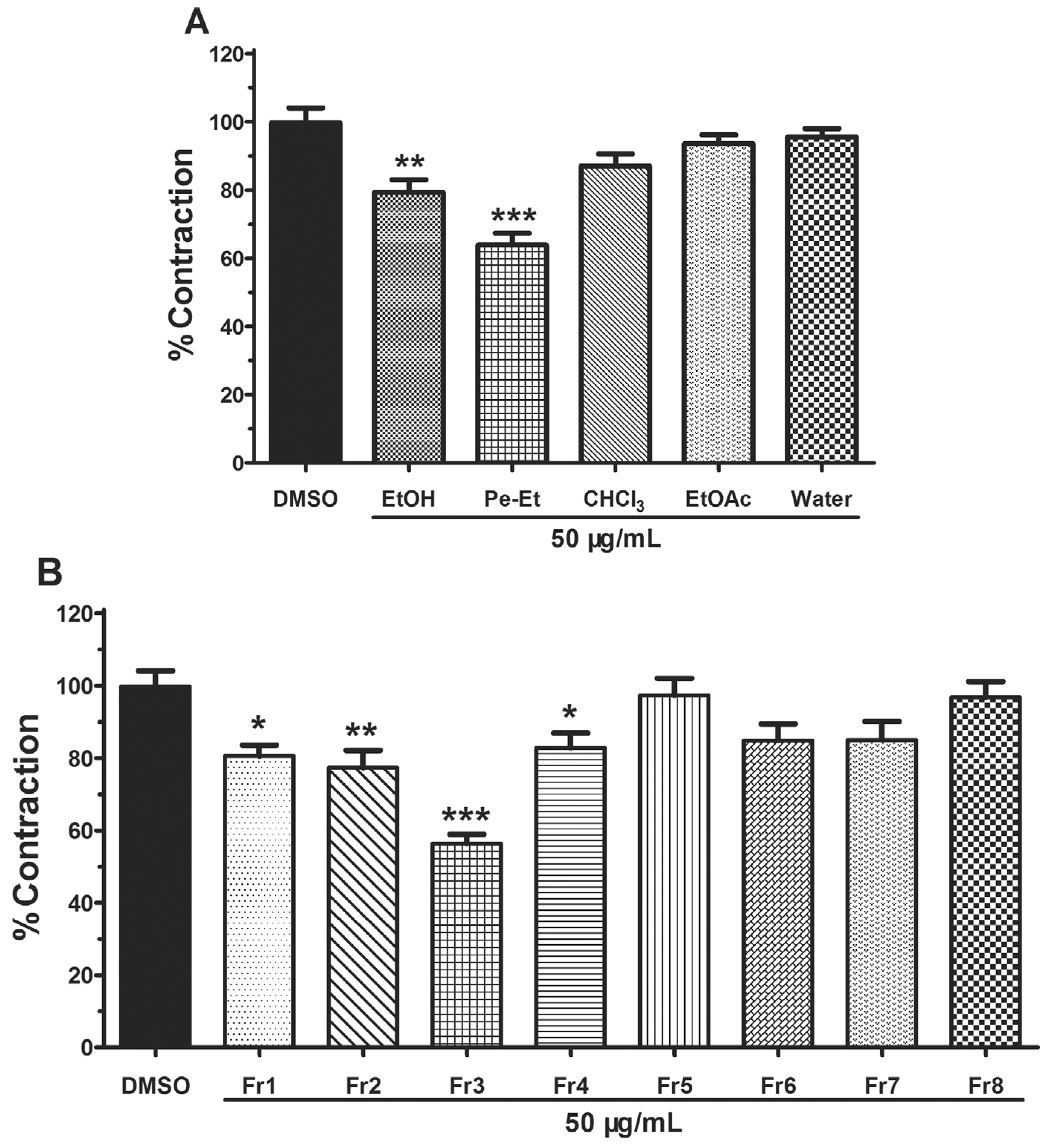
2.1. Bioassay-Guided Isolation

The bioassay-guided isolation results showed that the petroleum ether-soluble extracts could significantly relax PE-induced pulmonary artery rings at the concentration of 50 μg/mL, with 63.90% ± 7.78% contraction compared with vehicle control (DMSO) (Figure 2A). Each of the eight primary fractions (Fr1–Fr8) was also assessed using pulmonary artery rings (Figure 2B). At the same concentration (50 μg/mL), Fr1–4 could significantly inhibit the PE-induced contraction of pulmonary artery, especially Fr3, with the inhibition rate of 56.34% ± 5.82%. With further separation by a silica gel column, compounds 1 and 2 were obtained from Fr3.
Figure 2. Vasorelaxant effect of all parts from C. opobalsamum on the PE-induced constriction of rat pulmonary artery. (A) Vascular effect of EtOH extract (EtOH) and four partitions [petroleum ether-soluble (Pe-Et), chloroform-soluble (CHCl3), EtOAc-soluble (EtOAc) and water-soluble (Water)] obtained from that extract. (B) Eight fractions ofpetroleum ether-soluble. Results are presented as means ± SEM, n = 5. * p < 0.05, ** p < 0.01 and *** p < 0.001 compared to vehicle (DMSO).
Molecules 19 08503 g002

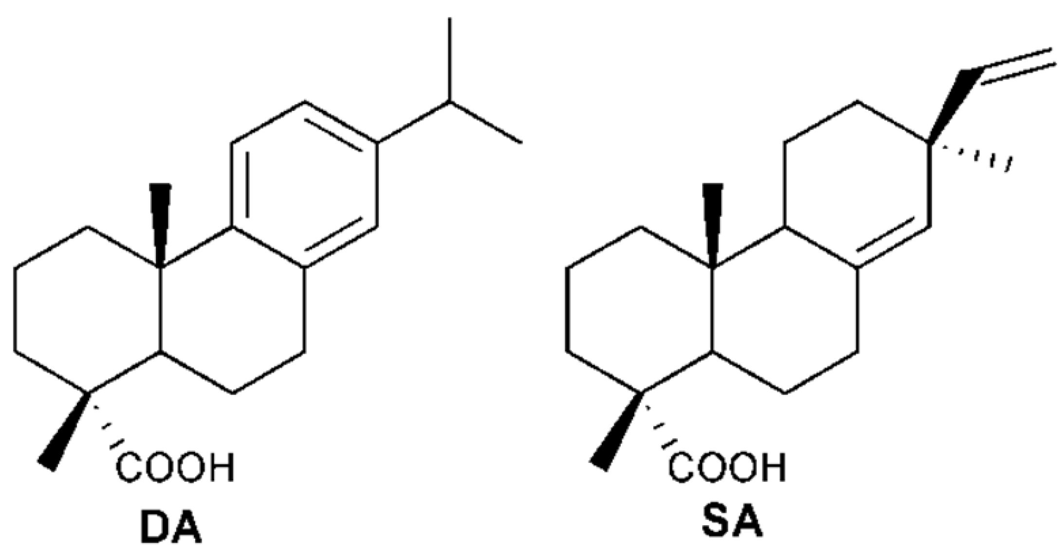
2.2. Elucidation of Compounds’ Structures

Compound 1 was obtained as a white powder, while compound 2 was obtained in the form of white needle-like crystals when recrystallised from EtOAc. For both compounds, a total of 20 carbons, including a carbonyl group, were found in the 13C-NMR spectrum, and a strong characteristic C=O peak (1,693 cm−1) appeared in the IR spectrum. There were four carbon double bond signals in compound 1 in the 13C-NMR spectrum, with a molecular ion peak [M − H] at 301.6214 in the HR-ESI-MS, and six carbon double bond signals in compound 2, with a molecular ion peak [M − H] at 299.6214. The optical rotations of compound 1 { Molecules 19 08503 i001: +2.0° (c 0.10 EtOH)} and 2{ Molecules 19 08503 i001: +63.0° (c 0.10 EtOH)} were in accordance with previous reported data [12]. By comparing their 1H- and 13C-NMR data with that of previous studies [13,14], compounds 1 and 2 were identified as SA and DA, respectively (Figure 1).

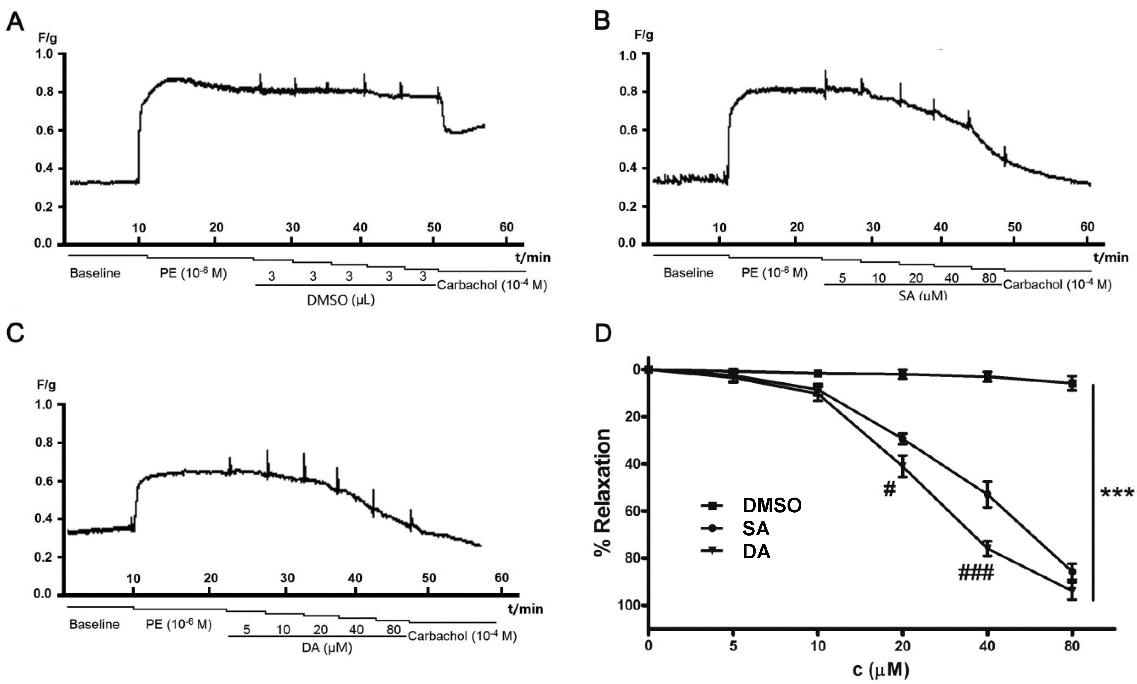
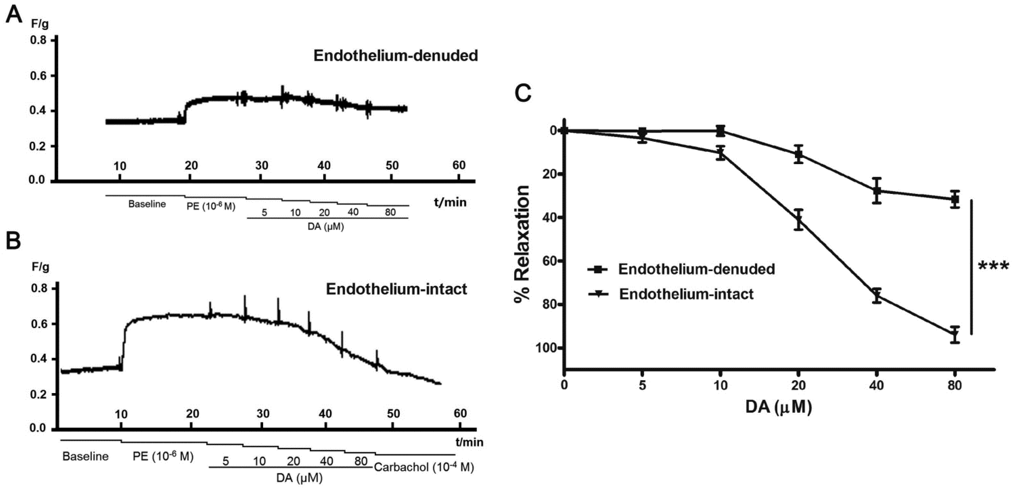
2.3. Effect of DA and SA on Pulmonary Arteries

Pulmonary artery rings were pretreated with PE (10−6 M), and then DA or SA (5, 10, 20, 40, 80 μM) was accumulatively added to the bath. As shown in Figure 3, we found that both SA and DA were effective against PE-induced contraction of pulmonary artery rings in a concentration-dependent manner.
Figure 3. Effect of DA and SA on rats pulmonary artery rings precontracted with PE (10−6 M). The representative recorded curves of tension were presented as AC, and D was the analysis results. Cumulative dosing of vehicle (DMSO) had no effect on rat pulmonary artery rings (A), however, dilatation effect of SA and DA on precontracted rat pulmonary artery rings were concentration-dependent (B,C). The results are represented as the means ± SEM (n = 5), *** p < 0.001 compared to corresponding control (DMSO), # p < 0.05 and ### p < 0.001 showed the difference between DA and SA at the same concentration.
Molecules 19 08503 g003
SA (5 μM) had a slight effect on the rest tone of the rings; the relaxation was only 2.46% ± 2.59%. However, when pulmonary artery rings were exposed to SA (20 μM), this effect was largely improved to 29.42% ± 2.25%. At a concentration of 80 μM, PE-induced contraction was suppressed by 85.75% ± 3.37%, which was close to the state of the pulmonary artery rings before being exposed to PE. The vasodilatory effect of DA was similar to that of SA, however, it was superior to that provided by SA, and EC50 value of DA and SA was 35.81 and 43.93 μM, respectively. DA and SA are both diterpenoids with a phenanthrene structure, but the vasodilatory effect of DA on the pulmonary artery was greater than that of SA at middle concentration (20–40 μM). The aromatic ring of DA may contribute to its effectiveness.

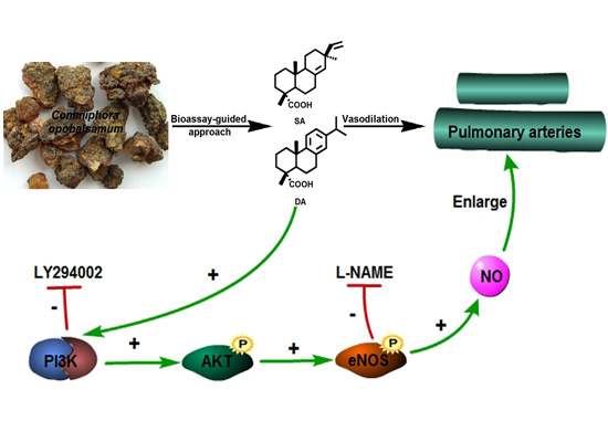
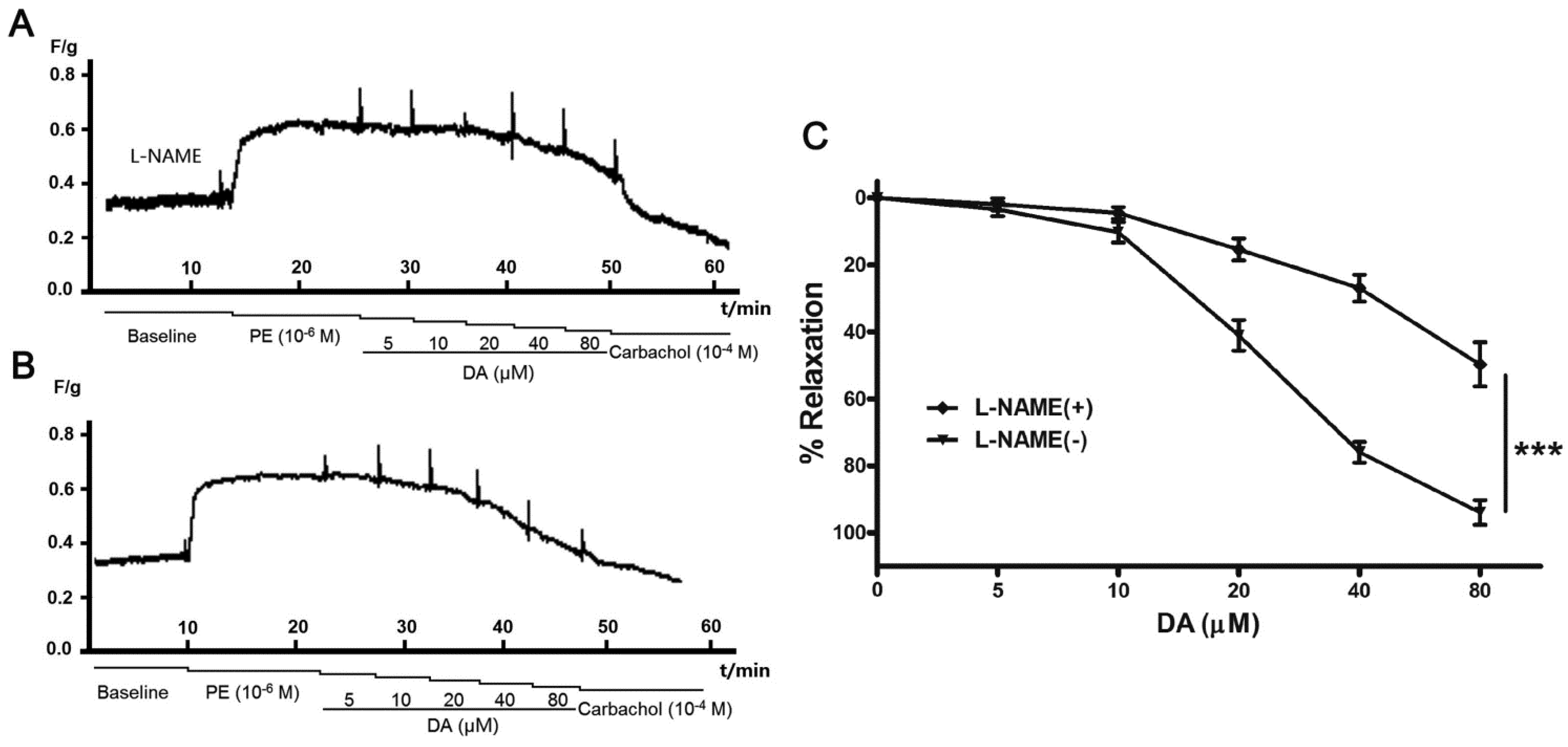
2.4. Effect of Endothelium and L-NAME on DA-Induced Vasorelaxation

To evaluate the involvement of endothelium in DA-induced vasorelaxation, we examined the vasodilatory effect of DA on endothelium-denuded pulmonary artery rings. As shown in Figure 4, when the endothelium of pulmonary artery rings was removed, the vasodilatory effect of DA on the pulmonary artery decreased distinctly compared with that of the endothelium-intact rings. The relaxation ratios were only 31.62% ± 3.75% at the concentration of 80 μM, less than that of endothelium-inract (relaxation ratios of 93.94% ± 3.69%). This finding suggested that the vasodilatory effect of DA was closely related to the endothelium. And additional vasodilatory effects may be the result of the open calcium-sensitive potassium ion channels induced by DA [15].
Figure 4. Effect of DA on endothelium-intact and endothelium-denuded rat pulmonary artery. Cumulative dosing of DA vasorelaxant effect on both endothelium-intact and endothelium-denuded rats pulmonary artery rings were detected (A,B). The results are represented as the means ± SEM (n = 5) (C), *** p < 0.001 for subdued effect of endothelium-denuded versus endothelium-intact group.
Molecules 19 08503 g004
The fact that DA showed relaxant effect on endothelium-dependent manner suggests that NO pathways maybe involved in the response [16]. NO is a crucial mediator in endothelial vasodilator function, which is synthesized from the terminal guanidino nitrogen of L-arginine by NOS enzymes [17]. Substantial evidence has shown that NO-mediated relaxation of several substances or plant extracts in rat aortic rings [18,19,20], so we decided to investigate the involvement of endothelial NOS (eNOS) in DA-induced pulmonary artery rings relaxation. Pulmonary artery rings were pretreated with L-NAME (10−4 M) for 30 min before being pre-contracted with PE. As illustrated in Figure 5, as NO-induced vasorelaxation was blocked by L-NAME, DA-induced vasodilatation was diminished by about 50% at the concentration of 80 μM. This suggested that the mechanism underlying DA-induced relaxation depended on eNOS activity. Why carbachol caused a profound relaxation vasodilatory (Figure 5A) may be that the activity of carbachol was so strong that its effect couldn’t be completely blocked by L-NAME (100 μM) [21].
Figure 5. Effect of NO in DA-induced pulmonary artery rings relaxation. L-NAME (10−4 M) significantly reduced DA-induced dilation effect on endothelium-intact artery rings (A,B). Results were expressed as mean ± SEM (n = 5) (C), *** p < 0.01 for inhibitory effect of L-NAME versus DA group.
Molecules 19 08503 g005

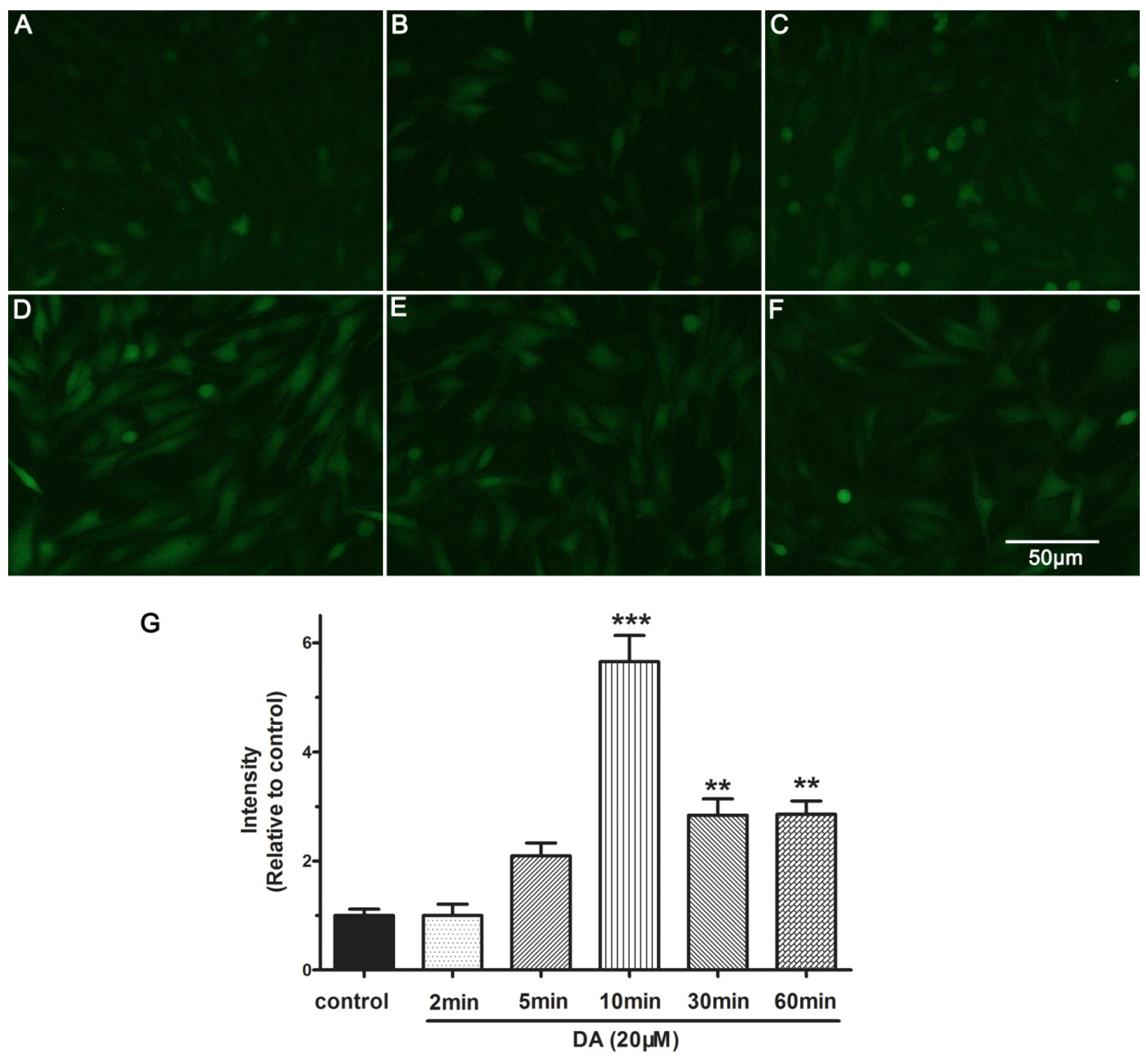
2.5. DA Promotes NO Release of PAECs

To determine whether NO release indeed is modulated by DA (20 μM), the intracellular NO production from 0 min to 60 min in PAECs was measured using DAF-FM DA fluorescence indicator. As shown in Figure 6A–F, DAF-FM DA fluorescence intensity was significantly increased from 2 min to 10 min, especially at 10 min, 5.65-fold compared with that at 0 min (control) (Figure 6D,G). However, the relative intensity of DAF-FM DA fluorescence rapidly decreased to 2.8-fold at 30 min post- exposure, and sustained to 60 min at this proximity level. The results indicated that DA stimulates NO release by achieving the maximum effect at 10 min. These demonstrated that DA might be a potent stimulus for eNOS activation in PAECs and took effect fast.
Figure 6. DA-induced NO production in PAECs within 60 min. NO production was visualized with fluorescence intensity. PAECs were treated with DA for different periods (A) 0 min (control); (B) 2 min; (C) 5 min; (D) 10 min; (E) 30 min; (F) 60 min. Results are presented as the mean ± SEM (n = 5) (G). ** p < 0.01 and *** p < 0.001 compared with the control group.
Molecules 19 08503 g006

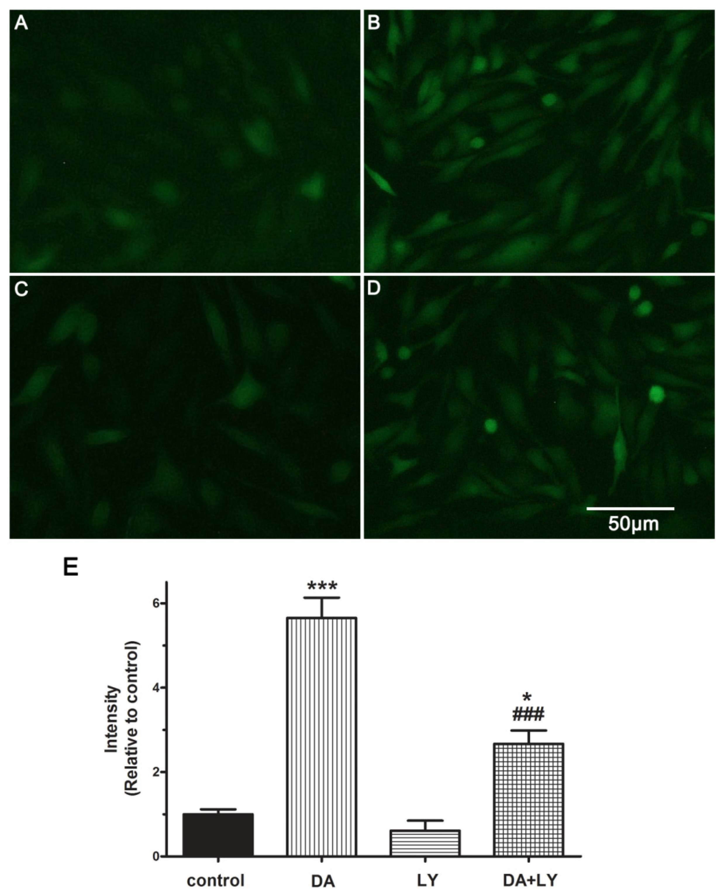
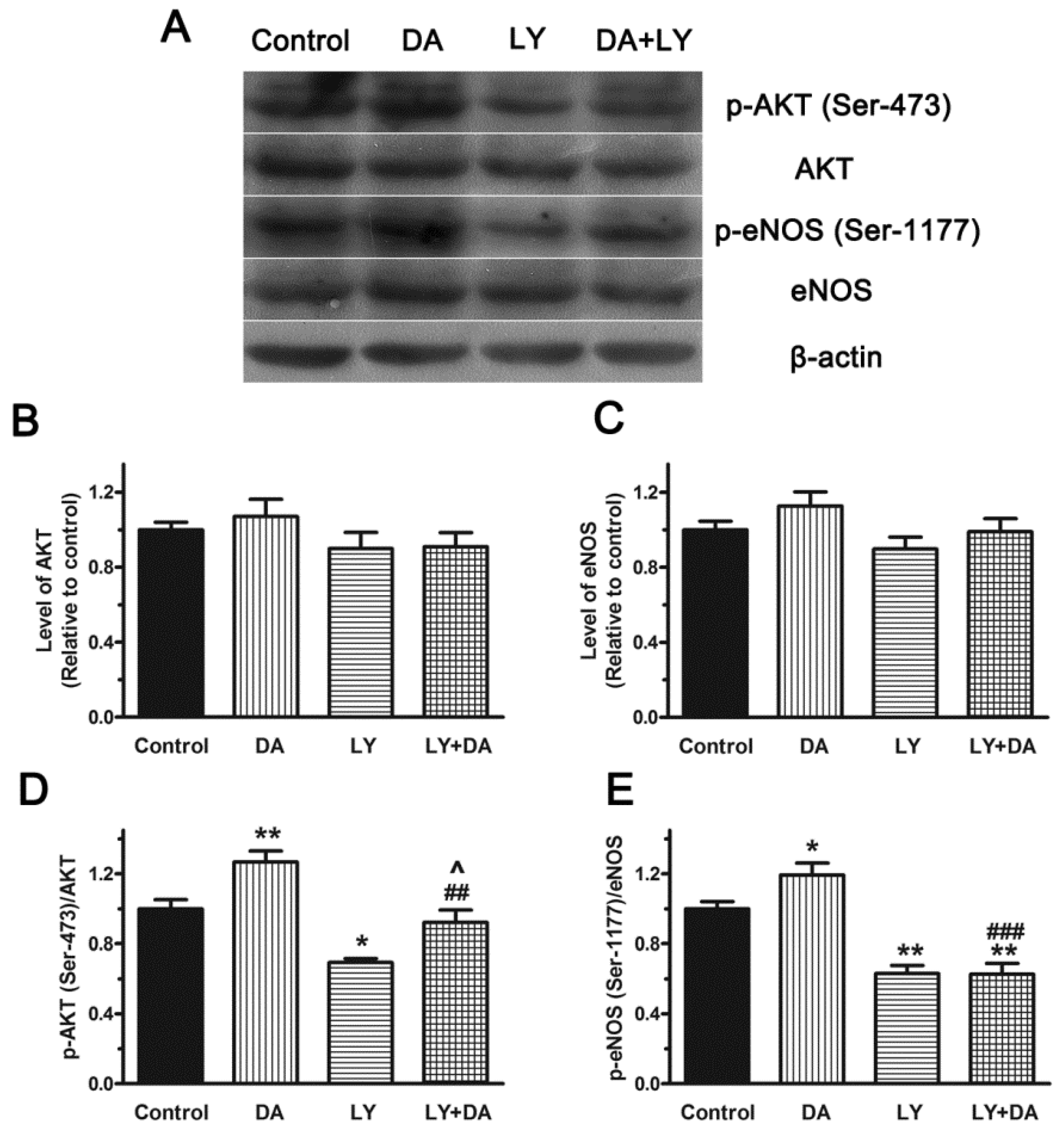
2.6. Role of the PI3K/Akt Pathway in DA-Induced Relaxation

Previous studies indicated that the activation of the PI3K/Akt pathway could promote eNOS phosphorylation leading to NO-induced relaxations [22,23]. To explore whether DA affects the PI3K/Akt pathway, we blocked the PI3K/Akt signaling pathway with LY294002 (LY, 10−6 M, Figure 7). The fluorescence intensity was increased by 5.65-fold compared with the control group after treatment with DA (20 μM) in 10 min. After the cells were exposed to LY for 10 min, the fluorescence intensity was reduced by 0.9-fold compared with the control group. When the PAECs were exposed to LY plus DA, the fluorescence intensity was decreased to 2.67-fold of control, significantly reduced compared with the DA group (Figure 7B,D,F, p < 0.01). The experimental data thus showed that PI3K/Akt inhibitor LY could reduce NO production enhanced by DA, which suggested that the PI3K/Akt pathway plays an important role in the NO synthesis.
Figure 7. DA (20 μM) stimulated NO production in PAECs via PI3K/Akt pathway in 10 min. DA stimulated the release of NO product (B) and LY294002 (LY 10−6 M) reduced NO production in PAECs (C) compared with the control group (A). However, LY could also reduce the stimulation of DA on NO production significantly (D). Results are presented as the mean ± SEM (n = 5) (E). * p < 0.05 and *** p < 0.001 compared with the control group. ### p < 0.001 versus DA group.
Molecules 19 08503 g007
In parallel, to better characterize the PI3K/AKT pathway involved in eNOS activation in response to DA, the expression of eNOS and Akt, as well as the phosphorylation level of eNOS (p-eNOS) and Akt (p-AKT) was assessed in PAECs using western blot analysis. Phosphorylation of Ser-473 on Akt is simultaneous with Akt activation and it was the most widely reported phosphorylation site according to previous reports [24,25]. Akt is an important determinant of eNOS phosphorylation at Ser-1177, leading to vascular smooth muscle relaxation [26]. So we firstly chose these phosphorylation sites for our study. Resullts showed that exposure of PAECs to DA (20 μM) in 10 min didn’t change the expression of Akt and eNOS (Figure 8B,C), but it caused the appearance of a strong phosphorylation signal of Akt (Ser-473) and eNOS (Ser-1177), and the values of Ser-473/AKT and Ser-1177/eNOS were 1.27 ± 0.11 and 1.19 ± 0.12 fold, respectively, compared with the corresponding control. When treated with LY (10−6 M) plus DA (20 μM), the value of Ser-473/AKT and Ser-1177/eNOS was reduced to 72.7% (p < 0.01) and 52.5% (p < 0.001) of control, respectively. It suggested that DA could activate eNOS through activation of the upstream PI3K/AKT signal pathway. However, the inhibitory effect of LY on the production of Ser-1177 was stronger than that of Ser-473, which implyed that other phosphorylation sites of AKT might also be activated by DA. All of these results indicated that phosphorylation activation of PI3K/Akt pathway is necessary for NO production induced by DA.
Figure 8. Role of the PI3K/Akt Pathway in DA-induced phosphorylation of AKT and eNOS in PAECs. PAECs were incubated with vehicle (DMSO), DA (20 μM), LY294002 (LY, 10−6 M) and DA (20 μM) plus LY (10−6 M) for 10 min. Then the levels of p-Akt (Ser-473), Akt, p-eNOS (Ser-1177) and eNOS (A) were determined by Western blot analysis. Results are presented as means ± SEM (n = 3) (B, C, D, E). * p < 0.05 and ** p < 0.01 versus control. ## p < 0.01 and ### p < 0.001 compared to corresponding DA group. ^ p < 0.05 compared to corresponding LY group.
Molecules 19 08503 g008

3. Experimental Section

3.1. General Information

The 1H-NMR and 13C-NMR spectra were recorded on a Bruker Avance 400 spectrometer (Bruker Company, Karlsruhe, Germany) in CDCl3 using tetramethylsilane (TMS) as an internal standard. The HR-ESI-MS data were obtained from a Waters LCT-Premier spectrometer (Waters Corporation, Milford, PA, USA) in ESI ionisation mode. The IR spectra were recorded on FTIR-8400S (Shimadzu Corporation, Kyoto, Japan). Optical rotations were determined with a WZZ-2B auto polarimeter (Shanghai Shenguang instrument Co. Ltd., Shanghai, China). Column chromatography was carried out on silica gel (200–400 mesh; Qingdao Haiyang Chemical Co. Ltd., Qingdao, China). Separations were monitored by TLC on GF254 plates (0.25 mm, Qingdao Haiyang Chemical Co. Ltd., Qingdao, China) and were visualized by UV inspection and or staining with 1% vanillin in concentrated sulphuric acid and heating. The tension data were recorded using the ALC-MPA force transducer (Shanghai Alcott Biotech Co, Ltd., Shanghai, China), which allowed eight rings to be recorded and analysed at the same time. The microscopic fluorescent images were taken by Nikon TE2000 Olympus fluorescent microscope (Nikon Corporation, Kyoto, Japan). Protein concentrations were measured with Bio-Rad protein assay kit (Bio-Rad Laboratories, Inc., Berkeley, CA, USA).

3.2. Materials and Reagents

The resin of C. opobalsamum was purchased in December 2011 from the AnGuo medicinal material market (Daqing, China). It was identified by Vice Professor Taiming Wei (Institute of Biological Pharmacy, Harbin Medical University, China). A voucher specimen (voucher number: ZY089) was deposited in the Institute of Biological Pharmacy, Harbin Medical University, China Herbarium. NG-Nitro-l-arginine Methyl Ester (l-NAME), fluorescent indicator 3-amino-4-aminomethyl-2',7'-difluorescein diacetate and LY294002 were purchased from Beyotime Institute of Biotechnology Co. Ltd. (Shanghai, China). Protein kinase B (Akt) antibody and phosphorylated Akt at Ser-473 (p-Akt) antibody, eNOS antibody and phosphorylated eNOS at Ser-1177 (p-eNOS) antibody were obtained from Cell Signaling Technology, Inc. (Danvers, MA, USA). All other reagents were from common commercial sources.

3.3. Extraction and Isolation

A bioassay-guided isolation approach was carried out following the earlier reported method [8]. The dried resin of C opobalsamum (4.0 kg) was exhaustively extracted with one volume of 95% EtOH under reflux (3 × 2 h). After removing the solvent under vacuum, the residue (722.0 g) was suspended in H2O and extracted successively with petroleum ether, CHCl3 and EtOAc to yield petroleum ether-soluble, CHCl3-soluble and EtOAc-soluble fractions. As the petroleum ether-soluble (112.3 g) fraction presented obvious PE-induced contraction activity on the pulmonary artery, this fraction was subjected to column chromatography on silica gel (4.0 kg) with a petroleum ether/ethyl acetate gradient (1:0 to 0:1, v/v). This generated eight fractions (Fr1–8) on the basis of the TLC profiles. The effects against PE-induced contraction of the pulmonary artery of all of the fractions were determined, and Fr3 showed a significant effect. Fr3 (16.9 g) was separated by silica gel column chromatography [petroleum ether–EtOAc (20:1, 10:1,4:1, 2:1, v/v)] to isolate compounds 1 (342 mg) and 2 (215 mg). Their structures were elucidated on the basis of spectroscopic methods (IR, 1H- and 13C-NMR, HR-ESI-MS).

3.4. Vessel Rings Preparation

The male Wistar rats (180–220 g) used in this study were treated as verified by the Institutional Animal Care and Use Committee (IACUC) of Harbin Medical University. According to previous literature [27], the rats were killed rapidly, and their mesentery and pulmonary arteries were quickly removed, cleaned of adherent connective tissues and cut into 3-mm-length rings under a dissecting microscope. The artery rings were maintained in ice-bathed Krebs solution containing, in mM, NaCl, 118.0; KCl, 4.7; KH2PO4, 1.2; MgSO4, 1.2; NaHCO3, 15.0; glucose, 5.5; and CaCl2, 2.5 (pH 7.4) throughout the whole process. Endothelium was damaged mechanically by a thin wire when preparing the endothelium-denuded rings. The endothelial integrity was assessed qualitatively by the degree of relaxation caused by carbachol (10−4 M) followed by treatment with phenylephrine (PE, 10−6 M). The rings were regarded as endothelium-denuded if they displayed less than 10% relaxation.

3.5. Effect of All the Parts on Pulmonary Artery Rings

All the fractions and compounds were dissolved in dimethyl sulfoxide (DMSO). PE and carbachol were dissolved in ultrapure water. Each artery ring was fixed on a force transducer and placed in a water-jacketed organ bath filled with 3 mL of Krebs solution and exposed to O2. The rings were initially stretched with a basal tension of 0.30 g for 30 min to achieve a state of equilibrium. After the vessel rings were contracted with 3 μL of PE for 15 min, the drugs (3 μL every time) were added to water-jacketed organ baths, and the tensions of each force transducer were recorded. The concentrations of the drugs were increased at 5 min increments. Carbachol (10−4 M) was used to detect the vasodilatory activity of the pulmonary artery after treated with drugs. Following are the formulas for the calculations of %contraction and %relaxation:
% Contraction = (F − F0)/(FPE − F0)
% Relaxation = 1 − % Contraction
F: Tension of the rings treated with drugs. F0: Pre-load. FPE: Tension of the rings treated with PE. (1): %Contraction (PE 10−6 M) = 100%; (2): %Relaxation (PE 10−6 M) = 0%.

3.6. Culture of Pulmonary Artery Endothelial Cells (PAECs)

PAECs were isolated from neonatal bovine obtained from a local abattoir. The calves’ use was in full compliance with the rules of the Ethics Committee of Laboratory Animals. PAECs were cultured in culture medium containing 20% fetal calf serum, penicillin (100 U/mL) and streptomycin (100 U/mL), supplemented with L-glutamine in 5% CO2 at 37 °C on culture flasks as previously described [28]. When the cells reached sub-confluence, they were pretreated with culture medium containing different concentrations of drugs that were tested in the experiments.

3.7. Measurement of NO Production in PAECs

DAF-FM DA as a fluorescence indicator was used to detect intracellular NO [29]. When PAECs grown on a microcoverglass of the 3 cm culture dish reached 80% confluence, cells were exposed to DA (20 μM) for 0 min, 2 min, 5 min, 10 min, 30 min and 60 min, respectively, then washed three times with phosphate-buffered (PBS, pH 7.4). After loading with 5 mM DAF-FM DA at 37 °C for 20 min in dark, the PAECs were rinsed three times with PBS and maintained in PBS throughout the experiments. The microscopic fluorescent images were taken by an Olympus fluorescent microscope at 495 nm (excitation) and 515 nm (emission) and analyzed with Image-Pro Plus software (Version 5.0). The average fluorescent density was measured to index the NO level [30].

3.8. Western Blotting

The expressions of nitric oxide synthase (eNOS) and protein kinase B (Akt), as well as the phosphorylation level of eNOS (Ser-1177) and Akt (Ser-473) in PAECs were assessed using western blotting as previously reported [25,31]. Briefly, after treatment with drugs, cells were washed three times with cold PBS and then lysed with alysis buffer (Tris 50 mM, pH 7.4, NaCl 150 mM, Triton X-100 1%, EDTA 1 mM and PMSF 2 mM) on ice. Lysates were sonicated for 1 min and then supernatant was collected by centrifugation at 13,500 rpm for 15 min at 4 °C. Total proteins (20 mg) were separated by SDS-PAGE and transferred to nitrocellulose membranes. After blocked with a blocking buffer containing 5% skimmed milk, the membranes were then incubated with the appropriate antibodies as follows: eNOS (1:1000), Akt (1:500), anti-phospho-Akt (Ser-473, 1:500), and anti-phospho-eNOS (Ser-1177, 1:1000). After maintained at 4 °C overnight, membranes were incubated with the secondary antibody (goat anti-rat IgG or goat anti-rabbit IgG) for 1 h at room temperature. Then the membranes were incubated with ECL and exposed to X-ray film in dark. Densitometric analyses were performed with the Bio-Rad Quality-One software.

3.9. Statistical Analysis

The data were expressed as the means ± SEM. Two-way ANOVA followed by Dunnett’s test was performed in the statistical analysis to study the effect of endothelium and L-NAME on DA-induced vasorelaxation and one-way ANOVA was used in other sections. A probability level of p < 0.05 was regarded as significantly different.

4. Conclusions

In conclusion, our study first found that DA and SA are the main constituents of C. opobalsamum that exhibits vascular effects, which may be regarded as an index for evaluating the quality of C. opobalsamum for blood stagnation. Both DA and SA could relax PE contracted pulmonary artery in a concentration-dependent manner. DA-induced vasodilation of pulmonary artery was endothelium-dependent and in accordance with the release of NO. It might be mediated by the activation of PI3K/AKT signaling pathway in endothelium.

Acknowledgments

This work was supported by the action plan project for the enterprise (2009CB522808) of the Ministry of Science and Technology of China and by the Innovation Fund Project for Graduate Student of Heilongjiang, China (Grant No.YJSCX20123-221HLJ).

Author Contributions

W.Y. Gao and T.M. Wei designed the research, supervised all the experiments and prepared the manuscript; X.Y. Dong, N. Xie, C.L. Zhou and Y.H. Fan were responsible for acquisition and analysis of the data; G.Y. Chen, Y.M Wang and D.L. Zhu contributed in guiding research and interpretating the data. All the authors have read and approved the final manuscript.

Conflicts of Interest

The authors declare no conflict of interest.

References

  1. Montani, D.; Gunther, S.; Dorfmuller, P. Pulmonary arterial hypertension. Orphanet J. Rare Dis. 2013, 8, 97. [Google Scholar] [CrossRef]
  2. Benza, R.; Miller, D.; Gomberg-Maitland, M.; Frantz, R.; Foreman, A.; Coffey, C.; Frost, A.; Barst, R.; Badesch, D.; Elliott, C.; et al. Predicting survival in pulmonary arterial hypertension: Insights from the registry to evaluate early and long-term pulmonary arterial hypertension disease management (REVEAL). Circulation 2010, 122, 164–172. [Google Scholar] [CrossRef]
  3. Al-Yahya, M.; Al-Meshal, L.; Mossa, J.; Al-Badr, A.; Tariq, M. Saudi Plants; King Saud University Press: Riyadh, Saudi Arabia, 1990. [Google Scholar]
  4. Shen, T.; Wan, W.; Yuan, H.; Kong, F.; Guo, H.; Fan, P.; Lou, H. Secondary metabolites from Commiphora opobalsamum and their antiproliferative effect on human prostate cancer cells. Phytochemistry 2007, 68, 1331–1337. [Google Scholar] [CrossRef]
  5. Abdul-Ghani, A.; Amin, R. Effect of aqueous extract of Commiphora opobalsamum on blood pressure and heart rate in rats. J. Ethnopharmacol. 1997, 57, 219–222. [Google Scholar] [CrossRef]
  6. Al-Howiriny, T.; Al-Sohaibani, M.; Al-Said, M.; Al-Yahya, M.; El-Tahir, K.; Rafatullah, S. Effect of Commiphora opobalsamum (L.) Engl. (Balessan) on experimental gastric ulcers and secretion in rats. J. Ethnopharmacol. 2005, 98, 287–294. [Google Scholar] [CrossRef]
  7. Al-Howiriny, T.; Al-Sohaibani, M.; Al-Said, M.; Al-Yahya, M.; El-Tahir, K.; Rafatullah, S. Hepatoprotective properties of Commiphora opobalsamum (“Balessan”), a traditional medicinal plant of Saudi Arabia. Drugs Exp. Clin. Res. 2004, 30, 213–220. [Google Scholar]
  8. Senejoux, F.; Demougeot, C.; Kerram, P.; Aisa, H.; Berthelot, A.; Bevalot, F.; Girard-Thernier, C. Bioassay-guided isolation of vasorelaxant compounds from Ziziphora clinopodioides Lam. (Lamiaceae). Fitoterapia 2012, 83, 377–382. [Google Scholar] [CrossRef]
  9. Hipolito, U.; Rodrigues, G.; Lunardi, C.; Bonaventura, D.; Ambrosio, S.; de Oliveira, A.; Bendhack, L.; da Costa, F.; Tirapelli, C. Mechanisms underlying the vasorelaxant action of the pimarane-ent-8(14),15-pimaradien-3β-ol in the isolated rat aorta. Eur. J. Pharmacol. 2009, 616, 183–191. [Google Scholar] [CrossRef]
  10. Ambrosio, S.; Tirapelli, C.; Bonaventura, D.; de Oliveira, A.; da Costa, F. Pimarane diterpene from Viguiera arenaria (Asteraceae) inhibit rat carotid contraction. Fitoterapia 2002, 73, 484–489. [Google Scholar] [CrossRef]
  11. Tirapelli, C.; dos Anjos Neto Filho, M.; Bonaventura, D.; Melo, M.; Ambrosio, S.; de Oliveira, A.; Bendhack, L.; da Costa, F. Pimaradienoic acid inhibits vascular contraction and induces hypotension in normotensive rats. J. Pharm. Pharmacol. 2008, 60, 453–459. [Google Scholar] [CrossRef]
  12. Su, S.; Duan, J.; Tang, Y.; Zhang, X.; Yu, L.; Jiang, F.; Zhou, W.; Luo, D.; Ding, A. Isolation and biological activities of neomyrrhaol and other terpenes from the resin of Commiphora myrrha. Planta Med. 2009, 75, 351–355. [Google Scholar]
  13. Fang, J.; Tsai, W.; Chebg, Y. Serratene triterpenes from Pinus armandii Bark. Phytochemistry 1991, 30, 1333–1335. [Google Scholar] [CrossRef]
  14. Cheung, H.; Toshio, M.; Mark, P. Further acidic constituents and neutral components of Pinus massoniana Resin. Tetrahedron 1993, 40, 7903–7907. [Google Scholar] [CrossRef]
  15. Ohwada, T.; Nonomura, T.; Maki, K.; Sakamoto, K. Dehydroabietic acid derivatives as a novel scaffold for large-conductance calcium-activated K+ channel openers. Bioorg. Med. Chem. Lett. 2003, 13, 3971–3974. [Google Scholar] [CrossRef]
  16. Rios, M.; Lopez-Martinez, S.; Lopez-Vallejo, F.; Medina-Franco, J.; Villalobos-Molina, R.; Ibarra-Barajas, M.; Navarrete-Vazquez, G.; Hidalgo-Figueroa, S.; Hernandez-Abreu, O.; Estrada-Soto, S. Vasorelaxant activity of some structurally related triterpenic acids from Phoradendron reichenbachianum (Viscaceae) mainly by NO production: Ex vivo and in silico studies. Fitoterapia 2012, 83, 1023–1029. [Google Scholar] [CrossRef]
  17. Scalera, F.; Martens-Lobenhoffer, J.; Tager, M.; Bukowska, A.; Lendeckel, U.; Bode-Boger, S. Effect of l-arginine on asymmetric dimethylarginine (ADMA) or homocysteine-accelerated endothelial cell aging. Biochem. Biophys. Res. Commun. 2006, 345, 1075–1082. [Google Scholar] [CrossRef]
  18. Koh, S.; Kang, M.; Kim, T.; Park, H.; Park, C.; Seong, Y.; Seong, H. Endothelium-dependent vasodilatory and hypotensive effects of Crotalaria sessiliflora L. in rats. Biol. Pharm. Bull. 2007, 30, 48–53. [Google Scholar] [CrossRef]
  19. Kwan, C.; Zhang, W.; Sim, S.; Deyama, T.; Nishibe, S. Vascular effects of Siberian ginseng (Eleutherococcus senticosus): Endothelium-dependent NO- and EDHF-mediated relaxation depending on vessel size. Naunyn Schmiedebergs Arch. Pharmacol. 2004, 369, 473–480. [Google Scholar] [CrossRef]
  20. Vasques, C.; Cortes, S.; Silva, M.; de Medeiros, I. Muscarinic agonist properties of the hydrobutanol extract from aerial parts of Waltheria viscosissima St. Hil. (Sterculiaceae) in rats. Phytother. Res. 1999, 13, 312–317. [Google Scholar] [CrossRef]
  21. Jiang, F.; Li, C.G.; Rand, M.J. Mechanisms of nitric oxide-independent relaxations induced by carbachol and acetylcholine in rat isolated renal arteries. Br. J. Pharmacol. 2000, 130, 1191–1200. [Google Scholar] [CrossRef]
  22. Kim, K.; Moriarty, K.; Bender, J. Vascular cell signaling by membrane estrogen receptors. Steroids 2008, 73, 864–869. [Google Scholar] [CrossRef]
  23. Ndiaye, M.; Chataigneau, M.; Lobysheva, I.; Chataigneau, T.; Schini-Kerth, V. Red wine polyphenol-induced, endothelium-dependent NO-mediated relaxation is due to the redox-sensitive PI3-kinase/Akt-dependent phosphorylation of endothelial NO-synthase in the isolated porcine coronary artery. FASEB J. 2005, 19, 455–457. [Google Scholar]
  24. Jin, S.N.; Wen, J.F.; Wang, T.T.; Kang, D.G.; Lee, H.S.; Cho, K.W. Vasodilatory effects of ethanol extract of Radix Paeoniae Rubra and its mechanism of action in the rat aorta. J. Ethnopharmacol. 2012, 142, 188–193. [Google Scholar] [CrossRef]
  25. Rattmann, Y.; Anselm, E.; Kim, J.; Dal-Ros, S.; Auger, C.; Miguel, O.; Santos, A.; Chataigneau, T.; Schini-Kerth, V. Natural product extract of Dicksonia sellowiana induces endothelium-dependent relaxations by a redox-sensitive Src- and Akt-dependent activation of eNOS in porcine coronary arteries. J. Vasc. Res. 2012, 49, 284–298. [Google Scholar] [CrossRef]
  26. Luo, Z.; Fujio, Y.; Kureishi, Y.; Rudic, R.D.; Daumerie, G.; Fulton, D.; Sessa, W.C.; Walsh, K. Acute modulation of endothelial Akt/PKB activity alters nitric oxide- dependent vasomotor activity in vivo. J. Clin. Investig. 2000, 106, 493–499. [Google Scholar] [CrossRef]
  27. Bai, Y.; Lu, P.; Han, C.; Yu, C.; Chen, M.; He, F.; Yi, D.; Wu, L. Hydroxysafflor Yellow A (HSYA) from Flowers of Carthamus tinctorius L. and Its Vasodilatation Effects on Pulmonary Artery. Molecules 2012, 17, 14918–14927. [Google Scholar] [CrossRef]
  28. Wang, Z.; Tang, X.; Li, Y.; Leu, C.; Guo, L.; Zheng, X.; Zhu, D. 20-Hydroxyeicosatetraenoic acid inhibits the apoptotic responses in pulmonary artery smooth muscle cells. Eur. J. Pharmacol. 2008, 588, 9–17. [Google Scholar] [CrossRef]
  29. Kojima, H.; Urano, Y.; Kikuchi, K.; Higuchi, T.; Hirata, Y.; Nagano, T. Fluorescent indicators for imaging nitric oxide production. Angew. Chem. Int. Ed. Engl. 1999, 38, 3209–3212. [Google Scholar] [CrossRef]
  30. Sheng, B.; Gong, K.; Niu, Y.; Liu, L.; Yan, Y.; Lu, G.; Zhang, L.; Hu, M.; Zhao, N.; Zhang, X.; et al. Inhibition of γ-secretase activity reduces Aβ production, reduces oxidative stress, increases mitochondrial activity and leads to reduced vulnerability to apoptosis:Implications for the treatment of Alzheimer’s disease. Free Radic. Biol. Med. 2009, 46, 1362–1375. [Google Scholar] [CrossRef]
  31. Yao, L.; Lu, P.; Li, Y.; Yang, L.; Feng, H.; Huang, Y.; Zhang, D.; Chen, J.; Zhu, D. Osthole relaxes pulmonary arteries through endothelial phosphatidylinositol 3-kinase/Akt-eNOS-NO signaling pathway in rats. Eur. J. Pharmacol. 2013, 699, 23–32. [Google Scholar] [CrossRef]
  • Sample Availability: Samples of the dehydroabietic acid (DA) and sandaracopimaric acid (SA) are available from the authors.

