Article

Intramolecular Hydrogen Bond in Biologically Active o-Carbonyl Hydroquinones

by
Maximiliano Martínez-Cifuentes
1,*,
Boris E. Weiss-López
2,
Leonardo S. Santos
1 and
Ramiro Araya-Maturana
3,*
1
Laboratorio de Síntesis Asimétrica, Instituto de Química de los Recursos Naturales, Universidad de Talca, Talca, Casilla 747, Chile
2
Departamento de Química, Facultad de Ciencias, Universidad de Chile, Santiago, Casilla 653, Chile
3
Departamento de Química Orgánica y Fisicoquímica, Facultad de Ciencias Químicas Y Farmacéuticas, Universidad de Chile, Santiago, Casilla 233, Chile
*
Authors to whom correspondence should be addressed.
Molecules 2014, 19(7), 9354-9368; https://doi.org/10.3390/molecules19079354
Submission received: 9 May 2014 / Revised: 18 June 2014 / Accepted: 27 June 2014 / Published: 3 July 2014
(This article belongs to the Special Issue Intramolecular Hydrogen Bonding)

Abstract

:
Intramolecular hydrogen bonds (IHBs) play a central role in the molecular structure, chemical reactivity and interactions of biologically active molecules. Here, we study the IHBs of seven related o-carbonyl hydroquinones and one structurally-related aromatic lactone, some of which have shown anticancer and antioxidant activity. Experimental NMR data were correlated with theoretical calculations at the DFT and ab initio levels. Natural bond orbital (NBO) and molecular electrostatic potential (MEP) calculations were used to study the electronic characteristics of these IHB. As expected, our results show that NBO calculations are better than MEP to describe the strength of the IHBs. NBO energies (∆Eij(2)) show that the main contributions to energy stabilization correspond to LP→σ* interactions for IHBs, O1O2-H2 and the delocalization LP→π* for O2-C2 = Cα(β). For the O1O2-H2 interaction, the values of ∆Eij(2) can be attributed to the difference in the overlap ability between orbitals i and j (Fij), instead of the energy difference between them. The large energy for the LP O2→π* C2 = Cα(β) interaction in the compounds 9-Hydroxy-5-oxo-4,8, 8-trimethyl-l,9(8H)-anthracenecarbolactone (VIII) and 9,10-dihydroxy-4,4-dimethylanthracen-1(4H)-one (VII) (55.49 and 60.70 kcal/mol, respectively) when compared with the remaining molecules (all less than 50 kcal/mol), suggests that the IHBs in VIII and VII are strongly resonance assisted.

Graphical Abstract

1. Introduction

Hydroquinones (HQ) and their oxidized form, quinones (Q), constitute a biologically relevant redox pair. A number of them come from natural sources [1,2] and exhibit a large number of biological activities related to their redox potential [3,4,5,6]. Although p-hydroquinone is more stable than p-quinone, usually substituted p-hydroquinones (p-HQ) are thermodynamically less stable than substituted p-quinones (p-Q), but p-Q can be effectively transformed into p-HQ by several mechanisms in biological systems [7], and therefore they can co-exist inside living organisms. The biological activity of hydroquinones has been related to their capability to lose an electron followed by deprotonation (or alternatively lose a hydrogen atom), to afford the corresponding semiquinone radical. These intermediates have been associated to biological properties, such as pro-oxidant activity, by interacting with several intracellular molecules, such as DNA and proteins.
Modulation of the electron-transfer capability is very important for the biological activity of quinones and hydroquinones. Among the interactions that play a central role in this issue, the formation of inter- or intramolecular hydrogen bonds in these molecules plays a key role [8,9,10]. A recent electrochemical study about quinones possessing intramolecular hydrogen bonds (IHBs) shows that this interaction stabilizes the anion radical structure, leading to a shift in reduction potentials toward less negative values when compared with quinones without IHBs [11]. IHBs have shown appreciable effects on the antioxidant properties of hydroquinones and related phenols [12,13].
The strength of a hydrogen bond lies between a weak covalent bond and Van der Waals interactions [14], and plays an important role in the geometry of single molecules as well as in the molecular structure of liquids and solids. Hydrogen bonds are important in areas as diverse as biology, chemistry and material science [15]. By definition, a hydrogen bond is an attractive interaction of the X-HY type, where the molecular fragment X-H acts as a hydrogen bond donor and Y acts as a hydrogen bond acceptor [16].
The o-carbonyl hydroquinone moiety is an important structural feature of several natural products with different biological activities, such as doxorubicin, daunorubicin [17], 2,5-dihydroxyacetophenone [18] and peyssonol A [19,20]. o-Carbonyl hydroquinones have also been used as building blocks for natural [21,22] and synthetic [23,24,25,26] compounds with a diversity of biological properties. In previous works, our group showed that some o-carbonyl hydroquinones can inhibit some tumor cell growth acting at the mitochondrial level [27,28,29]. Also theoretical and experimental NMR studies of some related hydroquinones has been carried out [30]. The IHBs present in these compounds, have been invoked as a key factor for their mitochondrial-mediated anti-cancer activity [31,32].
On the basis of the above considerations, it appears interesting to study the effect of the molecular structure on the characteristics of the IHBs present in a series of o-carbonyl hydroquinones (Figure 1). Therefore, the aim of this work is to study experimentally the IHBs in a series of structurally related o-carbonyl hydroquinones and one aromatic lactone, through the use of nuclear magnetic resonance (NMR). Several theoretical approaches can be used to study IHBs, for instance atoms in molecules (AIM) methodology [33,34]. Another scarcely explored methodology is through the use of molecular electrostatic potential (MEP), though this methodology has been mainly used for intermolecular HBs [35,36], more recently it has also been used to study intramolecular HBs [37]. The interesting results from this study made us decide the use this methodology. Besides, we have also used the natural bond orbitals (NBO) methodology, a widely used technique to study IHBs [38].
Figure 1. Structure of compounds studied in this work.
Molecules 19 09354 g001

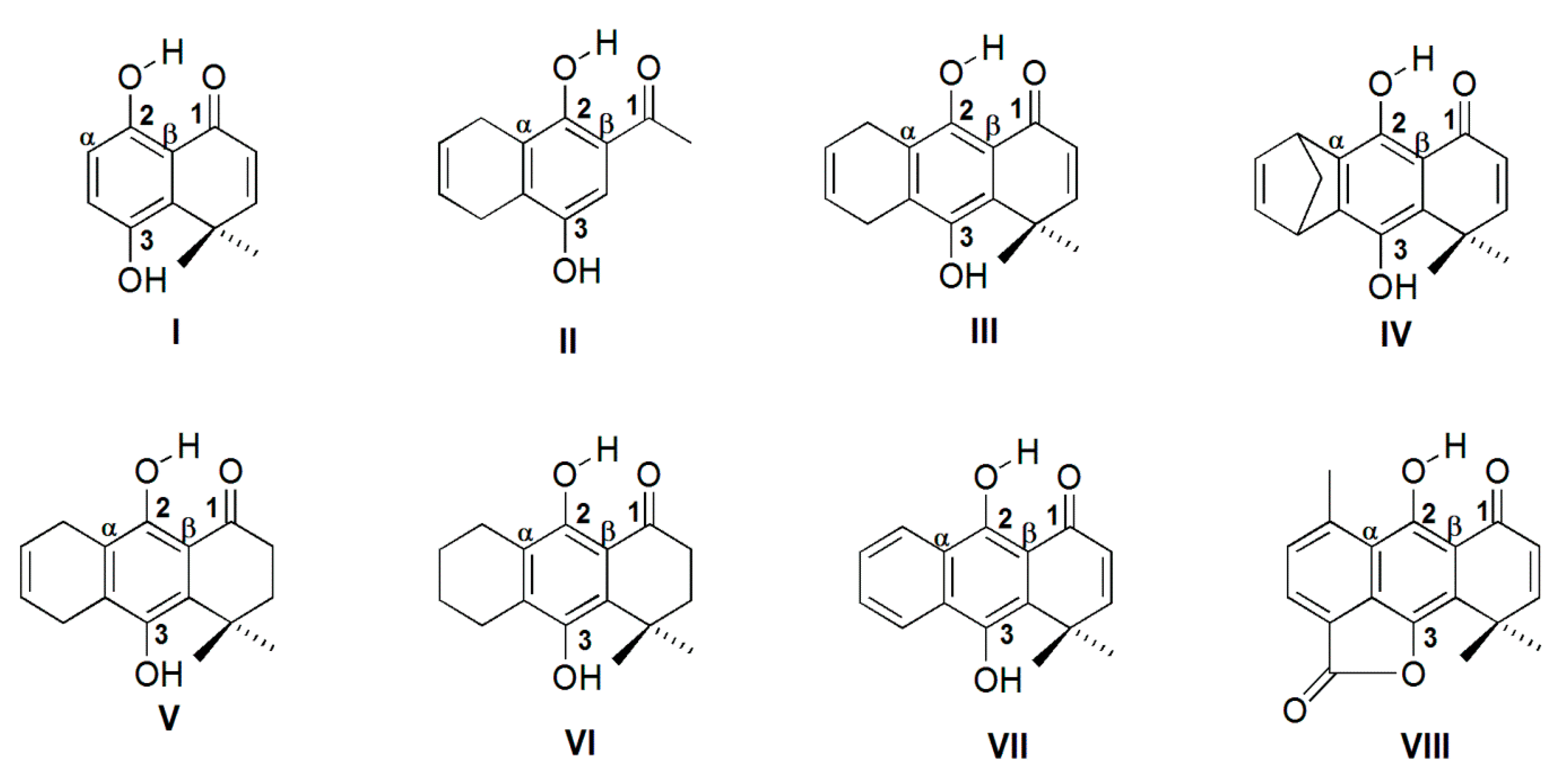
2. Results and Discussion

All the molecules studied here, containing IHBs (Figure 1), can be classified as resonance-assisted hydrogen bonds (RAHBs) [39,40] although this concept has been questioned in recent years [41,42,43,44]. RAHBs are characterized as conjugated molecular fragments connected through the hydrogen bond donor, which provokes a strong hydrogen bond compared with a system without the conjugation. We will take this definition into account in further analysis.

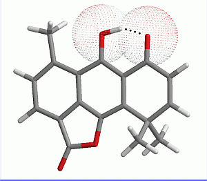
2.1. Geometry Optimization

The optimized geometry of all molecules have been obtained at the B3LYP/6-31++G(d,p) and MP2/6-31++G(d,p) levels of theory. The main calculated geometrical parameters for the characterization of IHBs, besides the experimental 1H-NMR shifts for H2 (Figure 1), are summarized in Table 1. While chloroform is a hydrogen bond donor, it is classified as a weak one (Abraham’s H donor parameter α = 0.15) [45], therefore it does not represent a significant competition to the strong IHBs present in this molecules. Therefore, the 1H-NMR chemical shift of H2 is a suitable parameter to represent the strength of the IHBs. We also measured the NMR spectra of compound I, which is not one of the strongest IHBs in the series, in DMSO-d6, a HB acceptor. The chemical shift for H2 was 12.55 ppm, only 0.01 ppm away from the value measured in chloroform (12.54 ppm). This observation shows that the studied IHBs remain unchanged, even in DMSO.
Table 1. 1H-NMR chemical shifts for H2 and geometrical parameters for hydrogen bonds calculated at B3LYP/6-31++G(d,p) and MP2/6-31++G(d,p) level of theory. The numbering of compounds is according to Figure 1.
MoleculeδH2B3LYP/6-31++G(d,p)MP2/6-31++G(d,p)
O1O2O2-H2O1H2< O2-H2O1O1O2O2-H2O1H2< O2-H2O1
I12.542.5400.9961.6381482.5730.9891.682148
II12.322.5560.9941.6571482.5920.9881.703148
III13.082.5330.9981.6241492.5670.9911.667149
IV12.702.5380.9961.6341492.5710.9901.679148
V12.952.5250.9971.6171492.5700.9891.676148
VI12.942.5210.9971.6131492.5210.9971.613149
VII14.532.5051.0051.5841502.5430.9951.637149
VIII15.602.4821.0141.5441522.5260.9991.608150
R2 0.890.980.940.920.390.840.500.59
Distances in Å, Angle in °, δ in ppm. R2 corresponds to correlation between NMR δH2 and geometrical parameters.
An inspection to Table 1 shows that the boundary cases are well described by both the MP2 and DFT methods. The largest chemical shift of VIII is in agreement with the shortest O1H2 distance, which indicates the strongest IHB. On the other hand, the lowest chemical shift of II accords with the largest O1H2 distance, showing that II has the weakest IHB among these HQs. When all molecules are compared, B3LYP/6-31++G(d,p) calculations are more suitable to describe the IHB geometrical parameters, according with the quantitative correlation of their strength with geometrical parameters and NMR chemical shift data (see correlation coefficients in Table 1). Because B3LYP/6-31++G(d,p) optimized geometries gave better correlations with experimental NMR data, we used these results for further calculations. The main features of the IHBs in this series of molecules, were explored through the use of NBO and MEP calculations.

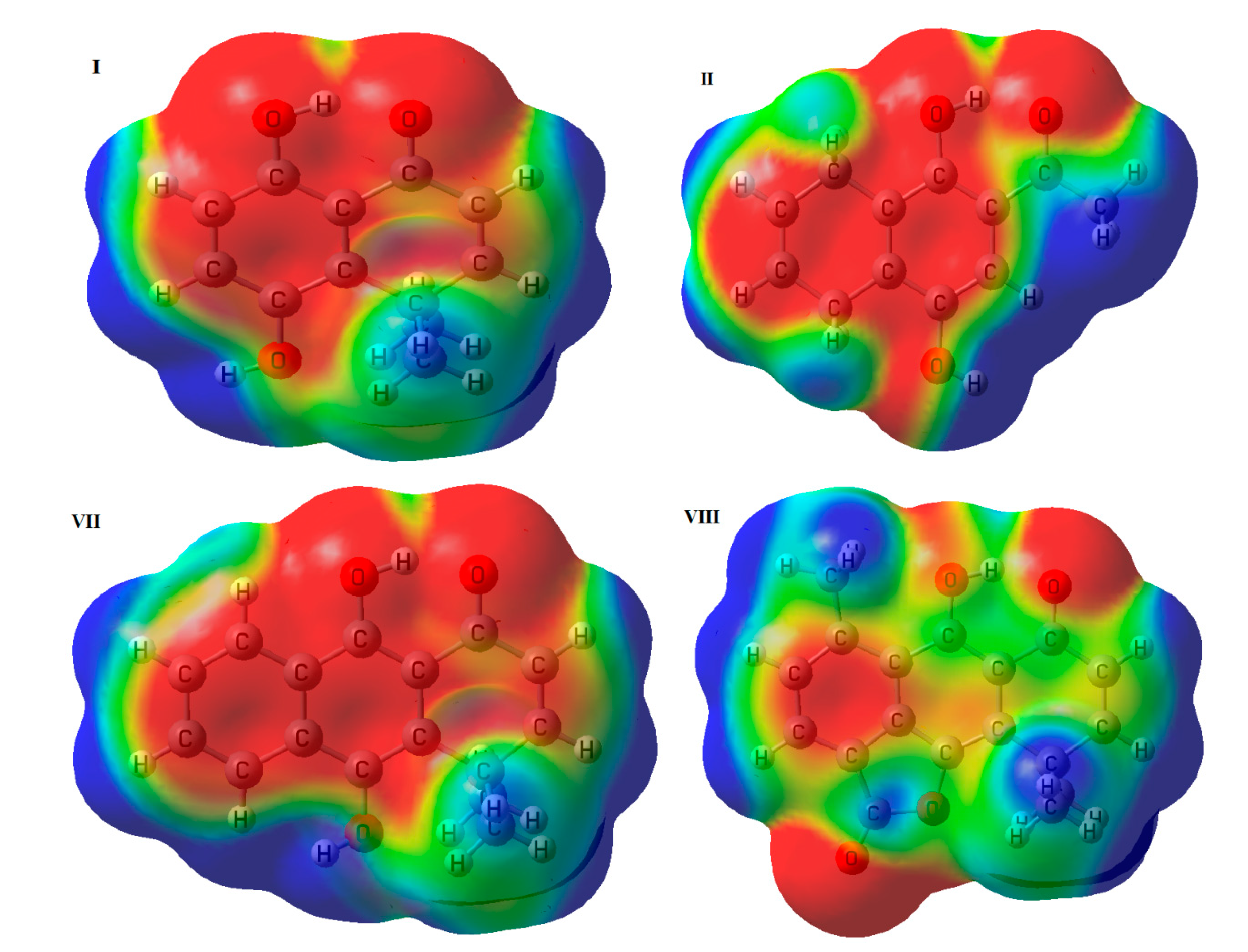
2.2. Molecular Electrostatic Potential

Because the electrostatic characteristic is always present in hydrogen bonds, several methods based in the electrostatic potential have been developed for their study [46,47,48,49]. MEP maps have been used to qualitatively rationalize trends observed in hydrogen bond donors and acceptors [50,51,52]. The MEP maps displayed in Figure 2, were generated projecting the color-coded values onto the 0.004 a.u. iso-potential energy surface. The red color indicates high electron-density sites, blue color indicates low electro-density sites and green-yellow color indicates neutral sites in the molecules. In this Figure, the MEPs of I and II, which present the weakest IHB, and the MEPs of VII and VIII, which present the strongest IHB, are shown as representative examples. Electron-rich sites are observed in the aromatic rings. The IHB site shows a remarkably electron-rich zone in both the donor and acceptor oxygens, while a small neutral zone appears on the hydrogen of the IHB from I, II and VII. Unlike the previous cases, VIII presents a more extended neutral zone on the hydrogen atom and the electron-rich zone around the oxygens is reduced.
Figure 2. Molecular electrostatic potential (0.004 a.u.) of I, II, VII and VIII.
Molecules 19 09354 g002
Quantitative MEP descriptors, such as the minimized electrostatic potential (Vmin) and the recently described Vα(r) parameter, have been used to characterize hydrogen bond basicity and acidity, respectively [53,54,55]. The Vα(r), calculated for H2 at r = 0.55Å, and the value of Vmin near O1 for all molecules are presented in Table 2. From this Table we observe, as a general tendency, that an increase of Vα(r), and therefore an increase of IHB donor strength, leads to a decrease in Vmin, and therefore lowering the IHB acceptor strength. This trend indicates that structural differences among the molecules, such as the presence of an additional aromatic ring in VII and VIII, affects both, the donor and acceptor hydrogen bond capabilities. The strong IHB exhibited by VIII, is in agreement with the higher value of Vα(r) (higher acidity of donor) and the lower value for Vmin(O2) (higher basicity of acceptor). Nevertheless, considering both parameters separately for all molecules, they do not correlate with the NMR data. In both cases, we could not find a lineal relationship between δH2 and Vα(r) or between δH2 and Vmin(O2), being R2 = 0.37 and R2 = 0.05 respectively. This can be explained because Vmin and the Vα(r) descriptors are significantly perturbed by the IHB. Regions of positive and negative MEP on the surfaces of hydrogen bond donors and acceptors, are influenced by the formation of intramolecular contacts in these molecules. The trends of Vα(r) and Vmin with the increase of HB interaction point in the opposite direction, and explain why MEP is not an appropriate descriptor for IHB strength.
Table 2. MEP values (B3LYP/6-31+G**//B3LYP/6-31++G**), Vmin and Vα(r) (kcal/mol).
MoleculeVα(r)Vmin(O1)
I165.0−48.9
II169.2−45.0
III163.2−50.5
IV161.7−51.3
V165.7−48.2
VI164.1−49.2
VII166.2−49.5
VIII174.9−43.6

2.3. NBO Analysis

The NBO analysis results, natural charges and Wiberg bond orders, are presented in Table 3. Table 4 shows the calculated stabilization energies. Correlations between natural charges and Wiberg bond order (WBO) for the atoms involved in the IHB, and the experimental δH2, were studied. The correlations between δH2 and natural charge on O1, O2 and H2 gave R2 values of 0.81, −0.09 and 0.90, respectively. These results show that the natural charge on the hydrogen atom involved in the IHB is a better parameter than the natural charge on the donor and acceptor oxygens in order to quantify the strength of the IHB. On the other hand, WBO for O2-H2 and H2O1 were shown to be excellent parameters for describing the strength of the IHB in this HQ series. In effect, the correlations of δH2 with O2-H2 and with H2O1 gave R2 = 0.99 and R2 = 0.98, respectively.
Table 3. Natural charges (NC) and Wiberg bond order (WBO) at HF/6-311G** //B3LYP/6-31++G** level for selected atoms in HQs.
MoleculeNC O1NC O2NC H2WBO O2-H2WBO H2O1
I−0.721−0.7530.5220.64700.0699
II−0.717−0.7590.5240.65010.0647
III−0.726−0.7650.5250.63950.0747
IV−0.727−0.7600.5220.64600.0712
V−0.725−0.7630.5240.63930.0751
VI−0.726−0.7650.5230.64020.0757
VII−0.734−0.7600.5300.61970.0890
VIII−0.736−0.7550.5320.60040.1051
Analyses of the second order stabilization energies ∆Eij(2) (Table 4) allow us to determine the orbital interaction responsible for the IHB. The main hyperconjugative interaction was LPO1→σ*O2-H2. Also, a significant hyperconjugative interaction of type LPO2→σ*C2=Cα(β) which accounts for the delocalization of phenolic oxygen electrons into the aromatic ring, is present. Accordingly, the main contributions to stabilization energy corresponds to the LP→σ* interaction for O1O2-H2 IHB as well as the LP→π* delocalization is the main contribution for the O2-C2 = Cα(β) fragment. It can be noticed that the stabilization energy ∆Eij(2) due the IHB formation is higher for VIII and VII, which present an additional aromatic ring fused to the hydroquinone ring. From the above, it is possible to argue that these IHBs are strongly assisted by resonance, involving the additional ring, which is supported by the high stabilization energy for LPtotal(1 + 2) O2→π* C2=Cα(β) in VIII and VII (55.49 and 60.70 Kcal/mol respectively) compared with values found in all remaining molecules, all with less than 50 kcal/mol stabilization energy. It is interesting to compare these results with a recently published work about 1-acylthiourea species, where two conformations with different competing IHB are feasible [56]. It was found that those conformation where IHB was assisted by resonance, presented a stabilization energy corresponding to LP O→σ*H-N around 12 kcal/mol higher than those conformation without resonance assisted IHB.
Table 4. Stabilization energies (kcal/mol) for selected NBO pairs (donor-acceptor) given by second order perturbation energies of the Fock matrix in the NBO basis for the HQs (HF/6-311G**//B3LYP/6-31++G**).
MoleculeΦiΦj∆Eij (2)εj–εi/auFij/auΦiΦj∆Eij (2)εj−εi/auFij/au
ILP1 O1σ* O2-H24.041.580.072LP1 O2σ* C2-Cα10.151.600.114
LP2 O1σ* O2-H228.331.180.165LP2 O2π* C2-Cα48.430.630.168
IILP1 O1σ* O2-H23.671.580.068LP1 O2σ* C2-Cα9.191.610.109
LP2 O1σ* O2-H226.021.181.159LP2 O2π* C2-Cα48.190.640.170
IIILP1 O1σ* O2-H24.081.570.072LP1 O2σ* C2-Cα9.421.610.110
LP2 O1σ* O2-H230.581.180.171LP2 O2π* C2-Cα47.570.640.167
IVLP1 O1σ* O2-H24.051.580.072LP1 O2σ* C2-Cα10.081.580.113
LP2 O1 σ* O2-H229.011.180.167LP2 O2π* C2-Cβ43.470.670.161
VLP1 O1σ* O2-H24.101.570.072LP1 O2σ* C2-Cα9.461.610.110
LP2 O1σ* O2-H230.881.180.173LP2 O2π* C2-Cα49.060.640.170
VILP1 O1σ* O2-H24.151.570.072LP1 O2σ* C2-Cα9.671.600.111
LP2 O1σ* O2-H231.311.190.174LP2 O2π* C2-Cα48.220.630.169
VIILP1 O1σ* O2-H24.361.540.074LP1 O2σ* C2-Cα9.701.630.112
LP2 O1σ* O2-H237.441.170.189LP2 O2π* C2-Cα55.490.650.178
VIIILP1 O1σ* O2-H24.641.510.076LP1 O2σ* C2-Cα9.821.600.112
LP2 O1σ* O2-H245.671.160.208LP2 O2π* C2-Cα60.700.640.184
A good correlation is observed between the NMR chemical shift of H2 and the stabilization energies for LPtotalO1→σ* interaction in the O2-H2 fragment (R2 = 0.98). The energy difference between the donor (Φi) and the acceptor (Φj) orbitals, and their overlap, determine the hyperconjugation energy. Lower difference in energy εj–εI as well as high overlap between them (Fij) favors hyperconjugation. For the LP O1→σ* O2-H2 interaction, εj–εI are very similar for all molecules, so that the difference among ∆Eij(2) can be attributed to the difference in the overlap ability between LP O1 and σ* O2-H2 orbitals, given by the Fij value (Table 4).

3. Experimental

3.1. General Information

Melting points were determined on a hot-stage apparatus and are uncorrected. The IR spectra were recorded on a FT-IR Bruker IFS 55 spectrophotometer from KBr discs; wave numbers are reported in cm−1. 1H-NMR and 13C-NMR spectra were obtained from a Bruker DRX-300 spectrometer (300 and 75 MHz, respectively) in CDCl3. Chemical shifts were recorded in ppm (δ) relative to TMS as internal standard. J-values are given in Hz. Electron impact (IE) high resolution mass spectra were recorded on a Thermo Finnigan model MAT 95XP Mass Spectrometer. Compounds IV, VII and VIII were previously described [57,58,59,60], and new compounds V and VI were synthesized as follows.
9,10-Dihydroxy-4,4-dimethyl-3,4,5,8-tetrahydroanthracen-1(2H)-one (V). Butadiene was bubbled through a solution of 8,8-dimethyl-6,7-dihydro-1,4,5(8H)-naphthalenetrione (100 mg, 0.49 mmol) in toluene (10 mL), and the mixture left in a sealed flask at room temperature for a week. Then silica gel (1 g) was added and the mixture stirred overnight at room temperature. The mixture was filtered and the solid washed with dichloromethane. Evaporation of the solvent gave crude of V. The pure product (72 mg, 57%) was obtained by column chromatography. 1H-NMR δ (CDCl3): 1.50 (s, 6H, 2 × CH3), 1.93 (t, 2H, J = 7 Hz, 3-CH2), 2.67 (t, 2H, J = 7 Hz, 2-CH2), 3.18–3.33 (m, 4H, 5- and 8-CH2), 4.29 (s, 1H, 10-OH), 5.83 (bd, 1H, J = 10 Hz, 6- or 7-H), 5.96 (bd, 1H, J = 10 Hz, 6- or 7-H), 12.95 (s, 1H, 9-OH). 13C-NMRδ (CDCl3): 27.21, 23.59, 24.84, 33.85, 34.98, 38.69,121.11, 121.82, 124.52, 142.98, 131.97, 155.23, 205.13. HRMS: m/z [M+] calcd. For C16H18O3: 258.1256; found: 258.1249. IR (KBr, cm−1): 1217, 1614, 2926, 3390. m.p. 130–132 °C.
9,10-Dihydroxy-4,4-dimethyl-3,4,5,6,7,8-hexahydroanthracen-1(2H)-one (VI). Hydroquinone VI was obtained by hydrogenation, stirring a mixture of III (100 mg, 0.39 mmol) and 10% Pd/C (25 mg) in ethanol (30 mL), for 4 h under 20 bar of H2. The crude product was purified by flash column chromatography, eluting with hexane-ethyl acetate 8:1 yielding pure VI (40 mg, 38%). 1H-NMR δ (CDCl3): 1.50 (s, 6H, 2 CH3), 1.69–1.87 (m, 4H, 6- and 7-CH2), 1.91 (t, 2H, J = 7 Hz, 3-CH2), 2.57 (t, 2H, J = 7 Hz, 2-CH2), 2.65 (t, 4H, J = 7 Hz, 5- and 8- CH2), 4.38 (s, 1H, 10-OH), 12.94 (s, 1H, 9-OH). HRMS: m/z [M+] calcd. For C16H20O3: 260.14124; found: 260.14045. IR(KBr): 1610, 2927, 3308 cm−1. m.p. 192–193.5 °C.

3.2. Theoretical Methods

The calculations were carried out using the Gaussian03 [61] program package, running in a Microsystem cluster of blades. Geometries were optimized at Møller-Plesset second-order-corrected [62] (MP2) ab-initio level, and Becke three-parameter Lee-Yang-Parr [63] (B3LYP) density functional theory (DFT) level. 6-31++G** basis set was used in both cases. We carried out the calculation in vacuum because this model is commonly associated to aprotic non-polar solvents, like chloroform and because the energies of molecules in both models are very similar [64]. No imaginary vibrational frequencies were found at the optimized molecular geometries, which indicate that they are true minima of the potential energy surface. The theoretical study of intramolecular hydrogen bond was carried out through the calculation of MEP and a NBO analysis.
The MEP is related to the electron density and it has been widely used to study hydrogen bonds [51], reactivity [65], and to correlate biological activity with molecular structure [66,67]. The MEP minimum (Vmin) is computed from the optimized geometries, using equation 1 at the B3LYP/6-31+G(d,p) level of theory, which has been described as adequate for this kind of calculations [55]:
Molecules 19 09354 i001
here ZA is the nuclear charge and ρ(r) the electron density. Vmin has been described [53,54] as a useful predictor of hydrogen bond acceptor basicity. Recently, it has been proposed that the Vα(r) descriptor [55], calculated at a distance of 0.55 Å from the hydrogen atom along the O-H bond, is also useful to predict hydrogen bond donor acidity.
On the other hand, the NBO method has been recognized as a powerful tool to gain insights into orbital interactions, such as stabilization energies caused by electron transfer and hyperconjugation stabilization energies [68,69]. The NBOs are one of the consequences of natural localized orbital sets that include natural atomic (NAO), hybrid (NHO) and semi-localized molecular orbital (NLMO) sets, intermediates between basis atomic orbitals (AOs) and canonical molecular orbitals (MOs) [34]. The NBO method involves population analysis, which distributes computed electron density into orbitals in the way chemist think, in terms of physical organic chemistry. The interaction between filled and antibonding orbitals represents the deviation of the molecule from the Lewis structure and can be used as a measure of delocalization due to the presence of hydrogen bonding interaction [34]. These hyperconjugative interactions play an important role in hydrogen bonding. The donor-acceptor interaction (stabilization energy) can be calculated with second-order perturbation theory analysis. The hiperconjugative interaction between lone pair (LP) on acceptor oxygen and sigma antibonding on donor H-O (LPO→σ*H-O) in OH-O’ complex, has been described as a major contribution to hydrogen bond interaction obtained by NBO analysis [70,71]. The NBO calculations were carried out at HF/6-311++G(d,p) level. The change of DFT to ab initio methods for NBO calculation has been described previously, to avoid possible unphysical results previously found when DFT method is used [72,73].

4. Conclusions

Differences in molecular structures among the members of this series have significant influence on the characteristics of IHB C-OH-O they present. The structures of these molecules were calculated using DFT and ab initio MO calculations, and contrasted with experimental data from 1H-NMR chemical shifts. The quantitative correlation between calculated geometrical parameters and 1H-NMR chemical shift in these IHBs was better described by DFT than ab initio molecular orbital calculations.
Maps of molecular electrostatic potential (MEP) showed a large negative area on the oxygen and a small neutral area on the hydrogen of the C-OH-O fragment. The neutral zone increased remarkably in structure VIII, which possess the strongest IHB. Quantitatively, MEP descriptors Vα(r) and Vmin exhibit a general tendency, where the increasing of IHB donor strength (reflected by Vα(r) leads to a decrease in the IHB acceptor strength (reflect by Vmin), but they do not correlate well with the 1H-NMR data. Natural bond orbital (NBO) analysis shows that in our case, Wiberg bond order is a better descriptor of IHB strength than natural charges.
Analyses of the second order stabilization NBO energies (∆Eij(2)) shows that the main contributions to stabilization energy correspond to LP→σ* interactions for IHB O1O2-H2 and the delocalization LP→π* for O2-C2 = Cα). The NMR chemical shift of H2 correlates well with the stabilization energies for LPtotalO2→σ* O1-H1. For the above interaction, the difference in ∆Eij(2) among the molecules can be attributed to the difference in the overlapping (Fij) ability between LP O1 and σ* O2-H2 orbitals, instead of the orbitals energy differences (εj–εI). The large energy for LP O2→π* C2 = Cα(β) in VIII and VII (55.49 and 60.70 kcal/mol, respectively), compared with the remaining molecules (all values less than 50 kcal/mol), suggests that the IHBs in VIII and VII are strongly resonance assisted hydrogen bonds.
In view of results of MEP and NBO calculations, we note that the latter provide a better quantitative description of the strength of IHBs in these molecules, and is more suitable to understand and predict the characteristics of this interaction. These results not only might be of interest to gain insight into intramolecular hydrogen bonds but also can help to rationalize the design of new hydroquinones with biological activity.

Supplementary Materials

Cartesian coordinates and energies for the calculated optimized structures. Supplementary Materials can be accessed at: https://www.mdpi.com/1420-3049/19/7/9354/s1.

Acknowlegments

We are grateful to FONDECYT grants 1140753, FONDECYT/POSTDOCTORADO 3140286 (M.M-C) and ACT grant 1107.

Author Contributions

MMC and RAM designed research; MMC performed research and analyzed the data; RAM, BEWL and LSS analyzed the data; MMC and RAM wrote the paper. All authors read and approved the final manuscript.

Conflicts of Interest

The authors declare no conflict of interest

References

  1. Goulart, M.O.F.; Zani, C.L.; Tonholo, J.; Freitas, L.R.; deAbreu, F.C.; Oliveira, A.B.; Raslan, D.S.; Starling, S.; Chiari, E. Trypanocidal activity and redox potential of heterocyclic- and 2-hydroxy-naphthoquinones. Bioorg. Med. Chem. Lett. 1997, 7, 2043–2048. [Google Scholar] [CrossRef]
  2. Thomson, R.H. Naturally Occurring Quinones; Academic Press: New York, NY, USA, 1971; pp. 1–38. [Google Scholar]
  3. Monks, T.J.; Hanzlik, R.P.; Cohen, G.M.; Ross, D.; Graham, D.G. Quinone chemistry and toxicity. Toxicol. Appl. Pharm. 1992, 112, 2–16. [Google Scholar]
  4. De Abreu, F.C.; Ferraz, P.A.D.; Goulart, M.O.F. Some applications of electrochemistry in biomedical chemistry. Emphasis on the correlation of electrochemical and bioactive properties. J. Brazil. Chem. Soc. 2002, 13, 19–35. [Google Scholar] [CrossRef]
  5. Nyland, R.L.; Luo, M.H.; Kelley, M.R.; Borch, R.F. Design and synthesis of novel quinone inhibitors targeted to the redox function of apurinic/apyrimidinic endonuclease 1/redox enhancing factor-1 (Ape1/ref-1). J. Med. Chem. 2010, 53, 1200–1210. [Google Scholar] [CrossRef]
  6. Pickhardt, M.; Gazova, Z.; von Bergen, M.; Khlistunova, I.; Wang, Y.P.; Hascher, A.; Mandelkow, E.M.; Biernat, J.; Mandelkow, E. Anthraquinones inhibit tau aggregation and dissolve Alzheimer’s paired helical filaments in vitro and in cells. J. Biol. Chem. 2005, 280, 3628–3635. [Google Scholar] [CrossRef]
  7. Roginsky, V.; Barsukova, T.; Loshadkin, D.; Pliss, E. Substituted p-hydroquinones as inhibitors of lipid peroxidation. Chem. Phys. Lipids 2003, 125, 49–58. [Google Scholar]
  8. Inbaraj, J.J.; Chignell, C.F. Cytotoxic action of juglone and plumbagin: A mechanistic study using HaCaT keratinocytes. Chem. Res. Toxicol. 2004, 17, 55–62. [Google Scholar]
  9. Assimopoulu, A.N.; Boskou, D.; Papageorgiou, V.P. Antioxidant activities of alkannin, shikonin and alkanna tinctoria root extracts in oil substrates. Food Chem. 2004, 87, 433–438. [Google Scholar] [CrossRef]
  10. Schreiber, J.; Mottley, C.; Sinha, B.K.; Kalyanaraman, B.; Mason, R.P. One-electron reduction of daunomycin, daunomycinone, and 7-deoxydaunomycinone by the xanthine/xanthine oxidase system: Detection of semiquinone free radicals by electron spin resonance. J. Am. Chem. Soc. 1987, 109, 348–351. [Google Scholar] [CrossRef]
  11. Armendáriz-Vidales, G.; Martínez-González, E.; Cuevas-Fernández, H.J.; Fernández-Campos, D.O.; Burgos-Castillo, R.C.; Frontana, C. In situ characterization by cyclic voltammetry and conductance of composites based on polypyrrole, multi-walled carbon nanotubes and cobalt phthalocyanine. Electrochim. Acta 2013, 89, 840–847. [Google Scholar]
  12. Foti, M.C.; Johnson, E.R.; Vinqvist, M.R.; Wright, J.S.; Barclay, L.R.C.; Ingold, K.U. Naphthalene diols: A new class of antioxidants intramolecular hydrogen bonding in catechols, naphthalene diols, and their aryloxyl radicals. J. Org. Chem. 2002, 67, 5190–5196. [Google Scholar] [CrossRef]
  13. Foti, M.C.; Amorati, R.; Pedulli, G.F.; Daquino, C.; Pratt, D.A.; Ingold, K.U. Influence of "Remote" intramolecular hydrogen bonds on the stabilities of phenoxyl radicals and benzyl cations. J. Org. Chem. 2010, 75, 4434–4440. [Google Scholar] [CrossRef]
  14. Pihko, P.M. Hydrogen Bonding in Organic Synthesis, 1st ed.; Wiley-VHC: Weinhein, Germany, 2009; p. 383. [Google Scholar]
  15. Gilli, G.; Gilli, P. The Nature of the Hydrogen Bond: Outline of a Comprehensive Hydrogen Bond Theory, 1st ed.; Oxford University Press: Oxford, UK, 2009; p. 317. [Google Scholar]
  16. Arunan, E.; Desiraju, G.R.; Klein, R.A.; Sadlej, J.; Scheiner, S.; Alkorta, I.; Clary, D.C.; Crabtree, R.H.; Dannenberg, J.J.; Hobza, P.; et al. Defining the hydrogen bond: An account (IUPAC Technical Report). Pure Appl. Chem. 2011, 83, 1619–1636. [Google Scholar]
  17. Lown, J.W. Molecular mechanisms of action of anticancer agents involving free radical intermediates. Adv. Free Radic. Biol. Med. 1985, 1, 225–264. [Google Scholar]
  18. Han, Y.; Jung, H.W.; Lee, J.Y.; Kim, J.S.; Kang, S.S.; Kim, Y.S.; Park, Y.K. 2,5-Dihydroxyacetophenone isolated from rehmanniae radix preparata inhibits inflammatory responses in lipopolysaccharide-stimulated RAW264.7 macrophages. J. Med. Food 2012, 15, 505–510. [Google Scholar]
  19. Talpir, R.; Rudi, A.; Kashman, Y.; Loya, Y.; Hizi, A. Three new sesquiterpene hydroquinones from marine origin. Tetrahedron 1994, 50, 4179–4184. [Google Scholar]
  20. Loya, S.; Bakhanashvili, M.; Kashman, Y.; Hizi, A. Peyssonol-a and peyssonal-b, 2 novel inhibitors of the reverse transcriptases of human-immunodeficiency-virus type-1 and type-2. Arch. Biochem. Biophys. 1995, 316, 789–796. [Google Scholar] [CrossRef]
  21. Maruyama, K.; Naruta, Y. A new method for the synthesis of β-glucosides using 2-chloro-3,5-dinitropyridine. Chem. Lett. 1979, 8, 847–848. [Google Scholar]
  22. Brimble, M.A.; Lynds, S.M. A short synthesis of deoxyfrenolicin. J. Chem. Soc. Perk. Trans. 1 1994, 5, 493–496. [Google Scholar] [CrossRef]
  23. Kraus, G.A.; Maeda, H. A direct preparation of 1,4-benzodiazepines - the synthesis of medazepam and related-compounds via a common intermediate. Tetrahedron Lett. 1994, 35, 9189–9190. [Google Scholar]
  24. Valderrama, J.A.; Benites, J.; Cortes, M.; Pessoa-Mahana, D.; Prina, E.; Fournet, A. Studies on quinones. Part 35: Access to antiprotozoal active euryfurylquinones and hydroquinones. Tetrahedron 2002, 58, 881–886. [Google Scholar]
  25. Valderrama, J.A.; Zamorano, C.; Gonzalez, M.F.; Prina, E.; Fournet, A. Studies on quinones. Part 39: Synthesis and leishmanicidal activity of acylchloroquinones and hydroquinones. Bioorg. Med. Chem. 2005, 13, 4153–4159. [Google Scholar] [CrossRef]
  26. Rios, D.; Benites, J.; Valderrama, J.A.; Farias, M.; Pedrosa, R.C.; Verrax, J.; Calderon, P.B. Biological evaluation of 3-acyl-2-arylamino-1,4-naphthoquinones as inhibitors of Hsp90 chaperoning function. Curr. Top. Med. Chem. 2012, 12, 2094–2102. [Google Scholar]
  27. Araya-Maturana, R.; Delgado-Castro, T.; Garate, M.; Ferreira, J.; Pavani, M.; Pessoa-Mahana, H.; Cassels, B.K. Effects of 4,4-dimethyl-5,8-dihydroxynaphtalene-1-one and 4,4-dimethyl-5,8-dihydroxytetralone derivatives on tumor cell respiration. Bioorg. Med. Chem. 2002, 10, 3057–3060. [Google Scholar]
  28. Araya-Maturana, R.; Cardona, W.; Cassels, B.K.; Delgado-Castro, T.; Ferreira, J.; Miranda, D.; Pavani, M.; Pessoa-Mahana, H.; Soto-Delgado, J.; Weiss-Lopez, B. Effects of 9,10-dihydroxy-4,4-dimethyl-5,8-dihydro-1(4H)-anthracenone derivatives on tumor cell respiration. Bioorg. Med. Chem. 2006, 14, 4664–4669. [Google Scholar]
  29. Urra, F.A.; Martínez-Cifuentes, M.; Pavani, M.; Lapier, M.; Jana-Prado, F.; Parra, E.; Maya, J.D.; Pessoa-Mahana, H.; Ferreira, J.; Araya-Maturana, R. An ortho-carbonyl substituted hydroquinone derivative is an anticancer agent that acts by inhibiting mitochondrial bioenergetics and by inducing G(2)/M-phase arrest in mammary adenocarcinoma TA3. Toxicol. Appl. Pharm. 2013, 267, 218–227. [Google Scholar]
  30. Dobado, J.A.; Gómez-Tamayo, J.C.; Calvo-Flores, F.G.; Martínez-García, H.; Cardona, W.; Weiss-López, B.; Ramírez-Rodríguez, O.; Pessoa-Mahana, H.; Araya-Maturana, R. NMR assignment in regioisomeric hydroquinones. Magn. Reson. Chem. 2011, 49, 358–365. [Google Scholar]
  31. Rodriguez, J.; Olea-Azar, C.; Cavieres, C.; Norambuena, E.; Delgado-Castro, T.; Soto-Delgado, J.; Araya-Maturana, R. Antioxidant properties and free radical-scavenging reactivity of a family of hydroxynaphthalenones and dihydroxyanthracenones. Bioorg. Med. Chem. 2007, 15, 7058–7065. [Google Scholar] [CrossRef]
  32. Soto-Delgado, J.; Bahamonde-Padilla, V.; Araya-Maturana, R.; Weiss-Lopez, B.E. On the mechanism of biological activity of hydroquinone derivatives that inhibit tumor cell respiration. A theoretical study. Comput. Theor. Chem. 2013, 1013, 97–101. [Google Scholar] [CrossRef]
  33. Graton, J.; Wang, Z.; Brossard, A.M.; Monteiro, D.G.; le Questel, J.Y.; Linclau, B. An Unexpected and Significantly Lower Hydrogen-Bond-Donating Capacity of Fluorohydrins Compared to Nonfluorinated Alcohols. Angew. Chem. Int. Ed. 2012, 51, 6176–6180. [Google Scholar]
  34. Afonin, A.V.; Vashchenko, A.V. Theoretical study of bifurcated hydrogen bonding effects on the 1J(N,H), 1hJ(N,H), 2hJ(N,N) couplings and 1H, 15N shieldings in model pyrroles. Magn. Reson. Chem. 2010, 48, 309–317. [Google Scholar]
  35. Luque, F.J.; Lopez, J.M.; Orozco, M. Perspective on “Electrostatic interactions of a solute with a continuum. A direct utilization of ab initio molecular potentials for the prevision of solvent effects”. Theor. Chem. Acc. 2000, 103, 343–345. [Google Scholar] [CrossRef]
  36. Politzer, P.; Murray, J.S. The fundamental nature and role of the electrostatic potential in atoms and molecules. Theor. Chem. Acc. 2002, 108, 134–142. [Google Scholar] [CrossRef]
  37. Paul, B.K.; Guchhait, N. Geometrical criteria versus quantum chemical criteria for assessment of intramolecular hydrogen bond (IMHB) interaction: A computational comparison into the effect of chlorine substitution on IMHB of salicylic acid in its lowest energy ground state conformer. Chem. Phys. 2013, 412, 58–67. [Google Scholar]
  38. Reed, A.E.; Curtiss, L.A.; Weinhold, F. Intermolecular interactions from a natural bond orbital, donor-acceptor viewpoint. Chem. Rev. 1988, 88, 899–926. [Google Scholar] [CrossRef]
  39. Gilli, G.; Bellucci, F.; Ferretti, V.; Bertolasi, V. Evidence for resonance-assisted hydrogen-bonding from crystal-structure correlations on the enol form of the beta-diketone fragment. J. Am. Chem. Soc. 1989, 111, 1023–1028. [Google Scholar] [CrossRef]
  40. Sobczyk, L.; Grabowski, S.J.; Krygowski, T.M. Interrelation between H-bond and Pi-electron delocalization. Chem. Rev. 2005, 105, 3513–3560. [Google Scholar] [CrossRef]
  41. Jablonski, M.; Kaczmarek, A.; Sadlej, A.J. Estimates of the energy of intramolecular hydrogen bonds. J. Phys. Chem. A 2006, 110, 10890–10898. [Google Scholar] [CrossRef]
  42. Woodford, J.N. Density functional theory and atoms-in-molecules investigation of intramolecular hydrogen bonding in derivatives of malonaldehyde and implications for resonance-assisted hydrogen bonding. J. Phys. Chem. A 2007, 111, 8519–8530. [Google Scholar]
  43. Sanz, P.; Mo, O.; Yanez, M.; Elguero, J. Non-resonance-assisted hydrogen bonding in hydroxymethylene and aminomethylene cyclobutanones and cyclobutenones and their nitrogen counterparts. ChemPhysChem 2007, 8, 1950–1958. [Google Scholar]
  44. Trujillo, C.; Sanchez-Sanz, G.; Alkorta, I.; Elguero, J.; Mo, O.; Yanez, M. Resonance assisted hydrogen bonds in open-chain and cyclic structures of malonaldehyde enol: A theoretical study. J. Mol. Struct. 2013, 1048, 138–151. [Google Scholar]
  45. Abraham, M.H. Scales of solute hydrogen-bonding-their construction and application to physicochemical and biochemical processes. Chem. Soc. Rev. 1993, 22, 73–83. [Google Scholar] [CrossRef]
  46. Devereux, M.; Popelier, P.L.A.; McLay, I.M. A refined model for prediction of hydrogen bond acidity and basicity parameters from quantum chemical molecular descriptors. Phys. Chem. Chem. Phys. 2009, 11, 1595–1603. [Google Scholar]
  47. Schwobel, J.; Ebert, R.U.; Kuhne, R.; Schuurmann, G. Prediction of the intrinsic hydrogen bond acceptor strength of chemical substances from molecular structure. J. Phys. Chem. A 2009, 113, 10104–10112. [Google Scholar]
  48. Lamarche, O.; Platts, J.A. Complementary nature of hydrogen bond basicity and acidity scales from electrostatic and atoms in molecules properties. Phys. Chem. Chem. Phys. 2003, 5, 677–684. [Google Scholar] [CrossRef]
  49. Politzer, P.; Murray, J.S.; Peralta-Inga, Z. Molecular surface electrostatic potentials in relation to noncovalent interactions in biological systems. Int. J. Quantum Chem. 2001, 85, 676–684. [Google Scholar]
  50. Atkinson, A.P.; Baguet, E.; Galland, N.; le Questel, J.Y.; Planchat, A.; Graton, J. Structural features and hydrogen-bond properties of galanthamine and codeine: An experimental and theoretical study. Chem. Eur. J. 2011, 17, 11637–11649. [Google Scholar]
  51. Tabatchnik, A.; Blot, V.; Pipelier, M.; Dubreuil, D.; Renault, E.; le Questel, J.Y. Theoretical study of the structures and hydrogen-bond properties of new alternated heterocyclic compounds. J. Phys. Chem. A 2010, 114, 6413–6422. [Google Scholar] [CrossRef]
  52. Parafiniuk, M.; Mitoraj, M.P. Origin of binding of ammonia-borane to transition-metal-based catalysts: An insight from the charge and energy decomposition method ETS-NOCV. Organometallics 2013, 32, 4103–4113. [Google Scholar] [CrossRef]
  53. Murray, J.S.; Ranganathan, S.; Politzer, P. Correlations between the solvent hydrogen-bond acceptor parameter beta and the calculated molecular electrostatic potential. J. Org. Chem. 1991, 56, 3734–3737. [Google Scholar]
  54. Kenny, P.W. Prediction of hydrogen-bond basicity from computed molecular electrostatic properties-implications for comparative molecular-field analysis. J. Chem. Soc. Perkin Trans. 2 1994, 199–202. [Google Scholar] [CrossRef]
  55. Kenny, P.W. Hydrogen Bonding, Electrostatic Potential, and Molecular Design. J. Chem. Inf. Model. 2009, 49, 1234–1244. [Google Scholar] [CrossRef]
  56. Saeed, A.; Khurshid, A.; Jasinski, J.P.; Pozzi, C.G.; Fantoni, A.C.; Erben, M.F. Competing intramolecular N-HO=C hydrogen bonds and extended intermolecular network in 1-(4-chlorobenzoyl)-3-(2-methyl-4-oxopentan-2-yl) thiourea analyzed by experimental and theoretical methods. Chem. Phys. 2014, 431–432, 39–46. [Google Scholar] [CrossRef]
  57. Cooper, S.C.; Sammes, P.G. (1,5)-Acetyl shifts in cycloadducts derived from 2-acetyl-1,4-benzoquinones. J. Chem. Soc. Chem. Comm. 1980, 13, 633–634. [Google Scholar]
  58. Castro, C.G.; Santos, J.G.; Valcarcel, J.C.; Valderrama, J.A. Kinetic study of the acid-catalyzed rearrangement of 4-acetyl-3,3-dimethyl-5-hydroxy-2-morpholino-2,3-dihydrobenzo[b]furan. J. Org. Chem. 1983, 48, 3026–3029. [Google Scholar]
  59. Valderrama, J.A.; Araya-Maturana, R.; Zuloaga, F. Studies on quinones. Part 27. Diels-Alder reaction of 8,8-dimethylnaphthalene-1,4,5(8H)-trione. J. Chem. Soc. Perkin Trans. 1 1993, 1103–1107. [Google Scholar]
  60. Araya-Maturana, R.; Cassels, B.K.; Delgado-Castro, T.; Valderrama, J.A.; Weiss-Lopez, B.E. Regioselectivity in the Diels-Alder reaction of 8,8-dimethylnaphthalene-1,4,5(8H)-trione with 2,4-hexadien-1-ol. Tetrahedron 1999, 55, 637–648. [Google Scholar]
  61. Frisch, M.J.; Trucks, G.W.; Schlegel, H.B.; Scuseria, G.E.; Robb, M.A.; Cheeseman, J.R.; Montgomery, J.A., Jr.; Vreven, T.; Kudin, K.N.; Burant, J.C.; et al. Gaussian 03; Revision E. 01; Gaussian, Inc.: Wallingford, CT, USA, 2004. [Google Scholar]
  62. Møller, C.; Plesset, M.S. Note on an approximation treatment for many-electron systems. Phys. Rev. 1934, 46, 5. [Google Scholar]
  63. Becke, A.D. Density-functional thermochemistry . 3. the role of exact exchange. J. Chem. Phys. 1993, 98, 5648–5652. [Google Scholar] [CrossRef]
  64. Siani, G.; Angelini, G.; de Maria, P.; Fontana, A.; Pierini, M. Solvent effects on the rate of the keto-enol interconversion of 2-nitrocyclohexanone. Org. Biomol. Chem. 2008, 6, 4236–4241. [Google Scholar] [CrossRef]
  65. Wilmot, N.; Marsella, M.J. Visualization method to predict the nucleophilic asymmetric induction of prochiral electrophiles. Org. Lett. 2006, 8, 3109–3112. [Google Scholar]
  66. Prasad, V.; Birzin, E.T.; McVaugh, C.T.; van Rijn, R.D.; Rohrer, S.P.; Chicchi, G.; Underwood, D.J.; Thornton, E.R.; Smith, A.B.; Hirschmann, R. Effects of heterocyclic aromatic substituents on binding affinities at two distinct sites of somatostatin receptors. Correlation with the electrostatic potential of the substituents. J. Med. Chem. 2003, 46, 1858–1869. [Google Scholar]
  67. Moro, S.; Bacilieri, M.; Cacciari, B.; Spalluto, G. Autocorrelation of molecular electrostatic potential surface properties combined with partial least squares analysis as new strategy for the prediction of the activity of human A(3) adenosine receptor antagonists. J. Med. Chem. 2005, 48, 5698–5704. [Google Scholar]
  68. Lämmermann, A.; Szatmari, I.; Fulop, F.; Kleinpeter, E. Inter- or Intramolecular N center dot center dot center dot H-O or N-H center dot center dot center dot O hydrogen bonding in 1,3-amino-alpha/beta-naphthols: An experimental NMR and computational study. J. Phys. Chem. A 2009, 113, 6197–6205. [Google Scholar] [CrossRef]
  69. Shchavlev, A.E.; Pankratov, A.N.; Enchev, V. Intramolecular hydrogen-bonding interactions in 2-nitrosophenol and nitrosonaphthols: Ab initio, density functional, and nuclear magnetic resonance theoretical study. J. Phys. Chem. A 2007, 111, 7112–7123. [Google Scholar]
  70. Weinhold, F. Chemistry—A new twist on molecular shape. Nature 2001, 411, 539–541. [Google Scholar] [CrossRef]
  71. Schreiner, P.R. Teaching the right reasons: Lessons from the mistaken origin of the rotational barrier in ethane. Angew. Chem. Int. Ed. 2002, 41, 3579–3581. [Google Scholar]
  72. Li, X.Y.; Wang, Y.; Zheng, S.J.; Meng, L.P. Substituent effects on the intramolecular hydrogen bond in 1-hydroxyanthraquinone: AIM and NBO analyses. Struct. Chem. 2012, 23, 1233–1240. [Google Scholar] [CrossRef]
  73. Haghdadi, M. DFT molecular orbital calculations and natural bond orbital analysis of 1,2,7-thiadiazepane conformers. Monatsh. Chem. 2013, 144, 1653–1661. [Google Scholar] [CrossRef]
  • Sample Availability: Samples of all compounds are available from the authors.

