An Unusual Piceatannol Dimer from Rheum austral D. Don with Antioxidant Activity

Abstract
:1. Introduction
2. Results and Discussion
2.1. Structural Elucidation of the New Compound

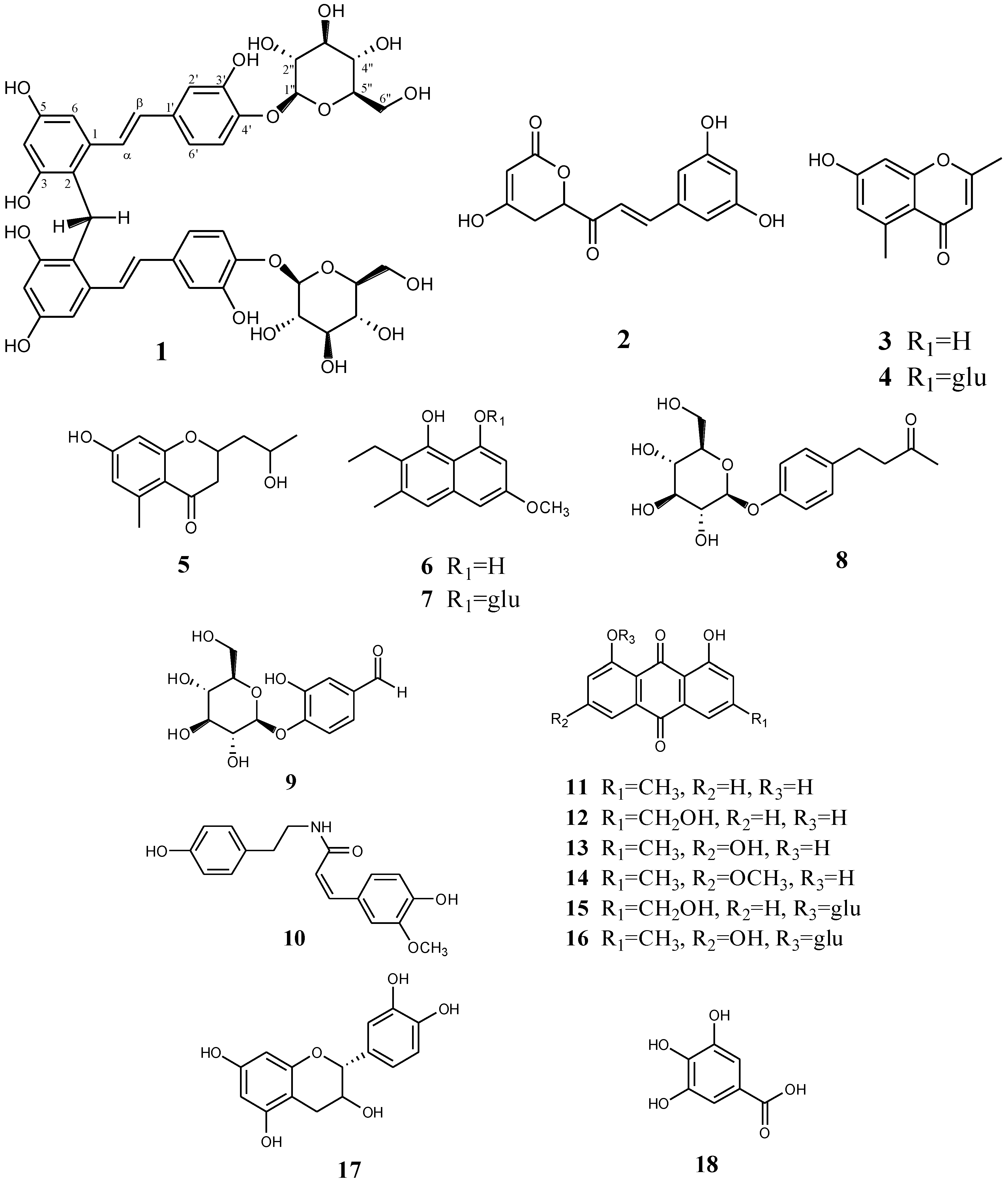
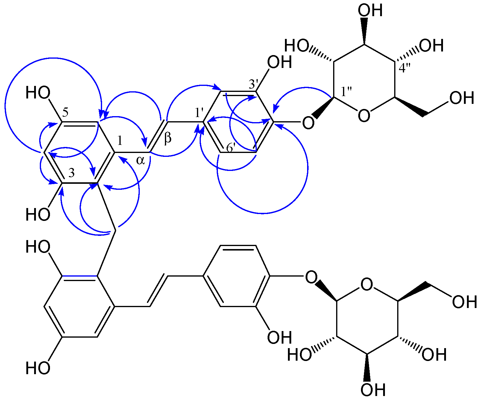
 +50.3030 (c = 0.22, MeOH). The molecular formula of compound 1, C41H44O18, was deduced from the quasimolecular ion peak at m/z 823.2437 [M]− (calcd. for C41H43O18, 823.2449) in the negative HR-ESI-MS, indicating 20 double bond equivalents. The IR (KBr) spectrum showed characteristic absorption bands for hydroxyl groups (3,440 cm−1), methylene groups (2,923 and 1,443 cm−1), aromatic rings (1,514 cm−1) and olefinic groups (1,630 and 986 cm−1). The 1H-NMR spectrum (Table 1) of compound 1 showed two sets of signals. The former set of signals, between δ = 6.0 and 7.5 ppm, was assigned to the protons of a trans-olefinic group (δ = 7.22 and 6.60 ppm, d, J = 16.0 Hz), two aromatic rings with 1,3,5-trisubstituted (δ = 7.06 ppm, d, J = 8.4 Hz; 6.90, d, J = 1.9 Hz and 6.82, dd, J = 8.4, 1.9 Hz) and 1,2,3,4-tetrasubstituted (δ = 6.51 ppm, d, J = 2.3 Hz; 6.28 ppm, d, J = 2.3 Hz) systems. The HMBC (Figure 2) correlations between δH 6.51 (H-6) with δC 120.2 (C-2), 102.8 (C-4), 156.6 (C-5) and 128.1 (C-α); δH7.22 (H-α) with δC 140.1 (C-1), 120.2 (C-2), 105.0 (C-6), 135.4 (C-1') and 129.4 (C-β); δH 6.60 (Η-β) with δC 140.1 (C-1), 115.0 (C-2'), 120.0 (C-6'), 135.4 (C-1') and 128.1 (C-α); indicated the presence of a stilbene skeleton. The latter set of signals, between δ = 3.0 and 5.0 ppm, was assigned to the glycosyl protons and the methylene protons (δ = 4.11 ppm, s), consistent with the 13C-NMR spectrum along with the DEPT spectra of compound 1 (Table 1), which showed six signals characteristic of a glucosyl group (δ = 104.3, 74.9, 77.5, 71.3, 78.3 and 62.4 ppm) and a methylene carbon (δ = 21.3 ppm). The sugar residue was identified as a d-glucopyranosyl unit by gas chromatography of the hydrolyzed product. The mode of the glucosyl linkage was determined to be β from the coupling constant value (d, J = 7 Hz) of the anomeric proton signal. The location of the glucosyl group is suggested to be C-4' by HMBC, which displayed a correlation from δH 4.75 (H-1'') to δC 146.1 (C-4'). These moieties account for ten degrees of unsaturation, only half of those in the molecular formula of compound 1. This result indicated that the structure of compound 1 was symmetrical. In summary, detailed analysis of 1D and 2D-NMR spectra suggested that the structural features of the symmetrical moieties of 1 was very similar to those of piceatannol-4'-O-β-d-glucopyranoside, which was previously isolated from R. australe as the main component, except for the presence of a CH2 group at position 2 (δ = 120.2 ppm). The connection of the symmetrical units was established by the HMBC experiment (Table 1), clearly indicating the correlation peaks from the methylene protons (δ = 4.11 ppm, s) to C-1 (δ = 140.1 ppm), C-2 (δ = 120.2 ppm) and C-3 (δ = 156.4 ppm), suggested that the connection group is the methylene at C-2. Therefore, the structure of compound 1 was established as shown in Figure 1. The 1H-NMR and 13C-NMR (100 MHz) spectral assignments performed by extensive 2D-NMR experiments (HSQC and HMBC) are summarized in Figure 2 and Table 1.

| Position | δH (Mult., J in Hz) | δC | DEPT | HMBC (Selected) |
|---|---|---|---|---|
| 1 | 140.1 | C | ||
| 2 | 120.2 | C | ||
| 3 | 156.4 | C | ||
| 4 | 6.28 (d, 2.3) | 102.8 | CH | C-2, 3, 5, 6 |
| 5 | 156.6 | C | ||
| 6 | 6.51 (d, 2.4) | 105.0 | CH | C-2, 4, 5 |
| 1' | 135.4 | C | ||
| 2' | 6.90 (d, 1.9) | 115.0 | CH | C-3', 4', 6' |
| 3' | 147.9 | C | ||
| 4' | 146.1 | C | ||
| 5' | 7.06 (d, 8.4) | 118.5 | CH | C-1', 3', 4' |
| 6' | 6.82 (dd, 8.5, 1.9) | 120.0 | CH | C-2′, 4′ |
| α | 7.22 (d, 16.0) | 128.1 | CH | C-1, 2, 6, 1' |
| β | 6.60 (d, 16.0) | 129.4 | CH | C-1, 2', 6', 1' |
| 1'' | 4.75 (d, 7.6) | 104.3 | CH | C-4' |
| 2'' | 3.49 (m) | 74.9 | CH2 | |
| 3'' | 3.49(m) | 77.5 | CH2 | |
| 4'' | 3.42 (m) | 71.3 | CH2 | |
| 5'' | 3.42 (m) | 78.3 | CH2 | |
| 6'' | 3.91 (brd) | 62.4 | CH2 | C-4'', 5'' |
| 3.74 (dd, 11.4, 4.6) | ||||
| CH2 | 4.11 (s) | 21.3 | CH2 | C-1, 2, 3 |
2.2. Antioxidant Activities by DPPH Scavenging Capacities
| Compounds | DPPH Radical IC50 (μM) a |
|---|---|
| 1 | 2.3 ± 0.5 |
| 2 | 31.7 ± 1.1 |
| 3 | 25.7 ± 0.7 |
| 4 | 66.9 ± 1.3 |
| 5 | 21.7 ± 1.1 |
| 6 | 32.1 ± 1.5 |
| 7 | 56.4 ± 0.9 |
| 8 | 109.7 ± 2.1 |
| 9 | 69.7 ± 1.5 |
| 10 | 23.4 ± 0.8 |
| resveratrol b | 15.6 ± 0.7 |
| piceatannol b | 0.14 ± 0.05 |
| ascorbic acid b | 19.7 ± 0.8 |
| BHA b | 18.7 ± 0.9 |
| α-tocopherol b | 25.1 ± 1.1 |
3. Experimental
3.1. General
3.2. Plant Materials
3.3. Extraction and Isolation of the Compounds
3.4. Acid Hydrolysis of Compound 1
3.5. Spectroscopic Data
= + 50.3030ο (c = 0.22, MeOH); IR (KBr) νmax 3440, 2922, 1630, 1514, 1443, 1349, 1272, 1090, 986, 803 cm−1; UV (MeOH) λmax (log ε) 223 (4.3), 325 (4.1) nm; positive ESI-MS [M+Na]+ at m/z 847; negative HR-ESI-MS [M−H]− at m/z 823.2437 (calcd for C41H43O18 823.2449); 1H- and 13C-NMR data (Table 1).3.6. DPPH Assays
4. Conclusions
Supplementary Materials
Supplementary Files
Supplementary File 1Acknowledgments
Author Contributions
Conflicts of Interest
References
- Bao, B.; Alisa, E.G. RHEUM Linnaeus. In Flora of China; Li, A., Bao, B., Alisa, E.G., Suk-pyo, H., John, M., Sergei, L.M., Hideaki, O., Chong-wook, P., Eds.; Science Press & Missouri Botanical Garden: St. Louis, MO, USA, 2003; Volume 5, pp. 277–350. [Google Scholar]
- Xiao, P.; He, L.; Wang, L. Ethnopharmacologic study of chinese rhubarb. J. Ethnopharmacol. 1984, 10, 275–293. [Google Scholar] [CrossRef]
- Rokaya, M.B.; Münzbergová, Z.; Timsina, B.; Bhattarai, K.R. Rheum australe D. Don: A review of its botany, ethnobotany, phytochemistry and pharmacology. J. Ethnopharmacol. 2012, 141, 761–774. [Google Scholar] [CrossRef]
- Zargar, B.A.; Masoodi, M.H.; Ahmed, B.; Ganie, S.A. Phytoconstituents and therapeutic uses of Rheum emodi wall. ex Meissn. Food Chem. 2011, 128, 585–589. [Google Scholar] [CrossRef]
- Liu, B.; Yang, J.; Wang, S. The chemical constituents in rhubarb rhizomes and roots derived from Rheum emodi Wall. Huaxi Yaoxue Zazhi 2007, 22, 33–35. [Google Scholar]
- Wang, A.Q.; Li, J.L.; Li, J.S. Chemical constituents of Rheum emodi. Zhong Cao Yao 2010, 41, 343–346. [Google Scholar]
- Chai, Y.Y.; Wang, F.; Li, Y.L.; Liu, K.; Xu, H. Antioxidant activities of stilbenoids from Rheum emodi Wall. Evid. Based Complement. Alternat. Med. 2012, 2012. [Google Scholar] [CrossRef]
- Matsuda, H.; Morikawa, T.; Toguchida, I.; Park, J.Y.; Harima, S.; Yoshikawa, M. Antioxidant constituents from rhubarb: Structural requirements of stilbenes for the activity and structures of two new anthraquinone glucosides. Bioorg. Med. Chem. Lett. 2001, 9, 41–50. [Google Scholar] [CrossRef]
- Agarwal, S.K.; Singh, S.S.; Verma, S.; Kumar, S. Antifungal activity of anthraquinone derivatives from Rheum emodi. J. Ethnopharmacol. 2000, 72, 43–46. [Google Scholar] [CrossRef]
- Shi, Y.Q.; Fukai, T.; Sakagami, H.; Kuroda, J.; Miyaoka, R.; Tamura, M.; Nomura, T. Cytotoxic and DNA damage-inducing activities of low molecular weight phenols from rhubarb. Anticancer Res. 2001, 21, 2847–2853. [Google Scholar]
- Suresh, B.K.; Tiwari, A.K.; Srinivas, P.V.; Ali, A.Z.; China, R.B.; Rao, J.M. Yeast and mammalian α-glucosidase inhibitory constituents from Himalayan rhubarb Rheum emodi Wall ex Meisson. Bioorg. Med. Chem. Lett. 2004, 14, 3841–3845. [Google Scholar] [CrossRef]
- Liang, H.X.; Dai, H.Q.; Fu, H.A.; Dong, X.P.; Adebayo, A.H.; Zhang, L.X.; Cheng, Y.X. Bioactive compounds from Rumex plants. Phytochem. Lett. 2010, 3, 181–184. [Google Scholar] [CrossRef]
- Xiang, L.; Lei, F.; Xing, D.; Wang, W.; Zheng, J. Neuron protective constituents from Rheum nanum and Rheum sublanceolatum. Tsinghua Sci. Technol. 2005, 10, 426–429. [Google Scholar] [CrossRef]
- Andersen, D.O.; Weber, N.D.; Wood, S.G.; Hughes, B.G.; Murray, B.K.; North, J.A. In vitro virucidal activity of selected anthraquinones and anthraquinone derivatives. Antivir. Res. 1991, 16, 185–196. [Google Scholar] [CrossRef]
- Liu, W.B.; Hu, L.; Hu, Q.; Chen, N.N.; Yang, Q.S.; Wang, F.F. New resveratrol oligomer derivatives from the roots of Rheum lhasaense. Molecules 2013, 18, 7093–7102. [Google Scholar] [CrossRef]
- Rajkumar, V.; Guha, G.; Ashok Kumar, R. Antioxidant and anti-cancer potentials of Rheum emodi rhizome extracts. Evid. Based Complement. Alternat. Med. 2011. [Google Scholar] [CrossRef]
- Fang, J.G.; Lu, M.; Chen, Z.H.; Zhu, H.H.; Li, Y.; Yang, L.; Wu, L.M.; Liu, Z.L. Antioxidant effects of resveratrol and its analogues against the free-radical-induced peroxidation of linoleic acid in micelles. Chem. Eur. J. 2002, 8, 4191–4198. [Google Scholar] [CrossRef]
- Hu, L.; Chen, N.N.; Feng, L.; Hu, Q.; Liu, W.B.; Yang, Q.S.; Wang, F.F. Piceatannol derivatives from Rheum austral D. Don and their chemotaxonomic significance. Biochem. Syst. Ecol. 2014, 55, 369–373. [Google Scholar] [CrossRef]
- Li., J.L.; Li., J.S.; He., W.Y.; Kong., M. Studies on the non-anthraquiones of Rheum hotaoense. Zhong Cao Yao 1998, 29, 721–723. [Google Scholar]
- Kjer, J.; Wray, V.; Edrada-Ebel, R.; Ebel, R.; Pretsch, A.; Lin, W.; Proksch, P. Xanalteric acids I and II and related phenolic compounds from an endophytic Alternaria sp. isolated from the mangrove plant Sonneratia alba. J. Nat. Prod. 2009, 72, 2053–2057. [Google Scholar] [CrossRef]
- Zhao, H.P.; Wang, Z.Y.; Chen, J.R.; Li, R.M.; Wang, Z.Q. New chromone glucoside from roots of Rumex gmelini. Nat. Prod. Res. Dev. 2009, 21, 189–191. [Google Scholar]
- Xu, J.; Kjer, J.; Sendker, J.; Wray, V.; Guan, H.; Edrada, R.; Proksch, P. Chromones from the endophytic fungus Pestalotiopsis sp. isolated from the Chinese mangrove plant Rhizophora mucronata. J. Nat. Prod. 2009, 72, 662–665. [Google Scholar] [CrossRef]
- Mei, R.; Liang, H.; Wang, J.; Zeng, L.; Lu, Q.; Cheng, Y. New seco-anthraquinone glucosides from Rumex nepalensis. Planta Med. 2009, 75, 1162. [Google Scholar] [CrossRef]
- Demirezer, Ö; Kuruüzüm, A.; Bergere, I.; Schiewe, H.J.; Zeeck, A. Five naphthalene glycosides from the roots of Rumex patientia. Phytochemistry 2001, 56, 399–402. [Google Scholar] [CrossRef]
- Shikishima, Y.; Takaishi, Y.; Honda, G.; Ito, M.; Takeda, Y.; Kodzhimatov, O.K.; Ashurmetov, O. Phenylbutanoids and stilbene derivatives of Rheum maximowiczii. Phytochemistry 2001, 56, 377–381. [Google Scholar] [CrossRef]
- Likhitwitayawuid, K.; Ruangrungsi, N.; Cordell, G.A. Amabiloside, a new glycoside from Crinum amabile. Nat. Prod. Lett. 1993, 3, 1–4. [Google Scholar] [CrossRef]
- Tanaka, H.; Nakamura, T.; Ichino, K.; Ito, K. A phenolic amide from Actinodaphne longifolia. Phytochemistry 1989, 28, 2516–2517. [Google Scholar] [CrossRef]
- Sharma, O.P.; Bhat, T.K. DPPH antioxidant assay revisited. Food Chem. 2009, 113, 1202–1205. [Google Scholar] [CrossRef]
- Rivière, C.; Pawlus, A.D.; Mérillon, J.M. Natural stilbenoids: Distribution in the plant kingdom and chemotaxonomic interest in Vitaceae. Nat. Prod. Rep. 2012, 29, 1317–1333. [Google Scholar] [CrossRef]
- Xiao, K.; Zhang, H.J.; Xuan, L.J.; Zhang, J.; Xu, Y.M.; Bai, D.L. Stilbenoids: Chemistry and bioactivities. In Studies in Natural Products Chemistry; Atta-ur-Rahman, Ed.; Elsevier Science: Amsterdam, The Netherlands, 2008; Volume 34, pp. 453–646. [Google Scholar]
- Shen, T.; Wang, X.N.; Lou, H.X. Natural stilbenes: An overview. Nat. Prod. Rep. 2009, 26, 916–935. [Google Scholar] [CrossRef]
- Sample Availability: Samples of the compounds 1–16 are available from the authors.
© 2014 by the authors. Licensee MDPI, Basel, Switzerland. This article is an open access article distributed under the terms and conditions of the Creative Commons Attribution license ( http://creativecommons.org/licenses/by/3.0/).
Share and Cite
Hu, L.; Chen, N.-N.; Hu, Q.; Yang, C.; Yang, Q.-S.; Wang, F.-F. An Unusual Piceatannol Dimer from Rheum austral D. Don with Antioxidant Activity. Molecules 2014, 19, 11453-11464. https://doi.org/10.3390/molecules190811453
Hu L, Chen N-N, Hu Q, Yang C, Yang Q-S, Wang F-F. An Unusual Piceatannol Dimer from Rheum austral D. Don with Antioxidant Activity. Molecules. 2014; 19(8):11453-11464. https://doi.org/10.3390/molecules190811453
Chicago/Turabian StyleHu, Lin, Na-Na Chen, Qun Hu, Cui Yang, Qing-Song Yang, and Fang-Fang Wang. 2014. "An Unusual Piceatannol Dimer from Rheum austral D. Don with Antioxidant Activity" Molecules 19, no. 8: 11453-11464. https://doi.org/10.3390/molecules190811453