Communication

A New Cycloartane-Type Triterpenoid Saponin Xanthine Oxidase Inhibitor from Homonoia riparia Lour

1
Kunming General Hospital of Chengdu Military Region, Kunming 650032, Yunnan, China
2
The First Affiliated Hospital of Kunming Medical University, Kunming 650031, Yunnan, China
*
Author to whom correspondence should be addressed.
Molecules 2014, 19(9), 13422-13431; https://doi.org/10.3390/molecules190913422
Submission received: 30 June 2014 / Revised: 6 August 2014 / Accepted: 7 August 2014 / Published: 29 August 2014
(This article belongs to the Section Natural Products Chemistry)

Abstract

:
A new cycloartane-type triterpenoid saponin named riparsaponin (1) was isolated from the stem of Homonoia riparia Lour together with six known compounds. The structure of riparsaponin was determined by using NMR and mass spectroscopy and X-ray crystallography techniques. Additionally, riparsaponin has a significant inhibitory effect on xanthine oxidase in vitro, and the IC50 was 11.16 nmol/mL.

1. Introduction

Homonoia riparia Lour, (family Euphorbiaceae), is widely distributed in the south part of China [1]. The roots of H. riparia are commonly used as an effective traditional Chinese herbal medicine for treating hepatitis and joint gall, stomach ache, and ambustion based on its antipyretic choleretic, anti-inflammatory, detoxification, and diuretic activities [1,2]. However, phytochemical and pharmacological investigations of this plant are currently lacking. Previous chemical research reported that it contains triterpenes, steroids, and phenolics [3,4,5]. In our present investigation, a new cycloartane-type triterpenoid saponin (compound 1, named riparsaponin) was isolated from the stems of H. riparia together with six known compounds (Figure 1). In addition, riparsaponin showed significant inhibitory activity on xanthine oxidase. Here, we report the isolation, identification and activity of the new compound, which could be helpful for treating gouty arthritis.
Figure 1. Structure of the compounds isolated from H. riparia.
Molecules 19 13422 g001

2. Results and Discussion

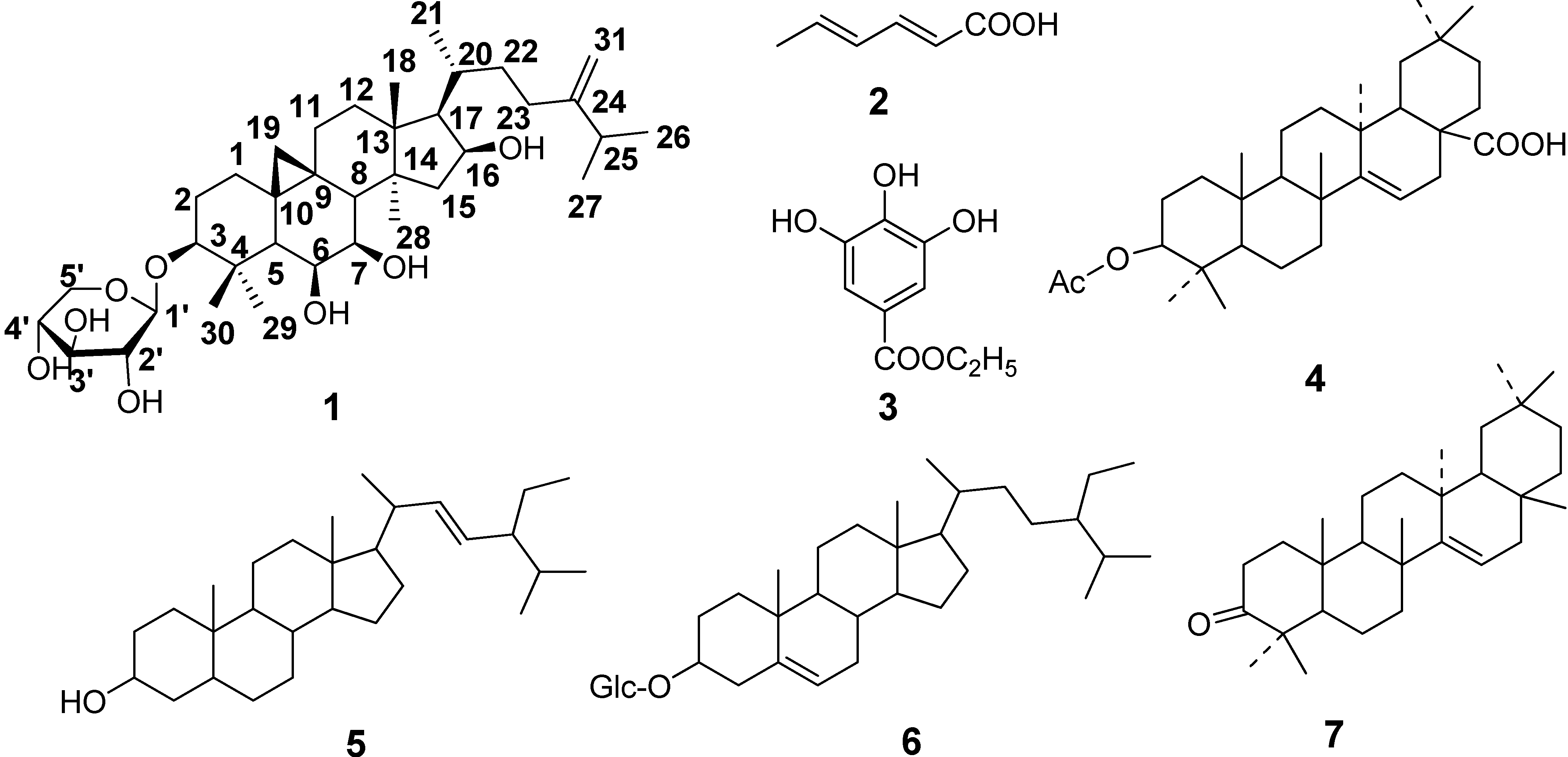
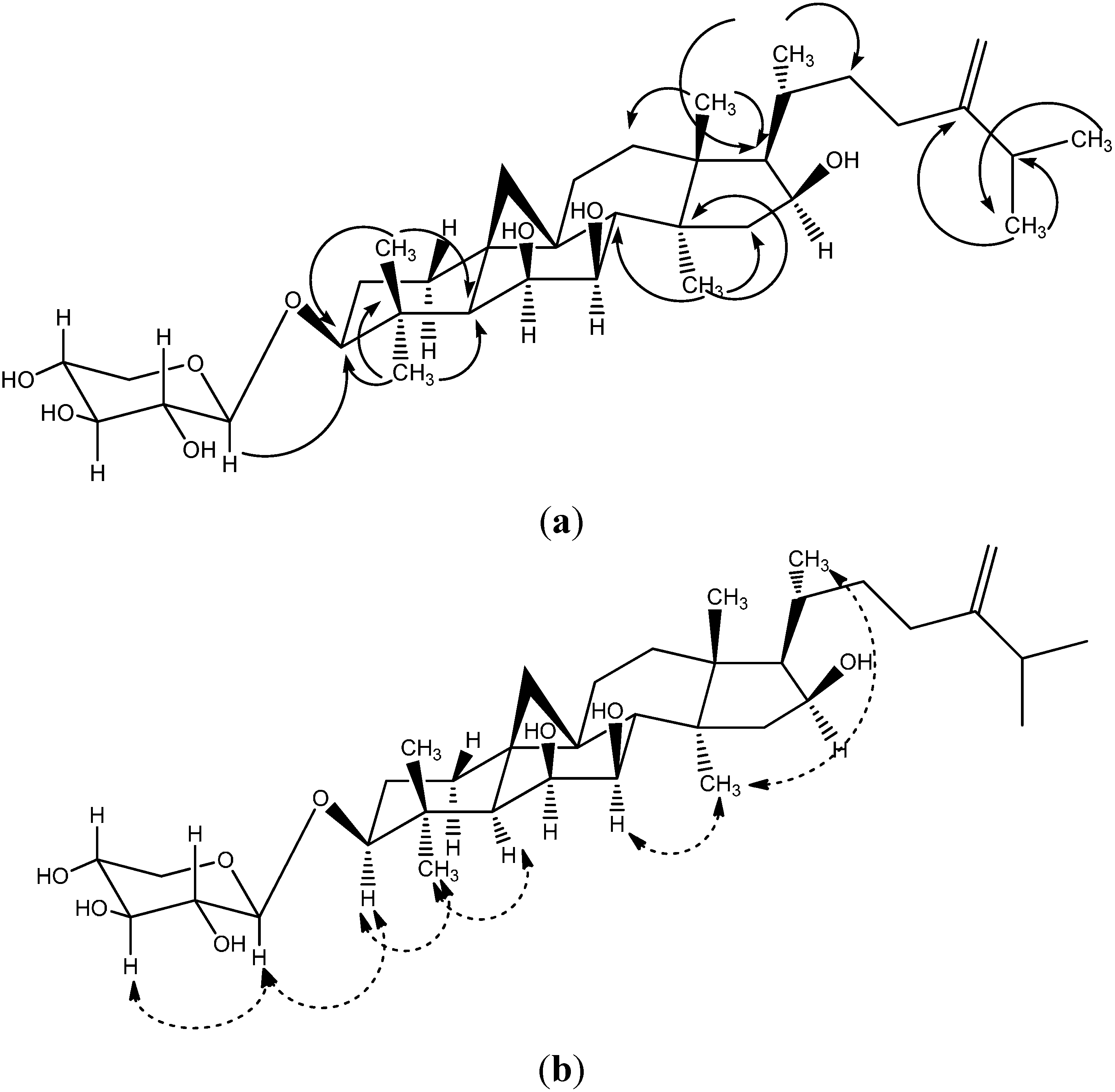
2.1. Identification of the Riparsaponin

The new compound riparsaponin (1) was identified by using NMR and mass spectroscopy, and X-ray crystallography techniques. Compound 1 was obtained as colorless prismatic crystal (MeOH); mp: 291–292 °C. Its Liebermann-Burchard response was positive. FAB-MS (m/z) (%): 620 (99), 712 (29), 487 (55). The 13C-NMR, 1H-NMR data are listed in Table 1. The 13C-NMR spectrum (100 MHz, DMSO-d6) showed 36 carbon signals, and lots of carbon signals were between 60–80 (δC), which indicated the existence of a saccharide group. The carbon signals of 105.9, 73.82, 105.9, 73.82, and 65.62 (δC) could further demonstrate the existence of pentose, and the carbon signal at 105.9 ppm is the terminal group carbon of the pentose (β-type) [6]. The two high-field doublets observed at δH 0.99 (1H, m) and 0.3 (1H, s) are characteristic of the two germinal protons of a cyclopropane moiety. What’s more, two olefinic protons at δH 4.65 (2H, brs) indicated the presence of an olefinic methylene moiety. There are two carbon signals at 106.13 and 156.22 (δC), suggesting a double bond. The 1H-NMR spectrum (400 MHZ, DMSO-d6) indicated seven methyl groups (δH 0.87, 0.88, 0.96, 0.98, 1.05, 1.08, 1.12). These spectroscopic data suggested this compound to be a cycloartane-type triterpene with an exocyclic methylene group. Combined the reference data [3], we found the 1H-NMR and 13C-NMR spectra of compound 1 were analogous to that reported for a cycloartane-type triterpene which was isolated from H. riparia previously. Furthermore, the structure of compound 1 was confirmed by HMBC, ROESY (Figure 2) and X-ray diffraction (CCDC deposition number is 1021164) (Figure 3, Table 2).
Figure 2. The key HMBC (a) and ROESY (b) connections of riparsaponin (1).
Molecules 19 13422 g002
Figure 3. Perspective drawing of compound 1 generated from X-ray crystal data.
Molecules 19 13422 g003
Table 1. 1H-NMR (400 Hz) and 13C-NMR (100 Hz) data of riparsaponin in DMSO-d6.
δCδH δCδH
132.62 t1.53 (2H, m)1931.41 t0.99 (1H, m)
0.30 (1H, s)
229.16 t1.72 (1H, m)2029.77 d1.77 (1H, m)
1.53 (1H, m)
387.52 d3.00 (1H, m)2117.92 q0.87 (3H, d, J = 8.6 Hz)
440.82 s 2234.54 t1.80 (1H, m)
1.08 (1H, m)
549.02 d1.22 (1H, s)2331.35 t2.07 (1H, m)
1.88 (1H, m)
671.38 d3.74 (1H, brs)24156.22 s
772.74 d3.21 (1H, m)2533.15 d2.22 (1H, m)
845.90 d1.88 (1H, d, J = 11.2 Hz)2621.76 q0.96 (3H, s)
924.04 s 2721.81 q0.98 (3H, s)
1018.72 s 2819.43 q0.88 (3H, s)
1125.43 t1.91 (1H, m)2924.07 q1.05 (3H, s)
0.92 (1H, m)
1232.27 t1.36 (1H, m)3016.40 q1.08 (3H, s)
1.08 (1H, m)
1345.64 s 31106.13 t4.65 (2H, brs)
1445.41 s 1'105.90 d4.09 (1H, d, J = 7.5 Hz)
1551.66 t2.16 (1H, m)2'73.82 d2.95 (1H, m)
1.41 (1H, m)
1670.72 d4.14 (1H, m)3'76.79 d3.04 (1H, m)
1755.44 d1.45 (1H, m)4'69.66 d3.24 (1H, m)
1819.39 q1.12 (3H, s)5'65.62 t3.61(1H, m)
2.97 (1H, m)
Table 2. Crystal data and structure refinement for riparsaponin.
Identification code070516a
Empirical formulaC36H57O8
Formula weight617.82
Temperature298 (2) K
Wave length0.71073 A
Crystal system, space groupOrthorhombic, P2(1)2(1)2(1)
Unit cell dimensionsa = 6.3405(9)Aalpha = 90 deg.
b = 12.7265(17)Abeta = 90 deg.
c = 41.573(6)Agamma = 90 deg.
Volume3354.6(8) A3
Z, Calculated density4, 1.223 Mg/m3
Absorption coefficient0.085 mm−1
F (000)1348
Crystalsize0.26 × 0.22 × 0.08 mm
The tarange for data collection1.67 to 28.31 deg.
Limiting indices−8 ≤ h ≤ 8,−16 ≤ k ≤ 16,−55 ≤ l ≤ 53
Reflections collected/unique29112/8026[R(int) = 0.0893]
Completeness to theta = 28.3198.9%
Absorption correctionMUTI-SCAN
Max. andmin. transmission1.000000 and 0.832723
Refinement methodFull-matrixleast-squaresonF2
Data/restraints/parameters8026/0/398
Goodness-of-fitonF20.786
Final Rindices [I > 2sigma(I)]R1 = 0.0666, wR2 = 0.1877
Rindices (alldata)R1 = 0.1410, wR2 = 0.2517
Absolute structure parameter0.7(17)
Extinction coefficient0.0040(15)
Largest diff. Peak and hole0.342 and −0.329 eA−3

2.2. Inhibitory Effect of Riparsaponin on Xanthine Oxidase Activity in Vitro

Gout is one of the common human metabolic diseases and caused by hyperuricemia, which can result in depositions of urate crystals in joints, leading to gouty arthritis [7]. Xanthine oxidase plays an important role during the formation of uric acid, and the accumulation of uric acid can result in hyperuricaemia, leading to gout [8]. Previous investigations revealed that inhibitors of xanthine oxidase could be potentially beneficial for treating gouty arthritis [9]. Herbal remedies have been used in China for more than millennium, and lots of investigations have reported that the herbal medicines and its derived compounds can safely and effectively in treatment of various diseases [10,11]. In our present study, the inhibitory activities of the six known compounds on xanthine oxidase were weak, but riparsaponin could significantly inhibit xanthine oxidase activity in vitro at the doses during 9.68 to 161.29 nmol/mL compared with the DMSO group (p < 0.01), in a dose-dependent manner (Table 3). In our present study, the IC50 of riparsaponin was 11.16 nmol/mL, which is a better value compared to allopurinol used as positive control drug (IC50 11.84 nmol/mL). The results above indicated that riparsaponin is a potential powerful xanthine oxidase inhibitor.
Table 3. Inhibitory effect of riparsaponin on xanthine oxidase activity (n = 5).
GroupConcentration (nmol/mL)Fluorescence UnitInhibition Ratio (%)IC50 Value (nmol/mL)
DMSO 8.291 ± 0.8920.07
Riparsaponin4.847.205 ± 0.86513.0911.16
9.684.815 ± 0.861 **41.93
19.363.248 ± 0.912 **60.82
40.322.362 ± 0.127 **71.52
80.651.872 ± 0.483 **77.42
161.291.474 ± 0.131 **82.22
Allopurinol2.206.796 ± 0.696 *18.0311.84
4.416.212 ± 0.580 **25.08
8.824.879 ± 0.410 **40.93
17.632.776 ± 0.217 **66.52
35.272.062 ± 0.370 **75.13
70.531.617 ± 0.220 **80.49
Data were expressed as Mean ± SD, * p < 0.05, ** p < 0.05, compared to the DMSO group.

3. Experimental Section

3.1. General Information

These following instruments were used: UV visible spectrophotometer (UV-1600) was made by Rayleigh Analytical Instrument Company (Beijing, China). Mass spectrometer (EI-MS) (VGAutoSpec-3000, Beckman, CA, USA). The NMR (AV-400, AV-500) and X-ray single crystal diffractometer (APEX II DUO) instruments were both made by Bruker (Bremen, Germany).

3.2. Plant Material

The stems of H. riparia were collected in Jinping County, Yunnan Province, China, in October 2010. The plant was identified by Jingxiu Li (Kunming Institute of Botany Chinese Academy of Science, Kunming, Yunnan, China). A voucher specimen was deposited in our laboratory.

3.3. Extraction and Isolation

Dried stems of H. riparia (58.8 kg) were powered and extracted three times with 60% ethanol (total 300 L) at reflux. Then the extracts were spray-dried to obtain a dry fine powder (4.8 kg). The powder was extracted three times by maceration with petroleum ether, CH2Cl2, EtOAc, and 95% ethanol, respectively (total 40 L, each extraction lasted 2 days).
After concentration, compound 2 was crystallized from the petroleum ether fraction, and the pure compound 2 (3.17 g) was obtained by recrystallization from EtOAc. Then, the petroleum ether fraction (73 g) was subjected to column chromatography (CC) over silica gel (200–300 mesh) eluting with petroleum ether–EtOAc (9:1, 8:1, 5:1, 3:1, 1:1, 1:3, 1:5, 1:9), and seven sub-fractions AG were obtained on the basis of TLC analysis. Compound 4 was crystallized from fraction B, and the pure compound 4 (60 mg) was obtained by recrystallization from petroleum ether–CH2Cl2 (9:1). Fraction C was subjected to CC over silica gel (200–300 mesh) eluting with petroleum ether–CH2Cl2 (9:1, 8:1, 5:1, 3:1, 1:1, 1:3, 1:5, 1:9), and combination of similar fractions after TLC comparison afforded five fractions C1C5. Compound 5 was crystallized from fraction C2, and the pure compound 5 (87 mg) was obtained by recrystallization from EtOAc.
The CH2Cl2 fraction (100 g) was subjected to AB-8 macroporous resin (The Chemical Plant of Nankai University, Tianjing, China) CC eluting with gradient ethanol (0%, 30%, 70%, 90%), and four fractions AD were obtained. After concentration, compound 3 was crystallized from fraction D, and the pure compound 3 (45 mg) was obtained by recrystallization in petroleum ether–EtOAC (9:1). Fraction C was subjected to CC over silica gel (200–300 mesh) eluting with petroleum ether–acetone (9:1, 8:1, 5:1, 3:1, 1:1, 1:3), and four sub-fractions C1, C2, C3, C4 were obtained on the basis of TLC. Sub-fraction C2 was subjected to CC over silica gel (200–300 mesh) eluting with petroleum ether–acetone (9:1, 8:1, 5:1, 3:1, 1:1, 1:3) again to afford four sub-fractions C21C24, and compound 6 (white powder, 144 mg) was separated out from fraction C22.
The EtOAc fraction (72.3 g) was subjected to CC over silica gel (200–300 mesh) eluting with petroleum ether–acetone (15:1, 10:1, 7:1, 5:1, 3:1, 1:1, 1:2), to give six sub-fractions AF on the basis of TLC analysis. After concentration, compound 7 (a white powder, 24 mg ) was separated out from fraction E. Fraction B was repeatedly subjected to CC over silica gel (200–300 mesh) eluting with petroleum ether–acetone (10:1, 7:1, 5:1, 3:1, 1:1, 1:2), and four sub-fractions B1B4 of B were thus obtained. Then sub-fraction B2 was subjected to CC over silica gel again eluting with a gradient of petroleum ether–acetone, and five fractions B21B25 were obtained; then compound 1 was crystallized from B22, and the pure compound 1 (213 mg) was obtained by recrystallization from petroleum ether–EtOAC.
2,4-Sorbic acid (2) was obtained as colorless crystals (EtOAc), mp: 134–136 °C; EI-MS (m/z) (%): 111 (38), 97 (71), 83 (80), 69 (90), 57 (100); 1H-NMR (400 MHz, CDCl3, ppm) δ: 5.75 (1H, d, J = 15.3 Hz, H-2), 7.33 (1H, dd, J = 15.4, 9.7 Hz, H-3), 6.26 (1H, m, H-4), 5.79 (1H, s, H-5), 1.87 (3H, d, J = 5.0 Hz, CH3-6); 13C-NMR (100 MHz, CDCL3, ppm) δ: 173.5 (C-1), 118.4 (C-2), 147.7 (C-3), 130.0 (C-4), 141.2 (C-5), 19.1 (C-6).
Ethyl gallate (3) was obtained as colorless crystals [petroleum ether‒EtOAc (9:1)], mp: 169–170 °C; FAB-MS (m/z) (%): 197 (90), 169 (5), 154 (100), 124 (3), 97 (3), 80 (15), 64 (3); 1H-NMR (400 MHz, CD3ODCD3, ppm) δ: 7.11 (2H, s, H-2, H-6), 4.26 (2H, q, J = 7.0 Hz, OCH2CH3), 1.27 (3H, t, J = 7.2 Hz, OCH2CH3), 6.94 (2H, d, J = 1.0 Hz), 8.12 (1H, s), 9.25 (2H, s); 13C-NMR (100 MHz, CD3ODCD3, ppm) δ: 122.0 (C-1), 109.7 (C-2), 146.0 (C-3), 138.6 (C-4), 146.0 (C-5), 109.7 (C-6), 166.6 (C-7), 60.9 (C-8), 14.6 (C-9).
Acetylaleuritolic acid (4) was obtained as colorless crystals [petroleum ether‒CH2Cl2 (9:1)], mp: 287–288 °C; EI-MS (m/z) (%): 498 (M+, 3), 439 (M+-COCH3, 2), 344 (RDA fragment, 5), 285 (344-COCH3), 189(100). 1H-NMR (400 MHz, CDCl3, ppm) δ: 0.75 (3H,s, CH3-26), 0.84 (3H, s, CH3-23), 0.88 (3H, s, CH3-24), 0.91 (3H, s, CH3-29), 0.93 (3H, s, CH3-30), 0.95 (3H, s, CH3-25), 1.13 (3H, s, CH3-27), 2.04 (3H, -COCH3), 4.48 (1H, t, J = 8.0 Hz, H-3), 5.50 (1H, t, J = 3.3 Hz, H-15); 13C-NMR (100 MHz, CDCl3, ppm) δ: 37.9 (C-1), 22.4 (C-2), 80.9 (C-3), 37.7 (C-4), 55.3 (C-5), 18.1 (C-6), 33.6 (C-7), 39.0 (C-8), 47.5 (C-9), 37.0 (C-10), 23.4 (C-11), 32.4 (C-12), 41.3 (C-13), 160.5 (C-14), 116.8 (C-15), 23.5 (C-16), 46.5 (C-17), 41.3 (C-18), 45.8 (C-19), 30.6 (C-20), 32.4 (C-21), 27.9 (C-22), 28.6 (C-23), 16.6 (C-24), 15.6 (C-25), 17.3 (C-26), 25.9 (C-27), 184.2 (C-28), 33.0 (C-29), 23.6 (C-30), 171.0 (-COOH), 21.3 (-COCH3) [12].
Spinasterol (5) was obtained as colorless crystals (CH2Cl2), mp: 162–163 °C; EI-MS (m/z) (%): 412 (M+), 396 (M+-O), 351 (369-OH), 271 (300-C2H5), 255 (271-O), 213 (255-C3H6); 1H-NMR (400 MHz, CDCl3, ppm) δ: 5.15 (1H, H-7), 5.03 (1H, dd, J = 8.6 Hz, H-22), 5.21 (1H, dd, J = 8.5 Hz, H-23), 0.70 (3H, s, CH3-18), 0.81 (3H, s, CH3-19), 0.83 (3H, t, J = 7.3 Hz, CH3-29), 0.85 (3H, d, J = 6.3 Hz, CH3-26), 0.86 (3H, d, J = 6.3 Hz, CH3-26), 1.04 (3H, d, J = 6.6 Hz, CH3-21), 3.54 (H, tt, J = 11.0 Hz, H-3); 13C-NMR (100 MHz, CDCl3, ppm) δ: 37.2 (C-1), 31.6 (C-2), 71.8 (C-3), 37.9 (C-4), 40.5 (C-5), 29.6 (C-6), 121.7 (C-7), 140.7 (C-8), 50.1 (C-9), 36.1 (C-10), 21.2 (C-11), 39.6 (C-12), 42.5 (C-13), 55.9 (C-14), 23.0 (C-15), 28.9 (C-16), 56.8 (C-17), 12.0 (C-18), 19.0 (C-19), 42.2 (C-20), 21.0 (C-21), 138.3 (C-22), 129.2 (C-23), 51.2 (C-24), 31.9 (C-25), 21.1 (C-26), 19.4 (C-27), 25.4 (C-28), 12.2 (C-29) [13].
Daucosterol (6) was obtained as white powders, mp: 288–289 °C; 1H-NMR (400 MHz, C5D5N, ppm) δ: 5.17 (1H, m, J = 5.2 Hz, H-6), 4.32 (1H, d, J = 7.2 Hz, H-1'), 4.07 (1H, m, H-3), 0.75 (3H, d, J = 8.0 Hz, CH3-21), 0.70 (3H, s, CH3-19), 0.65 (6H, d, J = 8.4 Hz, CH3-26, 27), 0.59 (3H, t, J = 4.0Hz, CH3-29), 0.42 (3H, s, CH3-18); 13C-NMR (100 MHz, C5D5N, ppm) δ: 37.5 (C-1), 32.0 (C-2), 78.6 (C-3), 39.9 (C-4), 140.9 (C-5), 121.9 (C-6), 32.2 (C-7), 32.0 (C-8), 50.3 (C-9), 36.4 (C-10), 21.3 (C-11), 39.3 (C-12), 42.5 (C-13), 56.8 (C-14), 23.4 (C-15), 27.3 (C-16), 56.2 (C-17), 12.0 (C-18), 19.4 (C-19), 36.4 (C-20), 19.0 (C-21), 34.2 (C-22), 26.3 (C-23), 45.8 (C-24), 29.4 (C-25), 19.2 (C-26), 20.0 (C-27), 22.8 (C-28), 12.0 (C-29), 102.6 (C-1'), 75.3 (C-2'), 78.5 (C-3'), 71.7 (C-4'),78.0 (C-5'), 62.8 (C-6').
Taraxerone (7) was obtained as a white powder, mp: 288–289 °C; EI-MS (m/z)(%): 424 (M+, 23), 409 (M+-CH3, 17), 300 (RDA fragment, 80), 285 (70), 272 (19), 257 (20), 205 (C ring fragment, 65), 204 (100), 189 (45), 133 (62); 1H-NMR (500 MHz, CDCl3, ppm) δ: 0.82 (3H, s, CH3), 0.91 (3H, s, CH3), 0.95 (3H, s, CH3), 0.98 (3H, s, CH3), 1.08 (3H, s, CH3), 1.10 (3H, s, CH3), 1.13 (3H, s, CH3), 1.15 (3H, s, CH3), 5.56 (1H, dd, J = 3.2, 8.2Hz, H-15);13C-NMR (125 MHz, CDCl3, ppm) δ: 38.4 (C-1), 33.6 (C-2), 217.5 (C-3), 47.6 (C-4), 55.8 (C-5), 20.0 (C-6), 37.7 (C-7), 38.9 (C-8), 48.8 (C-9), 37.5 (C-10), 17.4 (C-11), 34.1 (C-12), 29.8 (C-13), 157.6 (C-14), 117.1 (C-15), 36.7 (C-16), 35.7 (C-17), 48.8 (C-18), 40.0 (C-19), 28.8 (C-20), 33.1 (C-21), 35.1 (C-22), 26.1 (C-23), 21.5 (C-24), 14.8 (C-25), 29.9 (C-26), 25.6 (C-27), 29.9 (C-28), 33.4 (C-29), 21.3 (C-30) [14].

3.4. Xanthine Oxidase Inhibitory Assay

The inhibitory effect of compound 1 against xanthine oxidase activity was carried out according to the previous investigations [15]. The experiment was performed by using 96 holes plate, and the allopurinol and DMSO were used as the positive and negative control. Firstly, xanthine (50 μmol/L) was added into each reaction system, and then the tested samples were added. After that, the xanthine oxidase (0.1 U/mL), quinoline solution (5 μmol/L), (NH4)2Fe (SO4) solution (1 μmol/L), H2SO4 solution (50 μmol/L) were added subsequently. The reaction system incubated at room temperature for 18 min, then added NaOH solution (1 mol/L) into total reaction system to 150 μL. Finally, adding 150 μL ethanol to terminate the reaction, and the reaction production was determined by measuring the absorbance at 380 nm by using fluorescent chemiluminescence detector on a microplate spectrophotometer. The inhibitory percentage against xanthine oxidase (%) was calculated using the following equation:
inhibition (%) = [1 − (A sample/A control) × 100
In addition, the IC50 values were calculated

4. Conclusions

In the present study, a new cycloartane-type triterpenoid saponin named riparsaponin was isolated from the stem of H. riparia, and it has a significant inhibitory effect on xanthine oxidase.

Acknowledgments

This work was supported by the Key Project of Basic Research and Application Program of Yunnan Province in China (No. 2011FA036)

Author Contributions

Conceived and designed the experiments: JZ; performed the experiments: FX, XQZ, LLY, Analyzed the data: FX, XHW; wrote the paper: FX, JZ.

Conflicts of Interest

The authors declare no conflict of interest.

References

  1. Chinese Academy of Medical Sciences Research Institute. Higher Plant Figure of China; Beijing Science Press: Beijing, China, 1972; Volume 2, pp. 606–608. [Google Scholar]
  2. State Administration of Traditional Chinese Medicine. Chinese Material Medica; Science and Technology Press of Shanghai: Shanghai, China, 1999; Volume 6, pp. 824–825. [Google Scholar]
  3. Lee, I.; Kim, J.; Kim, Y.S.; Yoo, N.H.; Kim, C.S.; Jo, K.; Kim, J.H.; Bach, T.T.; Kim, J.S. Cycloartane-type triterpenes from the leaves of Homonoia Riparia with VEGF-induced angiogenesis inhibitor activity. J. Nat. Prod. 2012, 75, 1312–1318. [Google Scholar] [CrossRef]
  4. Parver, N.; Pal-Singh, M.; Khan, N.U. Chemical examination of the leaves of Homonoia riparia Lour ( Euphorbiaceae). J. Indian Chem. Soc. 1988, 65, 815–816. [Google Scholar]
  5. Yang, S.M.; Liu, X.K.; Qing, C.; Wu, D.G.; Zhu, D.Y. Chemical constituents from the roots of Homonoia riparia. Acta Pharm. Sin. 2007, 42, 292–296. [Google Scholar]
  6. Wu, L.J. Natural Pharmaceutical Chemistry, 5th ed; People’s Medical Publishing House: Beijing, China, 2004; pp. 93–103. [Google Scholar]
  7. Sowndhararajan, K.; Joseph, J.M.; Rajendrakumaran, D. In vitro xanthine oxidase inhibitory activity of methanol extracts of Erythrina indica Lam. leaves and stem bark. Asian Pac. J. Trop. Biomed. 2012, 2, S1415–S1417. [Google Scholar] [CrossRef]
  8. Masuoka, N.; Nihei, K.; Maeta, A.; Yamagiwa, Y.; Kubo, I. Inhibitory effects of cardols and related compounds on superoxide anion generation by xanthine oxidase. Food Chem. 2014, 166, 270–274. [Google Scholar]
  9. Sweeney, A.P.; Wyllie, S.G.; Shalliker, R.A.; Markham, J.L. Xanthine oxidase inhibitory activity of selected Australian native plants. J. Ethnopharmacol. 2001, 75, 273–277. [Google Scholar] [CrossRef]
  10. Peng, W.; Ming, Q.L.; Han, P.; Zhang, Q.Y.; Jiang, Y.P.; Zheng, C.J.; Han, T.; Qin, L.P. Anti-allergic rhinitis effect of caffeoylxanthiazonoside isolated fromfruits of Xanthium strumarium L. in rodent animals. Phytomedicine 2014, 21, 824–829. [Google Scholar] [CrossRef]
  11. Karki, R.; Park, C.H.; Kim, D.W. Extract of buckwheat sprouts scavenges oxidation and inhibits pro-inflammatory mediators in lipopolysaccharide-stimulated macrophages (RAW264.7). J. Integr. Med. 2013, 11, 246–252. [Google Scholar] [CrossRef]
  12. Nyasse, B.; Ngantchou, I.; Nono, J.J.; Schneider, B. Antifilarial activity in vitro of polycarpol and 3-O-acetyl aleuritolic acid from cameroonian medicinal plants against Onchocerca gutturosa. Nat. Prod. Res. 2006, 20, 391–397. [Google Scholar] [CrossRef]
  13. Villasenor, I.M.; Domingo, P.D. Antieareinogenicity potential of spinasterol isolated from squash flowers. Teratog. Carcinog. Mutagen. 2000, 20, 99–105. [Google Scholar] [CrossRef]
  14. Wang, G.K.; Lin, B.B.; Qin, M.J. Study on chemical constituents from leaf of Bombax ceiba (II). J. Chin. Med. Mater. 2014, 37, 240–242. [Google Scholar]
  15. Tsai, S.F.; Lee, S.S. Neolignans as xanthine oxidase inhibitors from Hyptis rhomboids. Phytochemistry 2014, 101, 121–127. [Google Scholar] [CrossRef]
  • Sample Availability: Contact the authors.

