Article

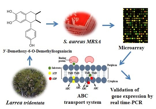
Potential Mechanism of Action of 3′-Demethoxy-6-O-demethyl-isoguaiacin on Methicillin Resistant Staphylococcus aureus

by
Juan Manuel J. Favela-Hernández
1,2,
Aldo F. Clemente-Soto
1,
Isaías Balderas-Rentería
1,
Elvira Garza-González
3 and
María Del Rayo Camacho-Corona
1,*
1
Facultad de Ciencias Químicas, Universidad Autónoma de Nuevo León, Av. Universidad S/N Ciudad Universitaria, San Nicolás de los Garza, Nuevo León C.P. 66451, Mexico
2
Facultad de Ciencia Químicas, Universidad Juárez del Estado de Durango, Av. Artículo 123 S/N, Núcleo Universitario, Col. Filadelfia, Gómez Palacio, Durango C.P. 35015, Mexico
3
Servicio de Gastroenterología y Departamento de Patología Clínica, Hospital Universitario, Dr. José Eleuterio González. Madero y Aguirre Pequeño, Universidad Autónoma de Nuevo León, Mitras Centro, Monterrey, Nuevo León C.P. 64460, Mexico
*
Author to whom correspondence should be addressed.
Molecules 2015, 20(7), 12450-12458; https://doi.org/10.3390/molecules200712450
Submission received: 18 March 2015 / Revised: 1 July 2015 / Accepted: 2 July 2015 / Published: 8 July 2015

Abstract

:
Bacterial infections represent one of the main threats to global public health. One of the major causative agents associated with high morbidity and mortality infections in hospitals worldwide is methicillin-resistant Staphylococcus aureus. Therefore, there is a need to develop new antibacterial agents to treat these infections, and natural products are a rich source of them. In previous studies, we reported that lignan 3′-demethoxy-6-O-demethylisoguaiacin, isolated and characterized from Larrea tridentate, showed the best activity towards methicillin-resistant S. aureus. Thus, the aim of this study was to determine the potential molecular mechanism of the antibacterial activity of 3′-demethoxy-6-O-demethylisoguaiacin against methicillin-resistant S. aureus using microarray technology. Results of microarray genome expression were validated by real-time polymerase chain reaction (RT-PCR). The genetic profile expression results showed that lignan 3′-demethoxy-6-O-demethylisoguaiacin had activity on cell membrane affecting proteins of the ATP-binding cassette (ABC) transport system causing bacteria death. This molecular mechanism is not present in any antibacterial commercial drug and could be a new target for the development of novel antibacterial agents.

Graphical Abstract

1. Introduction

Hospital-acquired Staphylococcus aureus infections cause some 292,000 hospitalizations and 19,000 deaths per year [1]. The worldwide emergence and dissemination of methicillin-resistant S. aureus (MRSA) has significantly reduced the therapeutic options and worsened clinical outcomes [2]. MRSA has spread to virtually all geographical areas for decades, arising as a major pathogen in both hospital and community settings. MRSA isolates are resistant to almost all β-lactams (except the newer anti-MRSA compounds) [3]. Therefore, there is a need to develop new antibacterial drugs with novel targets and/or mechanisms [4] and plants are a good source of them [5]. Larrea tridentata (Zygophyllaceae) is used in Mexico and the United States to treat bacterial infections [6]. From this plant the lignan 3′-demethoxy-6-O-demethylisoguaiacin (Figure 1) was isolated and characterized as the most active compound against MRSA [7]. The aim of this study was to determine the potential molecular mechanism of antibacterial activity of 3′-demethoxy-6-O-demethylisoguaiacin against methicillin-resistant S. aureus using microarray high-throughput technology and validate the results by real-time polymerase chain reaction (RT-PCR).
Figure 1. Chemical structure of 3′-demethoxy-6-O-demethylisoguaiacin.
Molecules 20 12450 g001

2. Results and Discussion

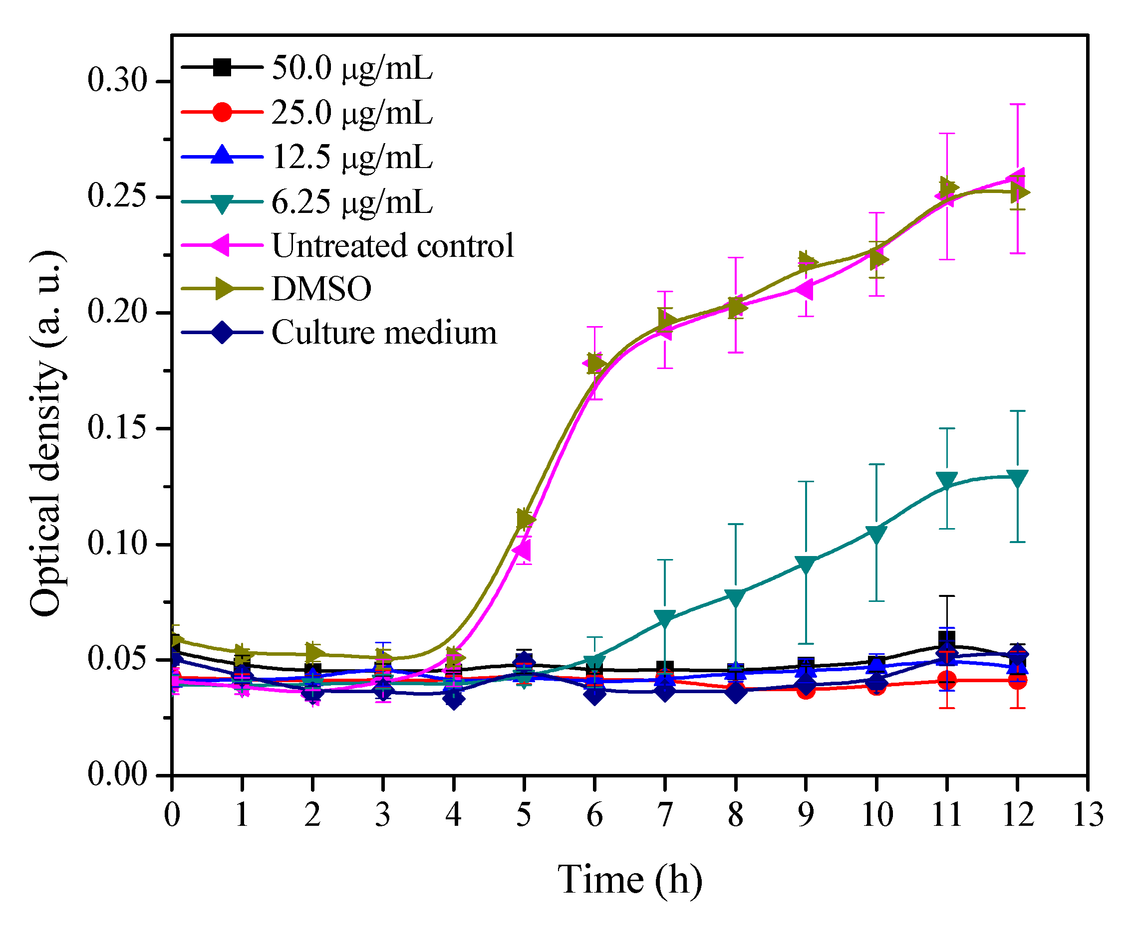
2.1. Growth Curve of MRSA with Different Concentrations of 3′-Demethoxy-6-O-demethylisoguaiacin

In order to determine the potential molecular mechanism of antibacterial activity of 3′-demethoxy-6-O-demethylisoguaiacin, a growth inhibitory curve of MRSA with different concentrations of 3′-demethoxy-6-O-demethylisoguaiacin was performed. Results showed that 3′-demethoxy-6-O-demethylisoguaiacin completely inhibited bacterial growth at concentrations of 50, 25 and 12.5 µg/mL, whereas a concentration of 6.25 µg/mL, there was growth at day 6 and growth continued until the end of the experiment but did not reach the optical density of the control (Figure 2). According to these results, the conditions (concentration and time) selected were 12.5 µg/mL and 1 h.
Figure 2. Bacterial growth-inhibitory concentration curve of 3′-demethoxy-6-O-demethylisoguaiacin against MRSA.
Molecules 20 12450 g002

2.2. Microarray Assay and RT-PCR

The microarray results showed a group of over-expressed (122) and repressed (84) genes with a Z score of ≥+1.5 and ≤−1.5, respectively. After microarray analysis, the analyzed genes were separated into groups according to their functional category (Table 1). The microarray results may be viewed at: http://www.ncbi.nlm.nih.gov/geo/query/acc.cgi?acc=GSE70309.
Table 1. Functional category of over expressed and repressed genes obtained from microarrays assay.
Functional CategoryOver Expressed Genes (%)Repressed Genes (%)
Translation/structural constituent of ribosome11.50
Pathogenesis2.50
Oxidation-reduction process3.30
Metabolic process 8.33.6
Unknow function, uncharacterized protein 14.337.8
Transcription3.30
Translation3.31.2
Catalytic activity2.50
Biosynthetic process7.58.4
Catabolic process20
Amino acid transport 2.50
Transport protein04.8
Glycolisis02.4
DNA repair02.4
Proteolysis02.4
Mechanism of defense04.8
Individual genes with different biological function3932.2
Some over-expressed genes are related to three metabolic pathways: pyruvate (SAR0217, SAR0824, and SAR0169), propanoate (SAR0217, SAR0169) and butanoate (SAR0217, SAR0169). These genes are related to catalytic activity, and in biological systems, they are essential for normal life processes. It is important to point out that the genes mentioned lower the energy required to begin a reaction, and decrease the reaction rate of enzymatic pathways at which cells perform essential chemical reactions [8].
Furthermore, catalysis is controlled by regulating enzymes, some of which are involved in enzyme structure changes, while others operate by altering the cell environment for catalytic activity. The molecular functions and the metabolic process involved in this biological process depend on the catalytic activity of enzymes that mediate most cell functions such as metabolism, which increases the rate of virtually all chemical reactions in cells in order to sustain life. Without enzymatic catalysis, most biochemical reactions would be so slow that they would not occur. Cells contain thousands of different enzymes, and their catalytic activities determine many chemical reactions that take place in the cell [9]. We propose that over-expression of the previously mentioned genes related with regard to catalytic activity affect main bacterial processes. Table 1 showed that the SAR1963 genes “Regulation of DNA repair” and SAR0785 “DNA replication” were also affected in MRSA. SAR1963 is part of a collection of processes by which a cell identifies and corrects damage to the DNA molecules that encode its genome, but genetic over-expression generates genetic diversity. This cause the losing of genomic stabilility as well as the lack of coordination and detection to a response to the damage, without effecting measures to prevent cell death [10,11]. The SAR0785 gene is directly related to the cellular metabolic process in which a cell duplicates one or more DNA molecules. Genetic over-expression affects the origin of replication particularly the genome sequence at which replication is initiated [12,13].
Analysis of repressed genes with the DAVID database resulted in some ATP-binding cassette (ABC) transport system proteins. This system is universally distributed among living organisms and functions in many different aspects of bacterial physiology [14]. This system is characterized by ABC protein transporters located in the cell membrane. Molecules are transported inside and outside the cell and this facilitate drugs release and elimination of toxic substances. In this way the therapeutic effects of antibacterial agents are avoided [15,16,17]. Zechini and Versace reported that some proteins of the ABC system are associated to drug-resistance [18]. Resistance of S. aureus RN4220 to vancomycin is due to the transport protein “msrA”; M. tuberculosis is resistant to tetracycline due to the protein “DrrAB”; E. coli is resistant to erythromycin due to the protein “MAcAB-TolC”. The ABC transport system of S. aureus is formed by 91 genes that express different proteins of this system. Of these 91 genes, we found six that were repressed (SAR0144, SAR1073, SAR1928, SAR0306, SAR0618, and SAR2267) after treatment with 3′-demethoxy-6-O-demethylisoguaiacin. With this we suggest that the above phytochemical compound could exert its effect on this membrane transport system.
The genes SAR0144, SAR1073, SAR1928 have a molecular function as ABC-type transporter ATP-binding protein, SAR0306 is a lipoprotein that is a releasing system ATP-binding protein, SAR0618 is a transport lipoprotein related to the union-substrate system and SAR2267 belongs to a subfamily of transport proteins, FecCD, and is related with ABC transporter permease system. 3′-demethoxy-6-O-demethylisoguaiacin repressed these MRSA proteins, and the bacteria could not expel the phytochemical, therefore this compound demonstrated its antibacterial activity.
In order to validate the expression level of six down-expressed genes (Table 2) corresponding to the ABC transport system, a RT-PCR assay was performed. In all six selected genes, repressed levels of mRNA as a result of exposure to the phytochemical compound were confirmed. This provides support to the microarray results and the above proposals.
Table 2. Real-Time PCR of six repressed genes involved in ABC transport system.
Gen IDDescriptionForward 5′→3′
Reverse 5′→3′
ZscoreRQTm Dissociation
SAR0144ABC transporter ATP-binding proteinGCACTAGAACGGGTCAACA
TGGGTCTAATGAAGCAACTGG
−1.507−5.922 ± 0.18678.12
SAR1073ABC transporter ATP-binding proteinATGTTGTTTAGAGGGGTCCAC
CCAACTTCGCTGCCTACT
−1.834−5.780 ± 0.32074.85
SAR1928ABC transporter ATP-binding proteinTGAAGTCGTTGCATTTGGAG
TCGCTTGGTTACGCATGT
−1.683−2.378 ± 0.04276.95
SAR0306ABC transporter ATP-binding proteinCGATTGGGTAGGAGGTGTA
CCAGAAGGTCCAACTAATGC
−1.782−2.039 ±0.30774.91
SAR0618Transport system lipoproteinGAACGCAGTTGGATGTAACC
CATACCACAGCCACTCAGAA
−1.917−1.392 ±0.06177.04
SAR2267FecCD transport family proteinGCGCCTTTATTGGTGGATTA
TGAACCAACAAGCCAAAACA
−1.955−4.205 ±0.02876.56
SARr01616S ribosomal RNA (Reference gen)CAGCATGCTACGGTGAATAC
GTTACGACTTCACCCCAATC
-176.75

3. Experimental Section

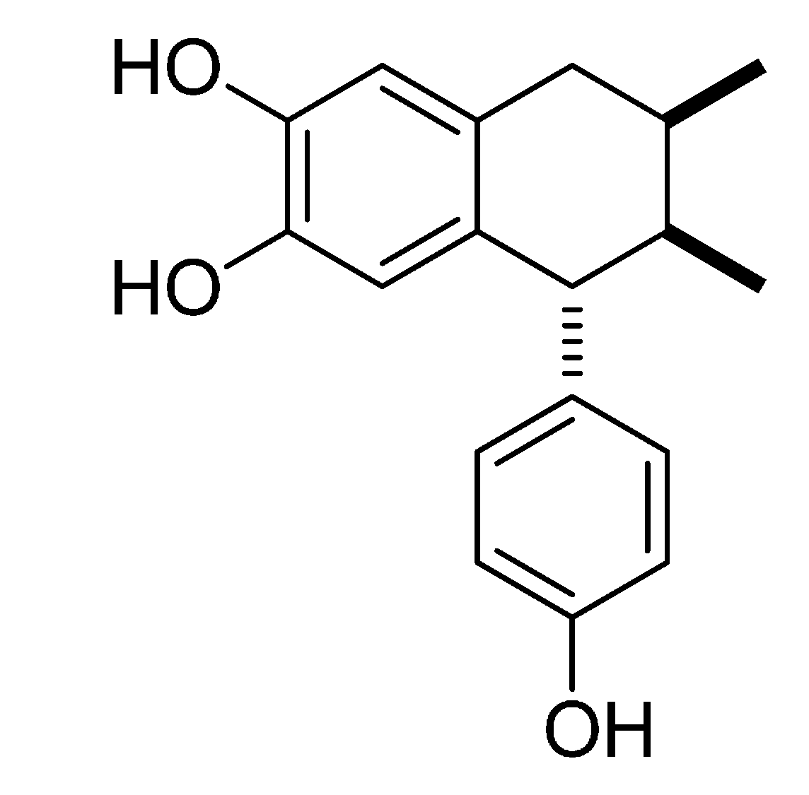
3.1. Isolation, Characterization and Evaluation of 3′-Demethoxy-6-O-demethylisoguaiacin

Dried and grounded leaves (500 g) of L. tridentata were extracted by maceration, first with hexane and then with CHCl3. Both extracts were separately distilled in vacuo until dryness. A total of 89 g of chloroform extract was obtained. A portion of this extract (80 g) was subjected to silica gel column chromatography (CC) and eluted with gradient of CHCl3/MeOH yielding 13 fractions. Fraction 10 (18 g) was chromatographed three times on silica gel CC, each column was eluted with a gradient of CHCl3/MeOH. From the sub-fraction 42–73 of the third column precipitated a brown solid which was purified by recrystallization with CHCl3/MeOH (98:2) yielding a beige solid (142.4 mg) [7]. This compound was characterized as 3′-demethoxy-6-O-demethylisoguaiacin by comparison of its spectral data with literature [19].
3′-Demethoxy-6-O-demethylisoguaiacin was evaluated against MRSA showing a MIC value of 12.5 µg/mL, which was the same as the positive control levofloxacin [7]. The cytotoxicity of the above natural product was determined using the WST-1 methodology [20]. Results showed that 3′-demethoxy-6-O-demethylisoguaiacin has an IC50 value of 31.10 µg/mL whereas the positive standard vincristine showed an IC50 value of 0.48 μg/mL against Chang liver cells (ATCC CCL-13) [21].

3.2. Growth Inhibitory Curve of MRSA Exposed to Different Concentrations of 3′-Demethoxy-6-O-demethylisoguaiacin

A stock solution was made by dissolving 1 mg of natural product in dimethylsulphoxide (DMSO) to reach a concentration of 20 mg/mL. A working solution was made with 3 μL of stock solution and 297 µL of Mueller-Hinton broth to give a concentration of 200 μg/mL. Then 100 µL of Mueller-Hinton broth was added to each well of a sterile 96 well microplate. Then, 100 μL of the working solution was added to row A. From this mixture 100 µL was taken with a multichannel pipet and transferred to row B to perform a 1:2 dilution. Then, further 1:2 dilutions were made until row D, finally the last 100 μL was discarded. Further, 100 µL of MRSA ATCC BAA-44 in Mueller-Hinton (105) was added to the wells with and without sample. The compound was tested in the concentrations of 50, 25, 12.5 and 6.25 µg/mL of 3′-demethoxy-6-O-demethylisoguaiacin. DMSO in each well did not exceed 0.05%, and this concentration does not affect bacterial growth. Each concentration was evaluated in triplicate. Two untreated controls were made, one with only bacteria and the other with bacteria and DMSO (≤0.083%). The plate was covered with a lid and incubated a 37 °C. Bacterial growth was determined by measuring the optical density at 620 nm at regular intervals of one hour until completing 12 h using a 96-well microtiter plate reader. The bacterial growth curve was performed in order to determinate exposure time and the concentration of 3′-demethoxy-6-O-demethylisoguaiacin needed to cause stress on MRSA. The experiment was done three times on different days.

3.3. RNA Isolation and Synthesis of Labeled cDNA

Two Erlenmeyer flasks with 100 mL of Mueller-Hinton broth each were inoculated with MRSA and the turbidity was adjusted at 0.5 MacFarland’s standard (1.5 × 10⁸ CFU). Both flasks were incubated for 4 h to achieve logarithmic phase (optical density 0.4 at 600 nm), then drug treatment was conducted by adding 3′-demethoxy-6-O-demethylisoguaiacin to one of the cultures to achieve a final concentration of 12.5 μg/mL. Untreated control bacteria was grown under identical conditions to treated bacteria, with the exception that no drug was added, only dimethyl sulfoxide (DMSO) which did not exceed 0.05% v/v. Both flasks were incubated 1 h during drug treatment. After incubation time the bacteria was harvested separately by centrifugation at 3500 rpm for 15 min. The bacterial pellets obtained were stored at −70 °C for RNA isolation. Total RNA of each bacterial pellet was isolated and purified with the RiboPure-Bacteria AM1925 kit (Ambion, Carlsbad, CA, USA) according to the manufacturer’s instructions, including a DNAse digestion step. Quantity and quality of RNA obtained were assessed by performing agarose gel electrophoresis and analyzing this in a biophotometer and Molecular Imager® Gel Doc™ XR+ System equipped with Image Lab™ software, respectively (BioRad, Hercules, CA, USA). To obtain the mRNA from each total RNA (10 µg), the MICROBExpress AM1905 (Ambion) kit was used according to the manufacturer’s protocol. The Labeled cDNA of each condition was obtained from mRNA using Amino Allyl cDNA Labeling AM 1705 (Ambion) kit and Amersham CyDye Post-Labeling Reactive Dye Packs (GE Healthcare Bio-Sciences AB, Piscataway, NJ, USA) according to the manufacturer’s instructions.

3.4. Microarray and Data Analysis

Microarray hybridization was performed using the procedure described in the protocol by MYcroarray Company (Ann Arbor, MI, USA). A whole genome of S. aureus MRSA252 (CAT-30K) consisting of 1 piece of sequence (BX571856 GeneBank) representing a total of 2744 transcripts was used. Dynamic hybridization was performed in a hybridization Chamber G2534A (Agilent, Santa Clara, CA, USA) and covered with G2534-60002 gasket slides (Agilent). Fluorophore incorporation efficiency was analyzed measuring absorbance at 555 nm for Cy3 and 655 nm for Cy5 using a Gene Pix 4000B scanner with Acuity 4.0 software (Molecular devices, Sunnyvale, CA, USA); scans were saved as TIFF images and quantified. Data were then normalized and analyzed with genArise software (version 2.2.0, Computing Unit, Cellular Physiology Institute, UNAM, Mexico City, Mexico). Genes with a Z score ± 1.5 were considered with altered expression. The list of genes with altered expression was analyzed with the following bioinformatic tools databases: Data base for Annotation Visualization and integrated Discovery (DAVID) [22], Kyoto Encyclopedia of Genes and Genomes (KEGG) [23], and GenBank, GenDB [24]. In order to perform this analysis, the list of genes of interest was inserted into these tools and databases. A group of genes with function was obtained and organized in Table 1.

3.5. Real Time-PCR Assay

Samples of both treated and untreated mRNA isolated from MRSA were employed in RT-PCR. cDNA was obtained using SuperScript® VILO™ cDNA Synthesis Kit (Invitrogen 11754050, Life Technologies, Carlsbad, CA, USA). RT-PCR was performed using SYBR® GreenER™ qPCR SuperMix Universal (Invitrogen 11780200, Life Technologies). The relative quantitation method employed was the comparative CT in StepOne™ Real-Time PCR system (Applied Biosystems, Foster City, CA, USA). The specific primers used in the experiment were designed in primer3 version 4. In order to assure specific amplification, melting curves were performed for all interest genes and the samples were run in triplicate in each reaction; 16S ribosomal RNA (SARr016) was used as housekeeping gene.

4. Conclusions

We propose a potential molecular mechanism for 3′-demethoxy-6-O-demethylisoguaiacin whereby it acts on the cellular membrane where the phytochemical represses some proteins of the ABC transport system of MRSA. Thus the bacteria could not release the phytochemical and this caused its antibacterial activity. It is important to point out that this molecular mechanism is not presented by any commercial drug and could be a new target for the development of novel antibacterial agents.

Acknowledgments

This work was supported by Consejo Nacional de Ciencia y Tecnología (CONACYT, grant number CB-2008-106107) and Secretaria de Educación Pública SEP PRODEP 103.5/15/3224. Juan Manuel J. Favela-Hernández received a scholarship from CONACYT to do Ph.D. studies. We thank to Lorena Chávez González, Simón Guzmán León, José Luis Santillán Torres and Jorge Ramírez, for technical assistance in the microarray determinations. Gerardo Coello, Gustavo Corral and Ana Patricia Gómez for genArise software assistance, Unidad de Microarreglos, Instituto de Fisiología Celular-UNAM. Authors acknowledge Sergio Lozano Rodríguez for improving English grammatical aspects of this manuscript.

Author Contributions

Juan Manuel J. Favela-Hernández carried out the MRSA growth inhibitory curve, performed the isolation of RNA, synthesis of labeled cDNA, microarray assay, the analysis and interpretation of data and drafted the manuscript. Aldo F. Clemente-Soto carried out RT-PCR. Isaías Balderas-Rentería supervised the experimental work. Elvira Garza-González gave the laboratory facilities to grow MRSA. María del Rayo Camacho-Corona carried out general supervision of the research group and acquisition of funding. All authors revised the intellectual content of article, read and approved the final manuscript.

Conflicts of Interest

The authors declare no conflict of interest.

References

  1. Ribeiro Lamblet, L.C.; Aparecida Barbosa, D.D. Prevalence of Staphylococcus aureus colonization in renal transplant patients. Rev. Esc. Enferm. USP 2014, 48, 824–830. [Google Scholar]
  2. Butterly, A.; Schmidt, U.; Wiener-Kronish, J. Methicillin-resistant Staphylococcus aureus colonization, its relationship to nosocomial infection, and efficacy of control methods. Anesthesiology 2010, 113, 1453–1459. [Google Scholar] [CrossRef] [PubMed]
  3. Stryjewski, M.E.; Corey, G.R. Methicillin-resistant Staphylococcus aureus: An evolving pathogen. Clin. Infect. Dis. 2014, 58, 10–19. [Google Scholar] [CrossRef] [PubMed]
  4. Svarovsky, S.A.; González-Mo, M.J. High-throughput platform for rapid deployment of antimicrobial agents. ACS. Comb. Sci. 2011, 13, 634–638. [Google Scholar] [CrossRef] [PubMed]
  5. Kingston, D.G.J. Modern natural products drug discovery and its relevance to biodiversity conservation. J. Nat. Prod. 2011, 74, 496–511. [Google Scholar] [CrossRef] [PubMed]
  6. Ruiz-Martínez, J.; Ascacio, J.A.; Rodríguez, R.; Morales, D.; Aguilar, C.N. Phytochemical screening of extracts from some Mexican plants used in traditional medicine. J. Med. Plants Res. 2011, 5, 2791–2797. [Google Scholar]
  7. Favela-Hernández, J.M.J.; García, A.; Garza-González, E.; Rivas-Galindo, V.M.; Camacho-Corona, M.R. Antibacterial and Antimycobacterial lignans and flavonoids from Larrea tridentata. Phytother. Res. 2012, 26, 1957–1960. [Google Scholar] [CrossRef] [PubMed]
  8. Jackson, C.J.; Foo, L.; Tokuriki, N.; Afriat, L.; Carr, P.D.; Kim, H.K.; Schenk, G.; Tawfik, D.S.; Ollis, D.L. Conformational sampling, catalysis, and evolution of the bacterial phosphotriesterase. Proc. Natl. Acad. Sci. USA 2009, 106, 21631–21636. [Google Scholar] [CrossRef] [PubMed]
  9. Seshasayee, A.S.; Fraser, G.M.; Luscombe, N.M. Comparative genomics of cyclic-di-GMP signalling in bacteria: Post-translational regulation and catalytic activity. Nucleic Acids Res. 2010, 38, 5970–5981. [Google Scholar] [CrossRef] [PubMed]
  10. Song, Y.; Rubio, A.; Jayaswal, R.K.; Silverman, J.A.; Wilkinson, B.J. Additional routes to Staphylococcus aureus daptomycin resistance as revealed by comparative genome sequencing, transcriptional profiling, and phenotypic studies. PLoS ONE 2013, 8, e58469. [Google Scholar] [CrossRef] [PubMed]
  11. Lenhart, J.S.; Schroeder, J.W.; Walsh, B.W.; Simmons, L.A. DNA repair and genome maintenance in Bacillus subtilis. Microbiol. Mol. Biol. Rev. 2012, 76, 530–564. [Google Scholar] [CrossRef] [PubMed]
  12. Rasigade, J.P.; Vandenesch, F. Staphylococcus aureus: A pathogen with still unresolved issues. Infect. Genet. Evol. 2014, 21, 510–514. [Google Scholar] [CrossRef] [PubMed]
  13. Kim, B.S.; Yi, H.; Chun, J.; Cha, C.J. Genome sequence of type strain of Staphylococcus aureus subsp. aureus. Gut Pathog. 2014, 17, 6. [Google Scholar] [CrossRef] [PubMed]
  14. Davidson, A.L.; Dassa, E.; Orelle, C.; Chen, J. Structure, function, and evolution of bacterial ATP-binding cassette systems. Microbiol. Mol. Biol. Rev. 2008, 72, 317–364. [Google Scholar] [CrossRef] [PubMed]
  15. Jonsson, I.M.; Juuti, J.T.; François, P.; Al Majidi, R.; Pietiäinen, M.; Girard, M.; Lindholm, C.; Saller, M.J.; Driessen, A.J.; Kuusela, P.; et al. Inactivation of the Ecs ABC transporter of Staphylococcus aureus attenuates virulence by altering composition and function of bacterial wall. PLoS ONE 2010, 5, e14209. [Google Scholar] [CrossRef] [PubMed]
  16. Henderson, D.P.; Payne, S.M. Vibrio cholerae iron transport systems: Roles of heme and siderophore iron transport in virulence and identification of a gene associated with multiple iron transport systems. Infect. Immun. 1994, 62, 5120–5125. [Google Scholar] [PubMed]
  17. Álvarez de Felipe, A.; Pulido-Duarte, M. Transportadores de tipo ABC: Consecuencias de su interacción con flavonoides. Bol. Latinoam. Caribe Plant Med. Aromat. 2008, 7, 296–311. [Google Scholar]
  18. Zechini, B.; Versace, I. Inhibitors of multidrug resistant efflux systems in bacteria. Anti-Infect. Drug Discov. 2009, 4, 37–50. [Google Scholar] [CrossRef]
  19. Konno, C.; Xue, H.Z.; Lu, Z.Z.; Ma, B.X.; Erdelmeier, C.A.J.; Che, C.T.; Cordell, G.A.; Soejarto, D.D.; Waller, D.P.; Fong, H.H.S. 1-Aryl Tetralin Lignans from Larrea tridentata. J. Nat. Prod. 1989, 52, 1113–1117. [Google Scholar] [CrossRef]
  20. Francoeur, A.M.; Assalian, A. Microcat: A novel cell proliferation and cytotoxicity assay based on WST-1. Biochemica 1996, 3, 19–25. [Google Scholar]
  21. Favela-Hernández, J.M.J. Aislamiento y Caracterización de los Compuestos Antibacterianos y Antituberculosos de Larrea Tridentata, Determinación de su Toxicidad y Mecanismo de Acción del Compuesto Más Activo. Ph.D. Thesis, Facultad de Ciencias Químicas, Universidad Autónoma de Nuevo León, San Nicolás de Los Garza, Mexico, 2012; p. 143. [Google Scholar]
  22. Huang, D.W.; Sherman, B.T.; Lempicki, R.A. Systematic and integrative analysis of large gene lists using DAVID Bioinformatics Resources. Nat. Protoc. 2009, 4, 44–57. [Google Scholar] [CrossRef] [PubMed]
  23. Altermann, E.; Klaenhammer, T.R. PathwayVoyager: Pathway mapping using the Kyoto Encyclopedia of Genes and Genomes (KEGG) database. BMC Genomics 2005, 3. [Google Scholar] [CrossRef] [Green Version]
  24. Meyer, F.; Goesmann, A.; McHardy, A.C.; Bartels, D.; Bekel, T.; Clausen, J.; Kalinowski, J.; Linke, B.; Rupp, O.; Giegerich, R.; et al. GenDB-an open source genome annotation system for prokaryote genomes. Nucleic Acids Res. 2003, 31, 2187–2195. [Google Scholar] [CrossRef] [PubMed]
  • Sample Availability: Sample of the compound is available from the authors.

