Article

Escherichia coli ASKA Clone Library Harboring tRNA-Specific Adenosine Deaminase (tadA) Reveals Resistance towards Xanthorrhizol

1
Department of Biotechnology, Yonsei University, 50-Yonsei-ro Seodaemun-gu, Seoul 120-749, Korea
2
Faculty of Biotechnology, Atma Jaya Catholic University of Indonesia, Jalan Jenderal Sudirman 51, Jakarta 12930, Indonesia
3
Superbacteria Research Center, Korea Research Institute of Bioscience and Biotechnology (KRIBB), 111 Gwahangno, Yuseong, Daejeon 305-806, Korea
*
Authors to whom correspondence should be addressed.
These authors contributed equally to this work.
Molecules 2015, 20(9), 16290-16305; https://doi.org/10.3390/molecules200916290
Submission received: 11 June 2015 / Revised: 27 August 2015 / Accepted: 31 August 2015 / Published: 9 September 2015

Abstract

:
Xanthorrhizol is a potent antimicrobial compound isolated from the rhizome of Curcuma xanthorrhiza. However, the mechanism of xanthorrhizol action is unknown. To screen for probable target(s), we introduced the ASKA pooled-plasmid library into Escherichia coli W3110 imp4213 and enriched the library for resistant clones with increasing concentrations of xanthorrhizol. After three rounds of enrichment, we found nine genes that increased xanthorrhizol resistance. The resistant clones were able to grow in LB medium containing 256 µg/mL xanthorrhizol, representing a 16-fold increase in the minimum inhibitory concentration. Subsequent DNA sequence analysis revealed that overexpression of tadA, galU, fucU, ydeA, ydaC, soxS, nrdH, yiiD, and mltF genes conferred increased resistance towards xanthorrhizol. Among these nine genes, tadA is the only essential gene. tadA encodes a tRNA-specific adenosine deaminase. Overexpression of E. coli W3110 imp4213 (pCA24N-tadA) conferred resistance to xanthorrhizol up to 128 µg/mL. Moreover, overexpression of two tadA mutant enzymes (A143V and F149G) led to a twofold increase in the MIC. These results suggest that the targets of xanthorrhizol may include tadA, which has never before been explored as an antibiotic target.

1. Introduction

Food material can be a promising resource for antimicrobial discovery. Xanthorrhizol, the main compound in the rhizome of Java turmeric (Curcuma xanthorrhiza), is known to exhibit antimicrobial activity against Gram-positive bacteria and against several Gram-negative bacteria, such as Bacillus cereus, Staphylococcus aureus, Streptococcus mutans, Salmonella typhimurium KCCM 11862 and Vibrio parahaemolyticus [1,2,3]. Furthermore, xanthorrhizol shows rapid bactericidal activity against S. mutans [1]. Xanthorrhizol was recently used to target the enoyl-ACP reductase (FabI) of E. coli [4]. However, targeting FabI, which is a bacteriostatic target [5,6], does not explain the rapid killing mode of action of this compound, thus implying that screening for other candidates may reveal additional xanthorrhizol targets.
Encompassing a complete set of E. coli genes, the ASKA clone (−) library (A complete Set of E. coli K-12 ORF Archive) comprises 4122 clones [7]) that are suitable for, but not limited to, systematic functional genomics study, including DNA microarrays and protein expression, protein localization, and protein–protein interaction studies [8]. The ASKA clone library has also been used to confirm or screen for antimicrobial targets [9,10,11]. Couce et al. used the ASKA clone library to overexpress genes that may confer resistance to fosfomycin [9]. Genome-wide screening of the ASKA clone library in LB medium containing fosfomycin resulted in the identification of a clone harboring pCA24N-murA as the sole survivor [9]. A study to identify triclosan targets using ASKA library enrichment revealed multiple triclosan resistance genes [11].
In this study, we used the ASKA clone (−) library to identify gene(s) that could increase resistance to xanthorrhizol. Unfortunately, xanthorrhizol is less active against several Gram-negative bacteria, including E. coli [3]. To overcome this limitation, we used E. coli W3110 imp4213 as a host for the ASKA plasmid library. This E. coli imp strain exhibits increased membrane permeability [12,13], rendering it more susceptible to ampicillin, carbenicillin, bacitracin, erythromycin, novobiocin, and rifampin [14]. We also found that E. coli W3110 imp4213 was susceptible to xanthorrhizol at a minimum inhibitory concentration (MIC) of 16 µg/mL. By using this ASKA library, we identified another xanthorrhizol target, tadA, a tRNA-specific adenine deaminase.

2. Results and Discussion

Wild-type E. coli W3110 cannot be killed by xanthorrhizol (MIC > 512 µg/mL). To use E. coli as a model for xanthorrhizol resistance, we used the E. coli W3110 imp4213 strain in our laboratory collection as a host for the ASKA pooled-plasmid library. E. coli W3110 imp4213 is susceptible to xanthorrhizol, with an MIC of 16 µg/mL.

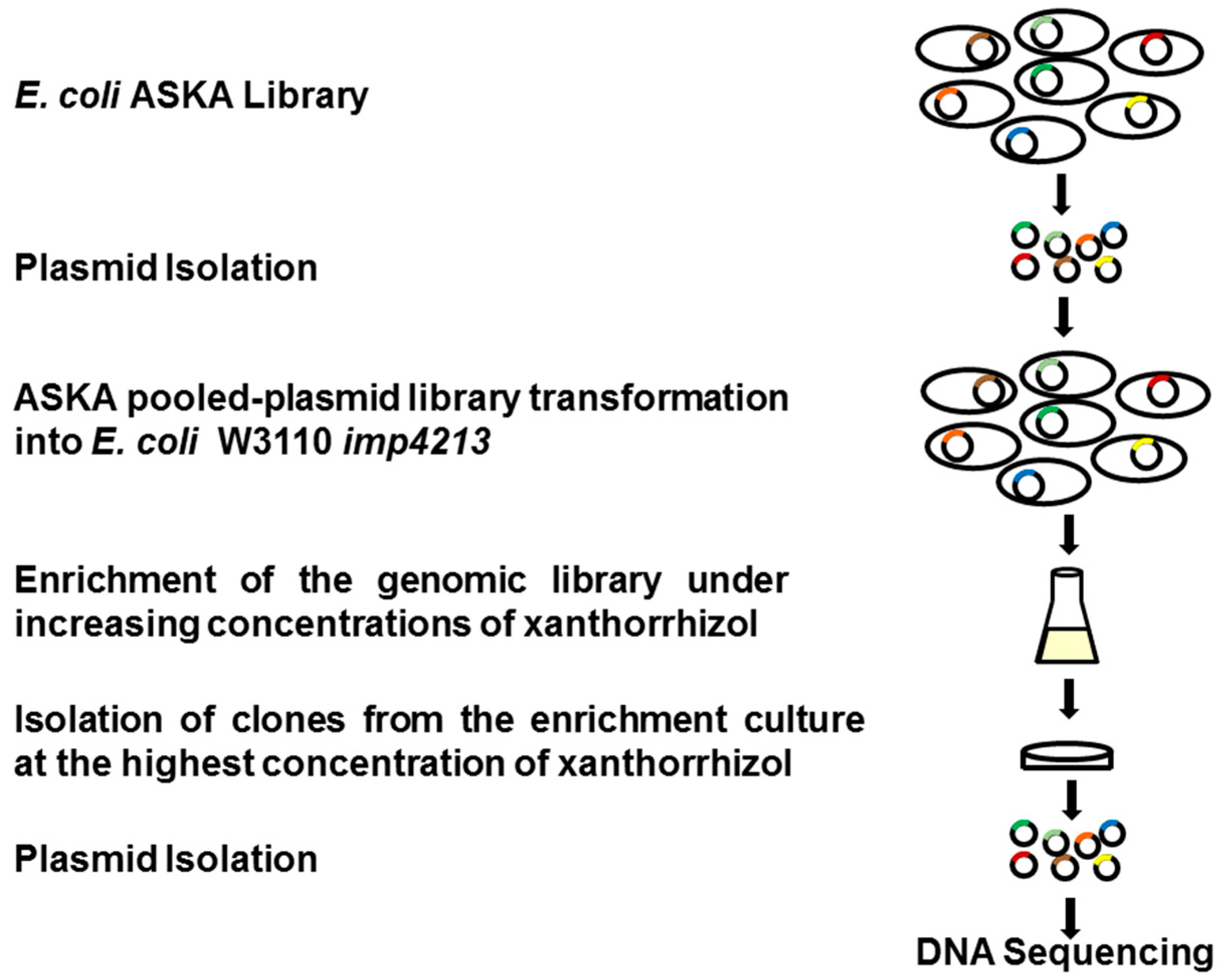
2.1. ASKA Library Enrichment and Screening for Xanthorrhizol Resistance

In this study, we used a complete set of ORF clones from the E. coli ASKA library to screen for gene(s) that confer resistance to xanthorrhizol. This is a plasmid-based genomic screening system that was modified from the protocol used for the triclosan target screen [11]. Cells that can grow at high concentrations of xanthorrhizol may bear plasmids containing genes that confer xanthorrhizol resistance. Such clones would likely out-compete others in the culture. The library grown in the absence of xanthorrhizol was used as a growth control. After three rounds of enrichment, clones from E. coli W3110 imp4213 harboring the ASKA pooled-plasmid library were isolated from the cultures grown with the highest concentrations of xanthorrhizol (Figure 1). The library culture showed delayed growth in LB medium supplemented with xanthorrhizol, compared to E. coli W3110 imp4213 harboring an empty plasmid pCA24N (Figure 1). After the final round of enrichment, the clones could grow at a xanthorrhizol concentration of 256 µg/mL within 20 h of inoculation.
Figure 1. Growth of the E. coli W3110 imp4213 (pCA24N) and E. coli W3110 imp4213 ASKA pooled-plasmid library culture in Luria broth media supplemented with chloramphenicol (30 µg/mL) and with various concentrations of xanthorrhizol. Growth was monitored for 20 h in an automatic incubator (TVS1 Bio-Photorecorder, Advantec, Shiga, Japan). (A) E. coli W3110 imp4213 (pCA24N) was grown in the present of xanthorrhizol 0, 8, 16 and 32 µg/mL. The growth was inhibited at 16 µg/mL; (B) E. coli W3110 imp4213 ASKA pooled-plasmid library was grown in LB media with various concentrations of xanthorrhizol after three rounds of enrichment. The xanthorrhizol concentrations used were 0, 32, 64, 128, and 256 µg/mL. The library clones that grew after the delay exhibited resistance towards xanthorrhizol.
Molecules 20 16290 g001
A total of 107 plasmids were isolated from the enriched library clones. Sequencing analysis of the isolated plasmids revealed nine different genes that may be related to xanthorrhizol resistance (Table 1). An ASKA clone harboring the tadA gene dominated the enriched culture (71 out of 107 isolated plasmids, or 66.4%), followed by the ASKA clones carrying galU (11.2%), fucU (7.5%), ydeA (5.6%), ydaC (3.7%), soxS (1.9%), nrdH (1.9%), yiiD (0.9%), and mltF (0.9%). Among these nine E. coli genes, only tadA is classified as an essential gene [15]. tadA encodes tRNA-specific adenosine deaminase A.
The tadA gene encodes a tRNA-specific adenosine deaminase that specifically converts A34 to I34 in bacterial tRNAArg2 [16]. The tadA gene is known to be essential in E. coli MG1665 [16,17,18] and Shewanella oneidensis MR-1 [19]. However, this gene is noted as non-essential in Bacillus subtilis 168 and Pseudomonas aeruginosa UCBPP-PA14 in the Online Gene Essentiality Database [20]. The tadA protein is conserved (>83%) among Gram-negative and Gram-positive bacteria, and yeast [16]. Prokaryotic tadA shares amino acid sequence similarity with the Tad2P enzymes in humans, Drosophila, and Saccharomyces [21]. Tad2P deaminates adenosine, but its deaminase domain is similar to that of the cytidine deaminase (CDA) superfamily [22]. However, tadA is specific for prokaryotic tRNA substrates and it specifically deaminates tRNAArg2. No eukaryotic tRNA substrates were found to be modified by tadA, except for yeast tRNAArg [16]. Because tadA is essential in E. coli, we used the tadA gene for further analysis.
Table 1. List of identified genes in the ASKA pooled-plasmid library enrichment.
Gene NameFunctionCOG Classification aEssentiality b
tadAtRNA-Specific Adenine DeaminaseTranslationEssential
galUGlucose-1-Phosphate UridylyltransferaseCell Envelope Biogenesis, Outer MembraneNon-essential
ydaCProtein Involved in Chromosome Maintenance/Rac Prophage GeneDNA Replication, Recombination, and RepairNon-essential
fucUl-Fucose MutarotaseCarbohydrate Transport and MetabolismNon-essential
mltF (yfhD)Membrane-Bound Lytic Murein Transglycosylase FCell Envelope Biogenesis, Outer MembraneNon-essential
ydeASugar Efflux TransporterCarbohydrate Transport and MetabolismNon-essential
soxSDNA-Binding Transcriptional RegulatorTranscriptionNon-essential
yiiDPredicted AcetyltransferaseGeneral Function Prediction OnlyNon-essential
nrdH (ygaN)Glutaredoxin-Like ProteinPost-Translational Modification, Protein Turnover, ChaperonesNon-essential c
a COG: Cluster of Orthologous Groups [23]; b These data were obtained from the Database of Essential Genes [15,24,25,26]; c The Database of Essential Genes classifies nrdH as an essential gene, but a knockout mutant is present in the KEIO collection [17].
The other eight genes that were isolated from the enrichment are listed as non-essential genes. However, among these genes, the essentiality of nrdH is ambiguous because, although Gerdes et al. classified nrdH as an essential gene [18], a knockout-mutant strain of nrdH has been constructed and is present in the KEIO non-essential gene-knockout collection [17]. Although galU is not essential for bacterial growth, this gene plays an important role in bacterial virulence, for example in Francisella tularensis [27]. ydaC (synonym: rcbA) encodes a protein that is involved in maintaining the integrity of the bacterial chromosome by lowering the steady-state level of double-strand breaks [28]. Overexpression of ydaC leads to increased resistance to erythromycin [10]. mltF encodes a membrane-bound lytic transglycosylase that is responsible for the release of 1,2-anhydromuropeptides from peptidoglycan [29]. MltF (YfhD) is located in the periplasmic space and is involved in organic solvent tolerance. However, the deletion or loss of mltF does not cause any loss of such tolerance [30]. SoxS is a DNA-binding transcriptional dual regulator. This regulator activates the transcription of the soxRS regulon, which is involved in the oxidative stress-defense system of E. coli [31]. Overexpression of soxS has also been reported to increase resistance to triclosan in E. coli [32]. This increased resistance is due to the expression control of genes involved in drug efflux, thereby leading to drug resistance [33]. Furthermore, modifications of lipopolysaccharide that are mediated by SoxS can protect E. coli against various compounds and increase resistance to multiple drugs [34]. The yiiD gene encodes a predicted acetyltransferase. Little information is available regarding this gene, but in a protein interaction study, Butland et al. revealed that yiiD interacts with an acyl carrier protein that is a key player in fatty-acid chain elongation [35].

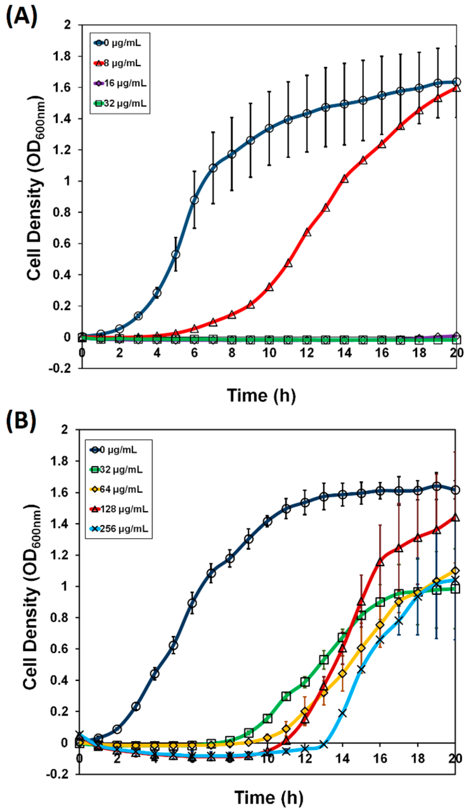
2.2. Verification of tadA Overexpression-Induced Resistance to Xanthorrhizol in E. coli W3110 imp4213 and Bacillus subtilis 168, Site-Directed Mutagenesis, and Binding Study

Our results revealed that an essential gene, tadA, was a candidate that might be responsible for increasing the resistance of the E. coli W3110 imp4213 ASKA pooled library to xanthorrhizol. The level of tadA gene overexpression was confirmed by cDNA analysis (Figure S1). tadA cDNA was increased approximately twofold compared to the control (Figure S1). We isolated plasmid pCA24N-tadA from the original ASKA library, re-introduced it into E. coli W3110 imp4213, and measured the MIC of xanthorrhizol against the resulting strain. E. coli W3110 imp4213 (pCA24N-tadA) exhibited xanthorrhizol resistance, with an MIC of 128 µg/mL (Table 2). This MIC was not as high as that of the enriched library culture, a result that is likely due to the library being a mixed culture in which each clone contributes to the overall resistance towards xanthorrhizol.
Table 2. Xanthorrhizol minimum inhibitory concentration s of wild-type and mutant tadA-overexpressing strains.
Bacterial StrainMIC (μg/mL)
E. coli W3110 imp421316
E. coli W3110 imp4213 (pCA24N)16
E. coli W3110 imp4213 (pCA24N-tadA)128
E. coli W3110 imp4213 (pCA24N-tadA (A143V))256
E. coli W3110 imp4213 (pCA24N-tadA (F149G))256
Bacillus subtilis 1688
B. subtilis 168 (pHT255)8
B. subtilis 168 (pHT255-tadA)32
As described in the Introduction Section, xanthorrhizol is more effective against Gram positive bacteria, such as B. subtilis. To confirm whether tadA is involved in increasing resistance towards xanthorrhizol, we treated B. subtilis 168 overexpressing tadA with xanthorrhizol. We observed that xanthorrhizol MICs increased fourfold (Table 2). This result suggests that tadA may be one of the xanthorrhizol targets in the bacterial cell.
Site-directed mutagenesis was used to confirm that tadA was the xanthorrhizol target. Using docking analysis, we found that substitutions at Ala143 and Phe149 would be likely to have only a small effect on overall protein folding (Figure S2). Ala143 and Phe149 are located at the C-terminus of the tadA protein. The subsequent determination of xanthorrhizol MICs confirmed that the tadA mutants A143V and F149G confer twofold increased resistance towards xanthorrhizol (Table 2).
The study of xanthorrhizol and tadA interaction was carried out using tryptophan quenching assay. Tryptophan quenching assay has been used to study binding activity of an enzyme inhibitor [36,37]. tadA protein contains three intrinsic tryptophan residues that make it suitable for quenching assay. The binding activity is indicated by the decreasing of fluorescence signal. In this study, 4 µM tadA protein was titrated with xanthorrhizol. Fluorescence titration demonstrated that xanthorrhizol was able to bind to the protein as revealed by fluorescence change with a Kd = 19.04 μM, 109.7 μM, and 32.78 μM for wild type tadA, A143V, and F149G, respectively (Figure 2). These results showed increasing Kd of xanthorrhizol in tadA mutants. However, the Kd for both tadA mutants was different even the MIC was the same. This could be because xanthorrhizol may have multiple intracellular targets that might contribute to its inhibition activity.
It should be noted that there have been no reports describing tRNA-specific adenosine deaminase as an antibiotic target. However, some information regarding antibiotics that target microbial RNA processing/editing enzymes is available. Among them, aminoacyl-tRNA synthetase (aaRS) is an essential enzyme and has for years been an attractive target for antibiotic development [38,39]. This enzyme catalyzes the synthesis of aminoacyl-tRNA, which plays an important role in protein biosynthesis. Targeting aaRS has also become one of the strategies to combat multidrug resistance Gram-negative bacteria [40]. Mupirocin, a natural product isolated from Pseudomonas fluorescens, is the only approved antibiotic targeting aaRS and is widely used as a topical antibiotic against S. aureus [41].
Figure 2. Interaction of Xanthorrhizol and tadA protein. Tryptophan quenching assay (AC) were used to observe interaction between xanthorrhizol and tadA protein; (A) wild type tadA; (B) tadA (A143V); and (C) tadA (F149G). The assays showed a response, as revealed by fluorescence signal quenching, that xanthorrhizol bound to tadA protein. Tryptophan quenching assay demonstrated that binding capacity of xanthorrhizol to tadA mutants were lower than wild type, as shown by calculated Kd 19.04 μM, 109.7 μM, and 32.78 μM for wild type, A143V, and F149G, respectively.
Molecules 20 16290 g002

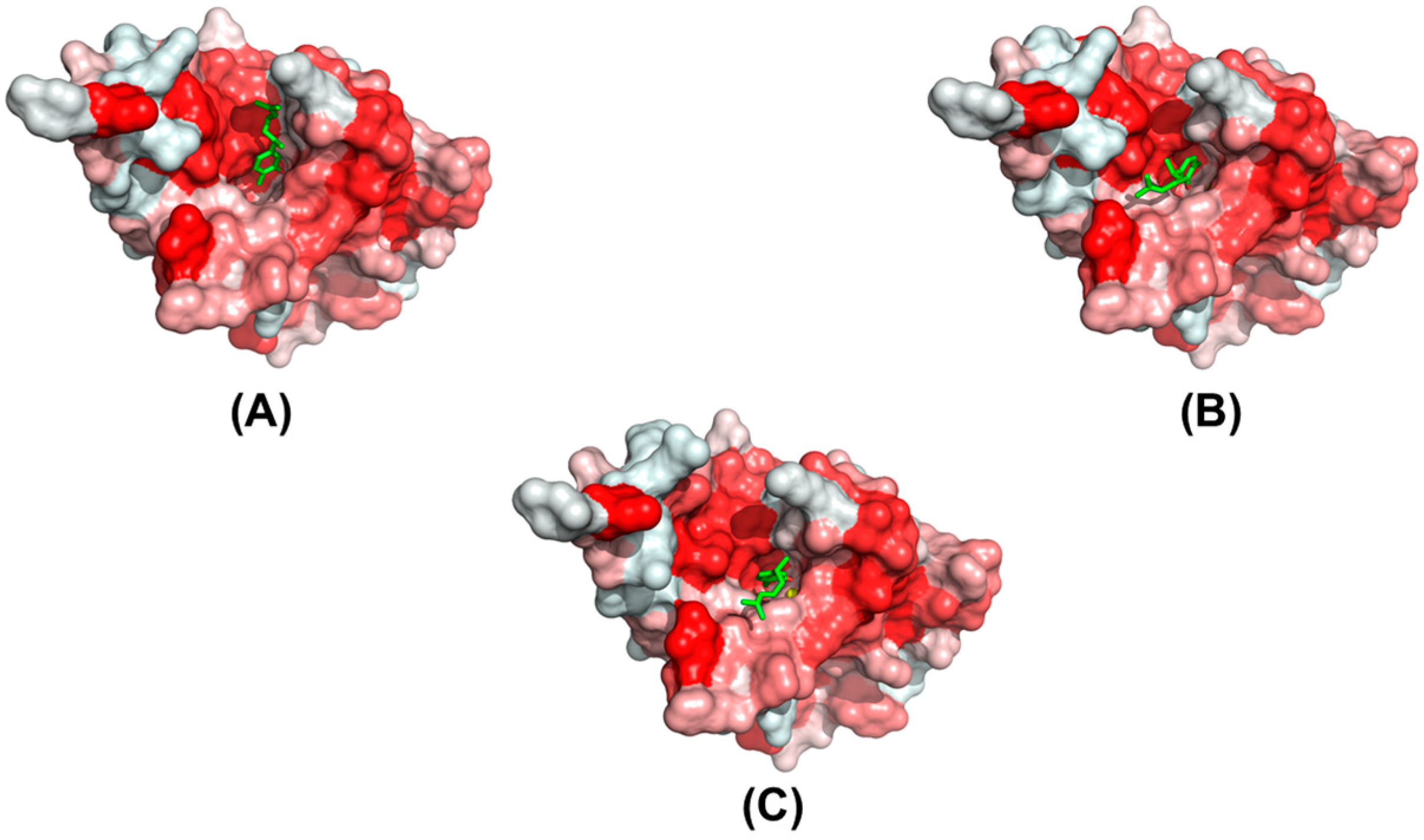
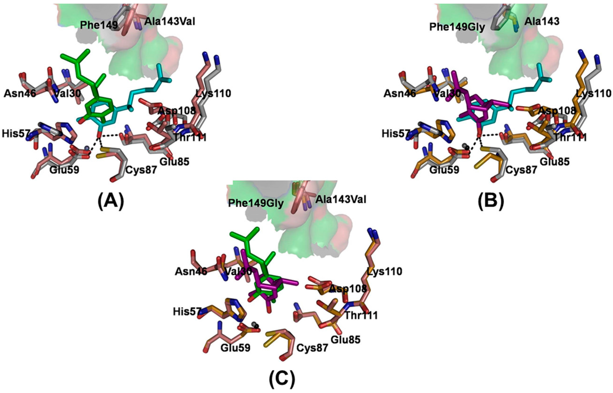
2.3. Modeling of the Binding Mechanism of Xanthorrhizol to Wild-Type and Mutant EcTadA

To identify the drug-resistance mechanism induced by the two mutations at sites 143 and 149, the interaction between EcTadA and xanthorrhizol was explored in detail from energetic and structural perspective. The binding free energies of xanthorrhizol with WT EcTadA and with the A143V and F149G mutants are −9.94 kcal/mol, −6.35 kcal/mol, and −6.69 kcal/mol, respectively (Table S1). Relative to wild-type EcTadA, the contribution of ΔGnonpolar is obviously decreased in the mutants (Table S1). The decrease in nonpolar interaction contribution can be caused by the loss of the van der Waals interaction contribution. The van der Waals contributions for both mutants decreased more than 5 kcal/mol relative to the wild type (Table S1).
Among the contributions of different energy components, the hydrophobic interaction, with a 50.82-kcal/mol contribution (Table S1), provides the main driving force for the binding of xanthorrhizol. A comparison of the hydrophobic surfaces (Figure 3) of the wild-type vs. either mutated EcTadA reveals that both the hydrophobicity and the size of the hydrophobic pocket are influenced by the mutations. In the wild type, the phenolic group of xanthorrhizol was embedded well into the corresponding hydrophobic pocket containing the catalytic active site. In the A143V mutant, with the substitution from a small, hydrophobic alanine to a large, hydrophobic valine, the relative cavity size of the hydrophobic pocket is decreased. Furthermore, the phenolic group of xanthorrhizol is moved away from the favorable binding pocket (Figure 3B). In the F149G mutant, the substitution of Phe149 with a side chain-lacking glycine also results in an unfavorable hydrophobic interaction; as with the A143V mutation, the xanthorrhizol is forced to leave the favorable position (Figure 3C). A superimposition of the representative structures of the mutants relative to the wild-type complex showed the evidence that, in the mutants, the phenolic group of xanthorrhizol is forced to leave the original and favorable substrate-binding (Figure 4). Such a change in position can influence the hydrophobic/hydrophilic interactions between the phenolic group and the residues (Asp53, His57, Glu59, Glu85, Cys87, and Cys90) of the binding pocket. Furthermore, the hydroxyl group of xanthorrhizol has moved away from the cysteine at position 87 in the mutant complexes. Thus, the hydrogen bonding interaction between the phenolic hydroxyl group and the sulfur hydrogen of Cys87 is obviously affected.
Figure 3. The hydrophobic surfaces of the wild-type and mutant EcTadA complexes: (A) wild type; (B) A143V; and (C) F149G. Representative structures extracted from the MD trajectories were used. The proteins are shown in a surface representation, with the hydrophobic scale in red (high values are in dark red). The xanthorrhizol is shown with a green stick model.
Molecules 20 16290 g003
Figure 4. A superimposition of structures of the mutants relative to wild type complex. Representative structures of the mutants (A) A143V and (B) F149G aligned to the WT structure for a 20-ns snapshot taken from the complex trajectories. The carbon atoms of both mutants are colored salmon (A143V) and bright orange (F149G), and the carbon atoms of the WT system are colored light gray. The carbon atoms of xanthorrhizol are colored green (A143V) and purple (F149G). Xanthorrhizol and amino acid residues are displayed as sticks; (C) Representative structure of the A143V mutant aligned to the F149G structure. The xanthorrhizol (cyan) and the involved amino acid residues (light gray) are displayed as sticks in the structure of the xanthorrhizol and wild-type EcTadA complex. The hydrogen bonds are displayed as black dashed lines.
Molecules 20 16290 g004
Moreover, in Figure 4, in the two studied mutant versions of EcTadA, xanthorrhizol is forced away from residues Asp108/Lys110/Thr111 (the favorable position) when compared with the wild-type system. In the mutant versions of EcTadA, the increased nonpolar interactions of residues A143V and F149G with their surrounding residues are likely responsible for a decreased binding-pocket size, and the xanthorrhizol is thus moved away from the binding pocket. Furthermore, the dimethylethenyl group of xanthorrhizol is flipped away from the binding pocket in the two mutant systems compared to wild-type EcTadA. As a result, the binding site of xanthorrhizol was moved by the mutations, and several important interactions were disturbed, including (1) the key hydrophobic interactions between the dimethylethenyl group and the surrounding hydrophobic residues; (2) the loss of the hydrogen bonds formed by the phenolic group with the side chains of Glu59/Glu85/Cys87; and (3) the gain of π-π stacking interactions between the phenolic benzene ring and His57, further decreasing binding affinity.
As described in the wet-experimental section, the mutants A143V and F149G each exhibit a twofold-decreased activity towards xanthorrhizol. Based on the above analysis, we speculate that the additional group in the side chain of each mutated residue will not form unfavorable contacts with the xanthorrhizol but will change its binding mode, which is in agreement with the root-mean-square deviation (RMSD) analysis of xanthorrhizol (see Supplemental Information Figure S3). As a result of this binding mode change, a π-π stacking interaction between the phenolic benzene moiety and His57 appears, and there is no obvious formation of the weaker hydrogen bonds between the hydroxyl group of Glu59, Glu85, and the sulfur hydrogens of Cys87.

3. Experimental Section

3.1. Chemicals, Bacterial Strains, and Plasmids

The ASKA clone (−) library and plasmid pCA24N were obtained from the National Bioresource Project, National Institute of Genetics, Japan. Plasmid pHT255 was obtained from MoBiTec GmbH (Goettingen, Germany). Bacterial strains used in this study are listed in Table 3. Xanthorrhizol was prepared in the Biomaterial Research Laboratory of Prof. Hwang at Yonsei University, Korea.
Table 3. Bacterial strains and primer pairs used for the site-directed mutagenesis of the tadA gene. The mutation site in the primer is underlined. Restriction enzyme site was written in bold letter.
NameDescriptionSource
E. coli Strain
DH5αF endA1 glnV44 thi-1 recA1 relA1 gyrA96 deoR nupG Φ80dlacZΔM15 Δ(lacZYA-argF)U169, hsdR17(rK mK+), λ–Laboratory Stock
W3110 imp4213F λ rph-1 INV(rrnD, rrnE) Δimp::TcRLaboratory Stock
W3110 imp4213 (pCA24N)W3110 Δimp + pCA24NLaboratory Stock
W3110 imp4213 + pCA24N-tadAtadA overexpressionThis Study
W3110 imp4213 + pCA24N-tadA (A143V)tadA (A143V) overexpressionThis Study
W3110 imp4213 + pCA24N-tadA (F149G)tadA (F149G) overexpressionThis Study
Bacillus strain
B. subtilis 168trpC2 (Trp)Laboratory Stock
B. subtilis 168 (pHT255)B. subtilis 168 + pHT255This Study
B. subtilis 168 (pHT255-tadA)tadA overexpressionThis Study
Primer
A143V-FGTGCGTGGCGTTGCTCAGTGACThis Study
A143-RTCATCCGCCAGTATTCCTTCCThis Study
F149G-FAGTGACGGCTTTCGCATGCGCCGCCAGThis Study
F149-RGAGCAACGCCGCGCACTCATCCGCThis Study
bstadA-FCCGGCTCTAGAATGACACAAGATG
AACTTTATATGAAAGAAGC
This Study
bstadA-RCCGGCGACGTCCTATTCAGACAAG
TTTTTCCTGGC
This Study

3.2. ASKA Pooled-Plasmid Library Construction

The ASKA clone (-) library was obtained from frozen stocks, and individual clones were re-grown in 96-deep-well plates (Bioneer, Daejeon, Korea) containing LB medium supplemented with chloramphenicol (30 μg/mL) overnight at 37 °C in a Megagrowth incubator (Bioneer). All of the cultures were pooled, and plasmid DNA was extracted using the HighGene™ Plasmid Miniprep Kit (SolGent Co., Ltd., Daejeon, Korea). The pooled plasmids were introduced into E. coli W3110 imp4213 via electroporation (Gene Pulser Xcell™ Electroporation System, BioRad Laboratories Inc., Hercules, CA, USA) and spread onto LB agar plates supplemented with chloramphenicol (30 μg/mL) and tetracycline (10 μg/mL). The plates were incubated at 37 °C overnight. The following day, E. coli W3110 imp4213 cells harboring the ASKA pooled-plasmid library were scraped up and pooled into 10 mL of LB.

3.3. Xanthorrhizol-Resistance Library Enrichment and Screening

The library enrichment was done according to the procedure described by Yu et al. [11] with some modifications. The expression of the genes in the plasmid library was induced by adding IPTG at a final concentration of 100 µM. The first round of enrichment included xanthorrhizol concentrations of 0, 8, 16, 32, 64, and 128 μg/mL. The bacterial cultures were incubated in a TVS1 Bio-Photorecorder, a temperature-controlled shaking incubator (Advantec), at 37 °C for 20 h. The library culture that grew at the highest xanthorrhizol concentration was used as the inoculum for the second and third rounds of enrichment with increasing xanthorrhizol concentrations. These enrichments were repeated for a total of 3 independent experiments. After the third enrichment, the xanthorrhizol-enriched cell cultures were spread onto LB agar media supplemented with tetracycline (10 μg/mL), chloramphenicol (30 μg/mL), and an appropriate xanthorrhizol concentration. Individual colonies were chosen for plasmid isolation followed by DNA sequencing to identify the gene inserts. An overall scheme for the ASKA pooled-library screen is presented in Figure 5.
Figure 5. A scheme of the ASKA pooled-plasmid library enrichment screen for targets that confer xanthorrhizol resistance on E. coli W3110 imp4213. The ASKA plasmid library was isolated and re-introduced into competent E. coli W3110 imp4213 cells by electroporation. The E. coli W3110 imp4213 ASKA plasmid library clones were pooled and cultivated in LB medium containing chloramphenicol (30 µg/mL) in the absence or presence of xanthorrhizol (8–512 µg/mL). Cells cultured from the highest xanthorrhizol concentration were spread onto LB agar medium containing chloramphenicol (30 µg/mL), tetracycline (10 µg/mL) and appropriate concentrations of xanthorrhizol. The resulting colonies were randomly chosen for plasmid isolation followed by DNA sequencing analysis.
Molecules 20 16290 g005

3.4. Verification of tadA Overexpression-Induced Resistance to Xanthorrhizol and Site-Directed Mutagenesis

To verify resistance towards xanthorrhizol, we isolated the pCA24N-tadA plasmid from the original ASKA clone and then re-introduced it into E. coli W3110 and E. coli W3110 imp4213. E. coli W3110 imp4213 (pCA24N-tadA) was grown overnight in 3 mL of LB supplemented with tetracycline (10 μg/mL) and chloramphenicol (30 μg/mL). Approximately 1% of the culture was inoculated into fresh medium with the same antibiotics and grown in a shaking incubator at 37 °C to an OD600nm of 0.5. Plasmid-insert expression was induced in the culture by the addition of IPTG at a final concentration of 100 μM. The cells were allowed to grow for 6 h. This culture was then used as the inoculum for an MIC assay. The microdilution broth method [42] was employed to determine the MIC of xanthorrhizol against E. coli W3110 imp4213 (pCA24N-tadA). The inoculum was adjusted to an OD625nm of 0.08, and the clone was tested against a range of xanthorrhizol concentrations (0, 4, 8 16, 32, 64, 128, 256, and 512 μg/mL). The MIC determination was performed in duplicate and repeated for a total of 3 independent experiments.
We predicted that substitutions of the amino acids at position A143 to V143 and at position F149 to G149 would increase resistance towards xanthorrhizol. Thus, we designed primer pairs to introduce these substitutions (Table 3). Plasmid pCA24N-tadA from the ASKA clone was used as the template. Site-directed mutagenesis was performed using the Exchange™ Site-Directed Mutagenesis Kit (Enzynomics, Daejeon, Korea). The mutant plasmid was introduced into chemically competent DH5α (Enzynomics) and spread onto LB supplemented with chloramphenicol (30 µg/mL). The culture was then incubated overnight at 37 °C. Colonies from each transformation were chosen for plasmid isolation followed by DNA sequencing analysis to confirm the mutation. DNA sequencing analysis was performed by SolGent Co. Ltd. Plasmid isolation was performed using the HiGene™ Plasmid Isolation Kit (SolGent Co. Ltd.). All mutants were tested for their resistance to xanthorrhizol by measuring MICs. To measure the MICs of the tadA mutants, we used the same protocol as described above. This experiment was performed in duplicate and repeated for a total of 3 independent experiments.

3.5. tadA Overexpression in B. subtilis 168

B. subtilis tadA gene was amplified from genomic DNA as a template employing PCR amplification. Primer used in the amplification was provided in Table 3. The amplified gene was digested and inserted into pHT255 vector plasmid at XbaI and AatII restriction site. Construction pHT255 carrying tadA gene was done in E. coli DH5α. Subsequently, pHT255 and pHT255-tadA was introduced into naturally competent B. subtilis 168 using Groningen method [43].
B. subtilis 168 harboring empty plasmid (pHT255) and tadA gene (pHT255-tadA) was grown on LB agar plate supplemented by Cm (5 µg/mL) overnight at 37 °C. On the following day, about 1-loop colony of the cultures were inoculated into a LB + Cm (5 µg/mL) and grown until OD600nm of 0.7–0.8. Subsequently, 1 mM IPTG was added to induce tadA gene expression. The cultures continued to grow for another 6 h. These induced cultures were used as inoculum for MIC determination. MIC determination was done as mentioned above. This experiment was performed in duplicate and repeated for a total of 3 independent experiments.

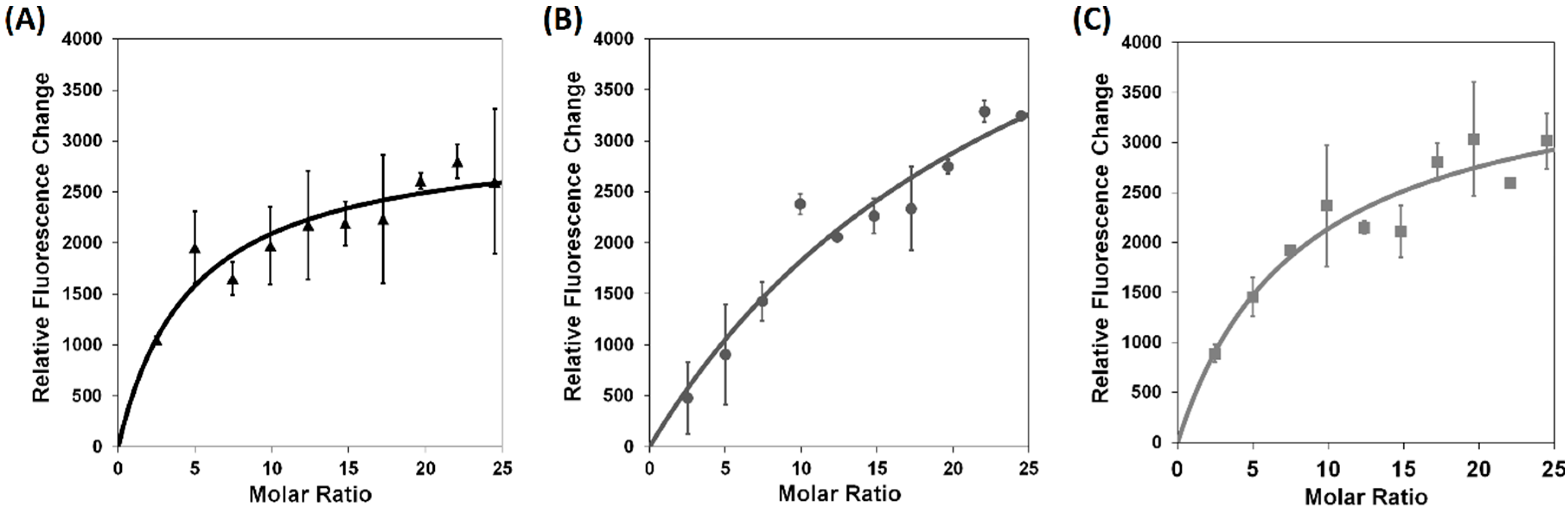
3.6. Xanthorrhizol-tadA Binding Study

E. coli strains producing wild-type tadA or the tadA (A143V) and tadA (F149G) mutants were grown overnight at 37 °C in LB supplemented with chloramphenicol (30 µg/mL). About 1% of each culture was inoculated into fresh medium and incubated for approximately 2–3 h until the OD600nm reached 0.5. The cultures were then induced by the addition of 100 µM IPTG and allowed to grow for an additional 8 h. To purify his-tagged tadA protein, cells were harvested and lysed by sonication. Proteins were bound to agarose resin (His•Bind Agarose Resin (Ni-IDA), Elpis Biotech, Daejeon, Korea) and purified through column chromatography as described in the kit protocol (His•Bind Agarose Resin (Ni-IDA) Elpis Biotech). Protein was desalted by using PD-10 desalting columns following the protocols described by the manufacturer (GE Healthcare Bio-Sciences AB, Uppsala, Sweden).
A tryptophan quenching assay was used to study the xanthorrhizol-tadA interaction. tadA (4 µM) in 50 mM Tris-Cl (pH 8) and 10% glycerol was titrated with 2 µL xanthorrhizol (5 mM) and was then immediately excited at 295 nm; the emission was recorded from 300 to 500 nm using a FluoroMate FS-2 fluorescence spectrophotometer (Scinco Co. Ltd., Seoul, Korea). The excitation and emission monochromator slit widths were 2.5 nm. The fluorescence decrease (F0F) caused by the addition of xanthorrhizol was fitted to the equation (F0F) = ΔFmax × [I]/(Kd + [I]) to construct the binding curve. The assay was repeated at least three times.

3.7. Computational Methods

3.7.1. Molecular Docking

The EcTadA crystal structure (PDB ID: 1Z3A) was used as the input for xanthorrhizol docking using two different docking programs, GLIDE [44] and GOLD [45]. The substrate and cofactor (tRNA and Zn2+, respectively) were imported from the crystal structure by structural superposition [46]. Proton addition was performed with the corresponding algorithms implemented in the different docking programs. In GLIDE (version 5.8), the considered search volume was confined within a cube of 20 Å3. A total of 5000 poses per xanthorrhizol were retained for the initial phase of docking, and the best 400 poses per xanthorrhizol were retained for energy minimization. In GOLD (version 5.1), the search was confined within a 10-Å-radius sphere around the center defined above with the slowest and most accurate genetic algorithm search options, generating 10 poses per xanthorrhizol. All 4 built-in scoring functions in GOLD (ChemPLP, GoldScore, ChemScore, and ASP) were used in parallel docking simulations. Finally, the xanthorrhizol structure was built using the Maestro software [47] included in the Schrödinger Suite 2011 package and was energy-minimized using Macromodel [48] with the OPLS_2005 force field.

3.7.2. Molecular Dynamics Simulations

Molecular dynamics simulations were performed using the program Q [49] with the OPLS-AA force field [50]. Subsequently, the binding free energy was calculated as described in the supplementary materials. The parameters needed for xanthorrhizol that were not present in the original version of the force field were retrieved from the automatic parametrization performed with Macromodel [48]. Spherical boundary conditions were used with a 20-Å-radius simulation sphere centered on the same point as defined for the docking calculations. This sphere was solvated with TIP3P [51] water molecules and subjected to polarization and radial constraints according to the surface-constrained all-atom solvent (SCAAS) model [49,52] at the sphere surface to mimic the properties of bulk water. Non-bonded interactions were calculated explicitly up to a 10-Å cutoff, except for the xanthorrhizol atoms, for which no cutoff was used. Beyond the cutoff, long-range electrostatics were treated with the local reaction field multipole-expansion method [53]. All solvent bonds and angles were constrained with the SHAKE algorithm [54], and a 1-fs molecular dynamics (MD) step size was used. The MD simulations were performed at a temperature of 298 K. Non-bonded pair lists were updated every 25 steps, and the same interval was used to sample the xanthorrhizol-surrounding residue interaction energies.

4. Conclusions

By enriching an E. coli W3110 imp4213 ASKA pooled-plasmid library, we found that TadA may be involved in the mechanism of xanthorrhizol action. We have shown that xanthorrhizol targets TadA and, more importantly, that tRNA-specific adenosine deaminase may be a new target for antibiotic development in the future.

Supplementary Materials

Supplementary materials can be accessed at: https://www.mdpi.com/1420-3049/20/09/16290/s1.

Author Contributions

Y. and D.K. performed the experiments. Y. and D.K. analyzed the data. J.-K.H. contributed materials. J.-K.H. and J.-G.P. conceived and designed the experiments. Y., D.K., J.-K.H. and J.-G.P. wrote the paper.

Conflicts of Interest

The authors declare no conflict of interest.

References

  1. Hwang, J.K.; Shim, J.S.; Baek, N.I.; Pyun, Y.R. Xanthorrhizol: A potential antibacterial agent from Curcuma xanthorrhiza against Streptococcus mutans. Planta Med. 2000, 66, 196–197. [Google Scholar] [CrossRef] [PubMed]
  2. Hwang, J.K.; Shim, J.S.; Pyun, Y.R. Antibacterial activity of xanthorrhizol from Curcuma xanthorrhiza against oral pathogens. Fitoterapia 2000, 71, 321–323. [Google Scholar] [CrossRef]
  3. Lee, Y.L.; Shim, J.S.; Rukayadi, Y.; Hwang, J.K. Antibacterial activity of xanthorrhizol isolated from Curcuma xanthorrhiza Roxb. against foodborne pathogens. J. Food Prot. 2008, 71, 1926–1930. [Google Scholar] [PubMed]
  4. Yogiara; Kim, D.; Hwang, J.K.; Pan, J.G. The food-grade antimicrobial xanthorrhizol targets the enoyl-ACP reductase or FabI in bacteria. Bioorg. Med. Chem. Lett. 2015. under review. [Google Scholar]
  5. Park, H.S.; Yoon, Y.M.; Jung, S.J.; Kim, C.M.; Kim, J.M.; Kwak, J.-H. Antistaphylococcal activities of CG400549, a new bacterial enoyl-acyl carrier protein reductase (FabI) inhibitor. J. Antimicrob. Chemother. 2007, 60, 568–574. [Google Scholar] [CrossRef] [PubMed]
  6. Park, H.S.; Yoon, Y.M.; Jung, S.J.; Yun, I.N.R.; Kim, C.M.; Kim, J.M.; Kwak, J.-H. CG400462, a new bacterial enoyl-acyl carrier protein reductase (FabI) inhibitor. Int. J. Antimicrob. Agents 2007, 30, 446–451. [Google Scholar] [CrossRef] [PubMed]
  7. NBRP E.coli Strain. Available online: http://www.shigen.nig.ac.jp/ecoli/strain (accessed on 2 October 2014).
  8. Kitagawa, M.; Ara, T.; Arifuzzaman, M.; Ioka-Nakamichi, T.; Inamoto, E.; Toyonaga, H.; Mori, H. Complete set of ORF clones of Escherichia coli ASKA library (a complete set of E. coli K-12 ORF archive): Unique resources for biological research. DNA Res. 2006, 12, 291–299. [Google Scholar] [CrossRef] [PubMed]
  9. Couce, A.; Briales, A.; Rodríguez-Rojas, A.; Costas, C.; Pascual, Á.; Blázquez, J. Genomewide overexpression screen for fosfomycin resistance in Escherichia coli: murA confers clinical resistance at low fitness cost. Antimicrob. Agents Chemother. 2012, 56, 2767–2769. [Google Scholar] [CrossRef] [PubMed]
  10. Soo, V.W.C.; Hanson-Manful, P.; Patrick, W.M. Artificial gene amplification reveals an abundance of promiscuous resistance determinants in Escherichia coli. Proc. Natl. Acad. Sci. USA 2011, 108, 1484–1489. [Google Scholar] [CrossRef] [PubMed]
  11. Yu, B.; Kim, J.; Ju, H.; Choi, S.-K.; Hwang, S.; Park, S.; Kim, E.; Pan, J.-G. Genome-wide enrichment screening reveals multiple targets and resistance genes for triclosan in Escherichia coli. J. Microbiol. 2012, 50, 785–791. [Google Scholar] [CrossRef] [PubMed]
  12. Braun, M.; Silhavy, T.J. Imp/OstA is required for cell envelope biogenesis in Escherichia coli. Mol. Microbiol. 2002, 45, 1289–1302. [Google Scholar] [CrossRef] [PubMed]
  13. Ruiz, N.; Falcone, B.; Kahne, D.; Silhavy, T.J. Chemical conditionality: A genetic strategy to probe organelle assembly. Cell 2005, 121, 307–317. [Google Scholar] [CrossRef] [PubMed]
  14. Sampson, B.A.; Misra, R.; Benson, S.A. Identification and characterization of a new gene of Escherichia coli K-12 involved in outer membrane permeability. Genetics 1989, 122, 491–501. [Google Scholar] [PubMed]
  15. Database of Essential Gene. Available online: http://www.essentialgene.org (accessed on 25 September 2013).
  16. Wolf, J.; Gerber, A.P.; Keller, W. tadA, an essential tRNA-specific adenosine deaminase from Escherichia coli. EMBO J. 2002, 21, 3841–3851. [Google Scholar] [CrossRef] [PubMed]
  17. Baba, T.; Ara, T.; Hasegawa, M.; Takai, Y.; Okumura, Y.; Baba, M.; Datsenko, K.A.; Tomita, M.; Wanner, B.L.; Mori, H. Construction of Escherichia coli K-12 in-frame, single-gene knockout mutants: The Keio collection. Mol. Syst. Biol. 2006, 2. [Google Scholar] [CrossRef] [PubMed]
  18. Gerdes, S.Y.; Scholle, M.D.; Campbell, J.W.; Balázsi, G.; Ravasz, E.; Daugherty, M.D.; Somera, A.L.; Kyrpides, N.C.; Anderson, I.; Gelfand, M.S.; et al. Experimental determination and system level analysis of essential genes in Escherichia coli MG1655. J. Bacteriol. 2003, 185, 5673–5684. [Google Scholar] [CrossRef] [PubMed]
  19. Deutschbauer, A.; Price, M.N.; Wetmore, K.M.; Shao, W.; Baumohl, J.K.; Xu, Z.; Nguyen, M.; Tamse, R.; Davis, R.W.; Arkin, A.P. Evidence-based annotation of gene function in Shewanella oneidensis MR-1 using genome-wide fitness profiling across 121 conditions. PLoS Genet. 2011, 7, e1002385. [Google Scholar] [CrossRef] [PubMed]
  20. OGEEdb. Available online: http://ogeedb.embl.de (accessed on 25 September 2013).
  21. Schaub, M.; Keller, W. RNA editing by adenosine deaminases generates RNA and protein diversity. Biochimie 2002, 84, 791–803. [Google Scholar] [CrossRef]
  22. Gerber, A.P.; Keller, W. RNA editing by base deamination: More enzymes, more targets, new mysteries. Trends Biochem. Sci. 2001, 26, 376–384. [Google Scholar] [CrossRef]
  23. Tatusov, R.L.; Koonin, E.V.; Lipman, D.I. A genomic perspective on protein families. Science 1997, 278, 631–637. [Google Scholar] [CrossRef] [PubMed]
  24. Luo, H.; Lin, Y.; Gao, F.; Zhang, C.-T.; Zhang, R. DEG 10, an update of the database of essential genes that includes both protein-coding genes and noncoding genomic elements. Nucleic Acids Res. 2014, 42, D574–D580. [Google Scholar] [CrossRef] [PubMed]
  25. Zhang, R.; Lin, Y. DEG 5.0, a database of essential genes in both prokaryotes and eukaryotes. Nucleic Acids Res. 2009, 37 (Suppl. 1), D455–D458. [Google Scholar] [CrossRef] [PubMed]
  26. Zhang, R.; Ou, H.Y.; Zhang, C.T. DEG: A database of essential genes. Nucleic Acids Res. 2004, 32 (Suppl. 1), D271–D272. [Google Scholar] [CrossRef] [PubMed]
  27. Jayakar, H.; Parvathareddy, J.; Fitzpatrick, E.; Bina, X.; Bina, J.; Re, F.; Emery, F.; Miller, M. A galU mutant of Francisella tularensis is attenuated for virulence in a murine pulmonary model of tularemia. BMC Microbiol. 2011, 11, 179. [Google Scholar] [CrossRef] [PubMed]
  28. Felczak, M.M.; Kaguni, J.M. The rcbA gene product reduces spontaneous and induced chromosome breaks in Escherichia coli. J. Bacteriol. 2012, 194, 2152–2164. [Google Scholar] [CrossRef] [PubMed]
  29. Scheurwater, E.M.; Clarke, A.J. The C-terminal domain of Escherichia coli YfhD functions as a lytic transglycosylase. J. Biol. Chem. 2008, 283, 8363–8373. [Google Scholar] [CrossRef] [PubMed]
  30. Bennik, M.H.J.; Pomposiello, P.J.; Thorne, D.F.; Demple, B. Defining a rob regulon in Escherichia coli by using transposon mutagenesis. J. Bacteriol. 2000, 182, 3794–3801. [Google Scholar] [CrossRef] [PubMed]
  31. Zafar, M.A.; Sanchez-Alberola, N.; Wolf, R.E., Jr. Genetic evidence for a novel interaction between transcriptional activator SoxS and region 4 of the σ70 subunit of RNA polymerase at class II SoxS-dependent promoters in Escherichia coli. J. Mol. Biol. 2011, 407, 333–353. [Google Scholar] [CrossRef] [PubMed]
  32. McMurry, L.M.; Oethinger, M.; Levy, S.B. Overexpression of marA, soxS, or acrAB produces resistance to triclosan in laboratory and clinical strains of Escherichia coli. FEMS Microbiol. Lett. 1998, 166, 305–309. [Google Scholar] [CrossRef] [PubMed]
  33. Alekshun, M.N.; Levy, S.B. Regulation of chromosomally mediated multiple antibiotic resistance: The mar regulon. Antimicrob. Agents Chemother. 1997, 41, 2067–2075. [Google Scholar] [PubMed]
  34. Lee, J.-H.; Lee, K.-L.; Yeo, W.-S.; Park, S.-J.; Roe, J.-H. SoxRS-Mediated lipopolysaccharide modification enhances resistance against multiple drugs in Escherichia coli. J. Bacteriol. 2009, 191, 4441–4450. [Google Scholar] [CrossRef] [PubMed]
  35. Butland, G.; Peregrin-Alvarez, J.M.; Li, J.; Yang, W.; Yang, X.; Canadien, V.; Starostine, A.; Richards, D.; Beattie, B.; Krogan, N.; et al. Interaction network containing conserved and essential protein complexes in Escherichia coli. Nature 2005, 433, 531–537. [Google Scholar] [CrossRef] [PubMed]
  36. Kapoor, M.; Reddy, C.C.; Krishnasastry, M.V.; Surolia, N.; Surolia, A. Slow-tight binding inhibition of enoyl acyl carrier protein reductase from Plasmodium falciparum by triclosan. Biochem. J. 2004, 381, 719–724. [Google Scholar] [CrossRef] [PubMed]
  37. Liu, N.; Cummings, J.E.; England, K.; Slayden, R.A.; Tonge, P.J. Mechanism and inhibition of the FabI enoyl-ACP reductase from Burkholderia pseudomallei. J. Antimrob. Chemother. 2011, 66, 564–573. [Google Scholar] [CrossRef] [PubMed]
  38. Ochsner, U.A.; Sun, X.; Jarvis, T.; Critchley, I.; Janjic, N. Aminoacyl-tRNA synthetases: Essential and still promising targets for new anti-infective agents. Expert Opin. Investig. Drugs 2007, 16, 573–593. [Google Scholar] [CrossRef] [PubMed]
  39. Zhao, Y.; Meng, Q.; Bai, L.; Zhou, H. In silico discovery of aminoacyl-tRNA synthetase inhibitors. Int. J. Mol. Sci. 2014, 15, 1358–1373. [Google Scholar] [CrossRef] [PubMed]
  40. Xu, Z.-Q.; Flavin, M.T.; Flavin, J. Combating multidrug-resistant Gram-negative bacteria infections. Expert Opin. Investig. Drugs 2014, 23, 163–182. [Google Scholar] [CrossRef] [PubMed]
  41. Boyce, J.M. MRSA patients: Proven methods to treat colonization and infection. J. Hosp. Infect. 2001, 48 (Suppl. A), S9–S14. [Google Scholar] [CrossRef]
  42. Wiegand, I.; Hilpert, K.; Hancock, R.E.W. Agar and broth dilution methods to determine the minimal inhibitory concentration (MIC) of antimicrobial substances. Nat. Protoc. 2008, 3, 163–175. [Google Scholar] [CrossRef] [PubMed]
  43. Bron, S. Plasmids. In Molecular Biological Methods for Bacillus; Harwood, C.R., Cutting, S.M., Eds.; John Wiley & Sons: Chichester, UK, 1990; pp. 75–174. [Google Scholar]
  44. Friesner, R.A.; Banks, J.L.; Murphy, R.B.; Halgren, T.A.; Klicic, J.J.; Mainz, D.T.; Repasky, M.P.; Knoll, E.H.; Shelley, M.; Perry, J.K.; et al. Glide: A new approach for rapid, accurate docking and scoring. 1. Method and assessment of docking accuracy. J. Med. Chem. 2004, 47, 1739–1749. [Google Scholar] [CrossRef] [PubMed]
  45. Verdonk, M.L.; Cole, J.C.; Hartshorn, M.J.; Murray, C.W.; Taylor, R.D. Improved protein-ligand docking using GOLD. Proteins 2003, 52, 609–623. [Google Scholar] [CrossRef] [PubMed]
  46. The PyMOL Molecular Graphics System, version 1.5.0.4; Schrödinger, LLC: New York, NY, USA, 2000.
  47. Maestro, version 9.2; Schrödinger, LLC: New York, NY, USA, 2011.
  48. MacroModel, version 9.9; Schrödinger, LLC: New York, NY, USA, 2011.
  49. Marelius, J.; Kolmodin, K.; Feierberg, I.; Åqvist, J. Q: A molecular dynamics program for free energy calculations and empirical valence bond simulations in biomolecular systems. J. Mol. Graph. Model. 1998, 16, 213–225. [Google Scholar] [CrossRef]
  50. Jorgensen, W.L.; Maxwell, D.S.; Tirado-Rives, J. Development and testing of the OPLS all-atom force field on conformational energetics and properties of organic liquids. J. Am. Chem. Soc. 1996, 118, 11225–11236. [Google Scholar] [CrossRef]
  51. Jorgensen, W.L.; Chandrasekhar, J.; Madura, J.D.; Impey, R.W.; Klein, M.L. Comparison of simple potential functions for simulating liquid water. J. Chem. Phys. 1983, 79, 926–935. [Google Scholar] [CrossRef]
  52. King, G.; Warshel, A. A surface constrained all-atom solvent model for effective simulations of polar solutions. J. Chem. Phys. 1989, 91, 3647–3661. [Google Scholar] [CrossRef]
  53. Lee, F.S.; Warshel, A. A local reaction field method for fast evaluation of long-range electrostatic interactions in molecular simulations. J. Chem. Phys. 1992, 97, 3100–3107. [Google Scholar] [CrossRef]
  54. Ryckaert, J.-P.; Ciccotti, G.; Berendsen, H. Numerical integration of the cartesian equations of motion of a system with constraints: Molecular dynamics of n-alkanes. J. Comput. Phys. 1977, 23, 327–341. [Google Scholar] [CrossRef]
  • Sample Availability: Samples of the compounds are available from the authors.

