Article

Separation and Identification of Four New Compounds with Antibacterial Activity from Portulaca oleracea L.

Basic research department of Jiamusi College, Heilongjiang University of Chinese Medicine, No. 39 Guang Hua Street, Qian Jin District, Jia Mu Si 154100, China
*
Author to whom correspondence should be addressed.
Molecules 2015, 20(9), 16375-16387; https://doi.org/10.3390/molecules200916375
Submission received: 21 July 2015 / Revised: 14 August 2015 / Accepted: 28 August 2015 / Published: 10 September 2015

Abstract

:
The Portulaca oleracea L. (P. oleracea) has been used to treat bacillary dysentery for thousands of years in China. Pharmacology studies on P. oleracea have also showed its significant antibacterial effects on the enteropathogenic bacteria, which might reveal the treatment of P. oleracea in cases of bacillary dysentery to some extent. To date, however, the therapeutic basis of P. oleracea treating on bacillary dysentery remains unknown. We determined the antibacterial effective fraction of P. oleracea in a previous study. The current study, which is based on our previous study, was first designed to isolate, identify and screen antibacterial active constituents from P. oleracea. As a result, four new compounds (14), portulacerebroside B (1), portulacerebroside C (2), portulacerebroside D (3) and portulaceramide A (4) along with five known compounds (59) were isolated, and structures were established by their physico-chemical constants and spectroscopic analysis. The antibacterial activities against common enteropathogenic bacteria were evaluated for all compounds and the new compounds 14 showed significant antibacterial effect on enteropathogenic bacteria in vitro, which might contribute to revealing the treatment of P. oleracea in cases of bacillary dysentery.

1. Introduction

The genus of Portulaca is an annual herb which taxonomically belongs to the family of Portulacaceae. Although it originates from India, Portulaca has been widely distributed in other temperate and tropical areas of the world [1,2]. In China, there are six species of Portulaca, among which Portulaca oleracea L. (P. oleracea) has been used as a traditional Chinese medicine (TCM) for thousands of years. P. oleracea, cold in nature and acid in flavor, possesses the efficacies of clearing away the heat evil and detoxifying and cooling blood to stop diarrhea. In clinical situations, P. oleracea has been used to treat acute appendicitis, scrofula ulcer, pediatric pertussis, burns and scalds, psoriasis [3], hemorrhinia, uterine bleeding, urinary tract infections, lung abscess, mumps, and especially more effective in bacillary dysentery which manifested feeling cold or fever, bellyache, diarrhea, tenesmus, and mucus pus blood stool [2]. Chemical studies on P. oleracea showed its main constituents of fatty acids, terpenes, alkaloids, coumarins, flavonoids, and volatile oil [2]. In addition, the only cerebroside of portulacerebroside A was reported in 2008 [4]. Pharmacology studies on P. oleracea showed its activities of antibacterial [5,6], hepatoprotective [7,8], anti-inflammatory, analgesia [2,9], muscle relaxant [10], neuroprotective [11], anti-oxidant [1], and anti-aging [12], but studies on the corresponding therapeutic basis were far from sufficient. Obviously, the treatment of P. oleracea on bacillary dysentery results from its antibacterial effect on the enteropathogenic bacteria; however, the corresponding active components in P. oleracea were unknown. Consequently, we screened and obtained the antibacterial fraction (EtOAc extract) from P. oleracea. The effective fraction could inhibit and kill the common enteropathogenic bacteria in vitro effectively, based on which a bioassay-guided isolation and phytochemical study of P. oleracea was performed and four new along with five known compounds were obtained from the effective fraction. The structures of known compounds 59 were determined by detailed 1D- and 2D-NMR analyses, ESI-MS and comparison of their spectral data with literature values was undertaken. In this paper, the isolation and structural elucidation of the new compounds 14 was described. We also investigated the antibacterial effects of compounds 19 against common enteropathogenic bacteria in vitro.

2. Results and Discussion
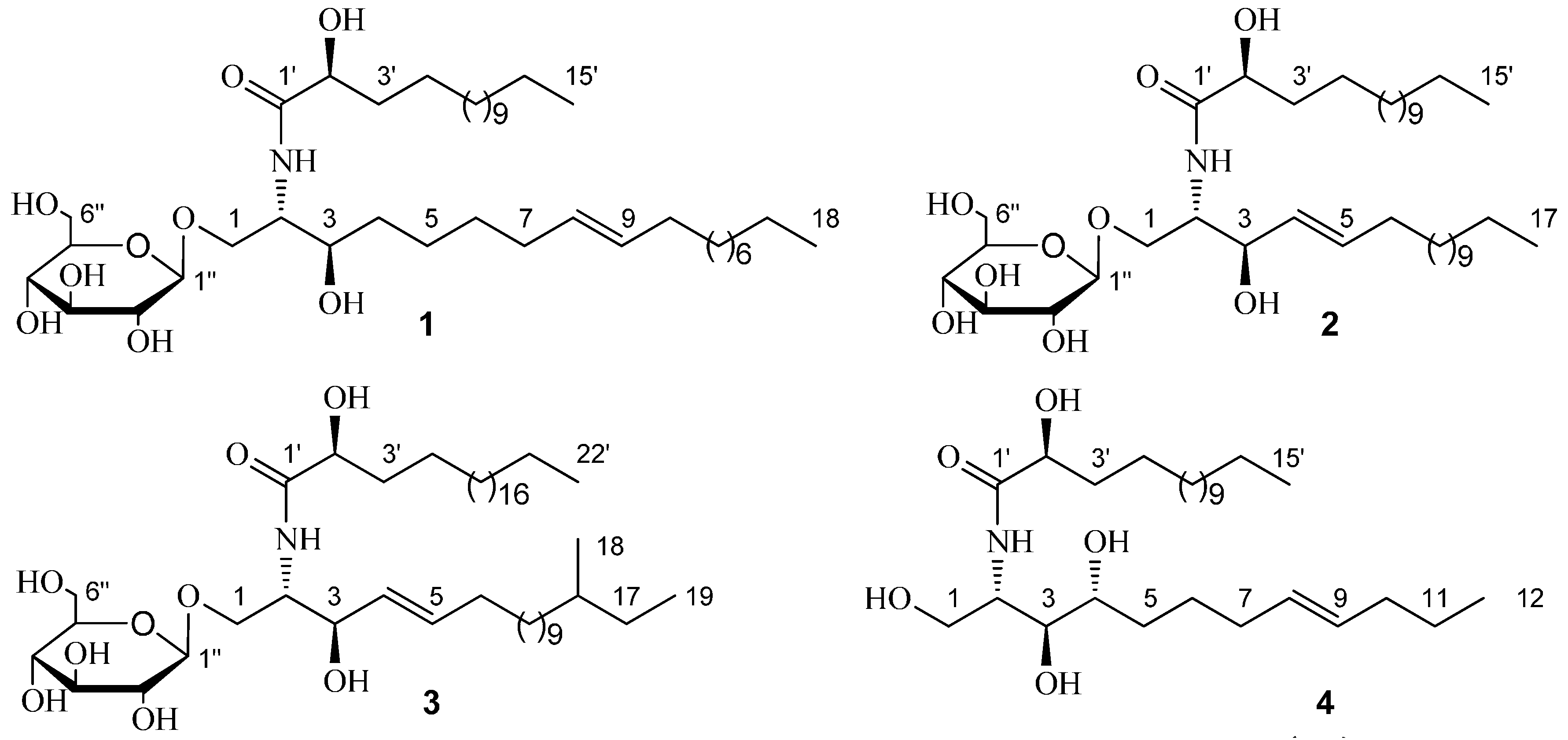
Compound 1 was obtained as white amorphous powder, [ α ] D 22 = +13.3° (c = 0.52, C5H5N). The molecular formula C39H75NO9 was established for 1 by HRESIMS m/z 702.5516 [M + H]+ (calc. for 702.5520), indicating three degrees of unsaturation. Methanolysis experiment of 1 liberated D-glucose suggested that 1 was a D-glucoside. The IR absorption bands at 3420 cm−1, 1645 and 1542 cm−1, and 722 cm−1 originated from hydroxyl, amide, and methylene groups, respectively.
The 1H- and 13C-NMR data (anomeric proton δH 4.90, 1H, d, J = 7.6 Hz; δC 105.7, 75.2, 78.5, 71.6, 78.6, and 62.7) of 1 suggested the presence of a β-d-glucopyranosyl moiety. The 1H- and 13C-NMR spectra (Table 1 and Table 2) showed characteristics of a cerebroside with a 2-hydroxy fatty acid fraction as the aglycone of 1. Methanolysis of 1 obtained a fatty acid methyl ester (FAME) and a long-chain base (LCB). The FAM was determined as 2-hydroxypentadecanoic acid methyl ester by Gas Chromatography-Mass Spectrometer (GC-MS) analysis. The 1D-TOCSY spectrum of 1 showed a correlation between δH 4.22 (1H, m, H-3) and 5.46 (2H, m, H-8, 9), which suggested the olefinic bond was located in the LCB. To determine the location of the olefinic bond in LCB, the dimethyl disulfide (DMDS) derivatives of the LCB was analyzed by ESIMS and characteristic fragment ion of m/z 187 [M + H]+ was obtained. Therefore, the olefinic bond was located at C-8 and C-9. The LCB was further determined as 2-aminooctadec-8-ene-1,3-diol by GC-MS analysis (Figure 1). The specific rotation [ α ] D 22 = −5.8° (c = 0.02, CHCl3) of the FAM confirmed that the absolute configuration of C-2′R [13]. The 2S, 3R stereochemistry was determined by comparing of the 13C-NMR data of C-2 and C-3 with those in references [14,15,16]. The trans-configuration (E) of the olefinic bond in 1 was determined by signals at δC 33.2/32.1 of the two carbons next to the olefinic bond in 13C-NMR spectrum [9]. The 1H- and 13C-NMR data (Table 1 and Table 2) were further assigned by the spectra of DEPT, HSQC, 1H-1H COSY, and HMBC. Consequently, 1 was established as 1-O-β-d-glucopyranosyl-(2S,3R,8E)-2-[(2′R)-2-hydroxylpentadecanoylamino]-8-octadecene-1,3-diol which is named as portulacerebroside B (Figure 2).
Table 1. 1H-NMR data of compounds 14 (400MHz, δ in ppm, J in Hz, in C5D5N-d5 at 30 °C).
1234
HδH (J, Hz)HδH (J, Hz)HδH (J, Hz)HδH (J, Hz)
NH8.40, d (8.4)NH8.36, d (8.0)NH8.36, d (8.0)NH8.55, d (8.0)
14.35, dd (11.6, 5.6)
4.73, m
14.22, m
4.72, m
14.22, m
4.72, m
14.49, dd (11.0, 4.6)
4.41, dd (11.0, 4.8)
24.60, m24.78, m24.78, m25.10, m
34.22, m34.76, m34.76, m34.34, dd (4.8, 6.4)
4–61.15–1.40, brs45.86, m45.86, m44.27, m
72.10, m55.98, m55.98, m52.22, 1.96, m
85.46, m62.05, m62.05, m61.67, m
95.46, m7–161.16–1.42, brs 7–171.16–1.42, brs 72.01, m
102.02, m170.88, t (6.4)180.88, d (7.4)85.52, m
11–171.15–1.40, brs2′4.60, m190.86, t (6.4)95.52, m
180.85, t (6.4)3′1.86, m2′4.60, m101.89, m
2′4.70, m4′1.73, m
1.16–1.42, brs
3′1.86, m111.26–1.38, brs
3′1.86, m5′–14′1.16–1.42, brs4′1.73, m
1.16–1.42, brs
120.88, t (6.8)
4′1.73, m
1.15–1.40, brs
15′0.88, t, 6.45′–21′1.16–1.42, brs2′4.60, dd (7.6, 3.2)
5′–14′1.15–1.40, brs1′′4.90, d, (7.6)22′0.88, t (6.4)3′2.18, 2.02, m
15′0.85, t (6.4)2′′4.01, m1′′4.90, d, (7.6)4′1.96, 1.73, m
1′′4.90, d (7.6)3′′4.22, m2′′4.01, m5′–14′1.26–1.38, brs
2′′3.91, m4′′4.22, m3′′4.22, m15′0.88, t (6.8)
3′′4.20, m5′′3.89, m4′′4.22, m
4′′4.03, m6′′4.34, 4.49, m5′′3.89, m
5′′4.12, m 6′′4.34, 4.49, m
6′′4.18, 4.52, m
Table 2. 13C-NMR data of compounds 14 (100 MHz, δ in ppm, in C5D5N-d5 at 30 °C).
1234
CδCCδCCδCCδC
170.4, CH2170.2, CH2170.4, CH2162.0, CH2
254.6, CH254.6, CH254.5, CH252.9, CH
371.3, CH372.4, CH372.4, CH376.8, CH
4–629.5–30.4, CH24131.6, CH4131.7, CH472.4, CH
733.25132.8, CH5132.8, CH533.8, CH2
8130.2, CH634.2, CH2634.2, CH2627.6, CH2
9130.7, CH7–1429.6–30.4, CH27–1529.6–30.5, CH2733.3, CH2
1032.1, CH21532.1, CH21635.6, CH8130.6, CH
11–1529.5–30.4, CH21622.8, CH21730.6, CH29130.6, CH
1632.1, CH21714.2, CH31819.6, CH31033.0, CH2
1722.9, CH218 1911.7, CH31130.4–29.5, CH2
1814.3, CH31′175.8, C1′175.8, C1214.3, CH3
1′175.5, C2′72.6, CH2′72.6, CH1′175.1, C
2′72.4, CH3′35.7, CH23′35.7, CH22′72.8, CH
3′35.7, CH24′26.3, CH24′26.3, CH23′35.6, CH2
4′26.3, CH25′–12′29.6–30.4, CH25′–19′29.6–30.4, CH24′26.8, CH2
5′–12′29.5–30.4, CH213′32.1, CH220′32.1, CH25′–12′30.4–29.5, CH2
13′32.1, CH214′22.8, CH221′22.8, CH213′32.1, CH2
14′22.9, CH215′14.2, CH322′14.2, CH314′22.8, CH2
15′14.3, CH31′′105.6, CH1′′105.6, CH15′14.3, CH3
1′′105.7, CH2′′75.1, CH2′′75.1, CH
2′′75.2, CH3′′78.6, CH3′′78.6, CH
3′′78.5, CH4′′71.5, CH4′′71.5, CH
4′′71.6, CH5′′78.6, CH5′′78.6, CH
5′′78.6, CH6′′62.6, CH26′′62.6, CH2
6′′62.7, CH2
Figure 1. GC-MS analysis long-chain base (LCB) 14 and dimethyl disulfide (DMDS) derivatives of LCBs from 1 and 4.
Molecules 20 16375 g001
Figure 2. Structures of compounds 14.
Molecules 20 16375 g002
Compound 2 was obtained as white amorphous powder, [ α ] D 22 = +14.8° (c = 0.40, C5H5N). The molecular formula was established as C38H73NO9 by HRESIMS m/z 688.5358 [M + H]+ (calc. for 688.5364), indicating three degrees of unsaturation. Methanolysis experiment of 2 liberated D-glucose suggested that 2 was a D-glucoside. The IR absorption bands at 3405 cm−1, 1634 and 1536 cm−1, and 718 cm−1 originated from hydroxyl, amide, and methylene groups, respectively.
The 1H- and 13C-NMR data of compound 2 were similar with 1, including the signals of a β-d-glucopyranosyl moiety. The position of olefinic bond in 2 was located at C-4 and C-5 by the spectra of HSQC, 1H-1H COSY, and HMBC. Methanolysis of 2 also obtained an FAM and an LCB. The FAM was determined as 2-hydroxypentadecanoic acid methyl ester by GC-MS analysis. The LCB of 2 was determined as 2-aminoheptadecenoic-4-ene-1,3-diol by GC-MS analysis (Figure 1). The specific rotation [ α ] D 22 = −6.8° (c 0.03, CHCl3) of the FAM confirmed that the absolute configuration of C-2′R, which is the same with 1. The 2S, 3R stereochemistry was determined by comparing of the 13C-NMR data of C-2 and C-3 with those of in references [17,18]. The trans-configuration (E) of the olefinic bond in 2 was determined by C-6 signal at δC 34.2 in 13C-NMR spectrum [16]. The 1H- and 13C-NMR data (Table 1 and Table 2) were further assigned by the spectra of DEPT, HSQC, 1H-1H COSY, and HMBC. Consequently, 2 was established as 1-O-β-d-glucopyranosyl-(2S,3R,4E)-2-[(2′R)-2-hydroxylpentadecanoylamino]-4-heptadecene-1,3-diol which is named as portulacerebroside C (Figure 2).
Compound 3 was obtained as white amorphous powder, [ α ] D 22 = +8.2° (c = 0.15, C5H5N). The molecular formula C47H91NO9 was established for 3 by HRESIMS m/z 814.6765 [M + H]+ (calc. for 814.6772), indicating three degrees of unsaturation. Methanolysis experiment of 3 liberated D-glucose suggested that 3 was a D-glucoside. The IR absorption bands at 3412 cm−1, 1638 and 1530 cm−1, and 722 cm−1 originated from hydroxyl, amide, and methylene groups, respectively.
The 1H- and 13C-NMR data of compound 3 were similar with 2 and the main difference lies in the methenyl group signal at δC 35.6 and methyl group signals at δC 11.7 and 19.6 in 13C-NMR spectrum of 3, which indicated the presence of a branched methyl group in 3. To determine the position of the branched methyl group, the 1D-TOCSY spectrum was used and correlations of δH 4.22 (1H, m, H-1) with 5.86 (1H, m, H-4), 0.88 (3H, d, J = 6.4 Hz), and 0.86, (3H, t, J = 6.4 Hz, 19-CH3) could be observed. Therefore, the branched methyl group was located in the LCB. Other methods to determine the structure of 3 were the same with 2. The 1H- and 13C-NMR data (Table 1 and Table 2) were further assigned by the spectra of DEPT, HSQC, 1H-1H COSY, and HMBC. Consequently, 3 was established as 1-O-β-d-glucopyranosyl-(2S,3R,4E)-2-[(2′R)-2-hydroxyldocosanamideamino]-16-methyl-4-nonadecene-1,3-diol which is named as portulacerebroside D (Figure 2).
Compound 4 was obtained as white amorphous powder, [ α ] D 22 = +9.5° (c = 0.30, C5H5N). The molecular formula C27H53NO5 was established for 1 by HRESIMS m/z 472.3995 [M + H]+ (calc. for 472.4002), indicating two degrees of unsaturation. The IR absorption bands at 3401 cm−1, 1621 and 1533 cm−1, and 723 cm−1 originated from hydroxyl, amide, and methylene groups, respectively.
The 1H- and 13C-NMR spectra (Table 1 and Table 2) of 4 showed characteristics of a sphingosine-type ceramide with a 2-hydroxy fatty acid fraction. Methanolysis of 4 obtained an FAM and an LCB. The FAM was determined as 2-hydroxypentadecanoic acid methyl ester by GC-MS analysis. The 1D-TOCSY spectrum of 4 showed a correlation between δH 4.27 (1H, m, H-4) and 5.52 (2H, m, H-8,9), which suggested the olefinic bond was located in the LCB. To determine the location of the olefinic bond in LCB, the DMDS derivatives of LCB was analyzed by ESIMS and characteristic fragment ion of m/z 117 [M + H]+ was obtained. Therefore, the olefinic bond was located at C-8 and C-9. The LCB was further determined as 2-aminododecane-8-ene-1,3,4-triol by GC-MS analysis (Figure 1). The specific rotation [ α ] D 22 = −4.7° (c 0.02, CHCl3) of the FAM confirmed that the absolute configuration of C-2′R [13]. The 2S, 3S, and 4R stereochemistry was determined by comparing of the 13C-NMR data of C-2, C-3, and C-4 with those of in reference [19]. The trans-configuration (E) of the olefinic bond in 4 was determined by signals at δC 33.3/33.0 of the two carbons next to the olefinic bond in 13C-NMR spectrum [16]. The 1H- and 13C-NMR data (Table 1 and Table 2) were further assigned by the spectra of DEPT, HSQC, 1H-1H COSY, and HMBC and 4 was established as (2S,3S,4R,8E)-2-[(2′R)-2-hydroxylpentadecanoylamino]-8-dodecene-1,3,4-triol which is named as portulaceramide A (Figure 2).
The known compounds were identified as friedelin (6) [20], 3-acetylaleuritolic acid (7) [21], 4α-methyl-3β-hydroxylfriedelan (8) [22], cycloartenol (9) [23], and lupeol (10) [24] by comparing their NMR spectroscopic and physical data with the literature values (Figure 3).
Figure 3. Structures of compounds 59.
Molecules 20 16375 g003
The antibacterial activities of compounds 19 against Escherichia coli (E. coli), Staphylococcus aureus (S. aureus), Shigella flexneri (S. flexneri), and Salmonella typhi (S. typhi) were investigated. Minimal inhibitory concentrations (MICs) and minimal bactericidal concentrations (MBCs) were determined. Compounds 14 showed significant antibacterial activity on common enteropathogenic bacteria in vitro (Table 3), while other compounds did not show any antibacterial effects on the enteropathogenic bacteria at the tested concentration (data not shown). The structures of compounds 14 involve a sphingoid base and an amide-linked fatty acyl chain. The amphipathic molecules exhibit diverse biological activity, including broad antibacterial activities against both Gram positive and negative bacteria in this study.
Table 3. The minimal inhibitory concentrations (MICs) (mg·mL−1) and minimal bactericidal concentrations (MBCs) (mg·mL−1) of compounds 14.
StrainsMICs (SD = 0)MBCs (SD = 0)
1 *2 *3 *4 *Amoxicillin1234Amoxicillin
E. coli0.18750.18750.18750.3752.34 × 10−20.250.250.250.503.12 × 10−2
S. aureus0.18750.18750.18750.3751.17 × 10−20.500.500.500.501.56 × 10−2
S. flexneri0.18750.18750.18750.18755.85 × 10−30.250.250.250.253.90 × 10−3
S. typhi0.18750.18750.18750.3751.17 × 10−20.250.250.250.507.80 × 10−3
Key: Effects of the tested compounds on enteropathogenic bacteria. MICs and MBCs were expressed as mean ± SD (n = 4) of three independent experiments. MIC was expressed as the mean concentration between the well showing growth and that showing no growth. The MBC was expressed as the lowest concentration of the compounds showing no any bacterial growth after incubating for 20 h; * Significant difference (p < 0.01) compared the MIC values of compounds 14 with 59 (MIC > 16.0 mg·mL−1, data not shown), respectively.

3. Experimental Section

3.1. General

The NMR spectra were measured on Bruker AVANCE 400 MHz NMR instrument (Bruker SpectroSpin, Karlsruhe, Germany), and chemical shifts are given as δ (ppm) while the coupling constants are given in Hz. Xero Q Tof MS spectrometer (Waters, Milford, MA, USA) was used to measure and analysis the HRESIMS data. Volatile derivatives from compounds were analyzed on a GC-MS (Angilent, California, CA, USA) instrument. Waters 2535 instrument coupled with a Waters Sunfire prep C18 OBD (19 × 250 mm i.d.) column, a UV-2998 (Waters, MA, USA), and RI-2414 detector as a Preparative HPLC (Waters, MA, USA) was used to prepare compounds. FTIR-8400S (Shimadzu, Kyoto, Japan) was used to record the IR Spectra data; Column chromatographies including Macroporous resin (AB-8 Crosslinked Polystyrene, Nankai Chemical Plant, Tianjin, China), silica gel (200–300 mesh, Haiyang Chemical Group Co. Ltd., Qingdao, China), and ODS-A (120A, 50 mm; YMC, Kyoto, Japan) were also employed. A microplate reader (BMG FLUOStar OPTIMA, Ortenberg, Germany) was used to monitor the growth of the bacterial strains.

3.2. Bacterial Strains and the Preparation of Inoculums

The bacterial strains of E. coli(ATCC25922), S. aureus (ATCC25923), S. flexneri (ATCC12022), and S. typhi (ATCC14028) from American Type Culture Collection were provided by Department of Microbiology and Immunology, Heilongjiang university of Chinese medicine. Strains from refrigerated stock cultures were inoculated into common agar plate and incubated at 37 °C for 18 h. The bacteria were activated in nutrient broth and incubated at 37 °C for another 18 h. The concentrations of strains for antibacterial test in vitro were 5 × 10⁵ CFU·mL−1.

3.3. Plant Materials

We collected the aerial part of P. oleracea from the Dongfanghong Forestry Agency (Jixi, China) and identified by Lianjie Su of Heilongjiang University of Chinese Medicine. The voucher specimen (No. 20130814) is deposited at the Herbarium of Heilongjiang University of Chinese Medicine, China.

3.4. Extraction and Isolation
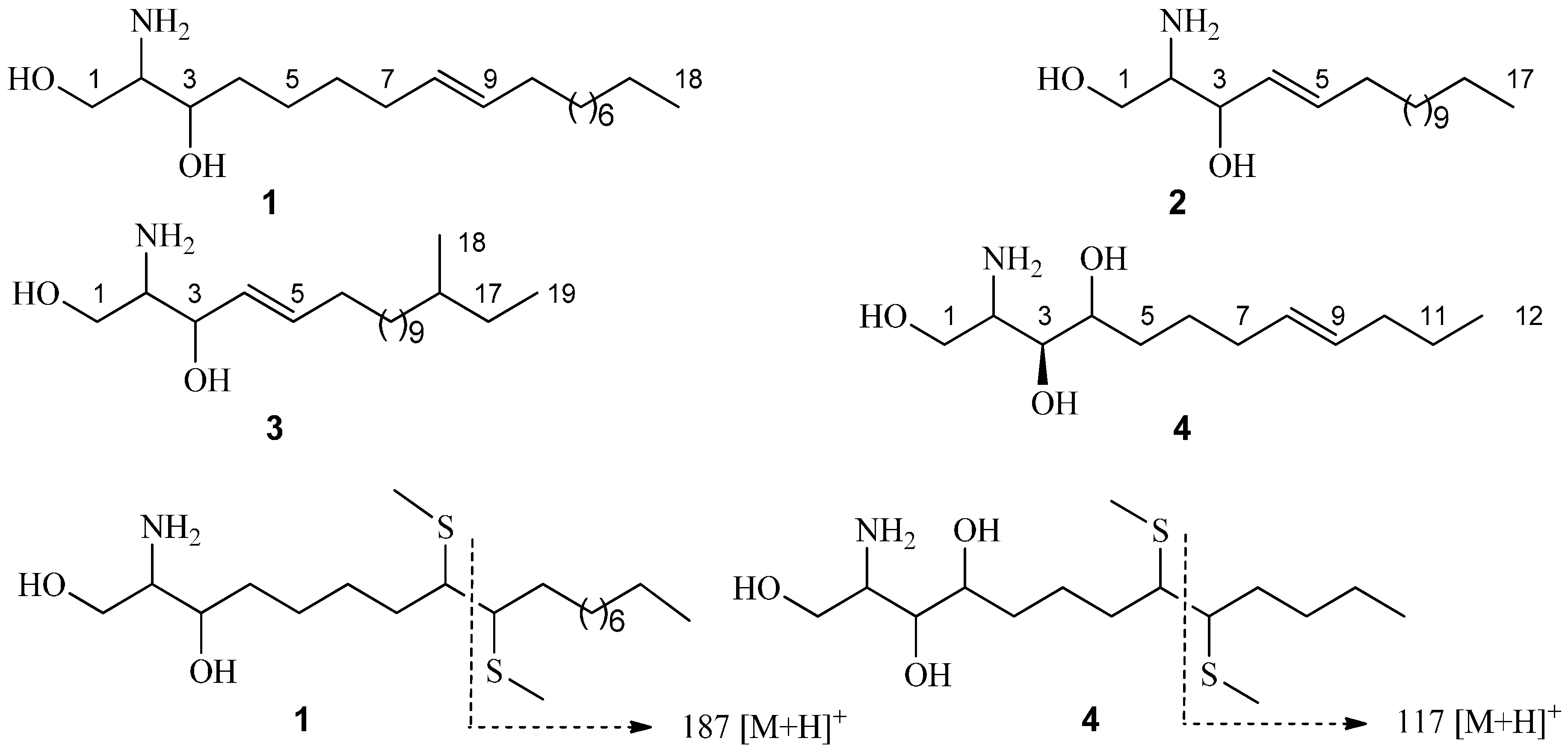
The dried P. oleracea (15.0 kg) extracted with 70% EtOH under reflux (2 × 120 L) for 2 h. The 70% EtOH extract (1770 g) was suspended in H2O (20 L), and successively extracted with petroleum ether (60–90 °C), EtOAc and n-butanol, respectively. Solvent was removed to give petroleum ether extract (125.3 g), EtOAc extract (173.7 g), n-butanol extract (184.8 g), and remained water extract (1267.2 g). The EtOAc extract is the antibacterial effective fraction as previous study. The EtOAc fraction (150.0 g) was repeatedly column chromatographed on silica gel with a gradient of CH2Cl2/MeOH (40:1 to 0:1) as eluents to afford seven fractions: F1–F7. F2 (13.2 g) continued silica gel chromatography and eluted with petroleum ether/EtOAc (15:1 to 1:1) to afford sub-fractions A1–A4. Compounds 4 (53 mg) and 9 (37 mg) were obtained by silica gel chromatography of the sub-fraction A2 (3.5 g) elution with petroleum ether/EtOAc (8:1), F3 (25.7 g) was subjected to column chromatography on silica gel with CH2Cl2/MeOH (30:1 to 10:1) to afford sub-fractions B1–B5. B3 (6.5 g) was separated on silica gel chromatography and eluted with CH2Cl2/MeOH (40:1 to 15:1) and further purified by preparative HPLC on a Hypersil-ODS II column (10 μm, 20 × 300 mm, flow rate 8 mL/min) with MeOH/H2O (48% and 52%) to afford compound 5 (43 mg, tR = 17 min), 7 (38 mg, tR = 24 min), and 8 (33 mg, tR= 28 min). F5 (32.6 g) was subjected to column chromatography on silica gel with CH2Cl2/MeOH (30:1–8:1) to produce sub-fractions C1–C5. The C3 (4.0 g) was repeatedly column chromatographed on silica gel with CH2Cl2/MeOH (15:1 to 6:1) as eluents to afford 2 (40 mg) and 6 (21 mg). The C4 (5.9 g) was repeatedly column chromatographed on silica gel with CH2Cl2/MeOH (15:1 to 6:1) as eluents and followed separated on column chromatography Sephadex LH-20 with MeOH as eluent to afford 1 (32 mg) and 3 (37 mg).
Portulacerebroside B (1): white amorphous powder, [ α ] D 22 = +13.3° (c = 0.52, C5H5N); IR (KBr) νmax 3420, 2933, 2837, 1645, 1542, 1471, 1280, 1158, 1082, 722 cm−1; ESIMS m/z 702 (100) [M + H]+; HRESIMS [M + H]+m/z 702.5516, calc. 702.5520 for C39H75NO9H; 1H- and 13C-NMR data, see Table 1 and Table 2.
Portulacerebroside C (2): white amorphous powder; [ α ] D 22 = +14.8° (c = 0.40, C5H5N); IR (KBr) νmax 3405, 2927, 2832, 1634, 1536, 1468, 1155, 718 cm−1; ESIMS m/z 688 (100) [M + H]+; HRESIMS [M + H]+m/z 688.5358 calc. 688.5364 for C38H73NO9H; 1H- and 13C-NMR data, see Table 1 and Table 2.
Portulacerebroside D (3): white amorphous powder; [ α ] D 22 = +8.2° (c = 0.15, C5H5N); IR (KBr) νmax 3412, 2941, 2838, 1638, 1530, 1455, 1162, 722 cm−1; ESIMS m/z 814 (100) [M + H]+; HRESIMS [M + H]+m/z 814.6765 calc. 814.6772 for C57H91NO9H; 1H- and 13C-NMR data, see Table 1 and Table 2.
Portulaceramide A (4): white amorphous powder; [ α ] D 22 = +9.5° (c = 0.30, C5H5N); IR (KBr) νmax 3401, 3212, 2921, 1621, 1533, 1474, 723 cm−1; ESIMS m/z 472 (100) [M + H]+; HRESIMS [M + H]+m/z 472.3995 calc. 472.4002 for C27H53NO5H; 1H- and 13C-NMR data, see Table 1 and Table 2.

3.5. Methanolysis of 14

Methanolysis experiment of 1 was conducted as previous studies [25,26]. Briefly, compound 1 (5.0 mg) was refluxed with 5% HCl in 82% aqueous MeOH (20 mL) for 18 h. After that the reaction mixture was extracted with n-hexane and the fatty acid methyl ester (FAME) was obtained as a white amorphous powder, [ α ] D 22 = −5.8° (c 0.02, CHCl3). The FAME was analyzed by GC-MS and the characteristic fragment ions (m/z 286 [M]+, 228 [M − COOMe]+) were obtained. Therefore, the FAME of 1 was determined as 2R-hydroxypentadecanoic acid methyl ester. The monosaccharide of 1 was determined as d-glucose (tR = 7.26 min) by analyzing the remained solution on a GC-MS. The remained solution was evaporated MeOH followed by adjusting pH 9 with aqueous ammonia. The solution was extracted with Et2O and the Et2O layer was dried to obtain the long-chain base (LCB) of 1. The LCB was analyzed by ESIMS to obtain the fragment ions of m/z 300 [M + H]+ and 282 [M − H2O + H]+. Thus, the LCB of 1 was determined as 2-aminooctadec-8-ene-1,3-diol (Figure 1).
Methanolysis of 2 was performed by the same method for 1 and an FAME and an LCB was obtained respectively. The FAME was a white amorphous powder, [ α ] D 22 =−6.8° (c 0.03, CHCl3), and the characteristic fragment ions (m/z 286 [M]+, 228 [M − COOMe]+) were obtained by GC-MS analysis. Therefore, the FAME of 2 was determined as 2R-hydroxypentadecanoic acid methyl ester. The monosaccharide of 2 was also determined as d-glucose. The LCB of 2 was analyzed by ESIMS to obtain the fragment ions of m/z 286 [M + H]+ and 268 [M − H2O + H]+. Thus, the LCB of 2 was determined as 2-aminoheptadecenoic-4-ene-1,3-diol (Figure 1).
Methanolysis of 3 was performed by the same method for 1 and an FAME and an LCB was also obtained respectively. The FAME was a white amorphous powder, [ α ] D 22 = −3.8° (c 0.02, CHCl3), and the characteristic fragment ions (m/z 384 [M]+, 326 [M − COOMe]+) were obtained by GC-MS analysis. Therefore, the FAME of 3 was determined as 2R-hydroxydocosanoic acid methyl ester. The monosaccharide of 3 was also determined as d-glucose. The LCB of 3 was analyzed by ESIMS to obtain the fragment ions of m/z 314 [M + H]+ and 296 [M − H2O + H]+. Thus, the LCB of 3 was determined as 2-aminononadecane-4-ene-1,3-diol (Figure 1).
Methanolysis of 4 was also performed to obtain the FAME and LCB. The FAME was a white amorphous powder, [ α ] D 22 = −4.7° (c 0.02, CHCl3), and the characteristic fragment ions (m/z 286 [M]+, 228 [M − COOMe]+) were obtained by GC-MS analysis. Therefore, the FAME of 4 was determined as 2R-hydroxypentadecanoic acid methyl ester. The LCB of 4 was analyzed by ESIMS to obtain the fragment ions of m/z 232 [M + H]+, 214 [M − H2O + H]+, 196 [M − 2H2O + H]+ and 178 [M − 3H2O + H]+. Thus, the LCB of 4 was determined as 2-aminododecane-8-ene-1,3,4-triol (Figure 1).

3.6. Dimethyl Disulfide Derivative of LCBs from 1 and 4

According to the reference [18], LCBs from 1 and 4 (0.5 mg) were dissolved in dimethyl disulfide (DMDS, 0.2 mL), respectively, and then iodine (1 mg) was added into the solutions. The mixtures were stored in a small-volume sealed vial at 60 °C for 40 h. The reaction was ended with aqueous Na2S2O3 (5%), and then we extracted the mixtures with n-hexane (0.3 mL). The extracts were concentrated respectively to give the DMDS derivatives of LCBs from 1 and 4. The derivatives were analyzed by ESIMS. As a result, the characteristic fragment ions of m/z 187 [M + H]+ for 1 and 117 [M + H]+ for 4 were observed respectively.

3.7. Antibacterial Test in Vitro
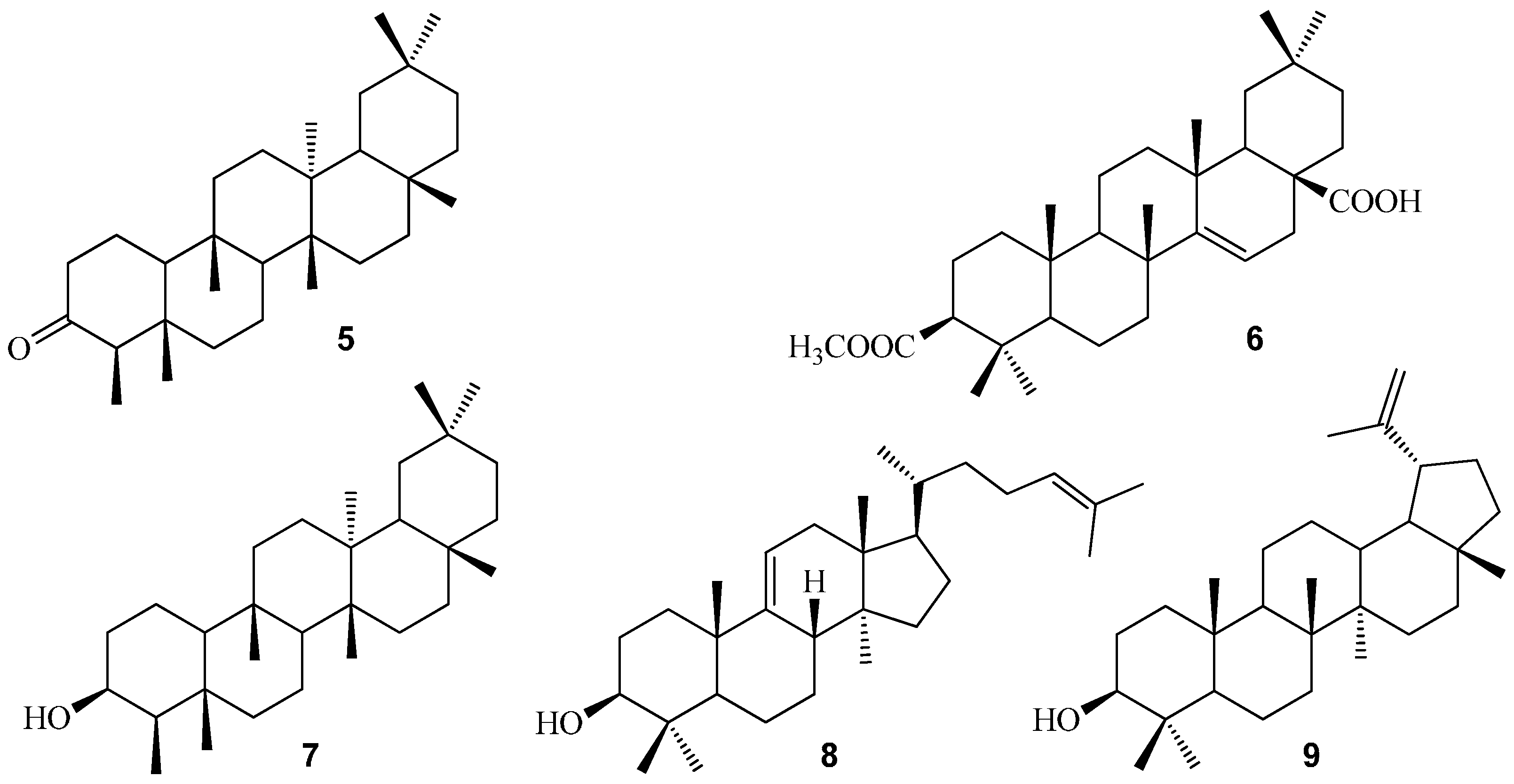
3.7.1. Compounds 19 Serial Dilution

A microdilution method was used to determine the MICs of the compounds on 96-well cultivated plates according to the previous report [27]. The compounds 19 were dissolved in nutrient broth with 10% DMSO and 32.0 mg·mL−1 solutions (pH 7.2) were obtained, respectively. There were 12 wells in each row of a microplate, to each of the first ones we added 100 μL compound solution, and to the remaining 11 wells we added 100 μL broth culture. For serial dilution, 100 μL each compound solution was added into the second well and then 100 μL was sequentially transferred to the following wells until the 10th well. The last two wells served as growth control and sterility check. After that, 100 μL of inoculum was added into each well except the last well in which 100 μL broth was added instead. Amoxicillin was used as a positive drug.

3.7.2. Determination of MICs and MBCs

The growth of the bacterial strains in the microplates was monitored at 37 °C for 20 h using a microplate reader. Standard antibacterial agent amoxicillin was also screened under identical conditions for comparison. Considering the role of DMSO, the same experiment was carried out with 10% DMSO and showed no activity against any bacterial strains. MIC was expressed as the mean concentration between the well showing growth and that showing no growth.
After MIC testing, the microplates set up for the MICs determination were used to determine the MBC as described previously [28]. For each well showing no bacterial growth, the entire volume was spread onto nutrient agar plates and subcultured. The MBC was defined as the lowest concentration of the compounds showing no bacterial growth after incubating for 20 h.

4. Conclusions

We investigated the chemical constituents of P. oleracea based on its antibacterial activity and nine compounds were obtained, including three new cerebrosides and a new ceramide. The structures of new compounds were identified as 1-O-β-d-glucopyranosyl-(2S,3R,8E)-2-[(2′R)-2-hydroxylpentadecanoylamino]-8-octadecene-1,3-diol (1), 1-O-β-d-glucopyranosyl-(2S,3R,4E)-2-[(2′R)-2-hydroxylpentadecanoyl-amino]-4-heptadecene-1,3-diol (2), 1-O-β-d-glucopyranosyl-(2S,3R,4E)-2-[(2′R)-2-hydroxyl-docosanamideamino]-16-methyl-4-nonadecene-1,3-diol (3), and (2S,3S,4R,8E)-2-[(2′R)-2-hydroxyl-pentadecanoylamino]-8-dodecene-1,3,4-triol (4), respectively. Antibacterial tests in vitro showed that new compounds could significantly inhibit or kill the common enteropathogenic bacteria, which might contribute to revealing the usefullness of P. oleracea as a treatment for bacillary dysentery.

Acknowledgments

This research was supported by the Program of Natural Science Foundation of Heilongjiang (No. H2013012).

Author Contributions

Xia Lei and Jianmin Li conceived and designed the experiments; Xia Lei performed the experiments; Bin Liu and Ning Zhang analyzed the data; Xia Lei contributed reagents/materials/analysis tools; Haiyang Liu wrote the paper.

Conflicts of Interest

The authors declare no conflict of interest.

References and Notes

  1. Liu, X.F.; Zheng, C.G.; Shi, H.G.; Tang, G.S.; Wang, W.Y.; Zhou, J.; Dong, L.W. Ethanol extract from Portulaca oleracea L. attenuated acetaminophen-induced mice liver injury. Am. J. Transl. Res. 2015, 7, 309–318. [Google Scholar] [PubMed]
  2. Zhou, Y.X.; Xin, H.L.; Rahman, K.; Wang, S.J.; Peng, C.; Zhang, H. Portulaca oleracea L.: A review of phytochemistry and pharmacological effects. Biomed. Res. Int. 2015, 2015, 925631. [Google Scholar] [CrossRef] [PubMed]
  3. Behravan, J.; Mosafa, F.; Soudmand, N.; Taghiabadi, E.; Razavi, B.M.; Karimi, G. Protective effects of aqueous and ethanolic extracts of Portulaca oleracea L. aerial parts on H2O2-induced DNA damage in lymphocytes by comet assay. J. Acupunct. Meridian Stud. 2011, 4, 193–197. [Google Scholar] [PubMed]
  4. Xin, H.L.; Hou, Y.H.; Xu, Y.F.; Yue, X.Q.; Li, M.; Lu, J.C.; Ling, C.Q. Portulacerebroside A: New cerebroside from Portulaca oleracea L. Chin. J. Nat. Med. 2008, 6, 401–403. [Google Scholar] [CrossRef]
  5. Elkhayat, E.S.; Ibrahim, S.R.; Aziz, M.A. Portulene, a new diterpene from Portulaca oleracea. J. Asian Nat. Prod. Res. 2008, 10, 1039–1043. [Google Scholar] [PubMed]
  6. Chan, B.C.; Han, X.Q.; Lui, S.L.; Wong, C.W.; Wang, T.B.; Cheung, D.W.; Cheng, S.W.; Ip, M.; Han, S.Q.; Yang, X.S.; et al. Combating against methicillin-resistant Staphylococcus aureus—Two fatty acids from Purslane ( Portulaca oleracea L.) exhibit synergistic effects with erythromycin. J. Pharm. Pharmacol. 2015, 67, 107–116. [Google Scholar] [PubMed]
  7. Noreen, S.; Hussain, I.; Tariq, M.I.; Ijaz, B.; Iqbal, S.; Qamar-Ul, Z.; Ashfaq, U.A.; Husnain, T. Portulaca oleracea L. as a prospective candidate inhibitor of hepatitis C virus NS3 serine protease. Viral Immunol. 2015, 28, 282–289. [Google Scholar] [PubMed]
  8. Al-Sheddi, E.S.; Farshori, N.N.; Al-Oqail, M.M.; Musarrat, J.; Al-Khedhairy, A.A.; Siddiqui, M.A. Portulaca oleracea seed oil exerts cytotoxic effects on human liver cancer (HepG2) and human lung cancer (A-549) cell lines. Asian Pac. J. Cancer Prev. 2015, 16, 3383–3387. [Google Scholar] [PubMed]
  9. Chan, K.; Islam, M.W.; Kamil, M.; Radhakrishnan, R.; Zakaria, M.N.; Habibullah, M.; Attas, A. The analgesic and anti-inflammatory effects of Portulaca oleracea L. subsp. sativa (haw.) Celak. J. Ethnopharmacol. 2000, 73, 445–451. [Google Scholar] [CrossRef]
  10. Radhakrishnan, R.; Zakaria, M.N.; Islam, M.W.; Chen, H.B.; Kamil, M.; Chan, K.; Al-Attas, A. Neuropharmacological actions of Portulaca oleraceae L. v. sativa (hawk). J. Ethnopharmacol. 2001, 76, 171–176. [Google Scholar] [CrossRef]
  11. Abdel Moneim, A.E. The neuroprotective effects of purslane (Portulaca oleracea) on rotenone-induced biochemical changes and apoptosis in brain of rat. Neurol. Disord. Drug Targets 2013, 12, 830–841. [Google Scholar] [CrossRef]
  12. Kim, J.Y.; Oh, H.M.; Kwak, S.C.; Cheon, Y.H.; Lee, M.S.; Rho, M.C.; Oh, J. Purslane suppresses osteoclast differentiation and bone resorbing activity via inhibition of Akt/GSK3 β-c-Fos-NFATc1 signaling in vitro and prevents lipopolysaccharide-induced bone loss in vivo. Biol. Pharm. Bull. 2015, 38, 66–74. [Google Scholar] [CrossRef] [PubMed]
  13. Shibuya, H.; Kawashima, K.; Sakagami, M.; Shimomura, M.; Ohashi, K.; Kitagawa, I. Sphingolipids and glycerolipids. Ι.: Chemical structures and ionophoretic activities of soyacerebrosides Ι and ΙΙ form soybean. Chem. Pharm. Bull. 1990, 38, 2933–2938. [Google Scholar] [CrossRef] [PubMed]
  14. Costantino, V.; Fattorusso, E.; Mangoni, A. Glycolipids from sponges. Part 9: Plakoside C and D, two further prenylated glycosphingolipids from the marine sponge Ectyoplasia ferox. Tetrahedron 2000, 56, 5953–5957. [Google Scholar] [CrossRef]
  15. Kang, S.S.; Kim, J.S.; Son, K.H.; Kim, H.P.; Chang, H.W. Cyclooxygenase-2 inhibitory cerebrosides from Phytolaccae Radix. Chem. Pharm. Bull. 2001, 49, 321–323. [Google Scholar] [CrossRef] [PubMed]
  16. Ling, T.J.; Xia, T.; Wan, X.C.; Li, D.X.; Wei, X.Y. Cerebrosides from the Roots of Serratula chinensis. Molecules 2006, 11, 677–683. [Google Scholar] [CrossRef] [PubMed]
  17. Yamada, K.; Wada, N.; Onaka, H.; Matsubara, R.; Isobe, R.; Inagaki, M.; Higuchi, R. Constituents of holothuroidea, isolation of ante-iso type regio isomer on long chain base moiety of glucocerebroside from the sea cucumber Holothuria leucospilota. Chem. Pharm. Bull. 2005, 53, 788–791. [Google Scholar] [CrossRef] [PubMed]
  18. Satoshi, K.; Kazufumi, N.; Masanori, I.; Ryuichi, H. Isolation and structure determination of six glucocerebrosides from the starfish Luidia maculate. Chem. Pharm. Bull. 2002, 50, 1091–1096. [Google Scholar]
  19. Yang, N.Y.; Ren, D.C.; Duan, J.A.; Xu, X.H.; Xie, N.; Tian, L.J. Ceramides and cerebrosides from Ligusticum chuanxiong Hort. Helv. Chim. Acta 2009, 92, 291–297. [Google Scholar] [CrossRef]
  20. Chen, C.; Wei, G.; Zhu, H.; Guo, Y.; Li, X.N.; Zhang, J.; Liu, Y.; Yao, G.; Luo, Z.; Xue, Y.; et al. A new 3,4- seco- oleanane-type triterpenoid with an unusual enedione moiety from Hypericum ascyron. Fitoterapia 2015, 103, 227–230. [Google Scholar] [CrossRef] [PubMed]
  21. Supaluk, P.; Saowapa, S.; Apilak, W.; Ratana, L.; Somsak, R.; Virapong, P. Bioactive metabolites from Spilanthes acmella Murr. Molecules 2009, 14, 850–867. [Google Scholar]
  22. Ding, H.W.; Li, F.F.; Song, S.J. Chemical constituents from Portulaca oleracea L. J. Shenyang Pharm. Univ. 2009, 26, 878–881. [Google Scholar]
  23. Tsai, P.W.; Kathlia, A.; Castro-Cruz, D.; Shen, C.C.; Chiou, C.T.; Ragasa, C.Y. Chemical constituents of Artocarpus camansi. Pharmacogn. J. 2013, 5, 80–82. [Google Scholar] [CrossRef]
  24. Rabia, R.; Zaitoon, I.; Sajid, A.; Muhammad, N.; Muhammad, Y.K.; Jamshed, I. Identification of highly potent and selective α-Glucosidase inhibitors with antiglycation potential, isolated from Rhododendron arboretum. R. Nat. Prod. 2015, 9, 262–266. [Google Scholar]
  25. Li, N.; Di, L.; Gao, W.C.; Wang, K.J.; Zu, L.B. Cytotoxic iridoids from the roots of Patrinia scabra. J. Nat. Prod. 2012, 75, 1723–1728. [Google Scholar] [CrossRef] [PubMed]
  26. Di, L.; Li, N.; Zu, L.B.; Wang, K.J.; Zhao, Y.X.; Wang, Z. Three new iridoid glucosides from the roots of Patrinia scabra. Bull. Korean Chem. Soc. 2011, 32, 3251–3254. [Google Scholar] [CrossRef]
  27. Lin, S.M.; Molan, P.C.; Cursons, R.T. The in vitro susceptibility of Campylobacter spp. to the antibacterial effect of manuka honey. Eur. J. Clin. Microbiol. 2009, 28, 339–344. [Google Scholar] [CrossRef] [PubMed]
  28. Lin, S.M.; Molan, P.C.; Cursons, R.T. The controlled in vitro susceptibility of gastrointestinal pathogens to the antibacterial effect of manuka honey. Eur. J. Clin. Microbiol. 2011, 30, 569–574. [Google Scholar] [CrossRef] [PubMed]
  • Sample Availability: Samples of the compounds of Portulacerebroside B–D and Portulaceramide A are available from the authors.

