Article

Isolation of Flavonoids from Deguelia duckeana and Their Effect on Cellular Viability, AMPK, eEF2, eIF2 and eIF4E

1
Bioprospecting and Biotechnology Lab, National Institute of Amazonian Research, Manaus 69067-375, Brazil
2
Institute of Pharmaceutical Science, Department of Pharmaceutical Biology and Biotechnology, University of Freiburg, Freiburg 79104, Germany
3
Escuela de Química and CIPRONA, Universidad de Costa Rica, 2060 San José, Costa Rica
*
Authors to whom correspondence should be addressed.
Molecules 2016, 21(2), 192; https://doi.org/10.3390/molecules21020192
Submission received: 4 November 2015 / Accepted: 29 January 2016 / Published: 6 February 2016
(This article belongs to the Collection Bioactive Compounds)

Abstract

:
Preparations of Deguelia duckeana, known in Brazil as timbó, are used by indigenous people to kill fish. Reinvestigation of its extracts resulted in the isolation and identification of 11 known flavonoids identified as 3,5,4’-trimethoxy-4-prenylstilbene (1), 4-methoxyderricidine (2), lonchocarpine (3), 4-hydroxylonchocarpine (4), 4-methoxylonchocarpine (5), 5-hydroxy-4’,7-dimethoxy-6-prenylflavanone (6), 4’-hydroxyisolonchocarpine (7), 4’-methoxyisolonchocarpine (8), 3’,4’,7-trimethoxyflavone (9), 3’,4’-methylenedioxy-7-methoxyflavone (10), and 2,2-dimethyl-chromone-5,4’-hydroxy-5’-methoxyflavone (11). Except for 1, 3, and 4 all of these flavonoids have been described for the first time in D. duckeana and the flavanone 6 for the first time in nature. Compounds 2, 3, 4, 7, 9, and 10 were studied for their potential to induce cell death in neuronal SK-N-SH cells. Only the chalcone 4 and the flavanone 7 significantly induced lactate dehydrogenase (LDH) release, which was accompanied by activation of caspase-3 and impairment of energy homeostasis in the MTT assay and may explain the killing effect on fish. Interestingly, the flavone 10 reduced cell metabolism in the MTT assay without inducing cytotoxicity in the LDH assay. Furthermore, the flavonoids 2, 3, 4, 7, and 10 induced phosphorylation of the AMP-activated protein kinase (AMPK) and the eukaryotic elongation factor 2 (eEF2). The initiation factor eIF4E was dephosphorylated in the presence of these compounds. The initiation factor eIF2alpha was not affected. Further studies are needed to elucidate the importance of the observed effects on protein synthesis and potential therapeutic perspectives.

1. Introduction

Flavonoids possess a broad variety of different biological activities, among which their antioxidative properties are of special interest. Reactive oxygen radicals are harmful to biomembranes, but can additionally play a role as mediators in different signaling pathways [1,2]. Depending on their structural features, flavonoids such as prenylated flavones or chalcones, can also be involved in cytotoxic processes [3,4].
The genus Deguelia which belongs to the Fabaceae family can be found in tropical South America and shows a predominance of prenylated flavonoids and stilbenes [5]. Prenylated stilbenes as well as chalcones and isoflavonoids have been isolated from Derris rariflora (syn. Deguelia nitidula) [6]. Similar compounds have also been found in the roots of Deguelia hatschbachii [7]. Stilbenes with a slight effect on seed germination and plant growth have been described from the leaves of Deguelia refuescens var. urucu [8], whereas prenylated isoflavonoids with a slight antibacterial and antifungal activity have been reported from Deguelia longeracemosa [9]. In leaves of Deguelia utilis prenylated chalcones and stilbenes, which have cytoprotective properties in a neuronal cell line, have been found [10].
Preparations of Deguelia duckeana (Fabaceae) are known in Brazil as timbó and used by indigenous people for killing fish. However, the compounds responsible for these cytotoxic effects as well as their mode(s) of action are still unknown. Initial phytochemical investigations revealed three chalcones and a stilbene derivative as constituents, but no studies on their cytotoxic activities have been performed [11]. This prompted us to reinvestigate D. duckeana and to perform the first studies on which way the isolated flavonoids may affect cell life using the neuronal cell line SK-N-SH.

2. Results and Discussion

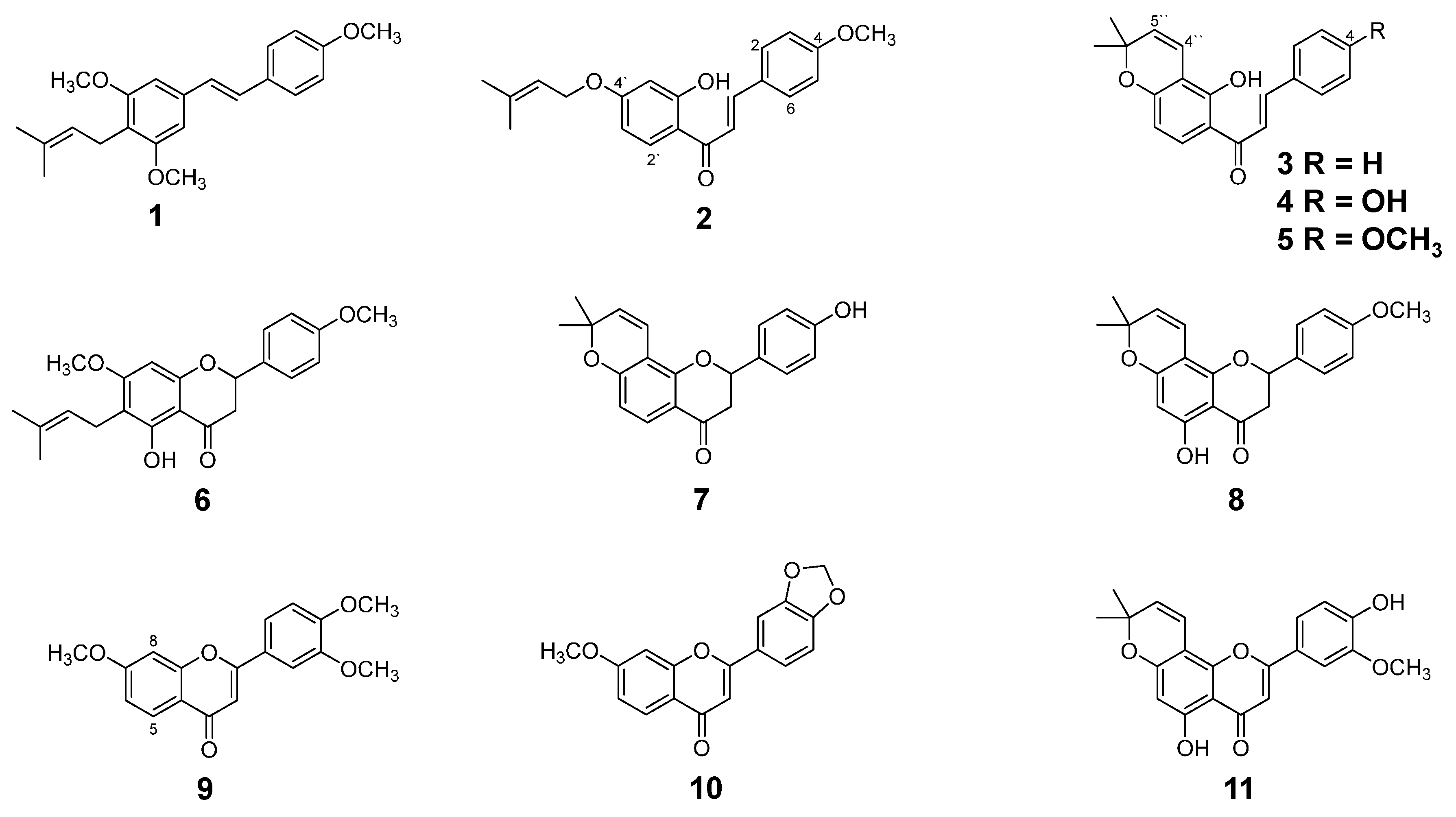
2.1. Isolation and Identification of Flavonoids from D. duckeana Including a New Flavanone

Fractionation of the CH2Cl2 extract from the roots of D. duckeana afforded eight known flavonoids. Based on 1H-, 13C-NMR and EI-MS data as well as comparison to spectral data from the literature these compounds were identified as 3,5,4’-trimethoxy-4-prenylstilbene (1) [11], 4-methoxy-derricidine (2) [11], 4-hydroxylonchocarpine (4) [11], 4-methoxylonchocarpine (5) [11], 4’-hydroxy-isolonchocarpine (7) [12], 4’-methoxyisolonchocarpine (8) [13], 3’,4’-methylenedioxy-7-methoxyflavone (10) [14] and 2,2-dimethylchromone-5,4’-dihydroxy-5’-methoxyflavone (11) [15]. The hexane extract from branches yielded two known flavonoids which were identified by the methods mentioned above as lonchocarpine (3) [11] and 3’,4’,7-trimethoxyflavone (9) [16]. Except for 1, 3, and 4 [11] all these compounds are described for the first time in D. duckeana. The structures are given in Figure 1.
Figure 1. Chemical structures of isolated flavonoids from D. duckeana.
Molecules 21 00192 g001
Additionally, a new flavanone has been isolated as a mixture with compound 8 from the CH2Cl2 extract. EI-MS gave a molecular ion peak at m/z 368, fragment ions at m/z 353 [M − 15]+, 352 [M − 15 − H]+ and 337 [M − 31]+ and a base peak at m/z 203, indicating a flavonoid skeleton. 1H-NMR data (see Table 1) suggested the presence of a flavanone skeleton because of the signals at δH 5.34 (1H, dd, H-2) and 2.76 (1H, dd, H-3eq) and 3.09 (1H, dd, H-3ax). The presence of a γ,γ-dimethyl allyl group was obvious from signals at δH 3.25 (2H, d, H-1’’), 5.19 (1H, m, H-2’’), 1.76 (3H, brs, H-4’’) and 1.81 (3H, brs, H-5’’). The occurrence of an AA’BB’ system with signals at δH 6.95 (2H, d, H-3’ and 5’) and 7.38 (2H, d, H-2’ and 6’) revealed a para-substituted B-ring. The 4’-OH was methylated, because of the slight downfield-shift of H-2’/6’ and H-3’/5’ and C-4’ compared to the NMR data reported for the unmethylated flavanone from Feronia limonia 5-hydroxy-2-(4-hydroxyphenyl)-7-methoxy-6-(3-methylbut-2-enyl)chroman-4-one [17]. Correlation in the HSQC and HMBC spectra allowed the unambiguous assignment of the 13C-NMR data (see Table 1) to the respective carbon atom and confirmed the substitution at C-6 with an isoprenyl side chain and at C-7 and C-4’ with methoxy groups. The substitution of the flavanone agreed also with the correlations in the NOESY spectrum (see Figure S3 in the Supplementary Materials). Hence, compound 6 is named as 5-hydroxy-4’,7-dimethoxy-6-prenylflavanone which has not been reported to the best of our knowledge, but was synthesized as a flavone [18].
Table 1. 1H-NMR and 13C-NMR spectroscopic data of compound 6 (CDCl3, 400 MHz, J in Hz).
CδC (ppm)δH (ppm)
279.15.34 (dd, 13.0 and 3.0)
343.32.76 (dd, 17.2 and 3.0) a and 3.09 (dd, 17.2 and 13.0) b
4196.0-
5160.0-
6109.9-
7165.4-
890.96.07 (1H, s)
9161.4-
10102.9-
1’130.3 -
2’/6’127.77.38 (d, 8.5)
3’/5’114.26.95 (d, 8.5)
4’160.2-
1”21.0
2”122.25.19 (1H, m)
3”131.6
4”25.81.76 (3H, s)
5”17.71.81 (3H, s)
5-OH160.212.06 (1H, s)
7-OCH3, 4’-OCH355.33.83 (6H, s)
a H-3eq; b H-3ax.

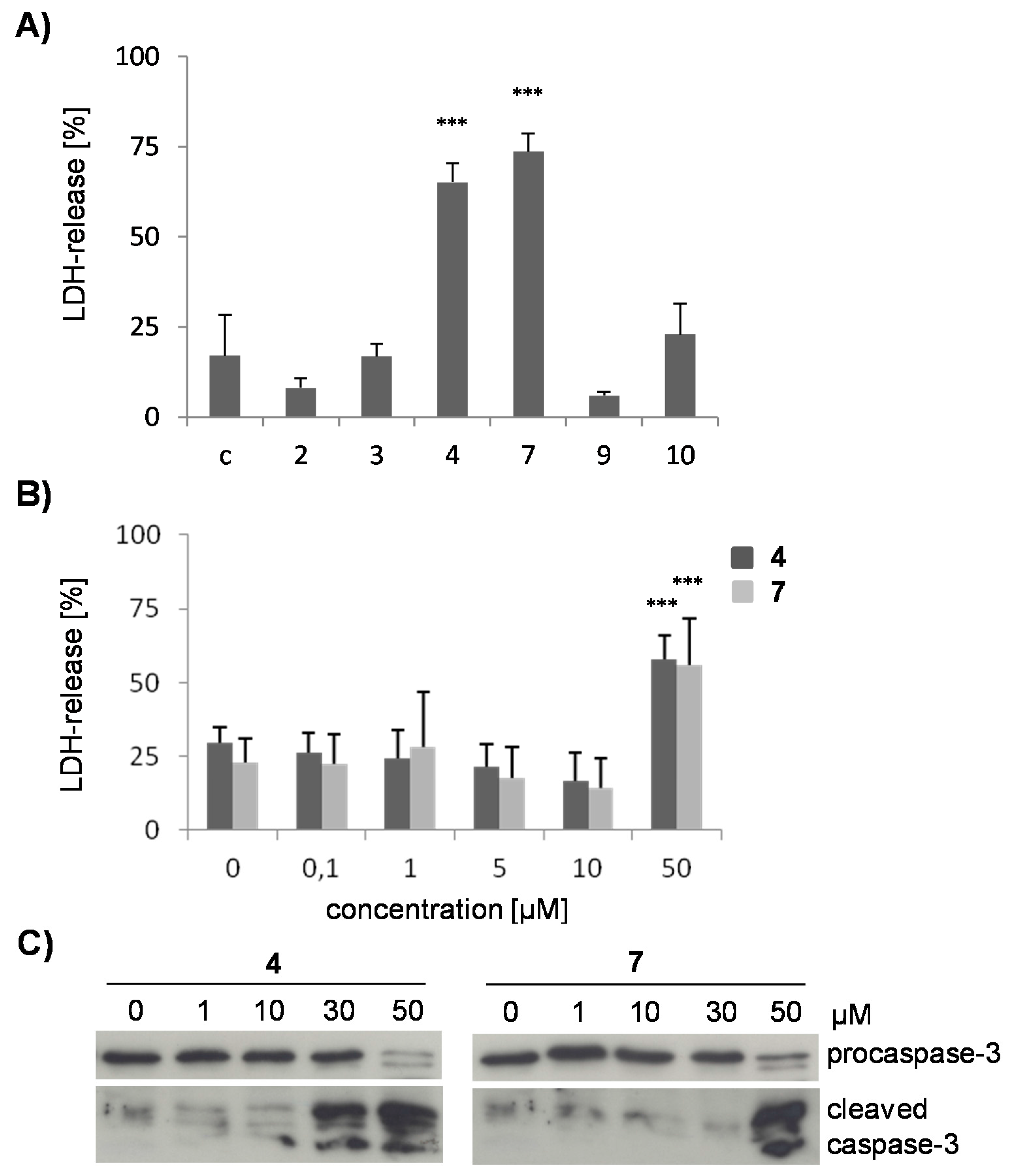
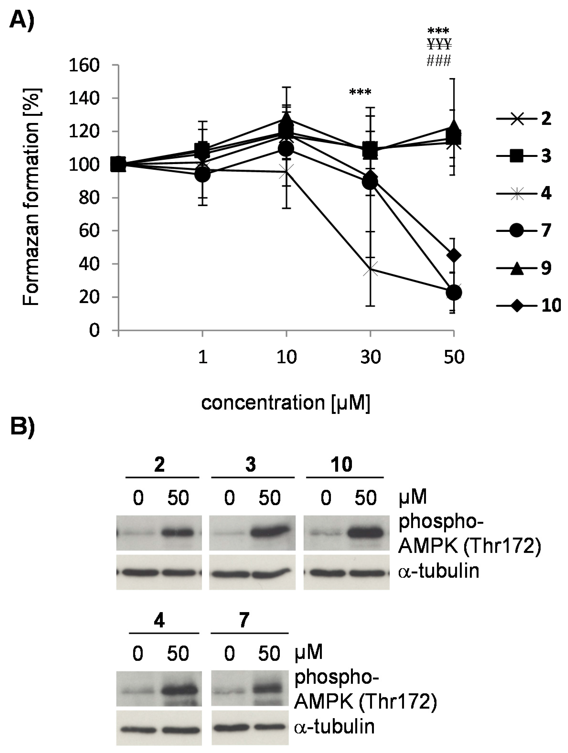
2.2. The Flavonoids from D. duckeana Differently Influence Cell Viability

Because timbó is considered as neurotoxic to fish, the biologic effects of isolated compounds were tested in the neuronal cell line SK-N-SH. Chalcones are known for their cytotoxic activity [4], and the compounds 2, 3 and 4 were first evaluated for their cytotoxic potential. To study whether the 2’’,2’’-dimethylpyrano- or the 3’,4’-methylenedioxy structural elements play a role in the cytotoxic activity compounds 7, 9 and 10 were included in the study. The cells were incubated with the respective flavonoid (50 μM) for 24 h and cell death was determined by measuring the release of intracellular lactate dehydrogenase into the supernatant (Figure 2A,B). Only the chalcone 4 and the flavanone 7, which both possess a 4’-hydroxy group and a 2’’,2’’-dimethylpyrano moiety, induced significant membrane damage, whereas the chalcones 2, 3 and the flavones 9 and 10 mediated no cytolytic effect. Compounds 4 and 7 were studied for their LDH release at different concentrations. No significant cell death was observed up to a 10 μM concentration, but only at 50 μM (Figure 2B).
Figure 2. Cytotoxicity of flavonoids from D. duckeana. Cell damage was determined by an LDH-release assay after incubating SK-N-SH cells with the flavonoids 24, 7, 9, and 10 at 50 µM (A) or with the compounds 4 and 7 at concentrations from 0.1–50 µM (B) for 24 h. Values represent means ± s.d. of three (A) or four (B) independent experiments. Statistical evaluation was performed by one-way ANOVA (in A) or two way ANOVA (in B), followed by the Bonferroni post hoc test. ***, p < 0.001 vs. untreated control cells. In (C), SK-N-SH cells were treated with 1–50 µM of compound 4 or 7 for 6 h before cellular lysates were analyzed for pro-caspase-3 cleavage by immunoblotting. A representative blot is shown (n = 3).
Molecules 21 00192 g002
To test whether cell death induced by compounds 4 and 7 is due to apoptosis, activation of caspase-3 was determined by immunoblotting. Cleavage of pro-caspase-3 to its active fragments was observed (Figure 2C), indicating that cytotoxicity, as determined in Figure 2A,B, may be due to secondary necrosis and a consequence of apoptosis.
Results were verified by the MTT cellular viability assay (Figure 3A). At concentrations ≥30 µM for compound 4 and 50 µM for compound 7, treated cells displayed a significantly reduced conversion of 3-(4,5-dimethylthiazol-2-yl)-2,5-diphenyltetrazolium bromide (MTT) to its formazan salt, indicating a decline in cellular viability. These concentrations are sufficient to induce apoptosis (Figure 2C) and secondary necrosis (Figure 2A,B). In contrast, compounds 2, 3, and 9 did not reduce conversion of the tetrazolium salt, confirming that these compounds have no effect on cellular viability (Figure 3A). Interestingly, the flavone 10 which differed from compound 9 by the 3’,4’-methylenedioxy group diminished the reducing capacity of SK-N-SH cells, as observed by a decline in absorbance (Figure 3A) without simultaneously killing the cells (Figure 2A). Reduced formazan conversion in the absence of cell death has been associated with impaired metabolism [19]. Despite the limiting data for drawing structure activity relationships the occurrence of the 3’,4’-methylenedioxy group may be important for a potential decrease of the metabolic activity in SK-N-SH cells.
Figure 3. Effect of flavonoids 2, 3, 4, 7, 9, and 10 on cellular metabolism. In (A), an MTT cell viability assay was performed. Cells were pretreated with 1–50 µM of the respective flavonoid for 24 h. Formazan formation was compared to DMSO-treated control cells (=100 %). Values represent means ± s.d. of five independent experiments. Statistical evaluation was performed by two-way ANOVA followed by the Bonferroni’s post hoc test. Statistically significant differences are shown for cells treated with compound 4 vs. untreated cells (***, p < 0.001), compound 7 vs. untreated cells (¥¥¥, p < 0.001) and compound 10 vs. untreated control (###, p < 0.001). In (B), SK-N-SH cells were treated with 50 µM of compound 2, 3, 4, 7 or 10 for 6 h, before cell lysates were analyzed for AMPK phosphorylation at threonine 172 by immunoblotting (upper blot). Immunoblots were reprobed for α-tubulin to demonstrate the analysis of equal amounts of protein in each sample (lower blot). Representative blots are shown (n = 3).
Molecules 21 00192 g003

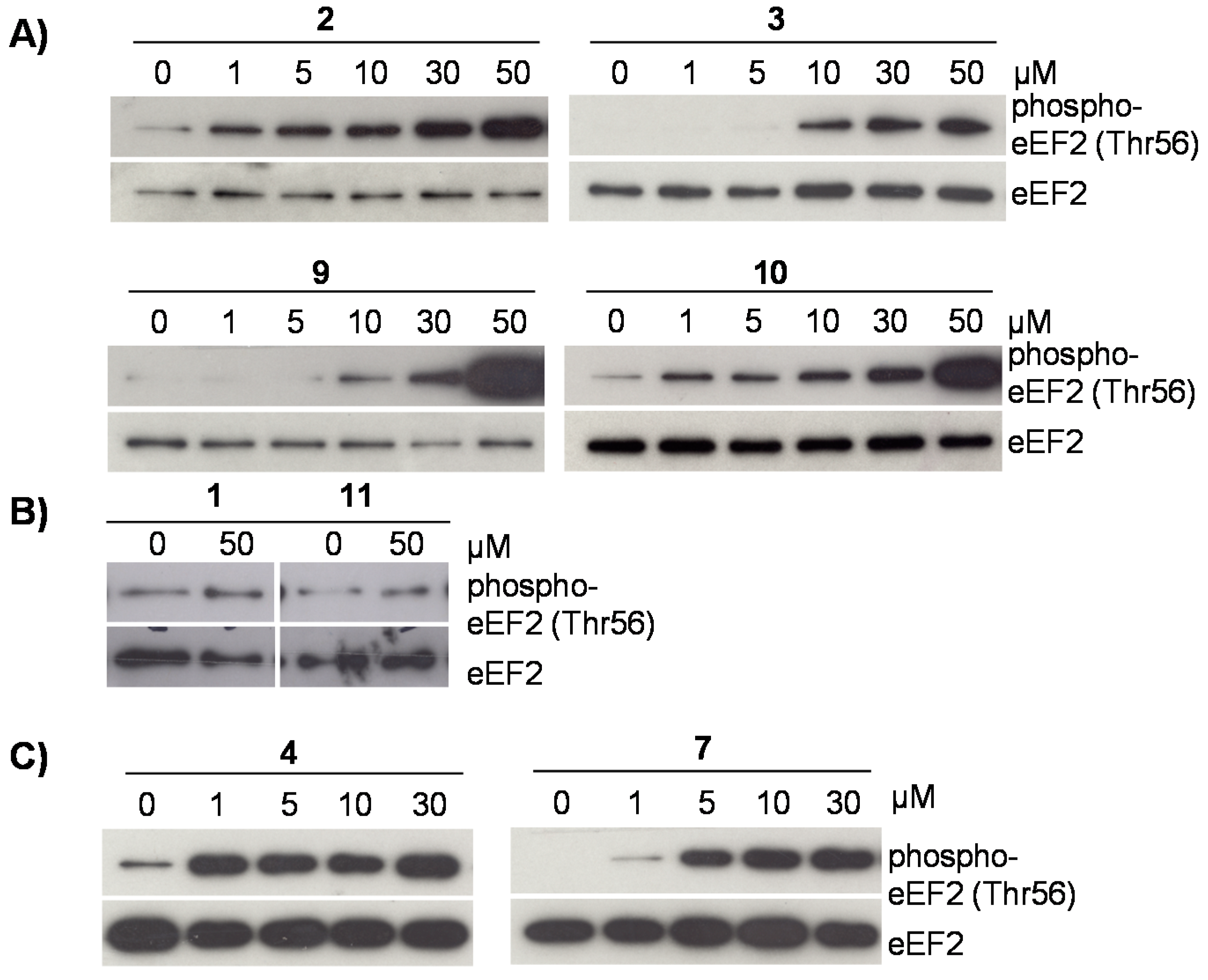
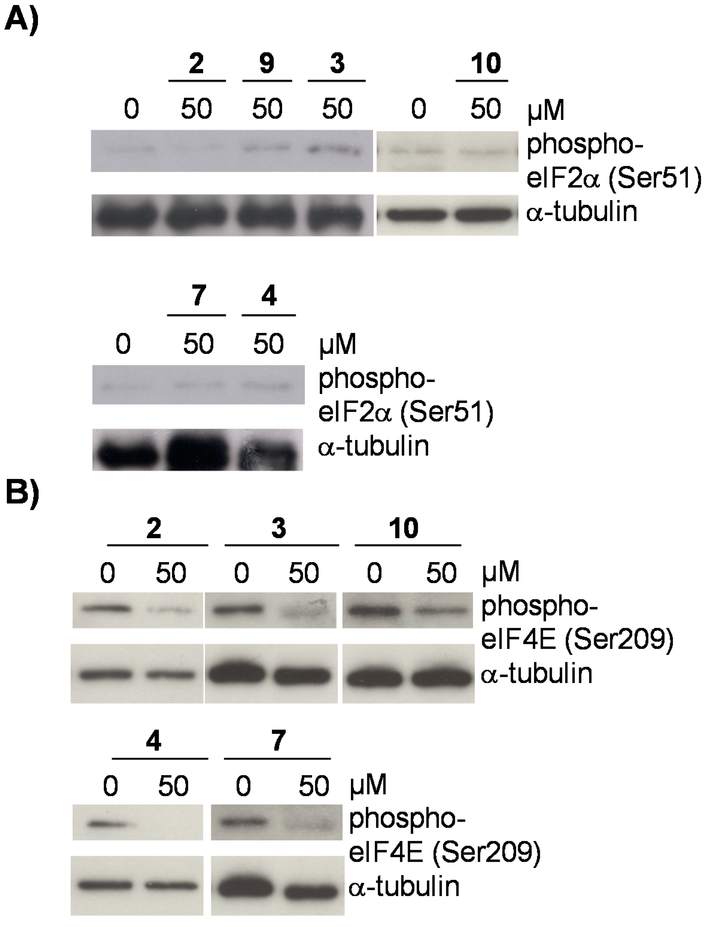
2.3. Flavonoids from D. duckeana Induce the Phosphorylation of AMPK and eEF2, But Not eIF2alpha

Cells with a low metabolism reduce very little MTT [20]. Therefore, we tested whether the flavone 10 attenuates the cellular metabolism by determining activation of AMPK, an energy-sensing enzyme closely involved in the regulation of energy homeostasis [21]. As shown in Figure 3B, treatment of cells with compound 10 resulted in an increase in AMPK-phosphorylation on its activating loop at threonine-172. Phosphorylation of AMPK at threonine-172 is a consequence of a conformational change due to increased AMP concentrations [22] and a prerequisite of AMPK to adjust intracellular energy levels by inhibiting energy consuming pathways such as global protein synthesis [23,24]. However, compounds 2, 3, 4 and 7 also induced phosphorylation of AMPK (Figure 3B) indicating that reduced formazan formation (Figure 3A) and AMPK activation are here independent processes.
Activation of AMPK indicates inhibition of global protein synthesis [23]. To study a possible impact of the tested compounds on translation, we determined their potential to induce phosphorylation of eEF2 at threonine 56, which blocks translational elongation by an AMPK/eEF2K-dependent mechanism and thus protein synthesis [23]. As shown in Figure 4A, the flavonoids 2, 3, 4, 7, and 10 markedly induced phosphorylation of eEF2, which couples AMPK activation with reduced translational elongation. The stilbene 1 and the flavone 11 did not mediate this posttranslational modification of eEF2 at threonine 56. However, up to now no structure activity relationships can be drawn.
The cytotoxic compounds 4 and 7 also induced posttranslational repression of eEF2 (Figure 4C), but at 50 µM eEF2 phosphorylation at threonine 56 declined in the presence of both compounds (data not shown). This is most probably due to the strong cytotoxic activity of these flavonoids (Figure 2A–C). Altogether, our results indicate, that the cytolytic potential of some flavonoids isolated from D. duckeana is not directly related to their potential to repress translational elongation.
Figure 4. (AC) Effect of flavonoids 14, 7, 911 on translational elongation. Phosphorylation of eEF2 at threonine 56 was analyzed in lysates of cells, pretreated with 1–50 µM of the indicated flavonoids for 6 h by immunoblotting. Immunoblots were reprobed for total eEF2 to visualize that similar amounts of proteins were analyzed in each sample. Representative blots are shown (n = 3).
Molecules 21 00192 g004
To test the influence of flavonoids from D. duckeana on the initiation phase in the translation process, phosphorylation of eIF2alpha was determined in the presence of compounds 2, 3, 4, 7, 9 and 10. eIF2alpha is required in the initiation of translation, as it mediates binding of GTP and the initiator Met-tRNA to the ribosome to form the 43S preinitiation complex. Phosphorylation at serine 51 inactivates eIF2alpha and as a consequence translation comes to halt because initiation is abrogated. As shown in Figure 5A, none of the tested flavonoids were able to mediate posttranslational phosphorylation of eIF2alpha at serine 51. Furthermore, we tested the potential of the flavonoids 2, 3, 4, 7, and 10 on the phosphorylation of eIF4E. Activation of the eukaryotic initiation factor eIF4E is a rate limiting step in cap-dependent translation and is regulated among others by AMPK under energy starvation [25]. Our results, shown in Figure 5B, demonstrate that the flavonoids 2, 3, 4, 7 and 10 repress phosphorylation of eIF4E at serine 209, which is associated with its reduced activity. Phosphorylation of eIF4E at serine 209 is mediated by the ras/MAP-kinase pathway [25], indicating that flavonoids from D. duckeana probably also affect mitogenic or stress stimuli via ERK1/2 or p38 MAP-kinase.
Figure 5. Effect of flavonoids 24, 7, 9, and 10 on translational initiation. Phosphorylation of eIF2alpha at serine 51 or eIF4E at serine 209 was analyzed in lysates of cells, pretreated with 50 µM of the indicated flavonoids for 6 h by immunoblotting. Immunoblots were reprobed for α-tubulin to visualize that similar amounts of proteins were analyzed in each sample. Representative blots are shown (n = 3).
Molecules 21 00192 g005

2.4. Conclusions

Altogether, we could extend the knowledge on the flavonoid profile of D. duckeana which fits those known from other species in the genus Deguelia. The isolated flavonoids were shown to have various effects on cells. Compounds 4 and 7, which both possess a 2”,2”-dimethylpyrano structure, may contribute to the reported killing of fish because of their proven cytotoxic effects. Moreover, we could demonstrate for the first time that flavonoids such as 2, 3, 4, 7, 9 and 10 affect phosphorylation of eEF2, AMPK and eIF4E and thus influence both translational initiation and elongation. Resulting consequences must be clarified by further experiments to elucidate the applicability of flavonoids in prospective therapeutic settings.

3. Experimental Section

3.1. General Procedures

NMR spectra were recorded on a DRX 400 NMR instrument (Bruker, Bremen, Germany) at 400 MHz (1H) and on an Avance III (Bruker) at 400 MHz (1H) and at 100 MHz (13C) either in CDCl3 and in methanol-d4. HR-APCI-MS spectra were measured on a Thermo Q Exactive mass spectrometer with an Orbitrap-Analyzer (Thermo Scientific, West Palm Beach, FL, USA). Open column chromatography was carried out by using silica gel 60 (230–400 and 70–230 mesh, Merck, Darmstadt, Germany), Sephadex LH-20 and Florisil (100–200 mesh, Merck). The fractions were monitored by analytical TLC (Silica gel 60 F254, aluminium sheets, Merck) using an Automatic TLC Sampler (CAMAG, Muttenz, Switzerland).

3.2. Plant Material

Roots and branches from Deguelia duckeana A. M. G. Azevedo (Fabaceae) were collected in Praia Dourada, Manaus, Amazonas. Identification was done by the botanist Ieda Leão do Amaral. Roots were collected in April, 2006 and branches in August, 2009. Voucher specimens are deposited at the Herbarium of the Instituto Federal de Educação do Amazonas (IFAM), under the numbers 10606 and 10613, respectively.

3.3. Extraction and Isolation

The material from D. duckeana were dried for three days in a hot-air oven 50 °C and separately extracted with CH2Cl2 for roots and with n-hexane for branches in a 2 L flask using an ultrasonic bath (Unique, Indaiatuba, São Paulo, Brazil) for 20 minutes and filtered. The extracts were concentrated in vacuo (40 °C). The CH2Cl2 extract from the roots (8.0 g) was fractionated by open column chromatography (CC) on SiO2 gel 60 (230–400 mesh, 138 × 3.2 cm, 104 g of SiO2) and eluted with a gradient of n-hexane/CH2Cl2, EtOAc/MeOH and MeOH 100%. 42 fractions were obtained which were monitored by TLC (n-hexane/CH2Cl2 8:2 and EtOAc/MeOH 7:3 with anisaldehyde/H2SO4 and Ce(SO4)2). Fraction 3 (200.0 mg) was separated by CC on SiO2 (70–230 mesh, 40 × 1.8 cm) with mixtures of CH2Cl2 and EtOAc yielding 10 fractions. Compound 1 (2.7 mg) was isolated from subfraction 6 (100.2 mg) by open CC on SiO2 (70–230 mesh, 26 × 1 cm, 20 g) with CHCl3 and a mixture of CHCl3/MeOH (75:5). Fractions 4 and 5 were combined (2.8 g) and were subjected to open CC on SiO2 (230–400 mesh, 74.5 × 3 cm) using mixtures of n-hexane, EtOAc and MeOH. 50 subfractions were obtained and monitored by TLC. Subfractions 13 to 15 (276.0 mg) afforded mainly compound 7, subfractions 16 to 18 (151 mg) were enriched with compound 2, subfraction 19 (30.0 mg) contained a mixture of compounds 2, 5 and 11 and subfraction 27 (15.0 mg) was enriched with compound 10. Compound 7 (9.6 mg) was purified on Sep-Pak Cartridges using Florisil (10.0 g, Phenomenex, Torrance, CA, USA) and a mixture of CH2Cl2 and MeOH, followed by open CC on SiO2 (230–400 mesh, 30.0 g) and n-hexane, CH2Cl2 and MeOH as solvent. Compound 2 (5.3 mg) was isolated from subfraction 16–18 by open CC on SiO2 (230–400 mesh, 40 × 1.8 cm) with mixtures of n-hexane, CH2Cl2 and EtOAc, followed by preparative TLC using the system CH2Cl2/EtOAc (95:5, v:v). Compound 11 (1.0 mg) was isolated from subfraction 19 by precipitation from MeOH. The remaining part of subfraction 19 was fractionated twice on Sephadex LH-20 column (2 × 10.5 cm) using MeOH followed by preparative TLC on SiO2 using the system CHCl3/MeOH (75:5, v:v). After open CC on Sephadex LH-20 (12.5 × 0.5 cm) a mixture (1.6 mg) of compounds 2 and 5 was obtained. Fraction 8 (30.2 mg) from the first separation procedure was again separated by CC on SiO2 (70–230 mesh, 20 × 1 cm) using mixtures of CHCl3/MeOH (78:2, 75:5) and MeOH 100% and afforded a fraction with a mixture of compounds 6 and 8. Fraction 21 (2.35 g) obtained from the first open CC was further fractionated by open CC on SiO2 (70–230 mesh, 20 × 3.6 cm) with a gradient of CH2Cl2, EtOAc and MeOH, followed by an open CC on SiO2 (230–400 mesh, 150 × 1.47 cm) and a gradient of n-hexane/CH2Cl2 and EtOAc and subsequently by an open CC on SiO2 (230–400 mesh, 140 × 1.8 cm, 88.75 g) and a gradient of CH2Cl2, EtOAc and MeOH. Compound 4 (1.0 mg) was finally obtained by precipitation from MeOH.
The hexane branch extract (2.0 g) was subjected to open CC on SiO2 (140 × 3.1 cm) using mixtures of n-hexane, EtOAc and MeOH and 112 fractions were yielded. Subfraction 25 (180.0 mg) was separated by open CC on neutral alumina (40 × 1.8 cm) using mixtures of n-hexane, CH2Cl2 and MeOH. Subfraction 9 (7.0 mg) was fractionated by preparative TLC (SiO2 gel 60 F254 plates) using n-hexane/CH2Cl2 (3:7, v:v) and afforded compound 3 (4.0 mg). Subfraction 63–66 (15.0 mg) was subjected to open CC on SiO2 (70–230 mesh, 20 × 0.5 cm, 5 g) using mixtures of CHCl3/MeOH (9:1, 8:2, 7:3) yielding compound 9 (3.1 mg). Fractionation schemes are shown in the Supplementary Materials (Figures S1 and S2).
Hydroxy-4’,7-dimethoxy-6-prenylflavanone (6): EI-MS: m/z (rel. int. %) 368 [M − 15]+ (33), 353 [M − 15]+ (16), 352 [M − 15 − H]+ (30), 337 [M − 31]+ (54), 218 (16), 203 (100), 179 (23); 1H-NMR, 13C-NMR: Table 1, HMBC and NOESY (Figure S3).

3.4. Cell Culture and Treatment

The human neuronal cell line SK-N-SH was obtained from the American Type Culture Collection (Manassas, VA, USA) and maintained in Eagle’s minimal essential medium (EMEM), supplemented with 10% fetal bovine serum (FBS), 100 IU/mL penicillin and 100 μg/mL streptomycin at 37 °C in a humidified atmosphere containing 5% CO2 (Thermo Fisher Scientific, Waltham, MA, USA). Cells were treated with 0.1–50 µM of the flavonoids after synchronization in FBS-free EMEM containing 100 IU/mL penicillin and 100 µg/mL streptomycin overnight. Control cells were treated with the highest concentration of DMSO as solvent (0.1%).

3.5. Cytotoxicity Assay

SK-N-SH cells (1 × 10⁶ cells per well of a 6 well plate) were treated with the flavonoids for 24 h, before cellular supernatants were analyzed for lactate dehydrogenase (LDH) content by the Cytotoxicity Detection Kit (Roche Applied Science, Mannheim, Germany) according to the protocol of the manufacturer. The absorbance at 490 nm was measured by a microplate reader (Model 680, BIO-RAD, Munich, Germany) with the reference wavelength of 690 nm. Total LDH release (100%) was obtained by the treatment of cells with 2% Triton-X100. The relative LDH release is defined by the ratio of LDH released over total LDH in the intact cells.

3.6. Metabolic Activity

The MTT viability assay was performed as described by Mosmann [20] Briefly, SK-N-SH cells (50000 cells per well of a 96 well plate) were treated with the respective flavonoid for 24 h and subsequently incubated with 1 mg/mL of 3-(4,5-dimethylthiazol-2-yl)-2,5-diphenyl tetrazolium bromide (MTT) for 2 h at 37 °C before excess MTT was aspirated. The residual formazan crystals were dissolved in 100 μL of dimethylsulfoxide and quantified at 595 nm, using a microplate reader (Model 680, BIO-RAD, Munich, Germany).

3.7. Immunoblot Analysis

SK-N-SH cells (50000 cells per well of a 96 well plate) were treated with the flavonoids for 6 h and subsequently lysed in 5 × SDS electrophoresis sample buffer. Lysates were sonicated and boiled for 3 min before proteins were separated by SDS-polyacrylamide gel electrophoresis, transferred onto a polyvinylidene difluoride membrane (EMD Millipore, Billerica, MA, USA) and blocked with 5% dry milk in Tris-buffered saline. Antibodies, raised against caspase-3, cleaved caspase-3, phospho-AMPK (Thr172), phospho-eEF2 (Thr56), eEF2, phospho-eIF2alpha (Ser51), phospho-eIF4E (Ser209) or α-tubulin were used for detection of specific proteins (Cell Signaling Technology, Danvers, MA, USA). After repeated washing, the specific bands were visualized using horseradish peroxidase-conjugated anti-rabbit IgG and enhanced chemiluminescence reagents (GE Healthcare, München, Germany).

3.8. Statistical Analysis

Data are shown as mean ± s.d. Statistical analysis was performed using the GraphPad Prism 5 software (GraphPad, La Jolla, CA, USA) and one-way or two way analysis of variance (ANOVA) followed by the Bonferroni post hoc test. Results were considered significant with p ≤ 0.05.

Supplementary Materials

Schemes of Deguelia duckeana fractionation are available online at https://www.mdpi.com/1420-3049/21/2/192/s1.

Acknowledgments

The authors (CVN and LMCC) would like to thank the Brazilian Research Agencies CNPq and CAPES for a grant. The authors are grateful to S. Ferlaino, Institute of Pharmaceutical Sciences, University of Freiburg and to Andersson Barison and Kahlil Salomé, University of Paraná, for obtaining the NMR spectra, to J. Wörth and C. Warth at the Institute of Organic Chemistry, University of Freiburg, for the HRMS data, to Christoph Nicolaus, Institute of Pharmaceutical Sciences, University of Freiburg for the help in literature research, to Jane Vasconcelos Neves Marinho, National Institute of Amazonian Research, Brazil, for the first plant material collection, and to the botanist Ieda Leão do Amaral for identification of the plant material. The article processing charge was funded by the German Research Foundation (DFG) and the Albert Ludwigs University Freiburg in the funding programme Open Access Publishing.

Author Contributions

Conceived and designed the experiments: Lorena M. C. Cursino, Cecilia V. Nunez, Matjaz Humar, Irmgard Merfort. Performed the experiments: Lorena M. C. Cursino, Matjaz Humar, Nerilson M. Lima. Analyzed the data: Lorena M. C. Cursino, Renato Murillo, Cecilia V. Nunez, Matjaz Humar, Irmgard Merfort. Wrote the paper: Lorena M. C. Cursino, Matjaz Humar, Irmgard Merfort.

Conflicts of Interest

The authors declare no conflict of interest.

References

  1. Ribeiro, D.; Freitas, M.; Lima, F.J.F.C.; Fernandes, E. Proinflammatory pathways: The modulation by flavonoids. Rev. Med. Res. 2015, 35, 877–936. [Google Scholar] [CrossRef] [PubMed]
  2. Nijveldt, R.J.; Nood, E.; Hoorn, D.E.C.; Boelen, P. G.; Norren, K.; Leeuwen, P.A.M. Flavonoids: A review of probable mechanisms of action and potential applications. Am. J. Clin. Nutr. 2001, 74, 418–425. [Google Scholar] [PubMed]
  3. Kuete, V.; Ngameni, B.; Wiench, B.; Krusche, B.; Horwedel, C.; Ngadjui, B.T.; Efferth, T. Cytotoxicity and mode of action of four naturally occurring flavonoids from the genus Dorstenia: Gancaonin Q, 4-hydroxylonchocarpin, 6-prenylapigenin and 6,8-diprenyleriodictyol. Planta Med. 2011, 77, 1984–1989. [Google Scholar] [CrossRef] [PubMed]
  4. Kuete, V.; Nkuete, A.H.L.; Mbaveng, A.T.; Wiench, B.; Wabo, H.K.; Tane, P.; Efferth, T. Cytotoxicity and modes of action of 4-hydroxy-2,6-dimethoxychalcone and other flavonoids toward drug-sensitive and multidrug-resistant cancer cell lines. Phytomedicine 2014, 21, 1651–1657. [Google Scholar] [CrossRef] [PubMed]
  5. Magalhães, A.F.; Tozzi, A.M.G.A.; Magalhães, E.V.; Moraes, V.R.S. Prenylated flavonoids from Deguelia hatschbachii and their systematic significance in Deguelia. Phytochemistry 2001, 57, 77–89. [Google Scholar] [CrossRef]
  6. Braz Filho, R.; Gottlieb, O.R.; Mourão, A.P.; Rocha, A.I.; Oliveira, F.S. Flavonoids from Derris species. Phytochem. Rep. 1975, 14, 1454–1456. [Google Scholar] [CrossRef]
  7. Magalhães, A.F.; Tozzi, A.M.G.A.; Magalhães, E.G.; Moraes, V.R.S. New spectral data of some flavonoids from Deguelia hatschbachii A.M.G. Azevedo. J. Braz. Chem. Soc. 2003, 14, 133–137. [Google Scholar] [CrossRef]
  8. Lobo, L.V.; Silva, G.A.; Freitas, M.C.C; Souza Filho, A.P.S.; Silva, M.N.; Arruda, A.C.; Guilhon, G.M.S.P.; Santos, L.S.; Santos, A.S.; Arruda, M.S.P. Stilbenes from Deguelia rufescens var. urucu (Ducke) A.M.G. Azevedo leaves: Effects on seed germination and plant growth. J. Braz. Chem. Soc. 2010, 21, 1838–1844. [Google Scholar] [CrossRef]
  9. Magalhães, A.F.; Tozzi, A.M.G.A.; Magalhães, E.G.; Souza-Neta, L.C. New prenylated metabolites of Deguelia longeracemosa and evaluation of their antimicrobial potential. Planta Med. 2006, 72, 358–363. [Google Scholar] [CrossRef] [PubMed]
  10. Oliveira, D.G.; Almeida, C.M.C.; Silva, C.Y.Y.; Arruda, M.S.P.; Arruda, A.C.; Lopes, D.C.F.; Yamada, E.S.; Costa, E.T.; Silva, M.N. Flavonoids from the leaves of Deguelia utilis (Leguminosae): Structural elucidation and neuroprotective properties. J. Braz. Chem. Soc. 2012, 23, 1933–1939. [Google Scholar] [CrossRef]
  11. Lima, N.M.; Andrade, J.I.A.; Lima, K.C.S.; Santos, F.N.; Barison, A.; Salomé, K.S.; Matsuura, T.; Nunez, C.V. Chemical profile and biological activities of Deguelia duckeana A.M.G. Azevedo (Fabaceae). Nat. Prod. Res. 2013, 27, 425–432. [Google Scholar] [CrossRef] [PubMed]
  12. Dagne, E.; Bekele, A.; Waterman, P.G. The flavonoids of Millettia ferruginosa and subsp. Darassana in Ethiopia. Phytochemistry 1989, 28, 1897–1900. [Google Scholar] [CrossRef]
  13. Jang, D.S.; Cuendet, M.; Hawthorne, M.E.; Kardono, L.B.S.; Kawanishi, K.; Fong, H.H.S.; Mehta, R.G.; Pezzuto, J.M.; Kinghorn, A.D. Prenylated flavonoids of the leaves of Macaranga conifera with inhibitory activity against cyclooxygenase-2. Phytochemistry 2002, 61, 867–872. [Google Scholar] [CrossRef]
  14. Mahmoud, E.N.; Waterman, G. Flavonoids from the stem bark of Millettia hemsleyana. Phytochemistry 1985, 24, 369–371. [Google Scholar] [CrossRef]
  15. Banerji, A.; Luthria, D.L.; Prabhu, B.R. Prenylated compounds from Atalantia racemosa: Isolation and synthesis of two pyranoflavones. Phytochemistry 1988, 27, 3637–3640. [Google Scholar] [CrossRef]
  16. Kato, M.J.; Yoshida, M.; Gottlieb, O.R. Flavones and lignans in flowers, fruits and seedlings of Virola venosa. Phytochemistry 1992, 31, 283–287. [Google Scholar] [CrossRef]
  17. Intekhab, J.; Mohammad, A. Isolation of a flavonoid from Feronia limonia. J. Saudi Chem. Soc. 2009, 13, 295–298. [Google Scholar] [CrossRef]
  18. Jain, A.C.; Khazanchi, R.; Kumar, A. Synthesis of carpachromene and related isopentenylated derivatives of apigenin. Tetrahedron 1978, 34, 3569–3573. [Google Scholar] [CrossRef]
  19. Berridge, M.V.; Herst, P.M.; Tan, A.S. Tetrazolium dyes as tools in cell biology: New insights into their cellular reduction. Biotechnol. Annu. Rev. 2005, 11, 127–152. [Google Scholar] [PubMed]
  20. Mosmann, T. Rapid colorimetric assay for cellular growth and survival: Application to proliferation and cytotoxicity assays. J. Immunol. Methods 1983, 65, 55–63. [Google Scholar] [CrossRef]
  21. Hardie, D.G. The AMP-activated protein kinase pathway-new players upstream and downstream. J. Cell Sci. 2004, 117, 5479–5487. [Google Scholar] [CrossRef] [PubMed]
  22. Sanz, P. AMP-Activated protein kinase: Structure and regulation. Curr. Protein Pept. Sci. 2008, 9, 478–492. [Google Scholar] [CrossRef] [PubMed]
  23. Horman, S.; Browne, G.; Krause, U.; Patel, J.; Vertommen, D.; Bertrand, L.; Lavoinne, A.; Hue, L.; Proud, C.; Rider, M. Activation of AMP-activated protein kinase leads to the phosphorylation of elongation factor 2 and an inhibition of protein synthesis. Curr. Biol. 2002, 12, 1419–1423. [Google Scholar] [CrossRef]
  24. Stein, S.C.; Woods, A.; Jones, N.A.; Davison, M.D.; Carling, D. The regulation of AMP-activated protein kinase by phosphorylation. Biochem. J. 2000, 345, 437–443. [Google Scholar] [CrossRef] [PubMed]
  25. Mamane, Y.; Petroulakis, E.; Rong, L; Yoshida, K.; Ler, L.W.; Sonnenberg, N. eIF4E—From translation to transformation. Oncogene 2004, 23, 3172–3179. [Google Scholar] [CrossRef] [PubMed]
  • Sample Availability: Samples of the compounds are not available.

