Article

Anti‐Inflammatory Effect of Quercetin on RAW 264.7 Mouse Macrophages Induced with Polyinosinic‐Polycytidylic Acid

Department of Pathology, College of Korean Medicine, Gachon University, Seong‐nam 13120, Korea
*
Author to whom correspondence should be addressed.
Molecules 2016, 21(4), 450; https://doi.org/10.3390/molecules21040450
Submission received: 3 February 2016 / Revised: 22 March 2016 / Accepted: 29 March 2016 / Published: 4 April 2016
(This article belongs to the Special Issue Natural Products and Inflammation)

Abstract

:
Quercetin (3,3′,4′,5,6-pentahydroxyflavone) is a well-known antioxidant and a flavonol found in many fruits, leaves, and vegetables. Quercetin also has known anti-inflammatory effects on lipopolysaccharide-induced macrophages. However, the effects of quercetin on virus-induced macrophages have not been fully reported. In this study, the anti-inflammatory effect of quercetin on double-stranded RNA (dsRNA)-induced macrophages was examined. Quercetin at concentrations up to 50 μM significantly inhibited the production of NO, IL-6, MCP-1, IP-10, RANTES, GM-CSF, G-CSF, TNF-α, LIF, LIX, and VEGF as well as calcium release in dsRNA (50 µg/mL of polyinosinic-polycytidylic acid)-induced RAW 264.7 mouse macrophages (p < 0.05). Quercetin at concentrations up to 50 μM also significantly inhibited mRNA expression of signal transducer and activated transcription 1 (STAT1) and STAT3 in dsRNA-induced RAW 264.7 cells (p < 0.05). In conclusion, quercetin had alleviating effects on viral inflammation based on inhibition of NO, cytokines, chemokines, and growth factors in dsRNA-induced macrophages via the calcium-STAT pathway.

Graphical Abstract

1. Introduction

Immunity is essential for life and innate immunity provides the first line of response to invading pathogens [1]. Inflammation is a primordial response that protects against infection and restores damaged tissue to its normal physiological functioning [2]. A recent study reported that it is becoming more important to maintain inflammatory homeostasis and to regulate the immune system [3]. Inflammation is an innate immune response by various immune cells, such as macrophages, to protect against harmful stimuli, such as viruses and bacteria [4]. During the inflammatory reaction, macrophages produce inflammatory mediators, such as nitric oxide (NO), cytokines, and growth factors. Calcium is released by macrophages during inflammatory processes. Therefore, modulating macrophage-mediated inflammatory responses is important for creating a new therapeutic approach against inflammatory diseases [5].
Viruses can induce tissue necrosis and inflammation and viral infections activate the immune response, trigger inflammatory diseases, and promote cancer growth [6]. dsRNA, which is recognized by Toll-like receptor-3, induces the production of inflammatory mediators, such as NO, interleukin (IL)-6, and tumor necrosis factor (TNF)-α, in macrophages; polyinosinic-polycytidylic acid (poly(I:C)) is considered a synthetic analog of dsRNA [7]. dsRNA activates nuclear factor kappa beta and the calcium signaling pathway [8].
Quercetin (3,3′,4′,5,6-pentahydroxyflavone, Figure 1) is a flavonol found in many fruits, vegetables, and leaves [9]. The biological activities of quercetin have been extensively demonstrated in various experimental models. For example, Liao and Lin have reported that quercetin significantly increased IL-10 secretions by peritoneal macrophages of the lipopolysaccharide (LPS)-induced septic mice [10]; Gardi et al. have reported that quercetin lowered levels of IL-1β, C-reactive protein, and monocyte chemotactic protein-1 (MCP-1) in a rat model of adjuvant arthritis [11]; Guazelli et al. have reported that the oral administration of quercetin -loaded microcapsules decreases neutrophil recruitment, attenuates histological alterations, and reduces macroscopical damage, edema, and IL-1β and IL-33 production in a mouse model of acetic acid -induced colitis [12]. Recently, Chiow et al. have reported that quercetin could inhibit both mouse hepatitis virus and dengue virus (type 2) [13]. Wo et al. have reported that quercetin may exert its antiviral activity against different influenza virus strains (including H1N1 and H3N2) via interaction with viral hemagglutinin protein and then inhibit virus entry into the cell [14]. However, the effects of quercetin on poly(I:C)-induced macrophages have not been fully reported.
In the present study, quercetin at concentrations up to 50 μM significantly inhibited the production of various factors, as well as calcium release and mRNA expression of signal transducer and activated transcription 1 (STAT1) and STAT3 in poly(I:C)-induced RAW 264.7 mouse macrophages.

2. Results

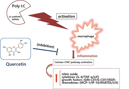
2.1. Effect of Quercetin on NO Production and Intracellular Calcium Release

NO is a major inflammatory mediator during the immuno-inflammatory reaction. NO concentration in culture medium can be determined by the Griess reaction. In this study, RAW 264.7 mouse macrophages were incubated with poly(I:C) and/or quercetin for 24 h, and 100 µL of supernatant from each well was mixed with 100 µL Griess reagent in a 96-well plate to determine NO concentration. The results show that quercetin significantly inhibited excessive production of NO in the cells at concentrations of 5, 10, 25, and 50 µM (Figure 2). No association was found between quercetin concentration and NO level. The inhibitory effect of 50 µM quercetin stronger than that at 5 and 10 µM. Therefore, quercetin concentrations of 10, 25, and 50 µM were chosen for the multiplex cytokine assay, and concentrations of 25 and 50 µM were chosen for the STAT mRNA expression evaluation.
Intracellular calcium release is related with many cellular processes including the inflammatory reaction. Calcium release from RAW 264.7 mouse macrophages can be determined by the Fluo-4 assay. Because calcium is involved in a signaling pathway, the cells were incubated with molecules for 18 h in this study. After incubating the cells with poly(I:C) and/or quercetin for 18 h, the medium was removed, and the cells were incubated with the Fluo-4 dye loading solution. After incubation, fluorescence intensity of each well was determined spectrofluorometrically with excitation and emission filters of 485 nm and 535 nm, respectively. The data show that quercetin inhibited significantly calcium release from poly(I:C)-induced RAW 264.7 mouse macrophages at concentrations of 5, 10, 25, and 50 µM (Figure 2). No significant association was found between quercetin concentration and intracellular calcium level.
The NO production and calcium release results are shown as a percentage relative to the control group. Indomethacin was used as a positive control.

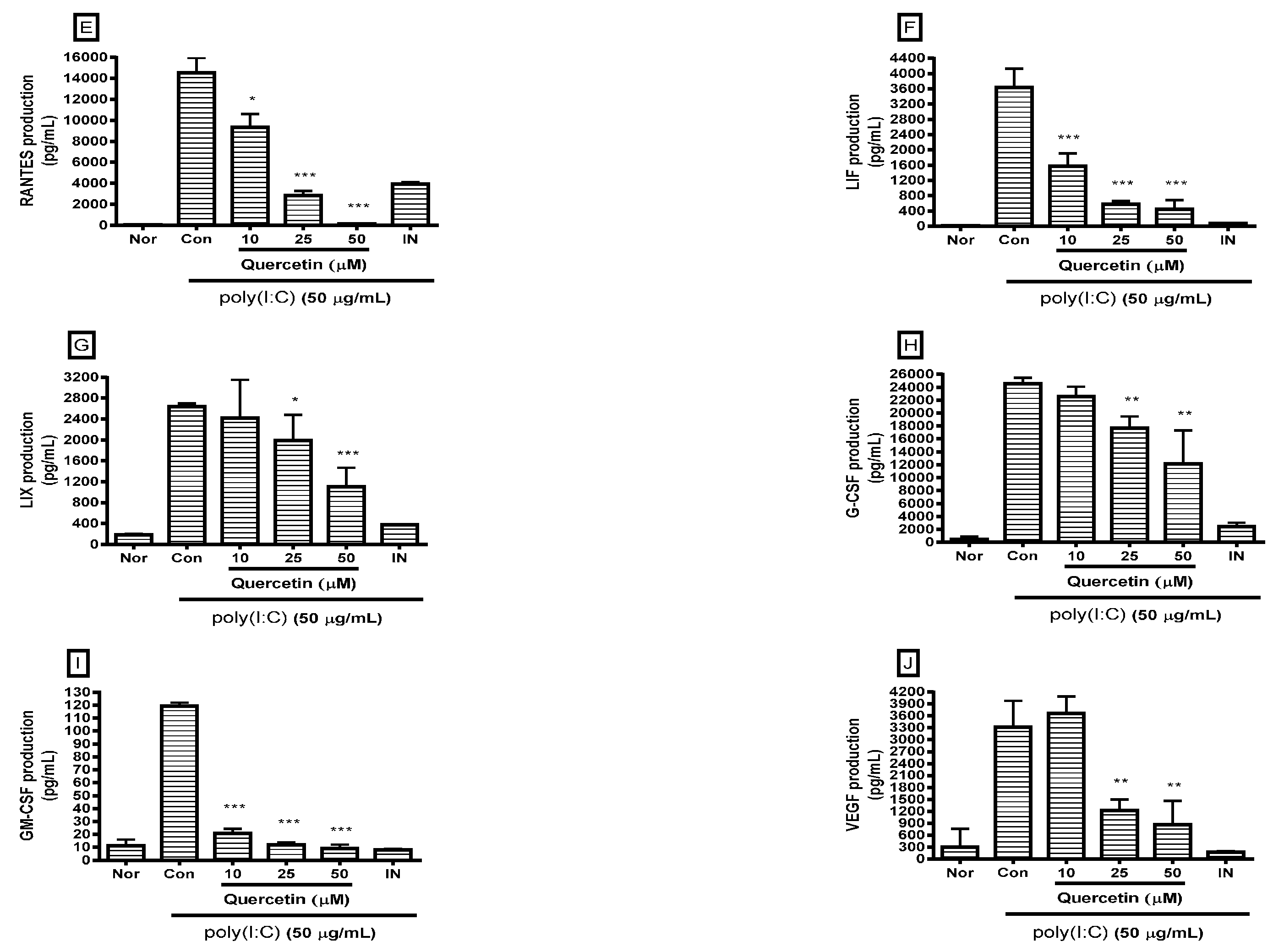
2.2. Effect of Quercetin on Cytokine Production

Cytokines are a distinct category of small (5–20 kDa) proteins that are important in inflammation and cell signaling. Cytokine production in culture medium was determined by the multiplex cytokine assay. After incubating the cells with poly(I:C) and/or quercetin for 24 h, the cytokines released from the cells were measured in cell culture supernatants using the Luminex assay based on xMAP technology. The result show that quercetin significantly reduced excess production of IL-6, TNF-α, MCP-1, interferon gamma-induced protein-10 (IP-10), regulated on activation, normal T cell expressed and secreted (RANTES), leukemia inhibitory factor (LIF), lipopolysaccharide-induced CXC chemokine (LIX), granulocyte-colony stimulating factor (G-CSF), granulocyte macrophage-colony stimulating factor (GM-CSF), and vascular endothelial growth factor (VEGF) in poly(I:C)-induced RAW 264.7 mouse macrophages (Figure 3).

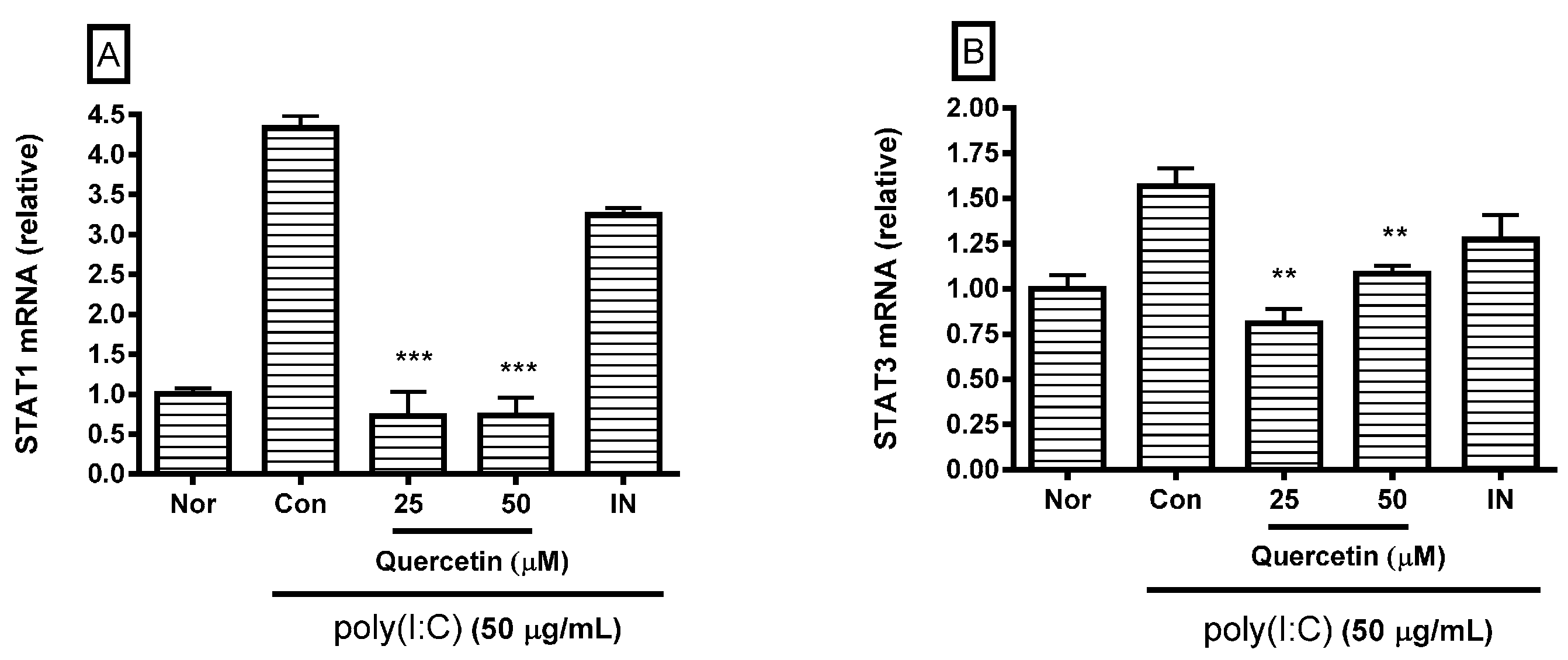
2.3. Effect of Quercetin on STAT1 and STAT3 mRNA Expression

Because STAT proteins are involved in a signaling pathway that activates macrophages, the cells were incubated with molecules for 18 h. After incubating the cells with poly(I:C) and/or quercetin for 18 h, real-time reverse-transcription polymerase chain reaction (RT-PCR) was performed to determine STAT1 (GenBank: NM_009283) and STAT3 (GenBank: NM_213659) mRNA expression. The GAPDH gene (GenBank: NM_001001303) was used for RNA normalization. The results show that quercetin significantly inhibited STAT1 and STAT3 mRNA expression in poly(I:C)-induced RAW 264.7 mouse macrophages at concentrations of 25 and 50 µM (Figure 4), indicating that quercetin modulates inflammatory reactions in poly(I:C)-induced macrophages via the calcium-STAT pathway.

3. Discussion

Inflammation in the immune system is essential to fight an infection and is a general term used to describe many diverse processes that tissues employ in response to infections by pathogens (such as bacteria, viruses, parasites, and fungi) and injuries caused by toxic molecules or physical damage such as burns or cuts [15].
Viral infections trigger anti-viral type I interferon responses by stimulating endosomal and cytosolic pattern recognition receptors, including Toll-like receptors, which are key initiators of inflammation [16]. Immunomodulatory approaches are being considered to combat a vast range of human and animal diseases including incurable viral diseases, cancers, autoimmune diseases, and inflammatory conditions [17]. Viral and bacterial infections exacerbate the immune system and increase morbidity of patients with chronic airways diseases; viral infections trigger an innate immune response through host detection of viral dsRNA via several different mechanisms, including activation of a member of the class of pattern recognition receptors, Toll-like receptor 3; lung inflammation associated with viral infections includes robust increase in cytokines, such as IL-1β, IL-6, IL-8, TNF-α, GM-CSF, IP-10, RANTES, and MIP-1α [18].
Among the many immuno-inflammatory leukocytes, macrophages and monocytes are of great importance [19]. Macrophages are found throughout all tissues and are a key component of the immune system. Recruitment of macrophages to specific sites is important for normal host defense [20], as they play an important role in inflammatory disease by releasing factors, such as NO, reactive oxygen species, inflammatory cytokines, chemokines, growth factors, and prostaglandin mediators involved in the immune response [21].
NO is a membrane-permeable signaling molecule involved in a broad array of biologic processes through its ability to modify proteins, lipids, and DNA and alter their functions and immunogenicity [22]. Appropriate levels of NO assist in mounting an effective defense against invading microbes, whereas the inability to generate NO results in serious, even fatal, susceptibility to infections. Furthermore, dysregulation or overproduction of NO has been implicated in the pathogenesis of many disorders, including atherosclerosis, neurodegenerative diseases, inflammatory autoimmune diseases, and cancers; therefore, the potential exists for NO to behave like a “double-edged” biological sword depending on it level. Thus, it is crucial to understand regulation of NO [23].
Some studies show that the regulation of inflammation is important for maintaining homeostasis [3]. Inflammation plays a critical defensive role in the human body, however, uncontrolled or aberrant inflammatory responses contribute to various acute and chronic inflammatory diseases [24]. Inflammation helps restore homeostasis after an insult but can be more damaging than the insult itself if uncontrolled, excessive, or prolonged [25]. For example, dysregulation of the inflammatory/immune responses is responsible for multiple organ failure in patients with sepsis, for which over expression of proinflammatory cytokines is a major mechanism [26]. It is well known that viral and bacterial infections contribute to the pathogenesis of severe sepsis, which is characterized by an overwhelming production of NO and proinflammatory cytokines, such as IL-6 [27]. Despite antibiotics, sepsis remains a clinical challenge, with high mortality rates and increasing prevalence [28]. The expression level of RANTES increases in lung tissue infected with influenza virus [29]. The robust, uncontrolled cytokine production, otherwise known as hypercytokinemia or a “cytokine storm”, is thought to initiate the severe systemic inflammatory response in various infections, such as those caused by influenza virus, cytomegalovirus, variola virus, severe acute respiratory syndrome coronavirus, and avian H5N1 influenza virus [30]. Thus, it is important to focus on therapeutics to modulate virus-induced hyperinflammation.
The poly(I:C)-induced activation of macrophages is regarded as an experimental model of viral inflammation in vitro. Although many studies on biological effects of quercetin have been reported, the effect of quercetin on poly(I:C)-induced macrophages has not been fully reported. The present study shows that quercetin may alleviate poly(I:C)-induced inflammation by decreasing levels of inflammatory mediators, such as NO, cytokines, chemokines, and growth factors, in RAW 264.7 mouse macrophages; quercetin is a candidate for modulating virus-hyperinflammation such as a cytokine storm.
Oxidative stress reduces endoplasmic reticulum (ER) calcium stores and increases intracellular calcium concentrations, resulting in ER stress-mediated STAT1 activation. Preapoptotic ER stress itself has proinflammatory effects in macrophages [31]. In the present study, quercetin significantly inhibited calcium release and STAT1 and STAT3 mRNA expression in poly(I:C)-induced RAW 264.7 mouse macrophages. Thus, quercetin might modulate poly(I:C)-induced macrophage activation via the calcium-STAT pathway.
Finally, the present study demonstrated that quercetin has anti-inflammatory effects related with inhibiting NO, IL-6, TNF-α, MCP-1, IP-10, RANTES, LIF, LIX, G-CSF, GM-CSF, and VEGF production in poly(I:C)-induced macrophages via the calcium-STAT pathway. Further study is needed to evaluate the clinical utility of quercetin for viral inflammation.

4. Materials and Methods

4.1. Materials

DMEM, fetal bovine serum, penicillin, streptomycin, PBS, and other tissue culture reagents were purchased from Gibco BRL (Grand Island, NY, USA). Quercetin, indomethacin, Griess reagent, and all other chemicals were purchased from Sigma-Aldrich (St. Louis, MO, USA).

4.2. Quantification of NO Production and Intracellular Calcium Release

NO concentration in culture medium was determined by the Griess reaction. Specifically, after incubating the cells with poly(I:C) and/or quercetin for 24 h, 100 µL of supernatant from each well was mixed with 100 µL Griess reagent in a 96-well plate. After a 15 min incubation at room temperature, optical density was determined at 540 nm with a microplate reader (Bio-Rad, Hercules, CA, USA) [32].
Calcium release from RAW 264.7 mouse macrophages was determined by the Fluo-4 assay. RAW 264.7 mouse macrophages in 96-well plates were incubated with poly(I:C) and/or quercetin for 18 h at 37 °C. Thereafter, the medium was removed, and the cells were incubated with 100 µL of the Fluo-4 dye loading solution (Molecular Probes, Eugene, OR, USA) for 30 min at 37 °C. After the incubation, fluorescence intensity in each well was determined spectrofluorometrically (Dynex, West Sussex, UK) with excitation and emission filters of 485 nm and 535 nm, respectively [33].

4.3. Multiplex Bead-Based Cytokine Assay

After 24 h treatment with poly(I:C) and/or quercetin, the cytokines released from treated cells were measured in cell culture supernatants using a Luminex assay based on xMAP technology. This assay was performed with Milliplex kits (Millipore, Billerica, MA, USA) and the Bio-Plex 200 suspension array system (Bio-Rad) as described previously [34,35,36]. Standard curves for each cytokine were generated using the kit-supplied reference cytokine samples.

4.4. RNA Isolation and Real Time RT-PCR Analysis

At the end of the 18 h incubation with poly(I:C) and/or quercetin, RAW 264.7 mouse macrophages were lysed and total RNA was isolated using the NucleoSpin RNA kit (Macherey-Nagel, Duren, Germany) according to the manufacturer’s instructions. RNA quantity and quality were confirmed using the Experion RNA StdSens Analysis kit (Bio-Rad) and Experion Automatic Electrophoresis System (Bio-Rad). cDNA was synthesized from 1 µg total RNA using the iScript cDNA Synthesis kit (Bio-Rad). Real-time RT-PCR was performed using the iQ SYBR Green Supermix (Bio-Rad). The GAPDH gene was used for RNA normalization. The analysis was performed on a Bio-Rad CFX 96 Real-Time PCR Detection System (Bio-Rad). Relative changes in gene expression were calculated using the comparative threshold cycle (Ct) method (Bio-Rad) [12]. The following primers were used: STAT1 (GenBank: NM_009283), sense: 5′-TGAGATGTCCCGGATAGTGG-3′, antisense: 5′-CGCCAGAGAGAAATTCGTGT-3′; STAT3 (GenBank: NM_213659), sense: 5′-GTCTGCAGAGT TCAAGCACCT-3′, antisense: 5′-TCCTCAGTCACGATCAAGGAG-3′; GAPDH (GenBank: NM_0010 01303), sense: 5′-AACCTGCCAAGTATGATGAC-3′, antisense: 5′-GGGAGTTGCTGTTGAAGT-3′.

4.5. Statistical Analysis

The results shown are summarized from three independent experiments and represent mean ± standard deviation. Significant differences were examined using one-way analysis of variance test followed by Tukey’s multiple comparison test with SPSS 11.0 software (SPSS Inc., Chicago, IL, USA). In addition, a correlation analysis was conducted to examine the relationships between quercetin concentration and the levels of NO and/or intracellular calcium. A p-value < 0.05 was considered significant.

5. Conclusions

In conclusion, this study demonstrated that quercetin has anti-inflammatory effects related with inhibiting NO, IL-6, TNF-α, MCP-1, IP-10, RANTES, LIF, LIX, G-CSF, GM-CSF, and VEGF production in poly(I:C)-induced macrophages via the calcium-STAT pathway. Further study is needed to evaluate the clinical utility of quercetin for viral inflammation.

Acknowledgments

The authors thank Ji Young Lee and Hyun Joo Kim (College of Korean Medicine, Gachon University) for their technical assistance.

Author Contributions

Y.K. and W.P. conceived and designed the experiments; Y.K. performed the experiments; W.P. analyzed the data; and W.P. wrote the manuscript.

Conflicts of Interest

The authors declare no conflicts of interest.

Abbreviations

The following abbreviations are used in this manuscript:
NOnitric oxide
dsRNAdouble-stranded RNA
TLRToll like receptor
poly(I:C)polyinosinic-polycytidylic acid
LPSlipopolysaccharide
ILinterleukin
TNFtumor necrosis factor
MCPmonocyte chemotactic protein
IP-10interferon inducible protein-10
LIXlipopolysaccharide-induced CXC chemokine
LIFleukemia inhibitory factor
G-CSFgranulocyte colony-stimulating factor
GM-CSFgranulocyte macrophage colony-stimulating factor
VEGFvascular endothelial growth factor
STAT1signal transducers and activators of transcription 1
STAT3signal transducers and activators of transcription 3
Nornormal
Concontrol
ERendoplasmic reticulum

References

  1. Bartemes, K.R.; Iijima, K.; Kobayashi, T.; Kephart, G.M.; McKenzie, A.N.; Kita, H. IL-33-Responsive Lineage- CD25+ CD44(Hi) Lymphoid Cells Mediate Innate Type 2 Immunity and Allergic Inflammation in the Lungs. J. Immunol. 2012, 188, 1503–1513. [Google Scholar] [CrossRef] [PubMed]
  2. Gilroy, D.W. Eicosanoids and the Endogenous Control of Acute Inflammatory Resolution. Int. J. Biochem. Cell Biol. 2010, 42, 524–528. [Google Scholar] [CrossRef] [PubMed]
  3. Legatzki, A.; Rösler, B.; von Mutius, E. Microbiome Diversity and Asthma and Allergy Risk. Curr. Allergy Asthma Rep. 2014, 14, 1–9. [Google Scholar] [CrossRef] [PubMed]
  4. Eo, H.J.; Park, J.H.; Park, G.H.; Lee, M.H.; Lee, J.R.; Koo, J.S.; Jeong, J.B. Anti-Inflammatory and Anti-Cancer Activity of Mulberry (Morus Alba L.) Root Bark. BMC Complement. Altern. Med. 2014, 14, 200. [Google Scholar] [CrossRef] [PubMed]
  5. O’Shea, J.J.; Ma, A.; Lipsky, P. Cytokines and Autoimmunity. Nat. Rev. Immunol. 2002, 2, 37–45. [Google Scholar] [CrossRef] [PubMed]
  6. Cho, Y.; Challa, S.; Moquin, D.; Genga, R.; Ray, T.D.; Guildford, M.; Chan, F.K. Phosphorylation-Driven Assembly of the RIP1-RIP3 Complex Regulates Programmed Necrosis and Virus-Induced Inflammation. Cell 2009, 137, 1112–1123. [Google Scholar] [CrossRef] [PubMed]
  7. Hasan, M.; Ruksznis, C.; Wang, Y.; Leifer, C.A. Antimicrobial Peptides Inhibit Polyinosinic-Polycytidylic Acid-Induced Immune Responses. J. Immunol. 2011, 187, 5653–5659. [Google Scholar] [CrossRef] [PubMed]
  8. Arsenault, R.J.; Kogut, M.H.; He, H. Combined CpG and Poly I:C Stimulation of Monocytes Results in Unique Signaling Activation Not Observed with the Individual Ligands. Cell Signal. 2013, 25, 2246–2254. [Google Scholar] [CrossRef] [PubMed]
  9. Anjaneyulu, M.; Chopra, K. Quercetin, an anti-oxidant Bioflavonoid, Attenuates Diabetic Nephropathy in Rats. Clin. Exp. Pharmacol. Phys. 2004, 31, 244–248. [Google Scholar] [CrossRef] [PubMed]
  10. Liao, Y.R.; Lin, J.Y. Quercetin Intraperitoneal Administration Ameliorates Lipopolysaccharide-Induced Systemic Inflammation in Mice. Life Sci. 2015, 137, 89–97. [Google Scholar] [CrossRef] [PubMed]
  11. Gardi, C.; Bauerova, K.; Stringa, B.; Kuncirova, V.; Slovak, L.; Ponist, S.; Drafi, F.; Bezakova, L.; Tedesco, I.; Acquaviva, A.; et al. Quercetin Reduced Inflammation and Increased Antioxidant Defense in Rat Adjuvant Arthritis. Arch. Biochem. Biophys. 2015, 583, 150–157. [Google Scholar] [CrossRef] [PubMed]
  12. Guazelli, C.F.; Fattori, V.; Colombo, B.B.; Georgetti, S.R.; Vicentini, F.T.; Casagrande, R.; Baracat, M.M.; Verri, W.A., Jr. Quercetin-Loaded Microcapsules Ameliorate Experimental Colitis in Mice by Anti-Inflammatory and Antioxidant Mechanisms. J. Nat. Prod. 2013, 76, 200–208. [Google Scholar] [CrossRef] [PubMed]
  13. Chiow, K.H.; Phoon, M.C.; Putti, T.; Tan, B.K.; Chow, V.T. Evaluation of Antiviral Activities of Houttuynia Cordata Thunb. Extract, Quercetin, Quercetrin and Cinanserin on Murine Coronavirus and Dengue Virus Infection. Asian Pac. J. Trop. Med. 2016, 9, 1–7. [Google Scholar] [CrossRef] [PubMed]
  14. Wu, W.; Li, R.; Li, X.; He, J.; Jiang, S.; Liu, S.; Yang, J. Quercetin as an Antiviral Agent Inhibits Influenza A Virus (IAV) Entry. Viruses 2015, 8. [Google Scholar] [CrossRef] [PubMed]
  15. Gadient, R.A.; Patterson, P.H. Leukemia Inhibitory Factor, Interleukin 6, and Other Cytokines using the GP130 Transducing Receptor: Roles in Inflammation and Injury. Stem Cells 1999, 17, 127–137. [Google Scholar] [CrossRef] [PubMed]
  16. Smith, J.A. A New Paradigm: Innate Immune Sensing of Viruses Via the Unfolded Protein Response. Front. Microbiol. 2014, 5. [Google Scholar] [CrossRef] [PubMed]
  17. Mahima; Ingle, A.M.; Verma, A.K.; Tiwari, R.; Karthik, K.; Chakraborty, S.; Deb, R.; Rajagunalan, S.; Rathore, R.; Dhama, K. Immunomodulators in Day to Day Life: A Review. Pak. J. Biol. Sci. 2013, 16, 826–843. [Google Scholar] [PubMed]
  18. Cooper, P.R.; Lamb, R.; Day, N.D.; Branigan, P.J.; Kajekar, R.; San Mateo, L.; Hornby, P.J.; Panettieri, R.A., Jr. TLR3 Activation Stimulates Cytokine Secretion without Altering Agonist-Induced Human Small Airway Contraction Or Relaxation. Am. J. Physiol. Lung Cell Mol. Physiol. 2009, 297, L530–L537. [Google Scholar] [CrossRef] [PubMed]
  19. Kang, X.; Kim, H.J.; Ramirez, M.; Salameh, S.; Ma, X. The Septic Shock-Associated IL-10 –1082 A > G Polymorphism Mediates Allele-Specific Transcription Via Poly(ADP-Ribose) Polymerase 1 in Macrophages Engulfing Apoptotic Cells. J. Immunol. 2010, 184, 3718–3724. [Google Scholar] [CrossRef] [PubMed]
  20. Cammer, M.; Cox, D. Chemotactic Responses by Macrophages to a Directional Source of a Cytokine Delivered by a Micropipette. Methods Mol. Biol. 2014, 1172, 125–135. [Google Scholar] [CrossRef] [PubMed]
  21. Medina, E.A.; Morris, I.R.; Berton, M.T. Phosphatidylinositol 3-Kinase Activation Attenuates the TLR2-Mediated Macrophage Proinflammatory Cytokine Response to Francisella Tularensis Live Vaccine Strain. J. Immunol. 2010, 185, 7562–7572. [Google Scholar] [CrossRef] [PubMed]
  22. Oates, J.C. The Biology of Reactive Intermediates in Systemic Lupus Erythematosus. Autoimmunity 2010, 43, 56–63. [Google Scholar] [CrossRef] [PubMed]
  23. Karpuzoglu, E.; Ahmed, S.A. Estrogen Regulation of Nitric Oxide and Inducible Nitric Oxide Synthase (iNOS) in Immune Cells: Implications for Immunity, Autoimmune Diseases, and Apoptosis. Nitric Oxide 2006, 15, 177–186. [Google Scholar] [CrossRef] [PubMed]
  24. Lee, J.H.; Lee, K.; Su, Z.; Boyanapalli, S.S.; Barman, D.N.; Huang, M.; Chen, L.; Magesh, S.; Hu, L.; Kong, A.T. In Vitro and in Vivo Anti-Inflammatory Effects of a Novel 4, 6-Bis ((E)-4-Hydroxy-3-Methoxystyryl)-1-Phenethylpyrimidine-2 (1H)-Thione. Chem. Res. Toxicol. 2013, 27, 34–41. [Google Scholar] [CrossRef] [PubMed]
  25. Dos Santos, G.; Kutuzov, M.A.; Ridge, K.M. The Inflammasome in Lung Diseases. Am. J. Physiol. Lung Cell Mol. Physiol. 2012, 303, L627–L633. [Google Scholar] [CrossRef] [PubMed]
  26. Hu, Z.; Murakami, T.; Suzuki, K.; Tamura, H.; Kuwahara-Arai, K.; Iba, T.; Nagaoka, I. Antimicrobial Cathelicidin Peptide LL-37 Inhibits the LPS/ATP-Induced Pyroptosis of Macrophages by Dual Mechanism. PLoS ONE 2014, 9, e85765. [Google Scholar] [CrossRef] [PubMed]
  27. Ulloa, L.; Tracey, K.J. The “Cytokine Profile”: A Code for Sepsis. Trends Mol. Med. 2005, 11, 56–63. [Google Scholar] [CrossRef] [PubMed]
  28. Sessler, C.N.; Shepherd, W. New Concepts in Sepsis. Curr. Opin. Crit. Care 2002, 8, 465–472. [Google Scholar] [CrossRef] [PubMed]
  29. Wareing, M.D.; Lyon, A.B.; Lu, B.; Gerard, C.; Sarawar, S.R. Chemokine Expression during the Development and Resolution of a Pulmonary Leukocyte Response to Influenza A Virus Infection in Mice. J. Leukoc. Biol. 2004, 76, 886–895. [Google Scholar] [CrossRef] [PubMed]
  30. Tisoncik, J.R.; Korth, M.J.; Simmons, C.P.; Farrar, J.; Martin, T.R.; Katze, M.G. Into the Eye of the Cytokine Storm. Microbiol. Mol. Biol. Rev. 2012, 76, 16–32. [Google Scholar] [CrossRef] [PubMed]
  31. Timmins, J.M.; Ozcan, L.; Seimon, T.A.; Li, G.; Malagelada, C.; Backs, J.; Backs, T.; Bassel-Duby, R.; Olson, E.N.; Anderson, M.E.; et al. Calcium/calmodulin-Dependent Protein Kinase II Links ER Stress with Fas and Mitochondrial Apoptosis Pathways. J. Clin. Investig. 2009, 119, 2925–2941. [Google Scholar] [CrossRef] [PubMed]
  32. Lee, J.Y.; Park, W. Anti-Inflammatory Effect of Myristicin on RAW 264.7 Macrophages Stimulated with Polyinosinic-Polycytidylic Acid. Molecules 2011, 16, 7132–7142. [Google Scholar] [CrossRef] [PubMed]
  33. Lee, J.Y.; Park, W.; Yi, D.K. Immunostimulatory Effects of Gold Nanorod and Silica-Coated Gold Nanorod on RAW 264.7 Mouse Macrophages. Toxicol. Lett. 2012, 209, 51–57. [Google Scholar] [CrossRef] [PubMed]
  34. Yoon, S.; Lee, Y.; Park, S.K.; Kim, H.; Bae, H.; Kim, H.M.; Ko, S.; Choi, H.Y.; Oh, M.S.; Park, W. Anti-Inflammatory Effects of Scutellaria Baicalensis Water Extract on LPS-Activated RAW 264.7 Macrophages. J. Ethnopharmacol. 2009, 125, 286–290. [Google Scholar] [CrossRef] [PubMed]
  35. Yuk, S.S.; Lim, E.M.; Lee, J.Y.; Lee, Y.J.; Kim, Y.S.; Lee, T.H.; Park, S.K.; Bae, H.; Kim, H.M.; Ko, S.G.; et al. Antiinflammatory Effects of Epimedium Brevicornum Water Extract on Lipopolysaccharide-Activated RAW264.7 Macrophages. Phytother. Res. 2010, 24, 1781–1787. [Google Scholar] [CrossRef] [PubMed]
  36. Lee, J.Y.; Park, W. Anti-Inflammatory Effect of Wogonin on RAW 264.7 Mouse Macrophages Induced with Polyinosinic-Polycytidylic Acid. Molecules 2015, 20, 6888–6900. [Google Scholar] [CrossRef] [PubMed]
  • Sample Availability: Quercetin samples are available from the authors.
Figure 1. Chemical structure of quercetin.
Molecules 21 00450 g001
Figure 2. Effect of quercetin on nitric oxide (NO) production (A) and calcium release (B) by dsRNA-induced RAW 264.7 mouse macrophages. After 24 h treatment, NO production was measured by the Griess reaction assay. Calcium release was measured with the Fluo-4 calcium assay after an 18 h treatment. The normal group (Nor) was treated with media only. The control group (Con) was treated with 50 µg/mL of the dsRNA synthetic analog polyinosinic-polycytidylic acid (poly(I:C)) alone. IN, indomethacin (0.5 µM). Values are mean ± standard deviation of three independent experiments. * p < 0.05 vs. Con; ** p < 0.01; *** p < 0.001. Significant differences were examined using one-way analysis of variance test followed by Tukey’s multiple comparison test.
Molecules 21 00450 g002
Figure 3. Effect of quercetin on production of cytokines, such as interleukin (IL)-6 (A); tumor necrosis factor (TNF)-α (B); monocyte chemoattractant protein-1 (MCP-1) (C); interferon gamma-induced protein-10 (IP-10) (D); RANTES (E), leukemia inhibitory factor (LIF) (F); lipopolysaccharide-induced CXC chemokine (LIX) (G); granulocyte-colony stimulating factor (G-CSF) (H); granulocyte macrophage-colony stimulating factor (GM-CSF) (I); and vascular endothelial growth factor (VEGF) (J) in dsRNA-induced RAW 264.7 mouse macrophages. Flourescene intensity of each cytokine in the culture medium was measured by a multiplex bead-based cytokine assay after the 24 h incubation. The normal group (Nor) was treated with media only. The control group (Con) was treated with 50 µg/mL of the dsRNA synthetic analog polyinosinic-polycytidylic acid (poly(I:C)) alone. IN, indomethacin (0.5 µM). Values are mean ± standard deviation of three independent experiments. * p < 0.05 vs. Con; ** p < 0.01; *** p < 0.001. Significant differences were examined using one-way analysis of variance test followed by Tukey’s multiple comparison test.
Molecules 21 00450 g003aMolecules 21 00450 g003b
Figure 4. Effect of quercetin on signal transducer and activated transcription 1 (STAT1) (A) and STAT3 mRNA expression (B) in dsRNA-induced RAW 264.7 mouse macrophages. After an 18 h treatment, STAT1 and STAT3 mRNA expression was measured by real-time reverse-transcription polymerase chain reaction assay. STAT1 and STAT3 mRNA were normalized to GAPDH mRNA. The normal group (Nor) was treated with media only. The control group (Con) was treated with 50 µg/mL of the dsRNA synthetic analog polyinosinic-polycytidylic acid (poly(I:C)) alone. IN, indomethacin (0.5 µM). Values are the mean ± standard deviation of three independent experiments. ** p < 0.01 vs. Con; *** p < 0.001. Significant differences were examined using one-way analysis of variance test followed by Tukey’s multiple comparison test.
Molecules 21 00450 g004

