Review

Nanoparticles: Alternatives Against Drug-Resistant Pathogenic Microbes

by
Gudepalya Renukaiah Rudramurthy
1,
Mallappa Kumara Swamy
2,*,
Uma Rani Sinniah
2,* and
Ali Ghasemzadeh
2
1
Department of Biotechnology, East-West College of Science, Bangalore-560091, Karnataka, India
2
Department of Crop Science, Faculty of Agriculture, Universiti Putra Malaysia, Serdang, Selangor, Darul Ehsan 43400, Malaysia
*
Authors to whom correspondence should be addressed.
Molecules 2016, 21(7), 836; https://doi.org/10.3390/molecules21070836
Submission received: 15 March 2016 / Revised: 17 June 2016 / Accepted: 20 June 2016 / Published: 27 June 2016
(This article belongs to the Collection Nanomedicine)

Abstract

:
Antimicrobial substances may be synthetic, semisynthetic, or of natural origin (i.e., from plants and animals). Antimicrobials are considered “miracle drugs” and can determine if an infected patient/animal recovers or dies. However, the misuse of antimicrobials has led to the development of multi-drug-resistant bacteria, which is one of the greatest challenges for healthcare practitioners and is a significant global threat. The major concern with the development of antimicrobial resistance is the spread of resistant organisms. The replacement of conventional antimicrobials by new technology to counteract antimicrobial resistance is ongoing. Nanotechnology-driven innovations provide hope for patients and practitioners in overcoming the problem of drug resistance. Nanomaterials have tremendous potential in both the medical and veterinary fields. Several nanostructures comprising metallic particles have been developed to counteract microbial pathogens. The effectiveness of nanoparticles (NPs) depends on the interaction between the microorganism and the NPs. The development of effective nanomaterials requires in-depth knowledge of the physicochemical properties of NPs and the biological aspects of microorganisms. However, the risks associated with using NPs in healthcare need to be addressed. The present review highlights the antimicrobial effects of various nanomaterials and their potential advantages, drawbacks, or side effects. In addition, this comprehensive information may be useful in the discovery of broad-spectrum antimicrobial drugs for use against multi-drug-resistant microbial pathogens in the near future.

Graphical Abstract

1. Introduction

Antimicrobial agents kill or inhibit the growth of a wide range of microbes such as bacteria (antibacterials), fungi (antifungals), and viruses (antivirals). Antimicrobials may be synthetic, may be of plant or animal origin, or may be chemically modified natural compounds [1], and can have a significant impact on the outcome of an infected patient/animal. They are used in the treatment (chemotherapy) and prevention (prophylaxis) of infections. Many infectious diseases have been combatted since the discovery of antimicrobial drugs in the 1960s [2]. The history of antimicrobial agents dates back to 1928, when penicillin was discovered; however, it did not come into use until 1942. Penicillin was the first antibiotic to be used in medicine [3], and the management of life-threatening bacterial infections improved significantly after its discovery. Moreover, the discovery of penicillin, which is considered one of the most significant advances in medicine, started what is known as “the antibiotic revolution” [3,4,5]. Antimicrobials can be classified into one of several categories such as disinfectants, antiseptics, or antibiotics. Disinfectants and antiseptics are extensively and commonly used in hospitals and healthcare units, and are essential for the control and prevention of microbial infections. Various active chemical agents/biocides are used in the preparation of antiseptics and disinfectants. Antiseptics destroy (cidal) or inhibit (static) the growth of microorganisms in or on living tissue, whereas disinfectants, which are similar to antiseptics, are used on inanimate objects or surfaces.
The mode of action of antibiotics varies and includes the inhibition of cell wall synthesis, inhibition of protein synthesis, inhibition of DNA replication, and inhibition/alteration of intermediary metabolism [6]. The penetration of antimicrobials into the cell is necessary if the target for antimicrobial action is located inside the bacterial cell wall. Hence, antimicrobial agents must be capable of penetrating to the site of action. Penetration through the cell wall and/or membrane is usually achieved by passive or facilitated diffusion, or by an active transport mechanism. However, the presence of lipopolysaccharide-lipoprotein complexes in the cell wall of Gram-negative organisms prevents many antibiotics from reaching the sensitive intracellular targets. Some antibacterial agents use aqueous transmembrane channels (porins) in the outer membrane to gain entry into Gram-negative organisms. Peptidoglycan, which forms a rigid layer, is similar in both Gram-positive and Gram-negative bacteria with some differences. However, Gram-positive organisms possess a very thick peptidoglycan coat (cross-linked with interpeptide bridges) and Gram-negative organisms have a very thin peptidoglycan layer. Many antibiotics, such as penicillins, cephalosporins, fosfomycin, bacitracin, cycloserine, vancomycin, and teicoplanin, selectively inhibit the synthesis of the peptidoglycan layer at different stages [6]. Antimicrobials such as ionophores interfere with the transport of cations through the cell membrane. Gramicidin A, monensin, and valinomycin disturb cation transport, and antimicrobial peptides, such as defensins, cecropins, and magainins have ionophoric properties [7,8,9,10]. The intracellular targets for antimicrobials include protein synthesis and DNA replication. Antibiotics such as puromycin, chloramphenicol, tetracyclines, aminoglycosides, fusidic acid, lincosamides, macrolides, streptogramins, mupirocin, and oxazolidinones interfere with the process of protein synthesis. Modulation of DNA supercoiling by topoisomerases is an essential step in DNA replication. Several antibiotics target topoisomerases and inhibit DNA synthesis, thereby combatting many bacterial infections. The inhibitors of DNA synthesis include quinolones, novobiocin, rifampicin, diaminopyrimidines, sulfonamides, and 5-nitroimidazoles [11]. However, microorganisms revealed a remarkable capability to adapt, survive, and evolve by developing resistance to antimicrobial compounds. The improper use of antimicrobial agents has led to the development of new resistance mechanisms followed by the global spread of resistant organisms, which threatens the effective treatment of common infectious diseases. One of the greatest challenges for practitioners is the emergence of multi-resistant bacteria, such as methicillin-resistant Staphylococcus aureus (MRSA) and vancomycin-resistant enterococci; this constitutes a global health threat. The health risk for patients infected with resistant bacteria is higher than those infected with non-resistant bacteria, and it results in prolonged illness and higher expenditure. Furthermore, the major concern/risk with the development of antimicrobial resistance is the spread of resistant organisms. MRSA strains, in hospitals or from clinical sources, have recently been detected in workers involved in animal production as well as within community settings [12].
The development of antimicrobial resistance may be caused by: (a) alteration or inactivation of the drug; (b) reduced binding capacity of the drug due to alteration in the binding sites; (c) reduction in the antimicrobial effect due to modification of the metabolic pathways; or (d) decreased permeability and/or increased active flux leading to reduced intracellular accumulation of antimicrobial agents [13]. However, antimicrobial resistance may be intrinsic or acquired; it can develop through the mutation of existing genes [14,15], or through the transfer of genes from other species or strains [16,17]. Antimicrobial resistance can be detected by growth inhibition assays in broth or agar disc diffusion [13]. Culture-based assays may be fast or slow depending on the doubling time of microbes. However, culture-based assays are not suitable for the vast majority of microbes that cannot grow outside the host, such as Mycobacterium leprae, Treponema pallidum, Corynebacterium diphtheria, Bartonella henselae, Tropheryma whippelii, and noroviruses. Molecular detection techniques such as diagnostic polymerase chain reaction (PCR) assays [18,19], quantitative PCR [20], and DNA microarrays [21,22] significantly improved disease diagnosis and the identification of resistance genes. Moreover, antibiotic resistance genes from unculturable microbes can be identified through metagenomics. The authors of several earlier studies report the identification of resistance genes such as those that encode β-lactams and bleomycin [23,24,25]. Fighting antibiotic resistance is a major priority in human and animal health. Several strategies are available to overcome antibiotic resistance, including a reduction in the extensive use of antimicrobials, collection and analysis of data, avoiding the inappropriate use of antimicrobials in farm animals, development of novel drugs, and nanotechnology [26,27]. Advances in nanotechnology have led to the synthesis of nano-sized organic and inorganic molecules with potential applications in industry, food packaging, textiles, medicine, and therapeutics. The development of novel nanoscale antimicrobial agents/nanocomposites can be used as an alternative strategy to overcome antimicrobial resistance [27].
The advent of nanotechnology, the biggest engineering innovation of recent times, has modernized medicine. The demand for nanotechnology-derived products is constantly increasing. Nanotechnology, which is the innovative technology in the present scenario, can have a profound influence on improving human health. Enhanced durability, performance, strength, flexibility, and the inimitable physicochemical properties of nanomaterials have been explored in the health industry. Nanomaterials can be used in treatment modalities including targeted drug delivery, prognostic visual monitoring of therapy, and even the detection of tumors [28,29]. However, continuous exposure of humans to nanoparticles (NPs) in the work place can cause unpredictable human health risks. Moreover, indirect exposure to NPs occurs when they are inhaled as air pollutants. Inhaled NPs sometimes evade the immune system and are distributed throughout the body, causing systemic health problems. The pollution of air by NPs is even detrimental to other biological species in the environment and disturbs the ecosystem [30]. Serious health problems due to ambient or occupational exposure may arise if these issues are not addressed in a cohesive and concerted manner by industries, governments, and scientists. In light of this, the present review was conducted to present complete information on the antimicrobial activity of different types of nanomaterials. In addition, we emphasized the application of NPs/nanocomposites in combating antimicrobial drug resistance. This review is a compiled survey of data retrieved from many search engines including ScienceDirect, Google Scholar, PubMed, Scopus, and SciFinder.

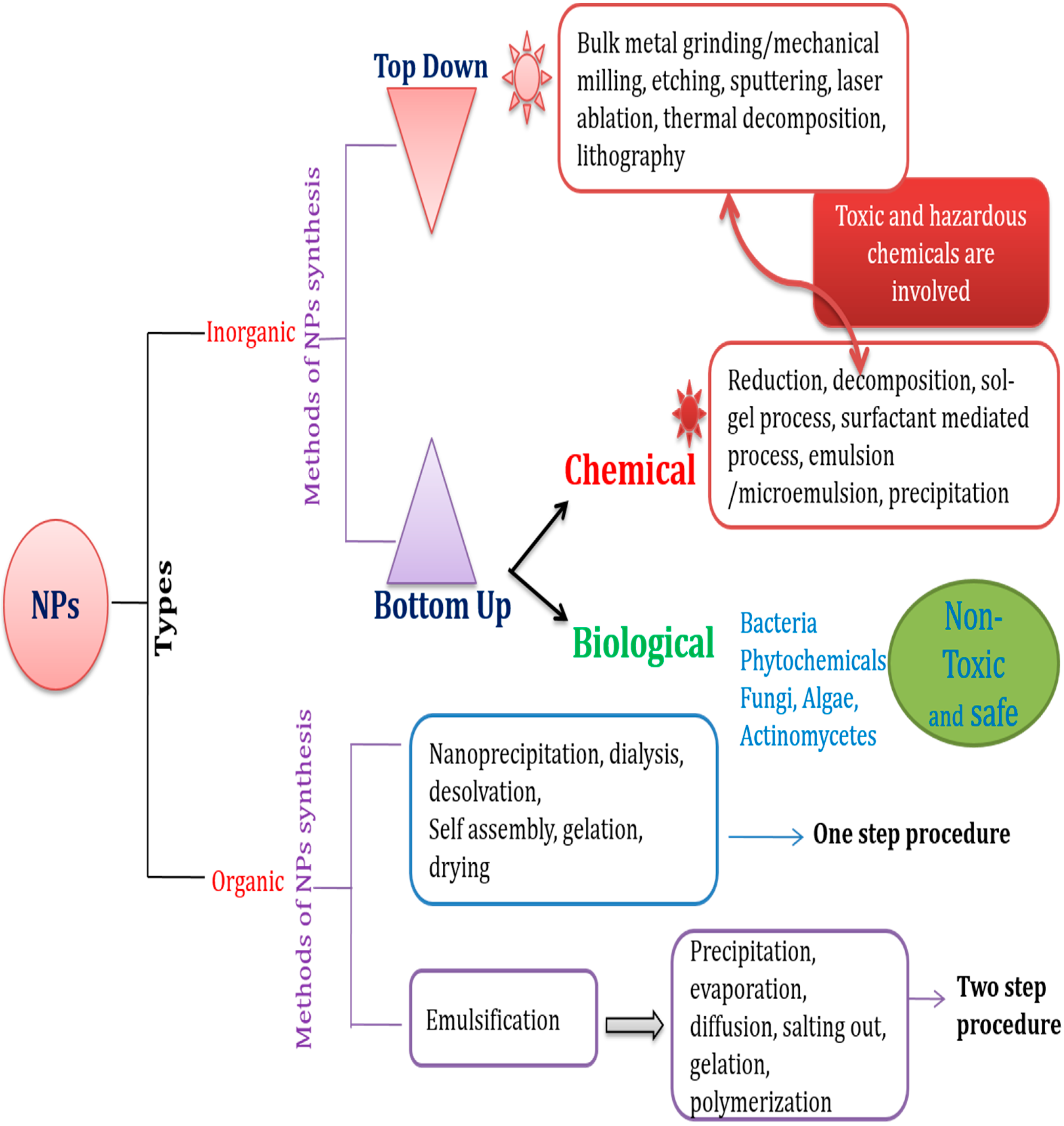
2. Nanoparticles/Nanocomposites

Antimicrobial drug resistance has prompted the development of several alternative strategies. Among these strategies, nanoscale materials/nanocomposites have emerged as significant and novel antimicrobial agents. Nanomaterials, typically 0.2–100 nm in size, have a high surface-to-volume ratio [31]; this increases their interaction with microorganisms, which in turn improves their antimicrobial activity. Transmission electron microscopy (TEM), low-resolution TEM (LRTEM), and high-resolution TEM (HRTEM) have helped in the characterization of NPs and revolutionized their use in various fields. The chemical, electrical, mechanical, optical, magnetic, and electro-potential properties of NPs differ from those of their bulk materials. This may be attributed to their high surface-to-volume ratio [32,33]. The physicochemical and biological properties of NPs can be manipulated according to the desired application [31,34]. NPs may be organic or inorganic; however, inorganic NPs are used more often owing to their ability to withstand adverse reaction conditions [31]. NPs have been used in optical, chemical, and biological fields. Their potential applications include many specific areas such as superconductors, optical devices, catalysts, fuel cells, gene and drug delivery, cell and tissue imaging, and biosensors [35,36,37,38,39]. Moreover, NPs have antimicrobial properties and have potential for use in diagnostic immunoassays [40,41,42]. Several types of NPs, including various metal and metal oxides, have been developed and evaluated by different research groups; examples include silver (Ag), gold (Au), Ag oxide (Ag2O), zinc oxide (ZnO), titanium dioxide (TiO2), calcium oxide (CaO), copper oxide (CuO), magnesium oxide (MgO), and silicon dioxide (SiO2) [39]. NPs act on microbes by several different methods and the mode of action of these NPs varies with each different type (Table 1 and Figure 1). Several bacterial strains are capable of adhering to any natural or artificial surface, and can even form biofilms on these surfaces. Many different factors are responsible for the adhesion and formation of biofilms by bacteria; these include the production of slime-like substances, electrostatic interactions, dipole-dipole and H-bond interactions, hydrophobic interactions, and van der Waals interactions. Therefore, nanomaterials that are used as antimicrobial agents must reduce microbial adhesion and biofilm formation. Hence, screening NPs for their anti-adhesion capability would increase their potential as antimicrobial agents. A schematic representation describing the various methods for the synthesis of different NPs is shown in Figure 2.

2.1. Inorganic NPs with Antibacterial and Antifungal Activities

Several inorganic metals and their oxides, including Ag, TiO2, CuO, iron oxide (Fe3O4), and ZnO, have been studied for their antimicrobial activities.

2.1.1. Silver NPs (AgNPs)

AgNPs are synthesized by physical, chemical, and biological methods. The physical method, which is also known as the “top-down” method, involves grinding the bulk metal, whereas the chemical method, widely called the “bottom-up” method, involves reduction, electrochemical processes, and decomposition by ultrasonic waves [43,44,45]. However, the synthesis of AgNPs by physical and chemical processes involves the use of toxic and hazardous chemicals, and the process is extremely expensive. The biological method, which is a “bottom-up” approach, exploits bacteria, fungi, and plant extracts to synthesize NPs. Recently, biologically synthesized NPs have received a great deal of attention, mainly in the field of biomedicine [46]. The biological method involves oxidation or reduction reactions by enzymes produced by microorganisms, or by phytochemicals. Several bacteria, fungi, and plants including Pseudomonas stutzeri, Bacillus megaterium, Escherichia coli [47,48,49], Aspergillus fumigatus, Fusarium solani [50,51], Aloe vera, Piper betle leaf, Leptadenia reticulata, and Momordica cymbalaria, have been explored for use in the synthesis of AgNPs [27,44,52,53,54,55,56]. The size of the AgNPs synthesized by biological methods varies between 1 and 600 nm [27,43,54].
The antimicrobial activities of Ag, Ag ions (Ag+), and Ag compounds have been known for many centuries. Ag has broad-spectrum antimicrobial activity against bacteria, fungi, and viruses, which is termed “oligodynamic activity”. Ag and its compounds undergo ionization in water and/or in body fluids, and the bioactive Ag+ interact with proteins and amino acids. Microorganisms are highly susceptible to the toxic effect of Ag+ and Ag compounds [57]. The mechanism of antimicrobial activity of Ag+ involves interference with the electron transport chain and the transfer of energy through the membrane, because Ag has an affinity for the sulfhydryl (thiol) groups in cell wall enzymes [57,58]. Ag+ also inhibit DNA replication and the respiratory chain in bacteria and fungi. However, the antimicrobial activity of Ag and its compounds is directly proportional to the number of biologically active Ag+ released, and its availability for interaction with the bacterial cell wall [57]. AgNPs are a good source of antimicrobial agents and possess antioxidant, anti-inflammatory, anticancer, and antiangiogenic activities [27,31,44,55]. The bactericidal activity of AgNPs against several pathogenic bacteria has been investigated by many research groups [27,31,43,44,54,59,60,61,62,63]. At present, AgNPs are widely considered an alternative antibacterial agent to Ag+. This is because the effects of Ag+ have a limited duration. AgNPs exhibit superior antimicrobial properties mediated by the synthesis of reactive oxygen species (ROS) including hydrogen peroxide [43,45,55]. Furthermore, the larger surface-to-volume ratio of AgNPs allows increased interactions with the cell membrane and easy penetration into the cell, leading to complete destruction of microbial cells compared with Ag+ [57,64].
Silver ions have a high affinity for sulfur and phosphate groups, which might explain their antimicrobial activity. Ag+ released from NPs react with sulfur-containing proteins, mainly on the cell surface, and phosphorous-containing nucleic acids. They are known to produce ROS inside the cell, eventually leading to cell death [31,57,59,92]. Nucleic acid damage and the alteration of the bacterial cell wall brought about by the attachment of AgNPs are considered the major reasons for bacterial cell death [45,92]. The size and shape of NPs play a significant role in their antimicrobial activity. AgNPs with a diameter of ≤10 nm form pores in the cell wall leading to the death of an organism [31,43,44,54,57]. The minimum inhibitory concentration (MIC) varies with the size of the NPs. The MIC of NPs smaller than 25 nm is 6.75–54 μg/mL, whereas 25-nm particles have a lower MIC of 1.69–13.5 μg/mL against methicillin-resistant Staphylococcus aureus and Staphylococcus epidermidis, and vancomycin-resistant Enterococcus faecium and Klebsiella pneumonia [31,57]. A Gram-positive bacterium, S. aureus, was effectively inhibited by AgNPs at higher concentrations (100 μg/mL) [60]. In addition, Rupareli et al. [82] observed strain-specific variations in the MIC/minimum bactericidal concentration (MBC) of E. coli when treated with AgNPs. The MIC values ranged from 40 to 180 μg/mL for different strains of E. coli (MTCC 443, MTCC 739, MTCC 1302, and MTCC 1687).
According to Lara et al. [93], AgNPs exhibited high bactericidal activity against multidrug-resistant Pseudomonas aeruginosa, ampicillin-resistant E. coli O157:H7, and erythromycin-resistant Streptococcus pyogenes. The MIC was 83.3 mM for P. aeruginosa and E. coli O157:H7, whereas it was 83.3 mM for S. pyogenes. They also suggested the possible use of AgNPs as a potential antimicrobial agent in medical devices, pharmaceutical products, and in the nosocomial environment. Likewise, Morones et al. [65] reported the antibacterial activity of AgNPs against Gram-negative bacteria, such as E. coli, P. aeruginosa, V. cholera, and S. typhus. Many other pathogenic bacteria are susceptible to AgNPs; they include Acinetobacter baumannii, Enterococcus faecalis, Klebsiella pneumoniae, Listeria monocytogenes, Micrococcus luteus, Proteus mirabilis, Salmonella typhi, Enterobacter aerogenes, Bacillus subtilis, Brucella abortus, Moraxella catarrhalis, Proteus mirabilis, Streptococcus viridans, Streptococcus pneumonia, Streptococcus mutans, Serratia proteamaculans, and Shigella flexneri [27,43,45,53,54,57,59,94]. More recently, it has been reported that AgNPs kill S. mutans isolated from clinical samples, suggesting their use for the treatment of dental caries [94]. Furthermore, fungal infections markedly contribute to increasing the mortality and morbidity of immunocompromised patients. Several studies have shown that AgNPs act as a potential antifungal agent. The antifungal activity of AgNPs is influenced by their size and zeta potential. Moreover, the mechanism of inhibition against fungi varies with particle size. AgNPs were found to be effective against various fungal pathogens, including Candida albicans, Candida tropicalis, Trichophyton rubrum, Penicillium brevicompactum, Cladosporium cladosporioides, Aspergillus fumigatus, Chaetomium globosum, Mortierella alpina, and Stachybotrys chartarum [95,96,97,98].

2.1.2. Magnesium Oxide (MgO) NPs

Inorganic metal oxides such as MgO, ZnO, and CaO are stable under harsh processing conditions and are generally considered safe for humans [99]. Moreover, MgO does not require photoactivation for its antimicrobial activity [100,101,102,103]. Various strategies have been developed for the synthesis of MgONPs that are similar to those used to develop AgNPs. The regulation of processing conditions allows the synthesis of MgONPs of various sizes and with different morphologies [104,105]. Several mechanisms have been proposed to explain the antibacterial activity of MgONPs, which include the formation of ROS, lipid peroxidation, electrostatic interactions, and alkaline effects. The strong electrostatic interaction between the bacterial cell surface and the MgONPs leads to the death of the bacteria [101]. The surface of MgONPs has a typically high pH due to the formation of a thin layer of water. When bacteria contact MgONPs, the high pH damages the bacterial cell membrane (the alkaline effect), ultimately leading to death. However, the antibacterial efficacy of MgONPs is size-dependent owing to changes in the surface energy. Particles smaller than 15 nm exhibit higher bactericidal activity than large, aggregated MgONPs [106,107]. MgONPs exhibit antibacterial activity against Gram-positive and Gram-negative bacteria such as S. aureus (MIC; 1000 μg/mL), E. coli (MIC; 500 μg/mL), and P. aeruginosa (MIC; 1000 μg/mL) [66]. MgONPs synthesized by the aerogel procedure exhibit biocidal activity against vegetative forms of Gram-positive and Gram-negative bacteria, and against spores, and can also be used as a potent disinfectant [67,108]. Moreover, MgONPs exhibit antimicrobial activity against E. coli, Bacillus megaterium, and B. subtilis [109].

2.1.3. Titanium Dioxide (TiO2) NPs

TiO2 is a non-toxic and chemically stable molecule with optical properties [109]. TiO2 NPs (TiO2NPs) can be used in pharmaceuticals, cosmetics, whiteners, food colorants, toothpaste, and to protect the skin against UV rays [110]. Moreover, they have significant antibacterial activity against certain microbes [111]. Several methods are available for the synthesis of TiO2 NPs including sol-gel and electrochemical techniques. Like other metals or metal oxides, TiO2 acts on bacteria through the generation of ROS. The oxidative stress on the crystal surfaces of anatase TiO2 generates ROS. The surface of anatase TiO2 reacts with water by photocatalysis and releases the hydroxyl radicals, which subsequently form superoxide radicals [112]. The ROS synergistically act on phospholipids (polyunsaturated) on the surface of bacteria [113], causing site-specific DNA damage [68,69]. The photocatalytic activity of TiO2 can be exploited in the preparation of biofilms, which have many applications such as the disinfection of contaminated surfaces in food processing industries. The antimicrobial activity of photocatalytic TiO2 against E. coli, S. aureus, and fungi was reported [31]. Researchers reported the light-induced biocidal activity of engineered TiO2 NPs against E. coli [114] and Aspergillus niger [115].

2.1.4. Zinc Oxide (ZnO) NPs

The U.S. Food and Drug Administration listed ZnO as “generally recognized as safe” (GRAS) [116]. The application of ZnO NPs (ZnONPs) depends on their shape, size, surface state, crystal structure, and dispensability. Several different techniques have been developed for the synthesis of ZnONPs, which include mechanochemical, precipitation, emulsion, and microemulsion processes. ZnONPs occur as different structures such as nanorods, needles, helixes, springs, rings, nanoplates/nanosheets, nanopellets, flowers, dandelions, and snowflakes. The mechanism of ZnONP antimicrobial activity involves the generation of hydrogen peroxide and the release of Zn2+ ions. ZnO generates highly reactive oxygen species such as OH and hydrogen peroxide (H2O2). Hydrogen peroxide generated on the surface of ZnO can penetrate the bacterial cells and effectively inhibit cell growth [70,71]. However, OH and superoxides are likely to remain on the surface of the cell because they cannot penetrate the cell membrane owing to their negative charge. The generation of hydrogen peroxide increases with the increasing surface area of the ZnONPs. However, Zn2+ ions released from the NPs damage the cell membrane and interact with intracellular components [72]. A wide range of Gram-positive and Gram-negative bacteria, including major foodborne pathogens, is susceptible to ZnONPs. Previous studies have shown that ZnONPs exhibit antibacterial activity against E. coli, Listeria monocytogenes, Salmonella, and Staphylococcus aureus [73,74]. In a study by Vidic et al. [117], ZnO nanostructures (100 nm) showed effective antibacterial activity against both Gram-positive (B. subtilis) and Gram-negative (E. coli) bacteria. However, at a lower concentration of 1 mg/mL, 100% inhibition was observed. Likewise, Reddy et al. [118] reported the inhibition of E. coli (~13 nm) by ZnONPs at ≥3.4 mM concentration, while S. aureus was completely inhibited at ≥1 mM concentration. Polyethylene glycol (PEG)-capped ZnONPs at above 5 mM concentration showed antibacterial activity against E. coli [119]. Later, Li et al. [120] suggested that the toxicity of ZnONPs arises mostly from the labile zinc complexes and free zinc ions. Pati et al. [121] reported the potential application of ZnONPs as antimicrobials against S. aureus. The mechanism of the ZnONP antimicrobial activity is related to the disruption of the bacterial cell membrane integrity, the diminishing cell surface hydrophobicity, and the downregulation of the transcription of oxidative stress resistance genes in bacteria. ZnONPs also induce ROS production to augment intracellular bacterial death. In another study, the MIC of ZnONPs for Campylobacter jejuni was reported at 0.05 to 0.025 mg/mL concentration [116].

2.1.5. Iron Oxide (Fe3O4) NPs

The intrinsic properties of iron-containing NPs increase their scientific, technological, and industrial value. Fe3O4 NPs are used in biosensors, food preservation agents, antimicrobial agents, magnetic refrigeration and storage media, ferrofluids, anti-cancer agents, magnetic resonance imaging (MRI), targeted drug delivery, and cell sorting. The biological compatibility and magnetic properties of Fe3O4 NPs make them attractive for applications in biomedical research [122,123]. Several different approaches are available for the synthesis and characterization of Fe3O4 NPs, including techniques such as sol-gel and forced hydrolysis, co-precipitation, hydrothermal processing, surfactant-mediated synthesis, laser pyrolysis, electrochemical processing, and microemulsion processing. Techniques such as absorption spectrophotometry, X-ray diffraction, and scanning electron microscopy (SEM) are available for the characterization of the synthesized NPs. The mode of antimicrobial action of Fe3O4 NPs might be through ROS, oxidative stress, superoxide radicals (O2), singlet oxygen (1O2), hydroxyl radicals (OH), or hydrogen peroxide (H2O2) [83]. The antimicrobial activity of Fe3O4 NPs against various bacteria including S. aureus, S. epidermidis, E. coli, Xanthomonas, and P. vulgaris has been established by several groups [124,125,126,127]. Chen et al. [123] demonstrated that immunoglobulin G-bound Fe3O4/titania core/magnetic shell NPs effectively inhibit the growth of various pathogenic multi-antibiotic-resistant bacteria such as Staphylococcus saprophyticus, S. pyogenes, and MRSA. Furthermore, Arokiyaraj et al. [128] evaluated the antimicrobial efficiency of Fe3O4 NPs, Argemone Mexicana L. plant leaf extract and Fe3O4 NPs treated with plant leaf extract against bacterial pathogens. Interestingly, they observed a considerable inhibition of E. coli MTCC 443 and P. mirabilis MTCC 425 strains by Fe3O4 NPs treated plant extract. According to Anghel et al. [129], Fe3O4 NP-coated textile dressings inhibit biofilm formation by C. albicans more than uncoated textile dressings. Moreover, Fe3O4 NPs coated with Rosmarinus officinalis essential oil had potent inhibitory activity against biofilm-forming C. albicans and C. tropicalis [130].

2.1.6. Gold (Au) NPs

Historically, colloidal Au was thought to have healing properties when consumed orally, and it is the earliest recognized form of AuNPs. The unique optical properties of AuNPs make them attractive potential tools in biomedicine. AuNPs are inert, biologically compatible, and have a high surface-to-volume ratio. The properties of AuNPs are very different from Au in its bulk form [131]. AuNPs can be prepared in a variety of different shapes or geometries such as nanorods, nanocages, nanocubes, and nanotriangles, which affect their optical features [132,133,134,135]. AuNPs in the size range 0.8–250 nm are regarded as the most popular NPs. However, nanoshells, which range in size from 80 to 150 nm, and smaller nanoshells (20–60 nm) containing a core of Fe3O4 nanocrystals, have also been extensively explored in biomedicine [136,137]. AuNPs can be synthesized by both chemical and biological methods. Chemical methods include the reduction of tetrachloroauric acid to produce colloidal AuNPs (size range 10–60 nm), the Brust–Schiffrin method for thiolated AuNP synthesis (size range 1–6 nm), Au nanoshell synthesis, and the seed-mediated method for the synthesis of nanorods (size range 1–2 nm) [133,134]. Though several chemical methods are available for synthesis, they are expensive and involve toxic chemicals. To overcome the limitations of chemical methods, several groups have developed economically feasible and eco-friendly biological methods for the synthesis of AuNPs [138]. Several researchers have used a biological method to synthesize AuNPs of various sizes and shapes, and tested their antimicrobial activity. For instance, they synthesized AuNPs using the bacteria Rhodopseudomonas capsulata or the fungus C. albicans, or used plant extracts as the reducing and stabilizing agents [138,139,140,141,142,143]. AuNPs are biologically inert, but they can be modified so that they have various functional groups, such as chemical or photothermal functionalities. Au nanorods are reported to have anti-cancerous and antimicrobial activity following photo-thermal heating [31]. AuNPs in combination with photosensitizers such as toluidine blue O are reported to exhibit antimicrobial activity against MRSA [75,76,77]. Biomolecules such as carbohydrates, antibodies, proteins, and oligonucleotides can be attached to AuNPs as functional moieties [144]. The addition of functional moieties increases the antimicrobial efficacy of NPs and several such modifications have been developed. AuNPs conjugated with specific antibodies have been reported to kill S. aureus (photothermally, using lasers) [145]. Antibiotics such as vancomycin, used to kill vancomycin-resistant enterococci [146,147], and aminoglycosidic antibiotics that act on both Gram-positive and Gram-negative bacteria [78,148,149], have been added to AuNPs. AuNPs act on bacteria through the generation of holes in the cell wall, which eventually lead to cell death due to the leakage of cell contents. Moreover, AuNPs can bind to DNA and inhibit the transcription process by preventing the uncoiling of DNA during transcription [78]. Khan et al. [150] reported the possible use of AuNPs (21 ± 2.5 nm and 0.2 mg/mL) conjugated with methylene blue (20 μg/mL) for preventing the formation of biofilm by the common nosocomial refractory fungus C. albicans. Several multidrug-resistant uropathogens, namely E. coli, E. cloacae complex, P. aeruginosa, S. aureus, and S. aureus-MRSA, were completely inhibited by AuNPs at nanomolar (8–32 nM) concentrations [151]. Mixed ligand-coated AuNPs have shown 99.9% growth inhibition against methicillin-susceptible S. aureus at a concentration of 10 μM [152]. Likewise, AgNPs synthesized biologically from the fungus Trichoderma viride showed MIC values of 40, 1.5, and 8 µg/mL against E. coli ATCC 8739, vancomycin-sensitive S. aureus ATCC 6538, and vancomycin-resistant S. aureus, respectively [153]. Au nanospheres conjugated with gentamycin showed enhanced antibacterial effect (0.0937 mg/mL MIC) against S. aureus compared with free gentamicin (0.18 mg/mL MIC) [154]. In a recent study, stable biofabricated AuNPs conjugated with gentamycin, ciprofloxacin, rifampicin, and vancomycin effectively inhibited S. epidermidis and Staphylococcus haemolyticus compared with antibiotics alone [155]. Dasari et al. [156] reported the effectiveness of AuNPs and Au ion complexes against three multidrug-resistant bacteria, namely E. coli, Salmonella typhimurium DT104, and S. aureus.

2.1.7. Copper Oxide (CuO) NPs

Copper and its compounds have wide potential applications in several fields owing to their wide range of physical properties, namely, superconductivity, high thermal conductivity, spin dynamics, and electron correlation effects. CuO is a semiconducting compound. Its monoclinic structure has photoconductive and photocatalytic or photovoltaic properties [31,157]. Several methods have been reported for the synthesis of CuONPs, which include laser irradiation, γ-radiolysis, thiol-induced reduction, reverse micelles, and green synthesis [79,158,159]. Several studies have reported that copper NPs exhibit antibacterial activity against Gram-positive bacteria, including B. subtilis and S. aureus, and Gram-negative bacteria, including E. coli [80,82]. Copper NPs synthesized by a biological process were reported to have antibacterial activity against the human pathogens E. coli and S. aureus [80]. The mechanism of the antibacterial activity of CuONPs involves the adhesion of NPs to bacterial cell walls, owing to opposite electric charges, which results in reduction at the cell wall of the bacteria. In addition, Cu2+ ions generate ROS resulting in oxidative stress-induced DNA and membrane damage to the bacteria [81,160,161]. Moreover, CuONPs have a strong affinity for the amines and carboxyl groups present on the cell surface of B. subtilis, which might explain their high antimicrobial activity against such organisms. More recently, many pathogens such as Klebsiella aerogenes, Pseudomonas desmolyticum, E. coli (Gram-negative) and S. aureus (Gram-positive) have been effectively inhibited using CuONPs synthesized from Gloriosa superba L. plant extracts [162]. Likewise, Khashan et al. [163] reported the antibacterial effect of CuONPs against E. coli, P. aeruginosa, P. vulgaris, and S. aureus. CuONPs, when combined with fluconazole, have shown improved antifungal activity against C. albicans [161].

2.1.8. Aluminum (Al) NPs

Aluminum oxide (Al2O3) or alumina, generally referred to as corundum (the crystalline form of alumina), is a white oxide with several phases: alpha, gamma, delta, and theta. Alpha-phase AlNPs are thermodynamically stable over a wide temperature range. In AlNPs, oxygen atoms adopt hexagonal close packing, and in the octahedral sites Al3+ ions fill two-thirds of the lattice to form a corundum-like structure [164]. Several different techniques are available for the synthesis of alumina NPs such as sol-gel pyrolysis, hydrothermal processing, sputtering, and laser ablation. The laser ablation technique is a rapid and high-purity process; hence, it is most widely used in the preparation of Al2O3 NPs [165]. Al2O3 and AlNPs have a wide range of applications in industry and medicine. Few studies have addressed the antimicrobial properties of AlNPs. According to Jing et al. [166], AlNPs had higher toxicity against B. subtilis, E. coli, and Pseudomonas fluorescens than their bulk materials. Similarly, alumina NPs have shown higher sensitivity and mutagenicity against P. fluorescens than the bulk materials [167]. AlNPs disrupt bacterial cell walls leading to cell death through ROS [168]. Sadiq et al. [84] reported that alumina NPs exhibit a growth-inhibitory effect on E. coli over a wide concentration range (10–1000 μg/mL).

2.1.9. Bismuth (Bi) NPs

Bismuth, a diamagnetic, crystalline, and brittle metal, is typically found as bismuth sulfide (bismuthinite), bismuth oxide (bismite), and bismuth carbonate (bismuthite) [169]. Bismuth and its compounds exhibit antimicrobial activity against various bacteria. Bismuth compounds are commonly employed in the treatment of gastrointestinal disorders. The antimicrobial activity of elemental bismuth is observed at relatively high concentrations owing to its limited water solubility. However, effective antimicrobial activity at lower concentrations can be achieved by increasing the solubility of bismuth with chelating agents such as dimercaptopropanol. Bismuth-dimercaptopropanol has high solubility and decreases antimicrobial activity for short periods. Hence, the slow dissolution of bismuth-dimercaptopropanol would enable antimicrobial activity for an extended period [170]. Bismuth NPs (BiNPs) are synthesized from commercial bismuth salts using surface modifiers and a suitable reducing agent. BiNPs exhibit antifungal, antibacterial, and antiviral activity. Earlier studies by Hernandez et al. [171,172] reported that BiNPs exhibit antibacterial (<1 mM) and antifungal (2 mM) activity at lower concentrations. BiNPs have potential in the treatment of drug-resistant bacteria [85], and inhibit the growth of Helicobacter pylori through alteration of the Krebs cycle and amino acid and nucleotide metabolism [86]. BiNPs are promising candidates for fighting many infectious diseases. However, their safe use in humans requires evaluation.

2.1.10. Carbon-Based NPs

Carbon-based nanomaterials play a significant role in biomedical applications such as tissue regeneration, advanced imaging, and drug or gene delivery. Several different types of carbon-based NPs have been developed including fullerenes, carbon nanotubes, and graphene oxide. The physical orientation of the carbon-based NPs and their particular use, depends on the conditions used to synthesize them [173]. The size and surface area of carbon-based NPs are relevant to their antibacterial activity; increased antimicrobial activity is associated with increased nanoparticle surface area and decreased size. Moreover, the antimicrobial activity also depends on the composition, intrinsic properties, and surface modification of the NPs [87,174]. The mode of action of carbon-based NPs includes severe damage to the bacterial membrane through oxidative stress [175,176] and/or physical interaction [88,176], the inhibition of energy metabolism [89,177], or impairment of the respiratory chain [178]. Direct contact between carbon nanomaterials and bacterial cells due to aggregation can also lead to cell death [87,88]. Carbon nanotubes are active against E. coli [87]. Single-walled carbon nanotubes (SWNTs) and multi-walled carbon nanotubes (MWNTs) have shown antimicrobial activity against both Gram-positive and Gram-negative bacteria owing to cell wall damage and the subsequent release of cell DNA [179]. Moreover, SWNTs exhibit antimicrobial activity against Salmonella enterica, E. coli, and E. faecium [90]. Fullerenes, which are ball-shaped carbon NPs, have antimicrobial activity against E. coli, Salmonella, Streptococcus spp., and Shewanella oneidensis [180,181]. Graphene, a two-dimensional crystal, and graphene oxide were shown to have an inhibitory effect on E. coli and S. aureus [182]. Carbon NPs complexed with Ag were found to have effective antimicrobial activity against MRSA, multidrug-resistant Acinetobacter baumannii, Burkholderia cepacia, Yersinia pestis, and Klebsiella pneumoniae [91]. The size of carbon nanotubes plays a significant role in governing their antibacterial properties [87,183,184]. Well-characterized SWNTs possess higher antibacterial activity than MWNTs. High levels of stress-related gene expression products were observed in E. coli when treated with MWNTs or SWNTs by Kang et al. [184]. Later, Vecitis et al. [185] proposed a possible antimicrobial mechanism of SWNTs involving the initial contact of SWNTs with bacteria (E. coli) followed by the perturbation of the cell membrane and electronic structure-dependent bacterial oxidation.

2.2. Organic NPs

Organic antimicrobials are considered less stable in nature compared with inorganic antimicrobials. Organic materials are highly susceptible to harsh process conditions, mainly at higher temperatures; this may lead to difficulties in designing and synthesizing organic NPs. However, several polymeric organic NPs that exhibit antimicrobial activity have been developed, including quaternary ammonium compounds, quaternary phosphoniums, and alkyl pyridiniums [186]. Polymeric NPs work either by releasing potent antimicrobial agents or by contact killing. They are chemically stable, nonvolatile, and can bind to the surface of cells. However, they cannot easily permeate through biological membranes [187]. Several organometallic polymers containing metals in the backbone chain or in the pendant groups have been synthesized by varying the size of the alkyl groups (methyl, ethyl, butyl, and octyl) [186]. These organometallic polymers exhibit effective inhibition against both DNA and RNA viruses. Several peptides have been synthesized using hydrophilic (lysine) and hydrophobic (alanine, phenylalanine, and leucine) amino acids. Peptides synthesized using lysine and phenylalanine exhibit broad and effective antimicrobial activity against E. coli, P. aeruginosa, Serratia marcescens, and C. albicans [186,188].

2.2.1. Quaternary Ammonium Compounds

Quaternary ammonium compounds (QAC) include well-known disinfectants such as cetrimonium chloride, benzalkonium chloride, and stearalkonium chloride. The N-alkyl chain length determines the antimicrobial activity of QACs. Alkyl chains with 12–14 carbons are effective against Gram-positive bacteria, whereas alkyl chains with 14–16 carbons have significant antibacterial activity against Gram-negative bacteria [189]. The mode of action of QACs is through electrostatic interaction between the negatively charged bacterial membrane and the positively charged QAC. Following the electrostatic interaction, the structural proteins and enzymes of the bacterial membrane are denatured owing to the integration of the hydrophobic tail of the QAC into the hydrophobic membrane. Nanoscale materials immobilized with N-alkylated polyethyleneimine (PEI) have bactericidal activity against Gram-positive and Gram-negative bacteria (water and airborne), and fungi, including antibiotic-resistant strains, through cell membrane rupture [186]. Substitution of PEIs with various groups also results in activity against C. albicans. In a report by Wan and Yeow [190], amine-functionalized TiO2 and AuNPs were shown to have excellent antibacterial properties without external excitation. In another study, quaternary ammonium compounds functionalized with SiNPs were reported to have improved bactericidal activity against E. coli (96.6%), S. aureus (98.5%), and Deinococcus geothermalis (99.6%) in comparison with pristine SiNPs [191]. Likewise, QAC-PEI-based NPs completely inhibited the growth of S. aureus at 80 g/mL, and E. coli at 320g/mL [192].

2.2.2. Triclosan and Polysiloxanes

Triclosan exhibits antimicrobial activity against Corynebacterium [193], and is one of the most commonly used antimicrobial agents. Polysiloxanes are linear polymers of silicon oxide. Siloxane copolymers substituted with quaternary ammonium groups are effective against E. coli and S. aureus [194].

2.2.3. Chitosan

Chitosan NPs exhibit a wide spectrum of activity against bacteria, viruses, and fungi. The characteristic features of chitosan (biocompatibility, non-toxicity, low-immunogenicity, antibacterial activity, and its ability to enhance absorption) increase its applicability in various fields [186,195]. The antibacterial activity of chitosan NPs depends on various factors including the pH and solvent [186]. Chitosan NPs exhibit good antifungal activity against C. albicans and Fusarium solani. However, A. niger is relatively resistant to chitosan NPs. The zeta potential is believed to have an effect on the negatively charged microbial surface, and contributes to the antifungal effect of chitosan [196].

2.3. Antiviral Properties of NPs

Viruses are one of the leading causes of disease and death globally. They can infect all life forms from prokaryotes to eukaryotes. The emergence of new resistant viral strains has become a global challenge for physicians and scientists alike. Vaccination programs have eradicated many viral diseases such as smallpox and paralytic poliomyelitis. Nanotechnology offers intriguing opportunities to overcome the problems associated with antiviral drug resistance through the re-exploration of known antimicrobial compounds by manipulation of their size [197]. Metal NPs kill various viruses such as human immunodeficiency virus 1 (HIV-1) [198,199,200], the hepatitis B virus [201], the influenza virus [202,203,204], the Tacaribe virus [205], the monkeypox virus [206], the respiratory syncytial virus [207], and herpes simplex virus type 1 (HSV-1) [208,209]. Although the mechanism of action of NPs is not clearly understood, reports indicate that NPs prevent infection by blocking viral entry into cells by competing with cellular heparan sulfate, and interfering with the beginning of the viral replication cycle [197,199,200]. AgNP/chitosan composites have antiviral activity against the H1N1 influenza A virus; however, chitosan alone did not exhibit any antiviral activity [210]. The inhibitory effect of AgNPs was attributed to the inhibition of virus attachment to the cell surface, and might involve modification of the viral protein by denaturation of its disulfide-bonded domain [197]. AuNPs stabilized with PEG have antiviral activity against HIV-1 and inhibit virus fusion; however, the mechanism of action is unclear [211]. Research has demonstrated that AuNPs bind with gp120, inhibit viral entry, and prevent CD4 attachment [211]. A study by Broglie et al. (2015) demonstrated the antiviral activity of a Au/copper sulfide core/NP shell (Au/CuS/NPs) system against norovirus virus-like particles (VLPs) [212]. Au/CuS/NPs may work by inactivating VLPs by direct contact, then destroying the capsid. The authors of an earlier study reported that copper and copper alloys degrade the GII.4 human norovirus genome and destroy the capsid [213]. Hence, it is believed that the copper component of Au/CuS/NPs may play a significant role in its antiviral activity. Copper iodide (CuI) NPs exhibit excellent antiviral activity against feline calicivirus; this may be due to Cu(+) ROS generation followed by the oxidation of capsid proteins [214]. It has been reported that the nanocomposite comprising TiO2, poly-l-lysine (PL), and DNA/RNA (TiO2, PL, DNA/RNA) exhibits antiviral activity against influenza A virus (H3N2 strain), as demonstrated by antisense technology [215].

3. Biological Compatibility of Nanoparticles (NPs)

The biological compatibility, or biocompatibility, of nanomaterials is their ability to function within a host system such that the host responds in an appropriate way in specific situations. Advances in nanotechnology have made possible the use of engineered NPs in the treatment of diseases. However, prior to the use of NPs in biomedical applications, their biological compatibility must be established [216]. Direct contact with cells, tissues, the extracellular environment, and physiological systems can trigger a sequence of biological effects that can be either beneficial or destructive. The physicochemical properties of NPs determine their biological compatibility as well as their efficacy. Hence, the establishment of standard evaluation criteria for biological compatibility is required for the biomedical application of NPs. The biological compatibility or toxicity of NPs can be studied with respect to cytotoxicity, hemocompatibility, histocompatibility, and neurotoxicity. NPs come into direct contact with blood when they are used as vectors in gene delivery, drug delivery, or as biosensors. Hence, the evaluation of blood-NP compatibility is essential before they can be used. Several techniques including hemolysis, blood cell aggregation, and coagulation behavior studies are available for the evaluation of blood-NP compatibility [217]. Blood-NP compatibility depends on several factors such as size, structure, and the surface properties of the NPs [217]. The increased and/or widespread use of NPs in biomedicine raises concerns about their access to different tissues and organs. NPs play a significant role in targeted drug delivery and diagnostics, and they have been explored intensively. However, histocompatibility needs to be evaluated prior to their application. Several NPs including dendrimers, AuNPs, carbon nanotubes, and superparamagnetic Fe3O4 (SPIONs) have been evaluated for histocompatibility. Both in vitro and in vivo toxicity studies have revealed that SPIONs are biocompatible and do not exhibit any severe toxic effects [218]. Silica-based NPs, which are known to be highly compatible, enter the cell without affecting cell survivability [217].
AgNPs induce cell toxicity by releasing Ag+ that cause cell shrinkage, affect membrane integrity, and induce apoptosis [219,220,221]. Research clearly indicates that AgNPs release free radicals, which induce cellular and DNA damage [222]. In addition, AgNPs show immunotoxicity in rats [223]. However, AgNPs are biocompatible and non-toxic under certain experimental conditions, and they are suitable for biological applications [224]. An earlier study revealed that cells treated with AgNPs in the presence of hydrogen peroxide exhibited no DNA damage [225]. According to earlier studies, based on inhalation or oral exposure, there was no severe toxicity from AgNPs in rat organs in vivo [223,226,227,228]. This was attributed to low absorption of the AgNPs from the lung and gastrointestinal tract. Munger et al. [229] reported that in vivo oral exposure to a commercial AgNP solution resulted in no clinical changes in human metabolism, hematology, urine, and imaging morphology. In vitro studies have indicated that the cell toxicity of AgNPs arises at concentrations of 5–50 μg/mL [230]. AgNP-biopolymer composites showed good antibacterial activity with no toxicity against three eukaryotic cell lines, namely, mouse fibroblasts (NIH-3T3), human osteosarcoma cells (MG63), and human hepatocarcinoma cells (HepG2) [231]. A 3-(4,5-dimethylthiazol-2-yl)-2,5-diphenyltetrazolium bromide (MTT) assay and a cell adhesion test confirmed that Ag/chitosan nanocomposites are compatible with L-929 normal cells [232]. AuNPs are less harmful to RAW264.7 cells then AgNPs [233]. Negligible cytotoxicity was observed in the cell lines when treated with Fe3O4-AuNPs [234]. TiO2 NPs are reported to be non-toxic at low doses (5 mg/kg body weight) and efforts have been made to increase their cytocompatibility [235,236]. More recently, a review by Shi et al. [237] on the toxicity of mesoporous silica NPs emphasized their biocompatibility. Various studies on the immune compatibility of NPs have been discussed in detail elsewhere [235]. More recently, 4T1 cells (a murine breast cancer cell line) exposed to below 125 µg/mL of manganese ferrite (MnFe2O4) NPs showed biocompatibility [238]. According to Tomitaka et al. [239], various ferrite NPs (Fe3O4, ZnFe3O4, and NiFe3O4) above 100 μg/mL show toxicity against HeLa cell lines. Similarly, TiO2 NPs at lower concentrations (100 μg/mL) are considered harmless to humans based on in vivo studies [240]. CaFe2O4 NPs were shown to exhibit high toxicity when used at concentrations above 250 μg/mL [241].
The severity of NP cytotoxicity depends on the route of administration and the site of accumulation. Polymeric NPs, which are surface modifiable and are capable of sustained drug release, are considered biocompatible and are used in the treatment of various pulmonary diseases such as asthma, tuberculosis, and lung cancer [242,243]. Several reports have demonstrated the neurotoxic potential of NPs. However, the mechanism and the pathways by which NPs exert neurotoxicity remain largely unclear. The high degree of biological compatibility of NPs indicates that they interact with the system without causing any unacceptable toxic, carcinogenic, immunogenic, or thrombogenic reactions. The biological compatibility of NPs depends on many factors including their size, structure, and formulation [225,244]. Some of the most important factors that influence the biocompatibility of NPs are as follows. First, application: NPs used in a particular application may be toxic in specific tissue types. However, they may not produce the same effect in different applications/tissue types. Second, the exposure half-life of NPs, i.e., the intrinsic characteristics of NPs, does not exclusively determine their biocompatibility. Third, biological compatibility is a relative factor and depends on the risk–benefit ratio. Fourth, the lack of adequate data: at present, the understanding of the biological compatibility of NPs is limited owing to a lack of information [244]. Hence, more research is necessary to evaluate the biological compatibility of NPs in a tissue- and application-specific manner.

4. Biodegradability and Encapsulation of Nanoparticles

The internal digestion and subsequent clearance of NPs from the body is commonly referred to as biodegradability. Biodegradable NPs (BNPs) are more appropriate for biomedical applications than non-degradable NPs. The clearance of NPs from the body after they have fulfilled their function is highly significant. Non-biodegradable NPs may have toxic side effects caused by their accumulation in mononuclear phagocytic cells such as those found in the liver and spleen [244]. Advances in the development of BNPs have revolutionized medicine. Polymer-based BNPs are submicron-sized colloidal particles; they can harbor a therapeutic agent embedded/encapsulated within their polymeric matrix or adsorbed/conjugated onto their surface [245]. The characteristic features of BNPs, such as their bioavailability, their suitability for the controlled release of drugs, genes, and other bioactive agents, their high encapsulation capabilities, and their low toxicity, have increased their applicability to the site-specific delivery of vaccines, drugs, and many other bioactive molecules. Proteins, polysaccharides, and synthetic biodegradable polymers are the source materials for the synthesis of BNPs. The selection of basic polymers for the synthesis of BNPs depends on the design and end application of the NPs. Several other factors are also important, including the degree of biodegradability and biological compatibility, the surface characteristics and functionality, the size of the desired NPs, and the properties of the encapsulated drugs [246]. Several methods are available for the synthesis of BNPs, including the dispersion of preformed polymers, ionic gelation, and the polymerization of monomers.
Several different biodegradable polymer matrices have been extensively used in the preparation of BNPs; these include chitosans, poly-alkyl-cyanoacrylates (PAC), gelatins, poly-d,l-lactide-co-glycolides (PLGA), polylactic acids (PLA), and poly-ε-caprolactones (PCL). PLGA undergoes hydrolysis in the body to produce metabolically biodegradable products, namely lactic and glycolic acids. Several different methods, such as solvent evaporation, emulsification-diffusion, and nanoprecipitation, are available for the preparation of PLGA NPs [247]. PLGA NPs have been used in the development of nanovaccines, gene delivery systems, and protein- and peptide-based nanomedicines [247,248]. PLA is considered a biocompatible and biodegradable polymer, and it is broken down in the body to lactic acid. PLA NPs can be prepared by solvent displacement, solvent evaporation or diffusion, and salting-out. PCL undergoes hydrolysis under normal physiological conditions with minimal or no toxicity. Hence, PCL is a potential candidate polymer for the preparation of BNPs, with applications in long-term implantable devices and drug delivery [246]. Various techniques such as solvent displacement/evaporation and nanoprecipitation have been used to prepare PLC NPs [246]. Gelatin is a polyampholyte and is extensively used in medical and food products. Gelatin NPs may be prepared by emulsion or desolvation/coacervation methods. Gelatin NPs can be used for drug delivery and the controlled release of drugs. Moreover, these NPs are nontoxic, biodegradable, and bioactive. Upon biodegradation, PACs produce several toxic compounds that have the potential to damage or stimulate the central nervous system. Hence, the use of PAC polymers in humans is not authorized [246].
Macrophages/phagocytic cells are a significant part of the mononuclear phagocytic system, and are involved in the removal of foreign particles including NPs (which may be considered foreign particles) from the circulation. Hence, the surface modification of NPs is necessary to allow them to evade the immune system of the body and succeed in vivo [249]. Surface-modified NPs (modified with biomolecules) remain in the vascular system for a prolonged period. Hence, surface-modified NPs may reach their target site safely and rapidly in comparison with non-modified NPs [246]. Several different polymers are available for the surface modification of NPs, including PEG, polysorbate 80, polysorbate 20, dextran, and tocopheryl polyethylene glycol 1000 succinate. However, PEGylation is the most common technique. PEGylation is the process of grafting or adsorbing PEG onto the surface of NPs. This increases the blood circulation half-life of the NPs. An earlier study by Leroux et al. [250] demonstrated that PEGylated PLGA NPs interact less with mononuclear phagocytes and remain in the systemic circulation longer. A study by Tobio et al. [251] revealed the decreased conversion of PEG-PLA NPs into lactic acid compared with PLA NPs. Tocopheryl polyethylene glycol 1000 succinate-modified NPs exhibit increased adhesion towards tumor cell surfaces [246]. A nano-suspension of paclitaxel (an anticancer agent) bound to biologically compatible proteins such as albumin (the system is commercially known as Abraxane) has been approved for the treatment of breast cancer [252].

5. Nanoparticles and Delivery Systems

One of the major obstacles associated with the treatment of many diseases is the effective delivery of a therapeutic compound to the target site. Conventional approaches to drug delivery have several drawbacks and limitations such as poor biodistribution, limited effectiveness, and a lack of selectivity [253,254]. Controlled drug delivery systems may overcome the drawbacks and limitations of conventional approaches. Such systems transport the drug/molecule to the site of action, protect the drug from rapid clearance/degradation from the body, and reduce undesirable side effects. The site-specific delivery of a drug is achieved through the attachment of the therapeutic drug to the NPs, which act as carriers, known as nanocarriers. Nanocarriers are readily absorbed by cells owing to optimized physicochemical and biological properties; hence, they can be successfully used to deliver therapeutic/bioactive compounds [252,254]. Many different nanocarriers are available for the development of drug delivery systems, including liposomes, dendrimers, polymers, carbon materials, solid lipid NPs, silicon, and magnetic NPs. Polysorbate-coated NPs attached to doxorubicin, an anti-cancer drug, are capable of crossing the intact blood-brain barrier (BBB), and are used in the treatment of brain cancer [255]. Moreover, a multifunctional system, known as Probes Encapsulated by Biologically Localized Embedding (PEBBLE), in which a variety of unique agents/molecules are encapsulated on the surface of NPs to perform multiple functions, has been designed by Summer and Kopelman [256]. One molecule guides the PEBBLE to a tumor, a second molecule helps visualize the target (by MRI), while a third molecule delivers a drug to the nearby cancer cells [256].
NPs can also be used as vectors in gene delivery or as biosensors. It is well known that most viral genomes undergo point mutation and recombination, which may lead to the emergence of new resistant virus strains. The treatment of such diseases is difficult because available drugs become ineffective. In such a situation, antisense technology may allow the selection of conservative regions in virus genomes. Peptide analogues of nucleic acids (PNA), which form complementary base pairs with RNA, are one of the most suitable candidates for inhibiting gene expression in vivo [215]. The successful delivery of PNA into the target site inhibits the expression of a target gene. Amirkhanov et al. [215] prepared a nanocomposite comprising TiO2, PL, and DNA/RNA using an electrostatic fixation method. These nanocomposites are capable of penetrating eukaryotic cells and effectively delivering PNA. Such nanocomposites exhibit antiviral activity against influenza A virus (H3N2 strain); the results indicate that TiO2 acts as a carrier and plays a significant role in the delivery of PNA into the cells. Several different polymer-based NPs have been devised for the delivery of RNA; they include PLGA- and PLA-based NPs for in vitro RNA interference (RNAi) delivery [252,257]. Chitosan-based NPs with encapsulated quantum dots (QD) have been successfully used to deliver small interfering RNA (siRNA), targeting human epidermal growth factor receptor-2 (HER2/neu) [258]. Moreover, chitosan/QD NPs surface-labeled with the HER2 antibody have been used in the targeted delivery of HER2 siRNA to SKBR3 breast cancer cells, which overexpress HER2 [252]. NPs labeled with fluorescent markers, such as Cy-5, help visualize the uptake and accumulation of nanotubes. Moreover, smart superparamagnetic Fe3O4 particle conjugates may be used to target and locate brain tumors earlier and more accurately than is possible using current techniques [252,259].

6. Limitations

NPs can be synthesized by several different methods and do not exceed 100 nm in size. The size, chemical composition, and shape of these NPs depend on the method of synthesis. They may be organic or inorganic, and may comprise metal, polymer, ceramic, latex, or carbon-based particles [174]. However, the major challenge commonly encountered is to control the size and shape of monodispersed NPs with higher stability during synthesis. Interestingly, the interaction between NPs and living cells/tissues is affected by several factors, including their size, shape, and composition [217]. The ability of NPs to permeate is essential if they are to be useful as antimicrobial or drug-delivery agents [31]. The majority of the NPs synthesized to date have the capacity to permeate membrane cells and spread to different regions of the body, such as blood vessels, nerve cells, and the lymphatic vascular system. However, NPs have potential health threats owing to selective accumulation in different cells, tissues, and certain cellular structures [174,217].
Several groups have studied the effect of NPs on biological systems and have reported that exposure to NPs with a diameter less than 100 nm may pose known and unknown risks. Moreover, risks associated with NPs vary with the type of NP [260]. NPs may escape the immune defense mechanism of the body owing to their small size and may cause inflammatory and/or toxic responses [217]. Experimental studies have revealed the relationship between pulmonary inflammation and toxic responses to ultrafine particles. The high surface area of these fine particles allows them to interact with cellular structures and is believed to increase oxidative stress. The ROS generated by NPs are major contributors to inflammation and toxicity, inducing oxidative stress, apoptosis, and the activation of signaling pathways, which may lead to the development of many conditions, such as pulmonary diseases. Moreover, exposure to NPs cause lung inflammation [261,262]. The translocation of ultrafine particles to extrapulmonary sites such as the liver, heart, and brain, and even to the systemic circulation, has been observed recently in both human and animal studies [261,262]. The effect of NPs on biological systems is not completely known. Hence, the harmful effects and limitations of NPs should be carefully investigated.

7. Conclusions

The evolution of drug-resistant microorganisms poses a great challenge for medical practitioners, and new drugs are urgently sought to treat many diseases. Microorganisms develop drug resistance by various mechanisms. Innovative advances in nanomedicine offer the possibility of NP-based bioimaging and early detection systems, and the diagnosis and treatment of diseases caused by drug-resistant microorganisms. The development of technology and techniques for the synthesis of NPs/nanocomposites has also revolutionized the field of biomedicine. Several such NPs have been developed by different research groups, and their antimicrobial activity has been tested on different microorganisms. The synthesis of NPs by biological methods reduces the environmental concerns associated with chemical synthesis. Several research groups have developed biological techniques for the synthesis of NPs that are environmentally friendly. The mode of action of NPs varies with the type of NP, their size, and composition. NPs have found applications in several other fields of biomedicine such as bioimaging, drug delivery, gene delivery, and cancer therapy. Though NPs have significant applications in biomedicine, the limitations, and/or health risks associated with these nano-sized particles cannot be ignored. Concrete guidelines need to be devised by the experts from industries, governments, and the scientific community to reduce the toxicity and other unpredictable effects of NPs on human and animal health. Although NPs pose some drawbacks, they provide hope for the development of effective antimicrobial agents for the future.

8. Future Prospects

NPs/nanocomposites have provided some hope for the treatment of many diseases that are difficult to manage/treat by currently available techniques. However, certain issues need to be addressed before NPs/nanocomposites can be used safely and effectively. Such issues include a more detailed understanding of the mechanism of action of NPs/nanocomposites, the development of eco-friendly methods for their synthesis, and the environmental and social implications of their use.

Acknowledgments

The authors are grateful to the Department of Biotechnology, East-West College of Science and the Department of Crop Science, University Putra Malaysia for supporting the research.

Authors Contribution

G.R.R., M.K.S. and U.R.S. together initiated, designed, and drafted the manuscript. G.R.R., M.K.S., U.R.S. and A.G. contributed to the literature collection. G.R.R, M.K.S and A.G. drew the figures. G.R.R. and M.K.S. revised the manuscript. All authors read and approved the final manuscript.

Conflicts of Interest

The authors declare no conflict of interest.

Abbreviations

The following abbreviations are used in this manuscript:
Ag2OSilver oxide
AgNPsSilver nanoparticles
Al2O3Alumina oxide
AuNPsGold nanoparticles
BINPsBismuth nanoparticles
BNPsBiodegradable nanoparticles
CaOCalcium oxide
CuOCopper oxide
CuFe2O4Copper ferrite
HIV-1Human immunodeficiency virus 1
HSV-1Herpes simplex virus type 1
HRTEMHigh-resolution transmission electron microscopy
LRTEMLow-resolution transmission electron microscopy
MBCminimum bactericidal concentration
MICMinimum inhibitory concentration
MgOMagnesium oxide
MnFe2O4Manganese ferrite
MRSAMethicillin-resistant Staphylococcus aureus
NPsNanoparticles
PACPoly-alkyl-cyanoacrylates
PCLPoly-ε-caprolactone
PEGPolyethylene glycol
PEIN-alkylated polyethyleneimine
PLGAPoly-d-l-lactide-co-glycolide
PLAPolylactic acid
PLPoly-l-lysine
PNAPeptide analogues of nucleic acids
QACQuaternary ammonium compounds
RNAiRNA interference
ROSReactive oxygen species
siRNASmall interfering RNA
TiO2Titanium dioxide
TiO2NPsTitanium dioxide nanoparticles
TEMTransmission electron microscopy
TPGSTocopheryl polyethylene glycol 1000 succinate
ZnOZinc oxide
ZnFe2O4Zinc ferrite

References

  1. Von Nussbaum, F.; Brands, M.; Hinzen, B.; Weigand, S.; Habich, D. Antibacterial natural products in medicinal chemistry—Exodus or revival? Angew. Chem. Int. Ed. 2006, 45, 5072–5129. [Google Scholar] [CrossRef] [PubMed]
  2. Coates, A.; Hu, Y.; Bax, R.; Page, C. The future challenges facing the development of new antimicrobial drugs. Nat. Rev. Drug. Discov. 2002, 1, 895–910. [Google Scholar] [CrossRef] [PubMed]
  3. Lemke, T.L.; Williams, D.A. Foye’s Principles of Medicinal Chemistry; Lippincott Williams and Wilkins: Baltimore, MD, USA, 2008. [Google Scholar]
  4. Verheij, T.J. The antibiotic revolution should be more focused. Br. J. Gen. Pract. 2009, 59, 716–717. [Google Scholar] [CrossRef] [PubMed]
  5. Davey, P.; Sneddon, J.; Nathwani, D. Overview of strategies for overcoming the challenge of antimicrobial resistance. Expert. Rev. Clin. Pharmacol. 2010, 3, 667–686. [Google Scholar] [CrossRef] [PubMed]
  6. Awad, H.M.; Kamal, Y.E.S.; Aziz, R.; Sarmidi, M.R.; El-Enshasy, H.A. Antibiotics as microbial secondary metabolites: Production and application. J. Teknol. 2012, 59, 101–111. [Google Scholar]
  7. Boman, H.G. Innate immunity and the normal micro flora. Immunol. Rev. 2000, 173, 5–16. [Google Scholar] [CrossRef] [PubMed]
  8. Gallo, R.L.; Huttner, K.M. Antimicrobial Peptides: An Emerging Concept in Cutaneous Biology. J. Investig. Dermatol. 1998, 111, 739–743. [Google Scholar] [CrossRef] [PubMed]
  9. Jenssen, H.; Hamill, P.; Hancock, R.E.W. Peptide Antimicrobial Agents. Clin. Microbiol. Rev. 2006, 19, 491–511. [Google Scholar] [CrossRef] [PubMed]
  10. Pushpanathan, M.; Gunasekaran, P.; Rajendhran, J. Antimicrobial Peptides: Versatile Biological Properties. Int. J. Pept. 2013. [Google Scholar] [CrossRef] [PubMed]
  11. Drlica, K.; Malik, M.; Kernsm, R.J.; Zhaom, X. Quinolone-mediated bacterial death. Antimicrob. Agents Chemother. 2008, 52, 385–392. [Google Scholar] [CrossRef] [PubMed]
  12. Eko, K.E.; Forshey, B.M.; Carrel, M.; Schweizer, M.L.; Perencevich, E.N.; Smith, T.C. Molecular characterization of methicillin-resistant Staphylococcus aureus (MRSA) nasal colonization and infection isolates in a Veterans Affairs hospital. Antimicrob. Resist. Infect. Control. 2015. [Google Scholar] [CrossRef] [PubMed]
  13. Schmieder, R.; Edwards, R. Insights into antibiotic resistance through metagenomic approaches. Future Microbiol. 2012, 7, 73–89. [Google Scholar] [CrossRef] [PubMed]
  14. Crumplin, G.C.; Odell, M. Development of resistance to ofloxacin. Drugs 1987, 34, 1–8. [Google Scholar] [CrossRef] [PubMed]
  15. Martinez, J.L.; Baquero, F. Mutation frequencies and antibiotic resistance. Antimicrob. Agents Chemother. 2000, 44, 1771–1777. [Google Scholar] [CrossRef] [PubMed]
  16. Palmer, K.L.; Kos, V.N.; Gilmore, M.S. Horizontal gene transfer and the genomics of enterococcal antibiotic resistance. Curr. Opin. Microbiol. 2010, 13, 632–639. [Google Scholar] [CrossRef] [PubMed]
  17. Hegstad, K.; Mikalsen, T.; Coque, T.M.; Werner, G.; Sundsfjord, A. Mobile genetic elements and their contribution to the emergence of antimicrobial resistant Enterococcus faecalis and Enterococcus faecium. Clin. Microbiol. Infect. 2010, 16, 541–554. [Google Scholar] [CrossRef] [PubMed]
  18. Sengupta, P.P.; Balumahendiran, M.; Suryanarayana, V.V.S.; Raghavendra, A.G.; Shome, B.R.; Ganjendragad, M.R.; Prabhudas, K. PCR-based diagnosis of surra-targeting VSG gene: Experimental studies in small laboratory rodents and buffalo. Vet. Parasitol. 2010, 171, 22–31. [Google Scholar] [CrossRef] [PubMed]
  19. Rudramurthy, G.R.; Sengupta, P.P.; Balamurugan, V.; Prabhudas, K.; Rahman, H. PCR based diagnosis of trypanosomiasis exploring invariant surface glycoprotein (ISG) 75 gene. Vet. Parasitol. 2013, 193, 47–58. [Google Scholar] [CrossRef] [PubMed]
  20. Shaheen, B.W.; Wang, C.; Johnson, C.M.; Kaltenboeck, B.; Boothe, D.M. Detection of fluoroquinolone resistance level in clinical canine and feline Escherichia coli pathogens using rapid real-time PCR assay. Vet. Microbiol. 2009, 139, 379–385. [Google Scholar] [CrossRef] [PubMed]
  21. Volokhov, D.; Chizhikov, V.; Chumakov, K.; Rasooly, A. Microarray analysis of erythromycin resistance determinants. J. Appl. Microbiol. 2003, 95, 787–798. [Google Scholar] [CrossRef] [PubMed]
  22. Zhu, L.X.; Zhang, Z.W.; Wang, C.; Yang, H.W.; Jiang, D.; Zhang, Q.; Mitchelson, K.; Cheng, J. Use of a DNA microarray for simultaneous detection of antibiotic resistance genes among staphylococcal clinical isolates. J. Clin. Microbiol. 2007, 45, 3514–3521. [Google Scholar] [CrossRef] [PubMed]
  23. Allen, H.K.; Moe, L.A.; Rodbumrer, J.; Gaarder, A.; Handelsman, J. Functional metagenomics reveals diverse β-lactamases in a remote Alaskan soil. ISME J. 2009, 3, 243–251. [Google Scholar] [CrossRef] [PubMed]
  24. Donato, J.J.; Moe, L.A.; Converse, B.J.; Smart, K.D.; Berklein, F.C.; McManus, P.S. Metagenomic analysis of apple orchard soil reveals antibiotic resistance genes encoding predicted bifunctional proteins. Appl. Environ. Microbiol. 2010, 76, 4396–4401. [Google Scholar] [CrossRef] [PubMed]
  25. Mori, T.; Mizuta, S.; Suenaga, H.; Miyazaki, K. Metagenomic screening for bleomycin resistance genes. Appl. Environ. Microbiol. 2008, 74, 6803–6805. [Google Scholar] [CrossRef] [PubMed]
  26. Laxminarayan, R.; Duse, A.; Wattal, C.; Zaidi, A.K.; Wertheim, H.F.; Sumpradit, N.; Vlieghe, E.; Hara, G.L.; Gould, I.M.; Goossens, H.; Greko, C. Antibiotic resistance—The need for global solutions. Lancet Infect. Dis. 2013, 13, 1057–1098. [Google Scholar] [CrossRef]
  27. Akhtar, M.; Swamy, M.K.; Umar, A.; Sahli, A.; Abdullah, A. Biosynthesis and characterization of silver nanoparticles from methanol leaf extract of Cassia didymobotyra and assessment of their antioxidant and antibacterial activities. J. Nanosci. Nanotechnol. 2015, 15, 9818–9823. [Google Scholar] [CrossRef] [PubMed]
  28. Wong, I.Y.; Bhatia, S.N.; Toner, M. Nanotechnology: Emerging tools for biology and medicine. Genes Dev. 2013, 27, 2397–2408. [Google Scholar] [CrossRef] [PubMed]
  29. Jena, M.; Mishra, S.; Jena, S.; Mishra, S.S. Nanotechnology-future prospect in recent medicine: A review. Int. J. Basic Clin. Pharmacol. 2013, 2, 353–359. [Google Scholar] [CrossRef]
  30. Fröhlich, E.; Salar-Behzadi, S. Toxicological assessment of inhaled nanoparticles: Role of in vivo, ex vivo, in vitro, and in silico studies. Int. J. Mol. Sci. 2014, 15, 4795–4822. [Google Scholar] [CrossRef] [PubMed]
  31. Ravishankar Rai, V.; Jamuna Bai, A. Nanoparticles and their potential application as antimicrobials. In Science against Microbial Pathogen: Communicating Current Research and Technological Advances; Méndez-Vilas, A., Ed.; Formatex Research Center: Badajoz, Spain, 2011; Volume 2, pp. 197–209. [Google Scholar]
  32. Hajipour, J.M.; Fromm, K.M.; Ashkarran, A.A.; de Aberasturi, D.J.; de Larramendi, I.R.; Rojo, T.; Serpooshan, V.; Parak, W.J.; Mahmoudi, M. Antibacterial properties of nanoparticles. Trend Biotechnol. 2012, 30, 499–511. [Google Scholar] [CrossRef] [PubMed]
  33. Whitesides, G.M. Nanoscience, nanotechnology, and chemistry. Small 2005, 1, 172–179. [Google Scholar] [CrossRef] [PubMed]
  34. Feynman, R. There’s plenty of room at the bottom. Science 1991, 254, 1300–1301. [Google Scholar]
  35. Adibkia, K.; Omidi, Y.; Siahi, M.R.; Javadzadeh, A.R.; Barzegar-Jalali, M.; Barar, J.; Maleki, N.; Mohammadi, G.; Nokhodchi, A. Inhibition of endotoxin-induced uveitis by methylprednisolone acetate nanosuspension in rabbits. J. Ocul. Pharmacol. Ther. 2007, 23, 421–432. [Google Scholar] [CrossRef] [PubMed]
  36. Tiwari, P.M.; Vig, K.; Dennis, V.A.; Singh, S.R. Functionalized gold nanoparticles and their biomedical applications. Nanomaterials 2011, 1, 31–63. [Google Scholar] [CrossRef]
  37. Zinjarde, S.S. Bio-inspired nanomaterials and their applications as antimicrobial agents. Chronic Young Sci. 2012, 3, 74–81. [Google Scholar] [CrossRef]
  38. Bahrami, K.; Nazari, P.; Nabavi, M.; Golkar, M.; Almasirad, A.; Shahverdi, A.R. Hydroxyl capped silver-gold alloy nanoparticles: Characterization and their combination effect with different antibiotics against Staphylococcus aureus. Nanomed. J. 2014, 1, 155–161. [Google Scholar]
  39. Dizaj, S.M.; Mennati, A.; Jafari, S.; Khezri, K.; Adibkia, K. Antimicrobial activity of carbon-based nanoparticles. Adv. Pharm. Bull. 2015, 5, 19–23. [Google Scholar]
  40. Nam, J.M.; Thaxton, C.S.; Mirkin, C.A. Nanoparticle-based bio-bar codes for the ultrasensitive detection of proteins. Science 2003, 301, 1884–1886. [Google Scholar] [CrossRef] [PubMed]
  41. Chen, H.; Kou, X.; Yang, Z.; Ni, W.; Wang, J. Shape- and size-dependent refractive index sensitivity of gold nanoparticles. Langmuir 2008, 24, 5233–5237. [Google Scholar] [CrossRef] [PubMed]
  42. Osterfeld, S.J.; Yub, H.; Gaster, R.S.; Caramuta, S.; Xu, L.; Han, S.J.; Hall, D.A.; Wilson, R.J.; Sun, S.; White, R.L.; et al. Multiplex protein assays based on real-time magnetic nanotag sensing. Proc. Natl. Acad. Sci. USA 2008, 105, 20637–20640. [Google Scholar] [CrossRef] [PubMed]
  43. Prabhu, S.; Poulose, E.K. Silver nanoparticles: Mechanism of antimicrobial action, synthesis, medical applications, and toxicity effects. Int. Nano Lett. 2012, 2. [Google Scholar] [CrossRef]
  44. Swamy, M.K.; Akhtarc, M.S.; Mohantya, S.K.; Sinniah, U.R. Synthesis and characterization of silver nanoparticles using fruit extract of Momordica cymbalaria and assessment of their in vitro antimicrobial, antioxidant and cytotoxicity activities. Spec. Acta Part A Mol. Biomol. Spectr. 2015, 151, 939–944. [Google Scholar]
  45. Haider, A.; Kang, I.K. Preparation of silver nanoparticles and their industrial and biomedical applications: A comprehensive review. Adv. Mater. Sci. Eng. 2015. [Google Scholar] [CrossRef]
  46. Malik, P.; Shankar, R.; Malik, V.; Sharma, N.; Mukherjee, T.K. Green chemistry based benign routes for nanoparticle synthesis. J. Nanopar. 2014. [Google Scholar] [CrossRef]
  47. Tanja, K.; Ralph, J.; Eva, O.; Claes-Göran, G. Silver-based crystalline nanoparticles, microbially fabricated. PNAS 1999, 96, 13611–13614. [Google Scholar]
  48. Fu, J.K.; Zhang, W.D.; Liu, Y.Y.; Lin, Z.Y.; Yao, B.X.; Weng, S.Z.; Zeng, J.L. Characterization of adsorption and reduction of noble metal ions by bacteria. Chem. J. Chin. Univ. 1999, 20, 1452–1454. [Google Scholar]
  49. El-Shanshoury, A.E.R.; ElSilk, S.E.; Ebeid, M.E. Extracellular biosynthesis of silver nanoparticles using Escherichia coli ATCC 8739, Bacillus subtilis ATCC 6633, and Streptococcus thermophilus ESh1 and their antimicrobial activities. ISRN Nanotechnol. 2011. [Google Scholar] [CrossRef]
  50. Bhainsa, C.K.; D’Souza, F.S. Extracellular biosynthesis of silver nanoparticles using the fungus Aspergillus funigatus. Colloids Surf. B Biointerfaces 2006, 47, 160–164. [Google Scholar] [CrossRef] [PubMed]
  51. Gade, A.; Ingle, A.; Bawaskar, M.; Rai, M. Fusarium solani: A novel biological agent for extracellular synthesis of nanoparticles. J. Nanopart. Res. 2009, 11, 2079–2085. [Google Scholar]
  52. Chandran, S.P.; Chaudhary, M.; Pasricha, R.; Ahmad, A.; Sastry, M. Synthesis of gold nanotriangles and silver nanoparticles using Aloe vera plant extract. Biotechnol. Prog. 2006, 22, 577–583. [Google Scholar] [CrossRef] [PubMed]
  53. Mallikarjuna, K.; Dillip, G.R.; Narasimha, G.; John Sushma, N.; Deva Prasad Raju, B. Phytofabrication and characterization of silver nanoparticles from Piper betle broth. Res. J. Nanosci. Nanotechnol. 2012, 2, 17–23. [Google Scholar]
  54. Swamy, M.K.; Sudipta, K.M.; Jayanta, K.; Balasubramanya, S. The green synthesis, characterization, and evaluation of the biological activities of silver nanoparticles synthesized from Leptadenia reticulata leaf extract. Appl. Nanosci. 2015, 5, 73–81. [Google Scholar] [CrossRef]
  55. Keat, C.L.; Aziz, A.; Eid, A.M.; Elmarzugi, N.A. Biosynthesis of nanoparticles and silver nanoparticles. Bioresour. Bioprocess. 2015, 2. [Google Scholar] [CrossRef]
  56. Mashwani, Z.R.; Khan, T.; Khan, M.A.; Nadhman, A. Synthesis in plants and plant extracts of silver nanoparticles with potent antimicrobial properties: Current status and future prospects. Appl. Microbiol. Biotechnol. 2015, 99, 9923–9934. [Google Scholar] [CrossRef] [PubMed]
  57. Franci, G.; Falanga, A.; Galdiero, S.; Palomba, L.; Rai, M.; Morelli, G.; Galdiero, M. Silver nanoparticles as potential antibacterial agents. Molecules 2015, 20, 8856–8874. [Google Scholar] [CrossRef] [PubMed]
  58. Lemire, J.A.; Harrison, J.J.; Turner, R.J. Antimicrobial activity of metals: Mechanisms, molecular targets and applications. Nat. Rev. Microbiol. 2013, 11, 371–384. [Google Scholar] [CrossRef] [PubMed]
  59. Reidy, B.; Haase, A.; Luch, A.; Dawson, K.A.; Lynch, I. Mechanisms of silver nanoparticle release, transformation and toxicity: A critical review of current knowledge and recommendations for future studies and applications. Materials 2013, 6, 2295–2350. [Google Scholar] [CrossRef]
  60. Yamanaka, M.; Hara, K.; Kudo, J. Bactericidal actions of a silver ion solution on Escherichia coli, studied by energy-filtering transmission electron microscopy and proteomic analysis. Appl. Environ. Microbiol. 2005, 71, 7589–7593. [Google Scholar] [CrossRef] [PubMed]
  61. Shahverdi, A.R.; Fakhimi, A.; Shahverdi, H.R.; Minaian, S. Synthesis and effect of silver nanoparticles on the antibacterial activity of different antibiotics against Staphylococcus aureus and Escherichia coli. Nanomed. Nanotechnol. Biol. Med. 2007, 3, 168–171. [Google Scholar] [CrossRef] [PubMed]
  62. Sarkar, S.; Jana, A.D.; Samanta, S.K.; Mostafa, G. Facile synthesis of silver nanoparticles with highly efficient anti-microbial property. Polyhedron 2007, 26, 4419–4426. [Google Scholar] [CrossRef]
  63. Shrivastava, S.; Bera, T.; Roy, A.; Singh, G. Ramachandra Rao P, Dash D. Characterization of enhanced antibacterial effects of novel silver nanoparticles. Nanotechnology 2007, 18. [Google Scholar] [CrossRef]
  64. Ramalingam, B.; Parandhaman, T.; Das, S.K. Antibacterial effects of biosynthesized silver nanoparticles on surface ultrastructure and nanomechanical properties of Gram-negative bacteria viz. Escherichia coli and Pseudomonas aeruginosa. ACS Appl. Mater. Interfaces 2016, 8, 4963–4976. [Google Scholar] [CrossRef] [PubMed]
  65. Morones, J.R.; Elechiguerra, J.L.; Camacho, A.; Holt, K.; Kouri, J.B.; Tapia, J.; Yacaman, M.J. The bactericidal effect of silver nanoparticles. Nanotechnology 2005, 16, 2346–2353. [Google Scholar] [CrossRef] [PubMed]
  66. Krishnamoorthy, K.; Manivannan, G.; Kim, S.J.; Jeyasubramanian, K.; Premanathan, M. Antibacterial activity of MgO nanoparticles based on lipid peroxidation by oxygen vacancy. J. Nano Res. 2012, 14. [Google Scholar] [CrossRef]
  67. Koper, O.; Klabunde, J.; Marchin, G.; Klabunde, K.J.; Stoimenov, P.; Bohra, L. Nanoscale powders and formulations with biocidal activity toward spores and vegetative cells of Bacillus species, viruses, and toxins. Curr. Microbiol. 2002, 44, 49–55. [Google Scholar] [CrossRef] [PubMed]
  68. Hirakawa, K.; Mori, M.; Yoshida, M.; Oikawa, S.; Kawanishi, S. Photo-irradiated titanium dioxide catalyzes site specific DNA damage via generation of hydrogen peroxide. Free Radic. Res. 2004, 38, 439–447. [Google Scholar] [CrossRef] [PubMed]
  69. Besinis, A.; de Peralta, T.; Handy, R.D. The antibacterial effects of silver, titanium dioxide and silica dioxide nanoparticles compared to the dental disinfectant chlorhexidine on Streptococcus mutans using a suite of bioassays. Nanotoxicology 2014, 8. [Google Scholar] [CrossRef] [PubMed]
  70. Yamamoto, O. Influence of particle size on the antibacterial activity of zinc oxide. Int. J. Inorg. Mater. 2011, 3, 643–646. [Google Scholar] [CrossRef]
  71. Padmavathy, N.; Vijayaraghavan, R. Enhanced bioactivity of ZnO nanoparticles—An antimicrobial study. Sci. Technol. Adv. Mater. 2008, 9. [Google Scholar] [CrossRef]
  72. Brayner, R.; Ferrari-Iliou, R.; Brivois, N.; Djediat, S.; Benedetti, M.F.; Fiévet, F. Toxicological impact studies based on Escherichia coli bacteria in ultrafine ZnO nanoparticles colloidal medium. Nano Lett. 2006, 6, 866–870. [Google Scholar] [CrossRef] [PubMed]
  73. Tayel, A.A.; El-Tras, W.F.; Moussa, S.; El-Baz, A.F.; Mahrous, H.; Salem, M.F.; Brimer, L. Antibacterial action of zinc oxide nanoparticles against foodborne pathogens. J. Food Saf. 2011, 31, 211–218. [Google Scholar] [CrossRef]
  74. Liu, Y.; He, L.; Mustapha, A.; Li, H.; Hu, ZQ.; Lin, M. Antibacterial activities of zinc oxide nanoparticles against Escherichia coli O157:H7. J. Appl. Microbiol. 2009, 107, 1193–1201. [Google Scholar] [CrossRef] [PubMed]
  75. Gil-Tomas, J. Lethal photosensitisation of Staphylococcus aureus using a toluidine blue O-tiopronin-gold nanoparticles conjugate. J. Mater. Chem. 2007, 17, 3739–3746. [Google Scholar] [CrossRef]
  76. Pissuwan, D.; Cortie, C.H.; Valenzuela, S.M.; Cortie, M.B. Functionalised gold nanoparticles for controlling pathogenic bacteria. Trends Biotechnol. 2009, 28, 207–213. [Google Scholar] [CrossRef] [PubMed]
  77. Perni, S. The antimicrobial properties of light-activated polymers containing methylene blue and gold nanoparticles. Biomaterials 2009, 30, 89–93. [Google Scholar] [CrossRef] [PubMed]
  78. Rai, A.; Prabhune, A.; Perry, C.C. Antibiotic mediated synthesis of gold nanoparticles with potent antimicrobial activity and their application in antimicrobial coatings. J. Mater. Chem. 2010, 20, 6789–6798. [Google Scholar] [CrossRef]
  79. Sampath, M.; Vijayan, R.; Tamilarasu, E.; Tamilselvan, A.; Sengottuvelan, B. Green synthesis of novel jasmine bud-shaped copper nanoparticles. J. Nanotechnol. 2014, 2014. [Google Scholar] [CrossRef]
  80. Chatterjee, A.K.; Sarkar, R.K.; Chattopadhyay, A.P.; Aich, P.; Chakraborty, R.; Basu, T. A simple robust method for synthesis of metallic copper nanoparticles of high antibacterial potency against E. coli. Nanotechnology 2012, 23, 85–103. [Google Scholar] [CrossRef] [PubMed]
  81. Raffi, M.; Mehrwan, S.; Bhatti, T.M.; Akhter, J.I.; Hameed, A.; Yawar, W.; ulHasan, M.M. Investigations into the antibacterial behavior of copper nanoparticles against Escherichia coli. Ann. Microbiol. 2010, 60, 75–80. [Google Scholar] [CrossRef]
  82. Rupareli, J.P.; Chatterjee, A.K.; Duttagupta, S.P.; Mukherji, S. Strain specificity in antimicrobial activity of silver and copper nanoparticles. Acta Biomater. 2008, 4, 707–771. [Google Scholar] [CrossRef] [PubMed]
  83. Behera, S.S.; Patra1, J.K.; Pramanik, K.; Panda, N.; Thatoi, H. Characterization and Evaluation of antibacterial activities of chemically synthesized Iron Oxide nanoparticles. World J. Nano Sci. Eng. 2012, 2, 196–200. [Google Scholar] [CrossRef]
  84. Sadiq, M.; Chowdhury, B.; Chandrasekaran, N.; Mukherjee, A. Antimicrobial sensitivity of Escherichia coli to alumina nanoparticles. Nanomed. Nanotechnol. Biol. Med. 2009, 5, 282–286. [Google Scholar] [CrossRef] [PubMed]
  85. Luo, Y.; Hossain, M.; Wang, C.; Qiao, Y.; An, J.; Ma, L.; Su, M. Targeted nanoparticles for enhanced X-ray radiation killing of multi drug resistant bacteria. Nanoscale 2013, 5, 687–694. [Google Scholar] [CrossRef] [PubMed]
  86. Nazari, P.; Dowlatabadi-Bazaz, R.; Mofid, M.R.; Pourmand, M.R.; Daryani, N.E.; Faramarzi, M.A.; Sepehrizadeh, Z.; Shahverdi, A.R. The Antimicrobial effects and metabolomic footprinting of carboxyl-capped bismuth nanoparticles against Helicobacter pylori. Appl. Biochem. Biotechnol. 2013, 172, 570–579. [Google Scholar] [CrossRef] [PubMed]
  87. Yang, S.; Herzberg, M.; Rodrigues, D.F.; Elimelech, M. Antibacterial effects of carbon nanotubes: Size does matter. Langmuir 2008, 24, 6409–6013. [Google Scholar]
  88. Yang, C.; Mamouni, J.; Tang, Y.; Yang, L. Antimicrobial activity of single-walled carbon nanotubes: Length effect. Langmuir 2010, 26, 6013–6019. [Google Scholar] [CrossRef] [PubMed]
  89. Shvedova, A.A.; Pietroiusti, A.; Fadeel, B.; Kagan, V.E. Mechanisms of carbon nanotube-induced toxicity: Focus on oxidative stress. Toxicol. Appl. Pharmacol. 2012, 261, 121–133. [Google Scholar] [CrossRef] [PubMed] [Green Version]
  90. Dong, L.; Henderson, A.; Field, C. Antimicrobial activity of single-walled carbon nanotubes suspended in different surfactants. J. Nanotechnol. 2012, 2012. [Google Scholar] [CrossRef]
  91. Leid, J.; Ditto, A.; Knapp, A.; Shah, P.; Wright, B.; Blust, R.; Christensen, L.; Clemons, C.B.; Wilber, J.P.; Young, G.W.; et al. In vitro antimicrobial studies of silver carbine complexes: Activity of free and nanoparticle carbene formulations against clinical isolates of pathogenic bacteria. J. Antimicrob. Chemother. 2012, 67, 138–148. [Google Scholar] [CrossRef] [PubMed]
  92. Fu, P.P.; Xia, Q.; Hwang, H.M.; Ray, P.C.; Yu, H. Mechanisms of nanotoxicity: Generation of reactive oxygen species. J. Food Drug Anal. 2014, 22, 64–75. [Google Scholar] [CrossRef] [PubMed]
  93. Lara, H.H.; Ayala-Núnez, N.V.; Turrent, L.D.C.I.; Padilla, C.R. Bactericidal effect of silver nanoparticles against multidrug-resistant bacteria. World J. Microbiol. Biotechnol. 2010, 26, 615–621. [Google Scholar] [CrossRef]
  94. Pérez-Díaz, M.A.; Boegli, L.; James, G.; Velasquillo, C.; Sánchez-Sánchez, R.; Martínez-Martínez, R.E.; Martínez-Castañón, G.A.; Martinez-Gutierrez, F. Silver nanoparticles with antimicrobial activities against Streptococcus mutans and their cytotoxic effect. Mater. Sci. Eng. C Mater. Biol. Appl. 2015, 55, 360–366. [Google Scholar] [CrossRef] [PubMed]
  95. Pereira, L.; Dias, N.; Carvalho, J.; Fernandes, S.; Santos, C.; Lima, N. Synthesis, characterization and antifungal activity of chemically and fungal-produced silver nanoparticles against Trichophyton rubrum. J. Appl. Microbiol. 2014, 117, 1601–1613. [Google Scholar] [CrossRef] [PubMed] [Green Version]
  96. Mallmann, E.J.J.; Cunha, F.A.; Castro, B.N.; Maciel, A.M.; Menezes, E.A.; Fechine, P.B.A. Antifungal activity of silver nanoparticles obtained by green synthesis. Rev. Inst. Med. Trop. Sao. Paul. 2015, 57, 165–167. [Google Scholar] [CrossRef] [PubMed]
  97. Ogar, A.; Tylko, G.; Turnau, K. Antifungal properties of silver nanoparticles against indoor mould growth. Sci. Total Environ. 2015, 521, 305–314. [Google Scholar] [CrossRef] [PubMed]
  98. Panáček, A.; Kolář, M.; Večeřová, R.; Prucek, R.; Soukupová, J.; Kryštof, V.; Hamal, P.; Zbořil, R.; Kvítek, L. Antifungal activity of silver nanoparticles against Candida spp. Biomaterials 2009, 30, 6333–6340. [Google Scholar] [CrossRef] [PubMed]
  99. Sundrarajan, M.; Suresh, J.; Gandhi, R.R. A comparative study on antibacterial properties of MgO nanoparticles prepared under different calcinations temperature. Dig. J. Nanomater. Biostruct. 2012, 7, 983–989. [Google Scholar]
  100. Fang, M.; Chen, J.H.; Xu, X.L.; Yang, P.H.; Hildebrand, H.F. Antibacterial activities of inorganic agents on six bacteria associated with oral infections by two susceptibility tests. Int. J. Antimicrob. Agents 2006, 27, 513–517. [Google Scholar] [CrossRef] [PubMed]
  101. Jones, N.; Ray, B.; Koodali, R.T.; Manna, A.C. Antibacterial activity of ZnO nanoparticles suspensions on a broad spectrum of microorganisms. FEMS Microbiol. Lett. 2008, 279, 71–76. [Google Scholar] [CrossRef] [PubMed]
  102. Manna, A.C. Synthesis, Characterization, and Antimicrobial Activity of Zinc Oxide Nanoparticles. In Nano-Antimicrobials: Progress and Prospects; Cioffi, N., Rai, M., Eds.; Springer Science & Business Media: Berlin/Heidelberg, Germany, 2012; pp. 151–180. [Google Scholar]
  103. Tang, Z.X.; Feng, B. MgO nanoparticles as antibacterial agent: Preparation and activity. Braz. J. Chem. Eng. 2014, 31, 591–601. [Google Scholar] [CrossRef]
  104. Kumar, A.; Kumar, J. On the synthesis and optical absorption studies of nano-size magnesium oxide powder. J. Phys. Chem. Sol. 2008, 69, 2764–2772. [Google Scholar] [CrossRef]
  105. Selvam, N.C.S.; Kumar, R.T.; Kennedy, L.J.; Vijaya, J.J. Comparative study of microwave and conventional methods for the preparation and optical properties of novel MgO-micro and nanostructures. J. Alloy Comp. 2011, 509, 9809–9815. [Google Scholar] [CrossRef]
  106. Jin, T.; He, Y. Antibacterial activities of magnesium oxide (MgO) nanoparticles against foodborne pathogens. J. Nanopart. Res. 2011, 13, 6877–6885. [Google Scholar] [CrossRef]
  107. Huang, L.; Li, D.Q.; Lin, Y.J.; Wei, M.; Evans, D.G.; Duan, X. Controllable preparation of Nano-MgO and investigation of its bactericidal properties. J. Inorg. Biochem. 2005, 99, 986–993. [Google Scholar] [CrossRef] [PubMed]
  108. Richards, R.; Li, W.; Decker, S.; Davidson, C.; Koper, O.; Zaikovski, V.; Volodin, A.; Rieker, T.; Klabunde, K. Consolidation of metal oxide nanocrystals. Reactive pellets with controllable pore structure that represent a new family of porous, inorganic materials. J. Am. Chem. Soc. 2000, 122, 4921–4925. [Google Scholar] [CrossRef]
  109. Zeng, H.C. Preparation and integration of nanostructured titanium dioxide. Curr. Opin. Chem. Eng. 2011, 1, 11–17. [Google Scholar] [CrossRef]
  110. Trouiller, B.; Reliene, R.; Westbrook, A.; Solaimani, P.; Schiest, R.H. Titanium dioxide nanoparticles induce DNA damage and genetic instability in vivo in mice. Cancer Res. 2009, 69, 8784–8789. [Google Scholar] [CrossRef] [PubMed]
  111. Allahverdiyev, A.M.; Abamor, E.S.; Bagirova, M.; Rafailovich, M. Antimicrobial effects of TiO(2) and Ag(2)O nanoparticles against drug-resistant bacteria and leishmania parasites. Future Microbiol. 2011, 8, 933–940. [Google Scholar] [CrossRef] [PubMed]
  112. Li, Y.F.; Liu, Z.P.; Liu, L.; Gao, W. Mechanism and activity of photocatalytic oxygen evolution on titania anatase in aqueous surroundings. J. Am. Chem. Soc. 2010, 132, 13008–13015. [Google Scholar] [CrossRef] [PubMed]
  113. Wong, M.S.; Chu, W.C.; Sun, D.S.; Huang, H.S.; Chen, J.H.; Tsai, P.J.; Lin, N.T.; Yu, M.S.; Hsu, S.F.; Wang, S.L.; Chang, H.H. Visible-light-induced bactericidal activity of a nitrogen-doped titanium photocatalyst against human pathogens. Appl. Environ. Microbiol. 2006, 72, 6111–6116. [Google Scholar] [CrossRef] [PubMed]
  114. Wu, P.; Xie, R.; Imlay, K.; Shang, J.K. Visible-light-induced bactericidal activity of titanium dioxide codoped with nitrogen and silver. Environ. Sci. Technol. 2010, 44, 6992–6997. [Google Scholar] [CrossRef] [PubMed]
  115. Yu, K.P.; Huang, Y.T.; Yang, S.C. The antifungal efficacy of nano-metals supported TiO2 and ozone on the resistant Aspergillus niger spore. J. Hazard. Mater. 2013, 261, 155–162. [Google Scholar] [CrossRef] [PubMed]
  116. Xie, Y.; He, Y.; Irwin, P.L.; Jin, T.; Shi, X. Antibacterial activity and mechanism of action of zinc oxide nanoparticles against Campylobacter jejuni. Appl. Environ. Microbiol. 2011, 77, 2325–2331. [Google Scholar] [CrossRef] [PubMed]
  117. Vidic, J.; Stankic, S.; Haque, F.; Ciric, D.; Le Goffic, R.; Vidy, A.; Jupille, J.; Delmas, B. Selective antibacterial effects of mixed ZnMgO nanoparticles. J. Nanopart. Res. 2013, 15, 1–10. [Google Scholar] [CrossRef] [PubMed]
  118. Reddy, K.M.; Feris, K.; Bell, J.; Wingett, D.G.; Hanley, C.; Punnoose, A. Selective toxicity of zinc oxide nanoparticles to prokaryotic and eukaryotic systems. Appl. Phys. Lett. 2007, 90. [Google Scholar] [CrossRef] [PubMed]
  119. Nair, S.; Sasidharan, A.; Rani, V.D.; Menon, D.; Nair, S.; Manzoor, K.; Raina, S. Role of size scale of ZnO nanoparticles and microparticles on toxicity toward bacteria and osteoblast cancer cells. J. Mater. Sci. Mater. Med. 2009, 20, 235–241. [Google Scholar] [CrossRef] [PubMed]
  120. Li, M.; Zhu, L.; Lin, D. Toxicity of ZnO nanoparticles to Escherichia coli: Mechanism and the influence of medium components. Environ. Sci. Technol. 2011, 45, 1977–1983. [Google Scholar] [CrossRef] [PubMed]
  121. Pati, R.; Mehta, R.K.; Mohanty, S.; Padhi, A.; Sengupta, M.; Vaseeharan, B.; Goswami, C.; Sonawane, A. Topical application of zinc oxide nanoparticles reduces bacterial skin infection in mice and exhibits antibacterial activity by inducing oxidative stress response and cell membrane disintegration in macrophages. Nanomed. Nanotech. Biol. Med. 2014, 10, 1195–1208. [Google Scholar] [CrossRef] [PubMed]
  122. Stoimenov, P.K.; Klinger, R.L.; Marchin, G.L.; Klabunde, K.J. Metal oxide nanoparticles as bactericidal agents. Langmuir 2002, 18, 6679–6686. [Google Scholar] [CrossRef]
  123. Chen, W.J.; Tsai, P.J.; Chen, Y.C. Functional Fe3O4/TiO2 core/shell magnetic nanoparticles as photokilling agents for pathogenic bacteria. Small 2008, 4, 485–491. [Google Scholar] [CrossRef] [PubMed]
  124. Tran, N.; Mir, A.; Mallik, D.; Sinha, A.; Nayar, S.; Webster, T.J. Bactericidal effect of iron oxide nanoparticles on Staphylococcus aureus. Int. J. Nanomed. 2010, 5, 277–283. [Google Scholar]
  125. Prabhu, Y.T.; Rao, K.V.; Kumari, B.S.; Kumar, V.S.S.; Pavani, T. Synthesis of Fe3O4 nanoparticles and its antibacterial application. Int. Nano Lett. 2015, 5, 85–92. [Google Scholar] [CrossRef]
  126. Lee, C.; Kim, J.Y.; Lee, W.I.; Nelson, K.L.; Yoon, J.; Sedlak, D.L. Bactericidal effect of zero-valent iron nanoparticles on Escherichia coli. Environ. Technol. 2008, 42, 4927–4933. [Google Scholar] [CrossRef]
  127. Taylor, E.N.; Webster, T.J. The use of superparamagnetic nanoparticles for prosthetic biofilm prevention. Int. J. Nanomed. 2009, 4, 145–152. [Google Scholar]
  128. Arokiyaraj, S.; Saravanan, M.; Prakash, N.U.; Arasu, M.V.; Vijayakumar, B.; Vincent, S. Enhanced antibacterial activity of iron oxide magnetic nanoparticles treated with Argemone mexicana L. leaf extract: An in vitro study. Mater. Res. Bull. 2013, 8, 3323–3327. [Google Scholar] [CrossRef]
  129. Anghel, I.; Grumezescu, A.M.; Andronescu, E.; Anghel, A.G.; Ficai, A.; Saviuc, C.; Grumezescu, V.; Vasile, B.S.; Chifiriuc, M.C. Magnetite nanoparticles for functionalized textile dressing to prevent fungal biofilms development. Nanoscale Res. Lett. 2012, 7. [Google Scholar] [CrossRef] [PubMed]
  130. Chifiriuc, C.; Grumezescu, V.; Grumezescu, A.M.; Saviuc, C.; Lazăr, V.; Andronescu, E. Hybrid magnetite nanoparticles/Rosmarinus officinalis essential oil nanobiosystem with antibiofilm activity. Nanoscale Res. Lett. 2012, 7. [Google Scholar] [CrossRef] [PubMed]
  131. Boisselier, E.; Astruc, D. Gold nanoparticles in nanomedicine: Preparations, imaging, diagnostics, therapies and toxicity. Chem. Soc. Rev. 2009, 38, 1759–1782. [Google Scholar] [CrossRef] [PubMed]
  132. Daniel, M.-C.; Astruc, D. Gold nanoparticles: Assembly, Supramolecular Chemistry, Quantum-size Related Properties, and Applications towards Biology, Catalysis and Nanotechnology. Chem. Rev. 2004, 104, 293–346. [Google Scholar] [CrossRef] [PubMed]
  133. Sanvicens, N.; Marco, M.P. Multifunctional nanoparticles—Properties and prospects for their use in human medicine. Trends Biotechnol. 2008, 26, 425–433. [Google Scholar] [CrossRef] [PubMed]
  134. Li, N.; Zhao, P.; Astruc, D. Anisotropic Gold Nanoparticles: Synthesis, Properties, Applications, and Toxicity. Angew. Chem. Int. Ed. 2014, 53, 1756–1789. [Google Scholar] [CrossRef] [PubMed]
  135. Patra, C.R.; Bhattacharya, R.; Mukhopadhyay, D.; Mukherjee, P. Fabrication of Gold Nanoparticles for targeted therapy in pancreatic cancer. Adv. Drug Deliv. Rev. 2009, 62, 346–361. [Google Scholar] [CrossRef] [PubMed]
  136. Radwan, S.H.; Azzay, H.M. Gold nanoparticles for molecular diagnosis. Expert Rev. Mol. Diagn. 2009, 9, 511–524. [Google Scholar] [CrossRef] [PubMed]
  137. Rasch, M.R.; Konstantin, V.; Brian, A. Limitations on the optical tunability of small diameter Gold nanoshells. Langmuir 2009, 25, 11777–11785. [Google Scholar] [CrossRef] [PubMed]
  138. Alex, S.; Tiwari, A. Functionalized gold nanoparticles: Synthesis, properties and applications—A review. J.Nanosci. Nanotechnol. 2015, 15, 1869–1894. [Google Scholar] [CrossRef] [PubMed]
  139. Nikoobakht, B.; El-Sayed, M.A. Preparation and growth mechanism of Gold nanorods (NRs) using seed-nediated growth method. Chem. Mater. 2003, 15, 1957–1962. [Google Scholar] [CrossRef]
  140. He, S.; Guo, Z.; Zhang, Y.; Zhang, S.; Wang, J.; Gu, N. Biosynthesis of gold nanoparticles using the bacteria Rhodopseudomonas capsulata. Mater. Lett. 2007, 61, 3984–3987. [Google Scholar] [CrossRef]
  141. Badri Narayanan, K.; Sakthivel, N. Coriander leaf mediated biosynthesis of gold nanoparticles. Mater. Lett. 2008, 62, 4588–4590. [Google Scholar] [CrossRef]
  142. Kasthuri, J.; Veerapandian, S.; Rajendiran, N. Biological synthesis of silver and gold nanoparticles using apiin as reducing agent. Colloids Surf. B 2009, 68, 55–60. [Google Scholar] [CrossRef] [PubMed]
  143. Chauhan, A.; Zubair, S.; Tufail, S.; Sherwani, A.; Sajid, M.; Raman, S.C.; Azam, A.; Owais, M. Fungus-mediated biological synthesis of gold nanoparticles: Potential in detection of liver cancer. Int. J. Nanomed. 2011, 6, 2305–2319. [Google Scholar]
  144. Bapista, P.; Periera, E.; Eaton, P.; Doria, G.; Miranda, A.; Gomes, I.; Quaresma, P.; Franco, R. Gold nanoparticles for the development of clinical diagnosis methods. Ann. Bioannal. Chem. 2008, 391, 943–950. [Google Scholar] [CrossRef] [PubMed]
  145. Zharov, V.P. Photothermal nanotherapeutics and nanodiagnostics for selective killing of bacteria targeted with gold nanoparticles. Biophys. J. 2006, 90, 619–627. [Google Scholar] [CrossRef] [PubMed]
  146. Gu, H.; Ho, P.L.; Tong, E.; Wang, L.; Xu, B. Presenting vancomycin on nanoparticles to enhance antimicrobial activities. Nano Lett. 2003, 3, 1261–1263. [Google Scholar] [CrossRef]
  147. Burygin, G.L. On the enhanced antibacterial activity of antibiotics mixed with gold nanoparticles. Nanoscale Res. Lett. 2009, 4, 794–801. [Google Scholar] [CrossRef] [PubMed]
  148. Grace, N.A.; Pandian, K. Antibacterial efficacy of aminoglycosidic antibiotics protected gold nanoparticles—A brief study. Colloids Surf. A Physicochem. Eng. Asp. 2007, 297, 63–70. [Google Scholar] [CrossRef]
  149. Saha, B. In vitro structural and functional evaluation of gold nanoparticles conjugated antibiotics. Nanoscale Res. Lett. 2007, 2, 614–622. [Google Scholar] [CrossRef]
  150. Khan, S.; Alam, F.; Azam, A.; Khan, A.U. Gold nanoparticles enhance methylene blue-induced photodynamic therapy: A novel therapeutic approach to inhibit Candida albicans biofilm. Int. J. Nanomed. 2012, 7, 3245–3257. [Google Scholar] [CrossRef] [PubMed]
  151. Li, X.; Robinson, S.M.; Gupta, A.; Saha, K.; Jiang, Z.; Moyano, D.F.; Sahar, A.; Riley, M.A.; Rotello, V.M. Functional gold nanoparticles as potent antimicrobial agents against multi-drug-resistant bacteria. ACS Nano 2014, 8, 10682–10686. [Google Scholar] [CrossRef] [PubMed]
  152. Bresee, J.; Maier, K.E.; Boncella, A.E.; Melander, C.; Feldheim, D.L. Growth inhibition of Staphylococcus aureus by mixed monolayer gold nanoparticles. Small 2011, 7, 2027–2031. [Google Scholar] [CrossRef] [PubMed]
  153. Fayaz, A.M.; Girilal, M.; Mahdy, S.A.; Somsundar, S.S.; Venkatesan, R.; Kalaichelvan, P.T. Vancomycin bound biogenic gold nanoparticles: A different perspective for development of anti VRSA agents. Process Biochem. 2011, 46, 636–641. [Google Scholar] [CrossRef]
  154. Ahangari, A.; Salouti, M.; Heidari, Z.; Kazemizadeh, A.R.; Safari, A.A. Development of gentamicin-gold nanospheres for antimicrobial drug delivery to Staphylococcal infected foci. Drug Deliv. 2013, 20, 34–39. [Google Scholar] [CrossRef] [PubMed]
  155. Roshmi, T.; Soumya, K.R.; Jyothis, M.; Radhakrishnan, E.K. Effect of biofabricated gold nanoparticle-based antibiotic conjugates on minimum inhibitory concentration of bacterial isolates of clinical origin. Gold Bull. 2015, 48, 63–71. [Google Scholar] [CrossRef]
  156. Dasari, T.S.; Zhang, Y.; Yu, H. Antibacterial Activity and Cytotoxicity of Gold (I) and (III) Ions and Gold Nanoparticles. Biochem. Pharmacol. 2015, 4. [Google Scholar] [CrossRef] [PubMed]
  157. Kwak, K.; Kim, C. Viscosity and thermal conductivity of copper oxide nanofluid dispersed in ethylene glycol. Korea Aust. Rheol. J. 2005, 17, 35–40. [Google Scholar]
  158. Phiwdang, K.; Suphankij, S.; Mekprasart, W.; Pecharapa, W. Synthesis of CuO nanoparticles by precipitation method using different precursors. Energy Procedia 2013, 34, 740–745. [Google Scholar] [CrossRef]
  159. Sastry, A.B.S.; Aamanchi, R.K.; Prasad, C.S.R.L.; Murty, B.S. Large-scale green synthesis of Cu nanoparticles. Environ. Chem. Lett. 2013, 11, 183–187. [Google Scholar] [CrossRef]
  160. Pramanik, A.; Laha, D.; Bhattacharya, D.; Pramanik, P.; Karmakar, P. A novel study of antibacterial activity of copper iodide nanoparticle mediated by DNA and membrane damage. Colloids Surf. B 2012, 96, 50–55. [Google Scholar] [CrossRef] [PubMed]
  161. Weitz, I.S.; Maoz, M.; Panitz, D.; Eichler, S.; Segal, E. Combination of CuO nanoparticles and fluconazole: Preparation, characterization, and antifungal activity against Candida albicans. J. Nanopart. Res. 2015. [Google Scholar] [CrossRef]
  162. Naika, H.R.; Lingaraju, K.; Manjunath, K.; Kumar, D.; Nagaraju, G.; Suresh, D.; Nagabhushana, H. Green synthesis of CuO nanoparticles using Gloriosa superba L. extract and their antibacterial activity. J. Taibah Univ. Sci. 2015, 9, 7–12. [Google Scholar] [CrossRef]
  163. Khashan, K.S.; Sulaiman, G.M.; Abdulameer, F.A. Synthesis and Antibacterial Activity of CuO Nanoparticles Suspension Induced by Laser Ablation in Liquid. Arab. J. Sci. Eng. 2016, 41, 301–310. [Google Scholar] [CrossRef]
  164. Martínez Flores, E.; Negrete, J.; Torres Villaseñor, G. Structure and properties of Zn-Al-Cu alloy reinforced with alumina particles. Mater. Des. 2003, 24, 281–286. [Google Scholar] [CrossRef]
  165. Ghorbani, H.R. A review of methods for synthesis of Al nanoparticles. Orient. J. Chem. 2014, 30, 1941–1949. [Google Scholar] [CrossRef]
  166. Mukherjee, A.; Mohammed Sadiq, I.; Prathna, T.C.; Chandrasekaran, N. Antimicrobial activity of aluminium oxide nanoparticles for potential clinical applications. Sci. Microb. Pathog. Commun. Curr. Res. Technol. Adv. 2011, 1, 245–251. [Google Scholar]
  167. Balasubramanyam, A.; Sailaja, N.; Mahboob, M.; Rahman, M.F.; Hussain, S.M.; Grover, P. In vitro mutagenicity assessment ofaluminium oxide nanomaterials using the Salmonella/microsome assay. Toxicol. In Vitro 2010, 24, 1871–1876. [Google Scholar] [CrossRef] [PubMed]
  168. Ansari, M.A.; Khan, H.M.; Khan, A.A.; Cameotra, S.S.; Saquib, Q.; Musarrat, J. Interaction of Al2O3 nanoparticles with Escherichia coli and their cell envelope biomolecules. J. Appl. Microbiol. 2014, 116, 772–783. [Google Scholar] [CrossRef] [PubMed]
  169. Claudio, C.R.; Chellam, S. Bismuth nanoparticles: Antimicrobials of broad-spectrum, low cost and safety. In Nanomedicine; Seifalian, A., de Mel, A., Kalaskar, D.M., Eds.; One Central Press: Manchester, UK, 2015; pp. 430–437. [Google Scholar]
  170. Badireddy, A.R.; Hernandez-Delgadillo, R.; Sanchez-Najera, R.I.; Chellam, S.; Cabral-Romero, C. Synthesis and characterization of lipophilic bismuth dimercaptopropanol nanoparticles and their effects on oral microorganisms growth and biofilm formation. J. Nanopart. Res. 2014, 16. [Google Scholar] [CrossRef]
  171. Hernandez-Delgadillo, R.; Velasco-Arias, D.; Diaz, D.; Arevalo-Niño, K.; Garza-Enriquez, M.; de la Garza-Ramos, M.A.; Cabral-Romero, C. Zerovalent bismuth nanoparticles inhibit Streptococcus mutans growth and formation of biofilm. Int. J. Nanomed. 2012, 7, 2109–2113. [Google Scholar]
  172. Hernandez-Delgadillo, R.; Velasco-Arias, D.; Martinez-Sanmiguel, J.J.; Diaz, D.; Zumeta-Dube, I.; Arevalo-Niño, K.; Cabral-Romero, C. Bismuth oxide aqueous colloidal nanoparticles inhibit Candida albicans growth and biofilm formation. Int. J. Nanomed. 2013, 8, 1645–1652. [Google Scholar]
  173. Ageitos, J.M.; Chuah, J.A.; Numata, K. Design Considerations for Properties of Nanocarriers on Disposition and Efficiency of Drug and Gene Delivery. In Nanomedicines: Design, Delivery and Detection; Braddock, M., Ed.; Royal Society of Chemistry: London, UK, 2016. [Google Scholar] [CrossRef]
  174. Buzea, C.; Pacheco, I.I.; Robbie, K. Nanomaterials and nanoparticles: Sources and toxicity. Biointerphases 2007, 2, 17–71. [Google Scholar] [CrossRef]
  175. Pacurar, M.; Qian, Y.; Fu, W.; Schwegler-Berry, D.; Ding, M.; Castranova, V.; Guo, N.L. Cell permeability, migration, and reactive oxygen species induced by multiwalled carbon nanotubes in human microvascular endothelial cells. J. Toxicol. Environ. Health 2012, 75, 129–147. [Google Scholar] [CrossRef] [PubMed]
  176. Manke, A.; Wang, L.; Rojanasakul, Y. Mechanisms of nanoparticle-induced oxidative stress and toxicity. Biomed. Res. Int. 2013, 2013. [Google Scholar] [CrossRef] [PubMed]
  177. Bellucci, S. Nanoparticles and Nanodevices in Biological Applications, Lecture Notes in Nanoscale Science and Technology; Springer-Verlag: Berlin/Heidelberg, Germany, 2009; Volume 1, pp. 47–67. [Google Scholar]
  178. Deryabin, D.G.; Davydova, O.K.; Yankina, Z.Z.; Vasilchenko, A.S.; Miroshnikov, S.A.; Kornev, A.B.; Ivanchikhina, A.V.; Troshin, P.A. The activity of [60] fullerene derivatives bearing amine and carboxylic solubilizing groups against Escherichia coli: A comparative study. J. Nanomater. 2014, 2014. [Google Scholar] [CrossRef]
  179. Arias, L.R.; Yang, L. Inactivation of bacterial pathogens by carbon nanotubes in suspensions. Langmuir 2009, 25, 3003–3012. [Google Scholar] [CrossRef] [PubMed]
  180. Cataldo, F.; da Ros, T. Medicinal Chemistry and Pharmacological Potential of Fullerenes and Carbon Nanotubes; Springer: Dordrecht, The Netherlands, 2008; Volume 1, pp. 23–50. [Google Scholar]
  181. Nakamura, S.; Mashino, T. Biological Activities of Water-Soluble Fullerene Derivatives; IOP Publishing: Philadelphia, PA, USA, 2009; Volume 159. [Google Scholar]
  182. Akhavan, O.; Ghaderi, E. Toxicity of graphene and graphene oxide nanowalls against bacteria. ACS Nano 2010, 4, 5731–5736. [Google Scholar] [CrossRef] [PubMed]
  183. Daima, H.K.; Bansal, V. Influence of Physicochemical Properties of Nanomaterials on Their Antibacterial Applications. In Nanotechnology in Diagnosis, Treatment and Prophylaxis of Infectious Diseases; Rai, M., Kon, K., Eds.; Academic Press/Elsevier: Boston, MA, USA, 2015. [Google Scholar] [CrossRef]
  184. Kang, S.; Pinault, M.; Pfefferle, L.D.; Elimelech, M. Single-walled carbon nanotubes exhibit strong antimicrobial activity. Langmuir 2007, 23, 8670–8673. [Google Scholar] [CrossRef] [PubMed]
  185. Vecitis, C.D.; Zodrow, K.R.; Kang, S.; Elimelech, M. Electronic-structure-dependent bacterial cytotoxicity of single-walled carbon nanotubes. ACS Nano 2010, 4, 5471–5479. [Google Scholar] [CrossRef] [PubMed]
  186. Beyth, N.; Houri-Haddad, Y.; Domb, A.; Khan, W.; Hazan, R. Alternative antimicrobial approach: Nano-antimicrobial materials. Evid. Based Complement. Altern. Med. 2015, 2015. [Google Scholar] [CrossRef] [PubMed]
  187. Kenawy, E.R.; Worley, S.D.; Broughton, R. The chemistry and applications of antimicrobial polymers: A state-of-the-art review. Biomacromolecules 2007, 8, 1359–1384. [Google Scholar] [CrossRef] [PubMed]
  188. Zhou, C.; Qi, X.; Li, P.; Chen, W.N.; Mouad, L.; Chang, M.W.; Leong, S.S.J.; Chan-Park, M.B. High potency and broad-spectrum antimicrobial peptides synthesized via ring-opening polymerization of α-aminoacid-N-carboxyanhydrides. Biomacromolecules 2010, 11, 60–67. [Google Scholar] [CrossRef] [PubMed]
  189. Xue, Y.; Xiao, H.; Zhang, Y. Antimicrobial Polymeric Materials with Quaternary Ammonium and Phosphonium Salts. Int. J. Mol. Sci. 2015, 16, 3626–3655. [Google Scholar] [CrossRef] [PubMed]
  190. Wan, W.; Yeow, J.T. Antibacterial properties of poly (quaternary ammonium) modified gold and titanium dioxide nanoparticles. J. Nanosci. Nnotechnol. 2012, 12, 4601–4606. [Google Scholar] [CrossRef]
  191. Song, J.; Kong, H.; Jang, J. Bacterial adhesion inhibition of the quaternary ammonium functionalized silica nanoparticles. Coll. Surf. B 2011, 82, 651–656. [Google Scholar] [CrossRef] [PubMed]
  192. Yudovin-Farber, I.; Golenser, J.; Beyth, N.; Weiss, E.I.; Domb, A.J. Quaternary ammonium polyethyleneimine: Antibacterial activity. J. Nanomater. 2010, 2010. [Google Scholar] [CrossRef]
  193. Zhang, H.; Wang, D.; Butler, R.; Campbell, N.L.; Long, J.; Tan, B.; Duncalf, D.J.; Foster, A.J.; Hopkinson, A.; Taylor, D.; et al. Formation and enhanced biocidal activity of water-dispersable organic nanoparticles. Nat. Nanotechnol. 2008, 3, 506–511. [Google Scholar] [CrossRef] [PubMed]
  194. Sauvet, G.; Fortuniak, W.; Kazmierski, K.; Chojnowski, J. Amphiphilic block and statistical siloxane copolymers with antimicrobial activity. J. Polym. Sci. Part A Polym. Chem. 2003, 41, 2939–2948. [Google Scholar] [CrossRef]
  195. Cheung, R.C.F.; Ng, T.B.; Wong, J.H.; Chan, W.Y. Chitosan: An update on potential biomedical and pharmaceutical applications. Mar. Drugs. 2015, 13, 5156–5186. [Google Scholar] [CrossRef] [PubMed]
  196. Yien, L.; Zin, N.M.; Sarwar, A.; Katas, H. Antifungal activity of chitosan nanoparticles and correlation with their physical properties. Int. J. Biomater. 2012, 2012. [Google Scholar] [CrossRef]
  197. Khandelwal, N.; Kaur, G.; Kumara, N.; Tiwari, A. Application of silver nanoparticles in viral inhibition: A new hope for antivirals. Dig. J. Nanomater. Biostruct. 2014, 9, 175–186. [Google Scholar]
  198. Sun, R.W.; Chen, R.; Chung, N.P.; Ho, C.M.; Lin, C.L.; Che, C.M. Silver nanoparticles fabricated in Hepes buffer exhibit cytoprotective activities toward HIV-1 infected cells. Chem. Commun. 2005, 40, 5059–5061. [Google Scholar] [CrossRef] [PubMed]
  199. Lara, H.H.; Ayala-Nuñez, N.V.; Ixtepan-Turrent, L.; Rodriguez-Padilla, C. Mode of antiviral action of silver nanoparticles against HIV-1. J. Nanobiotechnol. 2010, 8. [Google Scholar] [CrossRef] [PubMed]
  200. Lara, H.H.; Ixtepan-Turrent, L.; Garza-Treviño, E.N.; Rodriguez-Padilla, C. PVP-coated silver nanoparticles block the transmission of cell-free and cell-associated HIV-1 in human cervical culture. J. Nanobiotechnol. 2010, 8, 15–25. [Google Scholar] [CrossRef] [PubMed]
  201. Lu, L.; Sun, R.W.; Chen, R.; Hui, C.K.; Ho, C.M.; Luk, J.M.; Lau, G.K.; Che, C.M. Nanoparticles inhibit hepatitis B virus replication. Antivir. Ther. 2008, 13, 253–262. [Google Scholar] [PubMed]
  202. Mehrbod, P.; Motamed, N.; Tabatabaian, M.; Estyar, R.S.; Amini, E.; Shahidi, M.; Kheiri, M.T. In vitro antiviral effect of nanosilver on Influenza virus. DARU J. Pharm. Sci. 2009, 17, 88–93. [Google Scholar]
  203. Xiang, D.X.; Chen, Q.; Pang, L.; Zheng, C.L. Inhibitory effects of silver nanoparticles on H1N1 influenza A virus in vitro. J. Virol. Methods 2011, 78, 137–142. [Google Scholar] [CrossRef] [PubMed]
  204. Papp, I.; Sieben, C.; Ludwig, K.; Roskamp, M.; Böttcher, C.; Schlecht, S.; Herrmann, A.; Haag, R. Inhibition of Influenza virus infection by multivalent sialic-acid-functionalized gold nanoparticles. Small 2010, 6, 2900–2906. [Google Scholar] [CrossRef] [PubMed]
  205. Speshock, J.L.; Murdock, R.C.; Braydich-Stolle, L.K.; Schrand, A.M.; Hussain, S.M. Interaction of silver nanoparticles with Tacaribe virus. J. Nanobiotechnol. 2010, 8. [Google Scholar] [CrossRef] [PubMed]
  206. Rogers, J.V.; Parkinson, C.V.; Choi, Y.W.; Speshock, J.L.; Hussain, S.M. A preliminaryassessment of silver nanoparticles inhibition of Monkeypox virus plaque formation. Nanoscale Res. Lett. 2008, 3, 129–133. [Google Scholar] [CrossRef]
  207. Sun, L.; Singh, A.K.; Vig, K.; Pillai, S.; Shreekumar, R.; Singh, S.R. Silver nanoparticles inhibit replication of Respiratory Sincitial virus. J. Biomed. Biotechnol. 2008, 4, 149–158. [Google Scholar]
  208. Baram-Pinto, D.; Shukla, S.; Perkas, N.; Gedanken, A.; Sarid, R. Inhibition of herpes simplex virus type 1 infection by silver nanoparticles capped with mercaptoethane sulfonate. Bioconjug. Chem. 2009, 20, 1497–1502. [Google Scholar] [CrossRef] [PubMed]
  209. Baram-Pinto, D.; Shukla, S.; Gedanken, A.; Sarid, R. Inhibition of HSV-1 attachment, entry, and cell-to-cell spread by functionalized multivalent gold nanoparticles. Small 2010, 6, 1044–1050. [Google Scholar] [CrossRef] [PubMed]
  210. Mori, Y.; Ono, T.; Miyahira, Y.; Nguyen, V.Q.; Matsui, T.; Ishihara, M. Antiviral activity of silver nanoparticle/chitosan composites against H1N1 influenza A virus. Nanoscale Res. Lett. 2013, 8. [Google Scholar] [CrossRef] [PubMed]
  211. Vijayakumar, S.; Ganesan, S. Gold nanoparticles as an HIV entry inhibitor. Curr. HIV Res. 2012, 10, 643–646. [Google Scholar] [CrossRef] [PubMed]
  212. Broglie, J.J.; Alston, B.; Yang, C.; Ma, L.; Adcock, A.F.; Chen, W.; Yang, L. Antiviral Activity of Gold/Copper Sulfide Core/Shell Nanoparticles against Human Norovirus Virus-Like Particles. PLoS ONE 2015. [Google Scholar] [CrossRef] [PubMed]
  213. Manuel, C.S.; Moore, M.D.; Jaykus, L.A. Destruction of the Capsid and Genome of GII.4 Human Norovirus Occurs during Exposure to Metal Alloys Containing Copper. Appl. Environ. Microbiol. 2015, 81, 4940–4946. [Google Scholar] [CrossRef] [PubMed]
  214. Shionoiri, N.; Sato, T.; Fujimori, Y.; Nakayama, T.; Nemoto, M.; Matsunaga, T.; Tanaka, T. Investigation of the antiviral properties of copper iodide nanoparticles against feline calicivirus. J. Biosci. Bioeng. 2012, 113, 580–586. [Google Scholar] [CrossRef] [PubMed]
  215. Amirkhanov, R.N.; Mazurkova, N.A.; Amirkhanov, N.V.; Zarytova, V.F. Composites of peptide nucleic acids with titanium dioxide nanoparticles. IV. Antiviral activity of nanocomposites containing DNA/PNA duplexes. Russ. J. Bioorg. Chem. 2015, 41, 140–146. [Google Scholar] [CrossRef]
  216. Williams, D.F. On the mechanisms of biocompatibility. Biomaterials 2008, 29, 2941–2953. [Google Scholar] [CrossRef] [PubMed]
  217. Li, X.; Wang, L.; Fan, Y.; Feng, Q.; Cui, F.Z. Biocompatibility and toxicity of nanoparticles and nanotubes. J. Nanomater. 2012, 2012. [Google Scholar] [CrossRef]
  218. Reddy, N.N.; Varaprasad, K.; Ravindra, S. Evaluation of blood compatibility and drug release studies of gelatin based magnetic hydrogel nanocomposites. Colloids Surfaces A 2011, 385, 20–27. [Google Scholar] [CrossRef]
  219. Li, P.W.; Kuo, T.H.; Chang, J.H.; Yeh, J.M.; Chan, W.H. Induction of cytotoxicity and apoptosis in mouse blastocysts by silver nanoparticles. Toxicol. Lett. 2010, 197, 82–87. [Google Scholar] [CrossRef] [PubMed]
  220. Hadrup, N.; Lam, H.R. Oral toxicity of silver ions, silver nanoparticles and colloidal silver—A review. Regul. Toxicol. Pharmacol. 2014, 68. [Google Scholar] [CrossRef] [PubMed]
  221. Hadrup, N.; Loeschner, K.; Mortensen, A. The similar neurotoxic effects of nanoparticulate and ionic silver in vivo and in vitro. Neurotoxicology 2012, 33, 416–423. [Google Scholar] [CrossRef] [PubMed]
  222. Rim, K.T.; Song, S.W.; Kim, H.Y. Oxidative DNA damage from nanoparticle exposure and its application to workers’ health: A literature review. Saf. Health Work 2013, 4, 177–186. [Google Scholar] [CrossRef] [PubMed]
  223. De Jong, W.H.; Van Der Ven, L.T.; Sleijffers, A.; Park, M.V.; Jansen, E.H.; Van Loveren, H.; Vandebriel, R.J. Systemic and immunotoxicity of silver nanoparticles in an intravenous 28 days repeated dose toxicity study in rats. Biomaterials 2013, 34, 8333–8343. [Google Scholar] [CrossRef] [PubMed]
  224. Gautam, A.; van Veggel, F.C.J.M. Synthesis of nanoparticles, their biocompatibility, and toxicity behavior for biomedical applications. J. Mater. Chem. B 2013, 1, 5186–5200. [Google Scholar] [CrossRef]
  225. Flower, N.A.; Brabu, B.; Revathy, M.; Gopalakrishnan, C.; Raja, S.V.; Murugan, S.S.; Kumaravel, T.S. Characterization of synthesized silver nanoparticles and assessment of its genotoxicity potentials using the alkaline comet assay. Mutat. Res. Genet. Toxicol. Environ. Mutagen. 2012, 742, 61–65. [Google Scholar] [CrossRef] [PubMed]
  226. Kim, J.S.; Sung, J.H.; Ji, J.H.; Song, K.S.; Lee, J.H.; Kang, C.S.; Yu, I.J. In vivo genotoxicity of silver nanoparticles after 90-day silver nanoparticle inhalation exposure. Saf. Health Work 2011, 2, 34–38. [Google Scholar] [CrossRef] [PubMed]
  227. Kim, Y.S.; Kim, J.S.; Cho, H.S.; Rha, D.S.; Kim, J.M.; Park, J.D.; Choi, B.S.; Lim, R.; Chang, H.K.; Chung, Y.H.; et al. Twenty-eight-day oral toxicity, genotoxicity, and gender-related tissue distribution of silver nanoparticles in Sprague-Dawley rats. Inhal. Toxicol. 2008, 20, 575–583. [Google Scholar] [CrossRef] [PubMed]
  228. Sung, J.H.; Ji, J.H.; Song, K.S.; Lee, J.H.; Choi, K.H.; Lee, S.H.; Yu, I.J. Acute inhalation toxicity of silver nanoparticles. Toxicol. Ind. Health 2011, 27, 149–154. [Google Scholar] [CrossRef] [PubMed]
  229. Munger, M.A.; Radwanski, P.; Hadlock, G.C.; Stoddard, G.; Shaaban, A.; Falconer, J.; Grainger, D.W.; Deering-Rice, C.E. In vivo human time-exposure study of orally dosed commercial silver nanoparticles. Nanomed. Nanotechnol. Biol. Med. 2014, 10. [Google Scholar] [CrossRef] [PubMed]
  230. Espinosa-Cristobal, L.F.; Martinez-Castanon, G.A.; Loyola-Rodriguez, J.P.; Patino-Marin, N.; Reyes-Macias, J.F.; Vargas-Morales, J.M.; Ruiz, F. Toxicity, distribution, and accumulation of silver nanoparticles in Wistar rats. J. Nanopart. Res. 2013, 15. [Google Scholar] [CrossRef]
  231. Travan, A.; Pelillo, C.; Donati, I.; Marsich, E.; Benincasa, M.; Scarpa, T.; Semeraro, S.; Turco, G.; Gennaro, R.; Paoletti, S. Non-cytotoxic silver nanoparticle-polysaccharide nanocomposites with antimicrobial activity. Biomacromolecules 2009, 10, 1429–1435. [Google Scholar] [CrossRef] [PubMed]
  232. Cao, X.L.; Cheng, C.; Ma, Y.L.; Zhao, C.S. Preparation of silver nanoparticles with antimicrobial activities and the researches of their biocompatibilities. J. Mater. Sci. Mater. Med. 2010, 21, 2861–2868. [Google Scholar] [CrossRef] [PubMed]
  233. Hashimoto, M.; Toshima, H.; Yonezawa, T.; Kawai, K.; Narushima, T.; Kaga, M.; Endo, K. Responses of raw264.7 macrophages to water-dispersible gold and silver nanoparticles stabilized by metal-carbon sigma-bonds. J. Biomed. Mater. Res. A 2013, 102, 1838–1849. [Google Scholar] [CrossRef] [PubMed]
  234. Barnett, C.M.; Gueorguieva, M.; Lees, M.R.; McGarvey, D.J.; Hoskins, C. Physical stability, biocompatibility and potential use of hybrid iron oxide-gold nanoparticles as drug carriers. J. Nanopart. Res. 2013, 15. [Google Scholar] [CrossRef]
  235. Gamucci, O.; Bertero, A.; Gagliardi, M.; Bardi, G. Biomedical Nanoparticles: Overview of Their Surface Immune-Compatibility. Coatings 2014, 4, 139–159. [Google Scholar] [CrossRef]
  236. Fabian, E.; Landsiedel, R.; Ma-Hock, L.; Wiench, K.; Wohlleben, W.; van Ravenzwaay, B. Tissue distribution and toxicity of intravenously administered titanium dioxide nanoparticles in rats. Arch. Toxicol. 2008, 82. [Google Scholar] [CrossRef] [PubMed]
  237. Shi, Y.; Miller, M.L.; Di Pasqua, A.J. Biocompatibility of Mesoporous Silica Nanoparticles? Comm. Inorg. Chem. 2016, 36. [Google Scholar] [CrossRef]
  238. Kanagesan, S.; Hashim, M.; Aziz, S.A.B.; Ismail, I.; Tamilselvan, S.; Alitheen, N.B.; Swamy, M.K.; Rao, B.P.C. Synthesis, Characterization and in vitro evaluation of manganese ferrite (MnFe2O4) nanoparticles for their biocompatibility with murine breast cancer cells (4T1). Molecules 2016, 21. [Google Scholar] [CrossRef] [PubMed]
  239. Tomitaka, A.; Jeun, M.; Bae, S.; Takemura, Y. Evaluation of magnetic and thermal properties of ferrite nanoparticles for biomedical applications. J. Magnet. 2011, 16, 164–168. [Google Scholar] [CrossRef]
  240. Iavicoli, I.; Leso, V.; Fontana, L.; Bergamaschi, A. Toxicological effects of titanium dioxide nanoparticles: A review of in vitro mammalian studies. Eur. Rev. Med. Pharmacol. Sci. 2011, 15, 481–508. [Google Scholar] [PubMed]
  241. Khanna, L.; Verma, N.K. Synthesis, characterization and in vitro cytotoxicity study of calcium ferrite nanoparticles. Mater. Sci. Semiconduct. Process. 2013, 16, 1842–1848. [Google Scholar] [CrossRef]
  242. Sosnik, A.; Carcaboso, A.M.; Glisoni, R.J.; Moretton, M.A.; Chiappetta, D.A. New old challenges in tuberculosis: Potentially effective nanotechnologies in drug delivery. Adv. Drug Deliv. Rev. 2010, 62, 547–559. [Google Scholar] [CrossRef] [PubMed]
  243. Sarfati, G.; Dvir, T.; Elkabets, M.; Apte, R.N.; Cohen, S. Targeting of polymeric nanoparticles to lung metastases by surface-attachment of YIGSR peptide from laminin. Biomaterials 2011, 32, 152–161. [Google Scholar] [CrossRef] [PubMed]
  244. Naahidi, S.; Jafari, M.; Edalat, F.; Raymond, K.; Khademhosseini, A.; Chen, P. Biocompatibility of engineered nanoparticles for drug delivery. J. Control. Release 2013, 166, 182–194. [Google Scholar] [CrossRef] [PubMed]
  245. Labhasetwar, V.; Song, C.; Levy, R.J. Nanoparticle drug delivery system for restenosis. Adv. Drug Deliv. Rev. 1997, 24, 63–85. [Google Scholar] [CrossRef]
  246. Mahapatro, A.; Singh, D.K. Biodegradable nanoparticles are excellent vehicle for site directed in vivo delivery of drugs and vaccines. J. Nanobiotechnol. 2011, 9. [Google Scholar] [CrossRef] [PubMed]
  247. Kumari, A.; Yadav, S.K.; Yadav, S.C. Biodegradable polymeric nanoparticles based drug delivery systems. Colloids Surf. B Biointerfaces 2010, 75. [Google Scholar] [CrossRef] [PubMed]
  248. Carrasquillo, K.G.; Stanley, A.M.; Aponte-Carro, J.C.; de Jésus, P.; Costantino, H.R.; Bosques, C.J.; Griebenow, K. Non-aqueous encapsulation of excipient-stabilized spray-freeze dried BSA into poly(lactide-co-glycolide) microspheres results in release of native protein. J. Control. Release 2001, 76, 199–208. [Google Scholar] [CrossRef]
  249. Kim, D.; El-Shall, H.; Dennis, D.; Morey, T. Interaction of PLGA nanoparticles with human blood constituents. Colloids Surf. B Biointerfaces 2005, 40, 83–91. [Google Scholar] [CrossRef] [PubMed]
  250. Leroux, J.C.; Allémann, E.; de Jaeghere, F.; Doelker, E.; Gurny, R. Biodegradable nanoparticles from sustained release formulations to improved site specific drug delivery. J. Control. Release 1996, 39, 339–350. [Google Scholar] [CrossRef]
  251. Tobío, M.; Sánchez, A.; Vila, A.; Soriano, I.; Evora, C.; Vila-Jato, J.L.; Alonso, M.J. The role of PEG on the stability in digestive fluids and in vivo fate of PEGPLA nanoparticles following oral administration. Colloids Surf. B Biointerfaces 2000, 18, 315–323. [Google Scholar] [CrossRef]
  252. Suri, S.S.; Fenniri, H.; Singh, B. Nanotechnology-based drug delivery systems. J. Occup. Med. Toxicol. 2007, 2. [Google Scholar] [CrossRef] [PubMed]
  253. Nevozhay, D.; Kañska, U.; Budzyñska, R.; Boratyñski, J. Current status of research on conjugates and related drug delivery systems in the treatment of cancer and other diseases (Polish). Postepy Hig. Med. Dosw. 2007, 61, 350–360. [Google Scholar]
  254. Wilczewska, A.Z.; Katarzyna Niemirowicz, K.; Markiewicz, K.H.; Car, H. Nanoparticles as drug delivery systems. Pharmacol. Rep. 2012, 64, 1020–1037. [Google Scholar] [CrossRef]
  255. Steiniger, S.C.; Kreuter, J.; Khalansky, A.S.; Skidan, I.N.; Bobruskin, A.I.; Smirnova, Z.S.; Severin, S.E.; Uhl, R.; Kock, M.; Geiger, K.D.; et al. Chemotherapy of glioblastoma in rats using doxorubicinloaded nanoparticles. Int. J. Cancer 2004, 109, 759–767. [Google Scholar] [CrossRef] [PubMed]
  256. Sumner, J.P.; Kopelman, R. Alexa Fluor 488 as an iron sensing molecule and its application in PEBBLE nanosensors. Analyst 2005, 130, 528–533. [Google Scholar] [CrossRef] [PubMed]
  257. Shinde, R.R.; Bachmann, M.H.; Wang, Q.; Kasper, R.; Contag, C.H. PEGPLA/PLGA Nanoparticles for in vivo RNAi Delivery; NSTI Nano Tech.: Santa Clara, CA, USA, 2007. [Google Scholar]
  258. Tan, W.B.; Jiang, S.; Zhang, Y. Quantum-dot based nanoparticles for targeted silencing of HER2/neu gene via RNA interference. Biomaterials 2007, 28, 1565–1571. [Google Scholar] [CrossRef] [PubMed]
  259. Zhang, Y.; Sun, C.; Kohler, N.; Zhang, M. Self-Assembled Coatings on Individual Monodisperse Magnetite Nanoparticles for Efficient Intracellular Uptake. Biomed. Microdev. 2004, 6, 33–40. [Google Scholar] [CrossRef]
  260. Fakruddin, M.; Hossain, Z.; Afroz, H. Prospects and applications of nanobiotechnology: A medical perspective. J. Nanobiotechnol. 2012, 10. [Google Scholar] [CrossRef] [PubMed]
  261. Choi, J.; Wang, N.S. Nanoparticles in Biomedical Applications and Their Safety Concerns. In Biomedical Engineering—From Theory to Applications; Fazel-Rezai, R., Ed.; InTech Publisher: Rijeka, Croatia, 2011. [Google Scholar]
  262. Gwinn, M.R.; Vallyathan, V. Nanoparticles: Health effects—Pros and Cons. Environ. Health Perspect. 2006, 114, 1818–1825. [Google Scholar] [CrossRef] [PubMed]
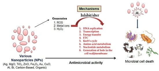
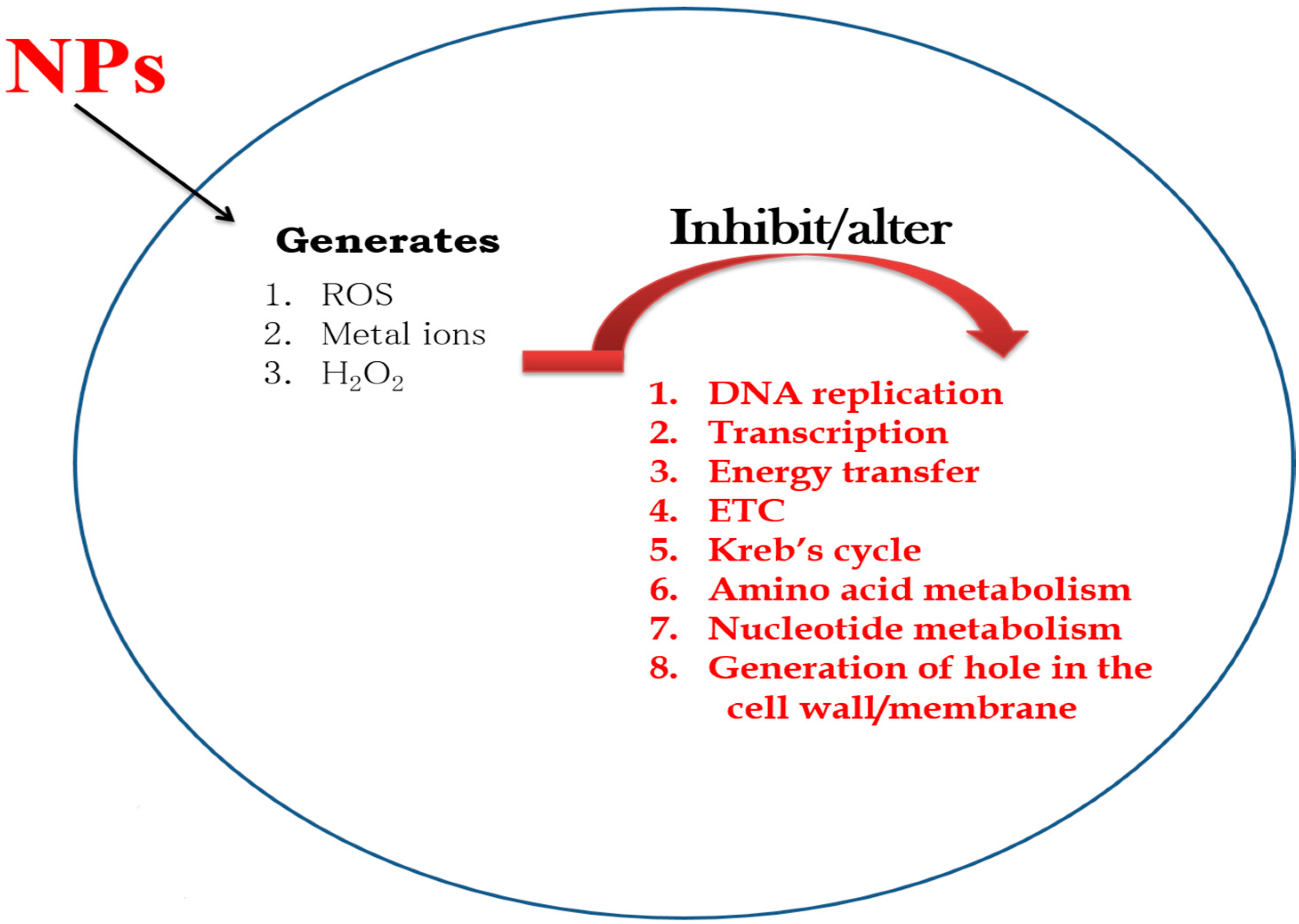
Figure 1. Mechanism of action of various nanoparticles (NPs) on microbial cells.
Molecules 21 00836 g001
Figure 2. Schematic representation of the synthesis of nanoparticles (NPs) by various methods.
Molecules 21 00836 g002
Table 1. Mode of action of various nanoparticles/nanocomposites against pathogenic microbes.
Type of NanoparticlesMode of ActionSusceptible MicrobesReferences
Silver (Ag) nanoparticlesInterfere with the electron transport chain and transfer of energy through the membrane.
Inhibit DNA replication and respiratory chain in bacteria and fungi.
Methicillin-resistant Staphylococcus aureus, Staphylococcus epidermidis. Vancomycin-resistant Enterococcus faecium and Klebsiella pneumoniae[31,57,65]
Magnesium oxide (MgO) nanoparticlesFormation of reactive oxygen species (ROS), lipid peroxidation, electrostatic interaction, alkaline effect.S. aureus, E. coli, Bacillus megaterium, Bacillus subtilis[66,67]
Titanium dioxide (TiO2) nanoparticlesFormation of superoxide radicals, ROS, and site-specific DNA damage.E. coli, S. aureus, and also against fungi[28,31,68,69]
Zinc oxide (ZnO) nanoparticlesHydrogen peroxide generated on the surface of ZnO penetrates the bacterial cells and effectively inhibits growth. Zn2+ ions released from the nanoparticles damage the cell membrane and interact with intracellular components.E. coli, Listeria monocytogenes, Salmonella, and S. aureus[70,71,72,73,74]
Gold (Au) nanoparticlesGenerate holes in the cell wall.
Bind to the DNA and inhibit the transcription process.
Methicillin-resistant S. aureus[75,76,77,78]
Copper oxide (CuO) nanoparticlesReduce bacteria at the cell wall.
Disrupt the biochemical processes inside bacterial cells.
B. subtilis, S. aureus, and E. coli[79,80,81,82]
Iron-containing nanoparticlesThrough ROS-generated oxidative stress. ROS, superoxide radicals (O2−), singlet oxygen (1O2), hydroxyl radicals (OH), and hydrogen peroxide (H2O2).S. aureus, S. epidermidis, and E. coli.[83]
Aluminum (Al) nanoparticlesDisrupt cell walls through ROS.E. coli[82,84]
Bismuth (Bi) nanoparticlesAlter the Krebs cycle, and amino acid and nucleotide metabolism.Multiple-antibiotic resistant Helicobacter pylori[85,86]
Carbon-based nanoparticlesSevere damage to the bacterial membrane, physical interaction, inhibition of energy metabolism, and impairment of the respiratory chain.E. coli, Salmonella enteric, E. faecium, Streptococcus spp., Shewanella oneidensis, Acinetobacter baumannii, Burkholderia cepacia, Yersinia pestis, and K. pneumonia[87,88,89,90,91]

