Article

Bioactive 2(1H)-Pyrazinones and Diketopiperazine Alkaloids from a Tunicate-Derived Actinomycete Streptomyces sp.

1
Natural Products Unit, King Fahd Medical Research Center, King Abdulaziz University, Jeddah 21589, Saudi Arabia
2
Suez Canal University Hospital, Suez Canal University, Ismailia 41522, Egypt
3
Department of Natural Products, Faculty of Pharmacy, King Abdulaziz University, Jeddah 21589, Saudi Arabia
4
Department of Pharmacognosy, Faculty of Pharmacy, Suez Canal University, Ismailia 41522, Egypt
5
Special Infectious Agents Unit, Fahd Medical Research Center, King Abdulaziz University, Jeddah 21589, Saudi Arabia
*
Author to whom correspondence should be addressed.
Molecules 2016, 21(9), 1116; https://doi.org/10.3390/molecules21091116
Submission received: 8 May 2016 / Revised: 11 August 2016 / Accepted: 16 August 2016 / Published: 24 August 2016
(This article belongs to the Collection Bioactive Compounds)

Abstract

:
As a part of our ongoing effort to allocate marine microbial bioactive leads, a tunicate-derived actinomycete, Streptomyces sp. Did-27, was investigated. Three new 2(1H)-pyrazinones derivatives, (S)-6-(sec-butyl)-3-isopropylpyrazin-2(1H)-one (1), (S)-3-(sec-butyl)-6-isopropylpyrazin-2(1H)-one (2) and (S)-6-(sec-butyl)-3-isobutylpyrazin-2(1H)-one (3), together with the known (1H)-pyrazinones analogues deoxymutaaspergillic acid (4), 3,6-diisobutyl-2(1H)-pyrazinone (5) and 3,6-di-sec-butyl-2(1H)-pyrazinone (6), and the diketopiperazine alkaloids cyclo(6-OH-d-Pro-l-Phe) (7), bacillusamide B (8), cyclo(l-Pro-l-Leu) and cyclo(l-Pro-l-Ile) (10) were isolated from this strain. The structures of the compounds were determined by study of their one- and two-dimensional NMR spectra as well as high-resolution mass spectral determinations. Compound 4 was reported previously as a synthetic product, while compound 6 was reported as 2-hydroxy-3,6-di-sec-butylpyrazine. Herein, we report the complete NMR data for compounds 4 and 6. The compounds were evaluated for their cytotoxic activities against three cell lines. Compound 5 showed potent and selective activity against HCT-116 cell line with IC50 of 1.5 μg/mL, while 110 showed variable cytotoxic activities against these cancer cell lines. These results provide further understanding about the chemistry and bioactivities of the alkylated 2(1H)-pyrazinone derivatives.

1. Introduction

The genus Streptomyces was first described by Waksman and Henrici [1] and is considered as a promising resource for bioactive natural products and drug discovery [2,3]. More than 75% of the important drugs are produced by members of the Streptomyces [4] including a wide array of antibiotics and anticancer drugs [5,6]. As a part of our ongoing effort to allocate bioactive leads from marine microbes [7,8,9], we have investigated a tunicate-derived actinomycete, Streptomyces sp. Did-27. Bioassay-guided fractionation of the active fractions of an organic extract of this strain resulted in the isolation and identification of three new alkylated 2(1H)-pyrazinone derivatives including (S)-6-(sec-butyl)-3-isopropylpyrazin-2(1H)-one (1), (S)-3-(sec-butyl)-6-isopropylpyrazin- 2(1H)-one (2) and (S)-6-(sec-butyl)-3-isobutylpyrazin-2(1H)-one (3), together with deoxymutaaspergillic acid (4) [10,11,12,13,14,15,16], 3,6-diisobutyl-2(1H)-pyrazinone (5) [15,16,17,18,19,20,21,22,23] and 3,6-di-sec-butyl-2(1H)-pyrazinone (6). Compound 6 was published before as 2-hydroxy-3,6-di-sec-butylpyrazine [23,24,25,26]. In addition, four diketopiperazine alkaloids including cyclo(6-OH-d-Pro-l-Phe) (7) [27], bacillusamide B (8) [28], cyclo(l-Pro-l-Leu) (9) [29] and cyclo(l-Pro-l-Ile) (10) [30] were isolated from the extract of the marine Streptomyces sp. The structures of the compounds were determined by extensive interpretation of their spectral data including 1D and 2D NMR and HRMS. Due to the lack or incomplete NMR data of compounds 4 and 6 in the literature, the complete NMR data of these compounds were presented. The isolated compounds were evaluated for their cytotoxic activity against colorectal carcinoma, hepatocellular carcinoma and breast cancer cell lines. Compound 5 showed potent and selective activity against HCT-116 cell line with IC50 of 1.5 μg/mL, while 110 showed variable cytotoxic activities against these cancer cell lines. These results provide further and deeper insight into the chemical diversity and biological activities the alkylated 2(1H)-pyrazinone derivatives.

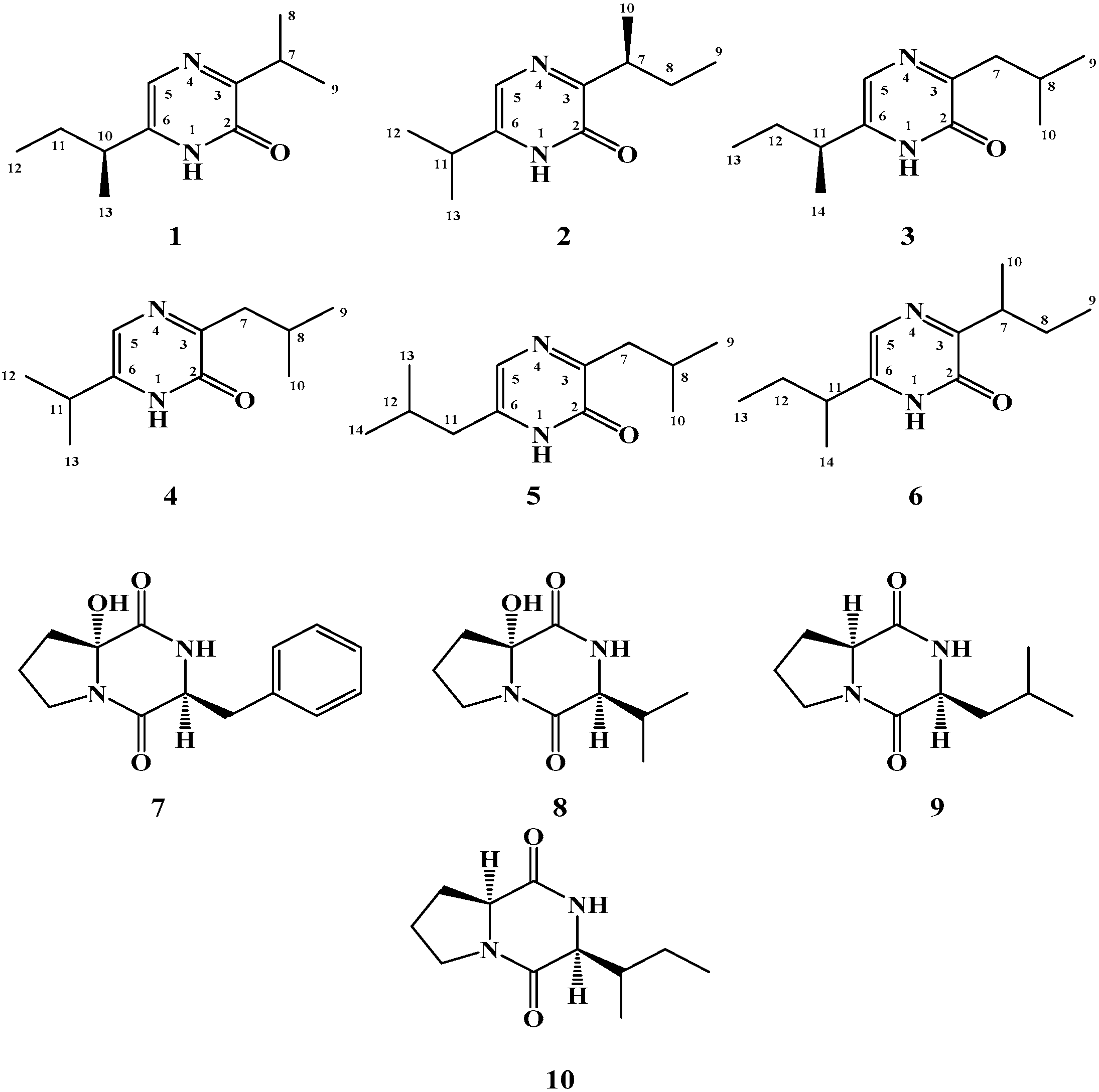
2. Results and Discussion

Compound 1 (Figure 1) possesses a molecular formula C11H18N2O as deduced from the HRESIMS pseudomolecular ion peak at m/z 195.1499 [M + H]+, requiring four degrees of unsaturation. The IR spectrum showed characteristic bands for an amidic carbonyl (1643 cm−1) and an amino group (3430 cm−1). The 1H and 13C NMR spectra of 1 together with the HSQC experiment displayed signals characteristic for a 3,6-disubstituted-2(1H)-pyrazinone skeleton [10,17]. This was evident from the 1H/13C signals at δH 11.19 (1H, s, NH), δC 156.9 (qC, C-2), δC 161.8 (qC, C-3), δHC 7.17 (1H, s, H-5)/120.9 (CH, C-5) and δC 141.6 (qC, C-6) (Table 1). In the COSY spectrum, two spin-spin coupling systems for isopropyl and sec-butyl subunits could be traced within 1. The signals at δHC 3.40 (1H, sept, J = 6.6 Hz, H-7)/30.1 (CH, C-7), 1.24 (3H, d, J = 6.6 Hz, H3-8)/19.9 (CH3, C-8) and 1.25 (3H, d, J = 6.6 Hz, H3-9)/20.0 (CH3, C-9) were assigned as an isopropyl group. While the presence of a sec-butyl group in 1 was supported by the signals at 2.51 (1H, sixth, J = 7.2 Hz, H-10)/37.1 (CH, C-10), 1.70 (1H, m, H-11a), 1.62 (1H, m, H-11b)/28.5 (CH2, C-11), 0.90 (3H, t, J = 7.2 Hz, H3-12)/11.8 (CH3, C-12) and 1.30 (3H, d, J = 6.6 Hz, H3-13)/18.8 (CH3, C-13). The placement of the isopropyl and sec-butyl subunits at C-3 and C-6, respectively, was supported by HMBC correlations of H-7/C-2, H-7/C-3, H3-8/C-3, H3-9/C-3, H-5/C-6, H-5/C-10, H-10/C-6, H-10/C-5, H3-13/C-6 (Figure 2). Additional HMBC correlation within the two alkyl moieties were shown in Figure 2. The configuration at C-10 in 1 was proposed to be 10S as established from the positive sign of the optical rotation of +12.5° (compared to +11.3° for the synthetic compound (S)-6-(sec-butyl)-3-isbutylpyrazin-2(1H)-one [31]. Thus, compound 1 was assigned as (S)-6-(sec-butyl)-3-isopropylpyrazin-2(1H)-one and is considered as a new natural compound.
Compound 2 (Figure 1) showed a molecular formula C11H18N2O as established from the HRESIMS pseudomolecular ion peak at m/z 195.1499 [M + H]+, requiring four degrees of unsaturation. The IR displayed bands for an amidic carbonyl (1640 cm−1) and an amino group (3435 cm−1). The 1H and 13C NMR spectra of 1 together with the HSQC experiment displayed signals characteristic for a 3,6-disubstituted-2(1H)-pyrazinone skeleton (Table 1). Investigation of the 1H and 13C NMR spectra, 1H-1H COSY and HSQC experiments of 2 supported the presence of three subunits including 3,6-disubstituted-2(1H)-pyrazinone, isopropyl and sec-butyl moieties as observed in 1. Since compounds 1 and 2 possess the same molecular formula, same number of degrees of unsaturation as well as same subunits, the difference between both compounds was in the placement of the alkyl subunits. In compound 2, the isopropyl and sec-butyl subunits exist at C-6 and C-3, respectively, instead of C-3 and C-6 in 1. These placements were unambiguously supported by HMBC correlations of H-11/C-5, H-11/C-6, H-5/C-11 as well as HMBC cross-peaks of H-7/C-2, H-7/C-3 and H3-10/C-3 (Figure 2). Additional HMBC correlation unambiguously supported the assignment of all carbon signals of 2 (Figure 2). Again, the configuration at C-7 in 2 was assigned as 7S based on the negative sign of the optical rotation. Compound 2 displayed a negative optical rotation of −3.4° (compared to +1.99° for paenibacillin A) [32]. Thus, compound 2 was considered as a new compound and was assigned (S)-3-(sec-butyl)-6-isopropylpyrazin-2(1H)-one.
Compound 3 (Figure 1) with a molecular formula C12H20N2O as established from the HRESIMS pseudomolecular ion peak at m/z 209.1655 [M + H]+. Compound 3 is 14 mass unit larger than 1 suggesting the presence of additional methylene group in 3. Its UV spectrum displayed absorption maxima at 325 and 227 nm. The IR spectrum showed absorption bands for an amidic carbonyl (1645 cm−1) and an amino group (3440 cm−1). Again, the 1H and 13C NMR spectra of 3 together with the HSQC experiment displayed signals characteristic for a 3,6-disubstituted-2(1H)-pyrazinone skeleton (Table 2). Study of the 1H and 13C NMR spectra, 1H-1H COSY and HSQC experiments of 3 supported the presence of three subunits including 3,6-disubstituted-2(1H)-pyrazinone, isobutyl and sec-butyl subunits. The placement of the isobutyl and sec-butyl subunits at C-3 and C-6 was unambiguously supported by HMBC correlations of H2-7/C-2, H2-7/C-3 and H-8/C-3, as well as HMBC cross-peaks of H-11/C-5, H-11/C-6, H2-12/C-6, H3-14/C-6 and H-5/C-11 (Figure 2). Additional HMBC correlations unambiguously supported the assignment of all carbon signals of 3 (Figure 2). Again, the configuration at C-11 in 3 was proposed to be 11S based on the positive sign of the optical rotation. Compound 3 displayed a positive optical rotation of +11.5° (compared to +11.3° for (S)-6-(sec-butyl)-3-isobutylpyrazin-2(1H)-one [31]. Compound 3 was reported before as a synthetic product [18], but this is the first report of this compound from a natural source. Accordingly, compound 3 is reported here as a new natural product and was assigned as (S)-6-(sec-butyl)-3-isobutylpyrazin-2(1H)-one.
Compound 4 (Figure 1) showed a molecular formula C11H18N2O as established from the HRESIMS pseudomolecular ion peak at m/z 195.1497 [M + H]+. It possesses the same molecular formula of 1. The 1H and 13C NMR spectra of 4 together with the HSQC experiment supported the presence of a 3,6-disubstituted-2(1H)-pyrazinone moiety (Table 2). Study of the 1H and 13C NMR spectra, 1H-1H COSY and HSQC experiments of 4 supported the presence of three subunits including 3,6-disubstituted-2(1H)-pyrazinone, isopropyl and isobutyl subunits. The placement of the isobutyl and isopropyl subunits at C-3 and C-6 was unambiguously supported by HMBC correlations (Figure 2). Therefore, compound 4 was assigned as deoxymutaaspergillic acid [10,11,12,13,14,15,16].
Compound 5 (Figure 1) with a molecular formula C12H20N2O as established by HRESIMS. It was identified as 3,6-diisobutyl-2(1H)-pyrazinone as established by study of its NMR data (Table 3) as well as by comparison with the literature [15,16,17,18,19,20,21,22,23]. Compound 6 (Figure 1) possesses a molecular formula of C12H20N2O as established by HRESIMS. Study of the 1H and 13C NMR spectra, 1H-1H COSY and HSQC experiments of 3 supported the presence of three subunits including 3,6-disubstituted-2(1H)-pyrazinone and two sec-butyl subunits (Table 3). Therefore, it was identified as 3,6-di-sec-butyl-2(1H)-pyrazinone as established by study of its NMR data (Table 3).
Compound 6 was reported before as 2-hydroxy-3,6-di-sec-butylpyrazine and was identified by mass spectroscopy only [23,24,25,26]. To the best of our knowledge, there is no available complete NMR data for compound 6. Thus, 6 was assigned as 3,6-di-sec-butyl-2(1H)-pyrazinone and its complete NMR data are presented in Table 3.
The diketopiperazine alkaloids 7–10 were identified by extensive study of their spectral data including HRESIMS, 1D (1H and 13C) and 2D (COSY, HSQC and HMBC) NMR data as well as by comparison with the literature. Thus, the compounds were identified as cyclo(6-OH-d-Pro-l-Phe) (7) [27], bacillusamide B (8) [28], cyclo(l-Pro-l-Leu) (9) [29] and cyclo(l-Pro-l-Ile) (10) [1].
Compounds 110 were evaluated for their antiproliferative and cytotoxic activities in the sulforhodamine B (SRB) assay against HCT-116 (colorectal carcinoma, ATCC CCL-247), HepG2 (hepatocellular carcinoma, ATCC HB-8065) and MCF-7 (breast cancer, ATCC HTB-22). Compound 5 showed potent and selective activity agaisnt HCT-116 cell line with IC50 of 1.5 µg/mL, while all other compounds were moderately active against this cell line with IC50 of 16–35 µg/mL. Similarly, all compounds were moderately active against MCF-7 with IC50 of 10–35 µg/mL (Table 4). Finally, all compounds were weakly active against HepG2 with IC50 ≥ 50 µg/mL when tested against HepG2 cell line. The results of the antiproliferative and cytotoxic activities of 110 are displayed in Table 4.

3. Materials and Methods

3.1 Experimental

General Experimental Procedures

Optical rotations were measured on a JASCO DIP-370 digital polarimeter at 25 °C at the sodium D line (589 nm). UV spectrum were recorded on a Hitachi 300 spectrometer. IR spectra were measured on a Shimadzu Infrared-400 spectrophotometer (Shimadzu, Kyoto, Japan). 1D and 2D NMR spectra (chemical shifts in ppm, coupling constants in Hz) were recorded on Bruker Avance DRX 600 MHz spectrometers (Bruker, Rheinstetten, Germany) using CDCl3 and CD3OD as solvents. NMR spectra were referenced to the residual protonated solvent signals (CHCl3: 7.26 ppm for 1H and 77.0 ppm for 13C; CH3OD: 3.30 ppm for 1H and 49.0 ppm for 13C). Positive ion HRESIMS data were obtained with a Micromass Q-ToF equipped with leucine enkaphalin lockspray, using m/z 556.2771 [M + H]+ as a reference mass. For column chromatography, silica gel (Merck, 70–230 mesh ASTM, Sigma-Aldrich, Darmstadt, Germany) and Sephadex LH-20 (0.25–0.1 mm, Pharmacia, Piscataway, NJ, USA) were used. Precoated silica gel 60 F-254 plates (Merck) were used for TLC. HPLC purifications were performed on a semi-preparative HPLC column (RP18, 5 μm, ARII Cosmosil, 250 × 10 mm, Waters, Nacalai Inc., San Diego, CA, USA).

3.2. Biological Materials

3.2.1. The Host Material, Didemnum sp.

The marine tunicate Didemnum sp. was collected in November 2013 by hands using SCUBA at depths between 15 and 20 m near Obhur, Saudi Arabia. The tunicate material was identified by Dr. Francoise Monniot at Muséum National d’Histoire Naturelle (MNHN), Paris. A voucher specimen was deposited in the MNHN, Paris, under the Registration Number A2-Did c-476.

3.2.2. Actinomycete Material

The actinomycete strain was identified as a member of the genus Streptomyces on the basis of 16S rRNA gene sequence analysis. Genomic DNA isolation, PCR amplification of 16S rRNA gene and sequence alignment of the strain were performed as described previously [33]. Its 16S rRNA gene sequence showed 98% similarity with type strains of Streptomyces flocculus (DQ442498) and Streptomyces rangoonensis (NR_041110).

3.3. Fermentation and Extraction

The spores of Streptomyces sp. Did-27 were directly cultured in 2000 mL Erlenmeyer flasks containing 500 mL of ISP-2 (ISP2, medium 2 of the International Streptomyces Project) [34] fermentation media consisted of yeast extract 4.0 g, malt extract 10.0 g and dextrose 4.0 g and 3.3% sea salt in 1 L distilled water (pH 7.2). The cultures were incubated on a rotatory shaker at 180 rpm at 28 °C for eight days. The whole fermentation broth (20 L) was extracted three times with EtOAc three times. The combined EtOAc solutions were combined and evaporated under reduced pressure to give a dark brown gum (4.3 g).

3.4. Isolation and Purification of Compounds 110

The EtOAc extract (4.3 g) was subjected to SiO2 VLC eluting with n-hexane/CH2Cl2/MeOH gradients to give six fractions (A–F). Fraction B (390 mg) was subjected to gel filtration on Sephadex LH-20 using MeOH as eluent to give five subfractions (B1–B5). Fraction B3 (139 mg) was further subjected to C18 HPLC separation eluting with 30% ACN to yield 1 (4.5 mg), 2 (1.6 mg), and 3 (3.9 mg). Fraction B4 (180 mg) was subjected to C18 HPLC separation eluting with 35% ACN to yield 7 (6.5 mg), 8 (5.3 mg), 9 (10 mg) and 10 (4.8 mg). Fraction E (320 mg) was purified by gel filtration over Sephadex LH-20 using MeOH giving four subfractions (E1–E4). Fraction E2 (130 mg) was purified by C18 HPLC eluting with 35% ACN to yield 4 (2.9 mg), 5 (4.9 mg) and 6 (4.6 mg).

3.5. Spectral Data of the Compounds

Compound 1: White solid; [ α ] D 25 +12.5 (c 0.1, CHCl3); UV (MeOH) λmax (log ε): 329 (3.65), 227 (3.50) nm; IR (film) νmax 3430, 1643 cm−1; NMR data: Table 1; HRESIMS m/z 195.1499 (calcd for C11H19N2O, [M + H]+, 195.1497).
Compound 2: White solid; [ α ] D 25 −3.4 (c 0.1, CHCl3); UV (MeOH) λmax (log ε): 328 (3.65), 227 (3.52) nm; IR (film) νmax 3435, 1640 cm−1; NMR data: Table 1; HRESIMS m/z 195.1499 (calcd for C11H19N2O, [M + H]+, 195.1497).
Compound 3: White solid; [ α ] D 25 +11.5 (c 0.1, CHCl3), UV (MeOH) λmax (log ε): 325 (3.55), 227 (3.50) nm; IR (film) νmax 3440, 1645 cm−1; NMR data: Table 2; HRESIMS m/z 209.1655 (calcd for C12H21N2O, [M + H]+, 209.1654).
Compound 4: White solid; UV (MeOH) λmax (log ε): 325 (3.53), 227 (3.50) nm; IR (film) νmax 3440, 1645 cm−1; NMR data: Table 2; HRESIMS m/z 195.1497 (calcd for C11H19N2O, [M + H]+, 195.1497).

3.6. Evaluation of Antiproliferative and Cytotoxic Activities of the Compounds

The in vitro antiproliferative and cytotoxic activities of the compounds was evaluated against three human tumor cells including HCT-116 (colorectal carcinoma, CCL-247, ATCC, Manassas, VA, USA), HepG2 (hepatocellular carcinoma, HB-8065, ATCC, Manassas, VA, USA) and MCF-7 (breast cancer, HTB-22, ATCC, Manassas, VA, USA). The effect of compounds 1-10 on cell proliferation and cytotoxicity were evaluated using the sulforhodamine B (SRB) assay as described previously [35]. Doxorubicin were used as positive control drug. The results of the cytotoxic and antiproliferative activities of 110 are displayed in Table 4.

4. Conclusions

In conclusion, investigation of a tunicate-derived actinomycete, Streptomyces sp. Did-27, afforded three new compounds, namely (S)-6-(sec-butyl)-3-isopropylpyrazin-2(1H)-one (1), (S)-3-(sec-butyl)-6-isopropylpyrazin-2(1H)-one (2) and (S)-6-(sec-butyl)-3-isobutylpyrazin-2(1H)-one (3) and six previously reported ones including deoxymutaaspergillic acid (4), 3,6-diisobutyl-2(1H)-pyrazinone (5), 3,6-di-sec-butyl-2(1H)-pyrazinone (6), cyclo(6-OH-d-Pro-l-Phe) (7), bacillusamide B (8), cyclo(l-Pro-l-Leu) and cyclo(l-Pro-l-Ile) (10). Their structures were assigned by interpretation of their spectral data. In addition, the complete NMR data for compounds 4 and 6 were reported here for the first time. Compound 5 showed selective and potent active agaisnt colorectal carcinoma cell line (HCT-116) with with IC50 of 1.5 µg/mL. All other compounds were moderatly active against MCF-7 and weakly active against HepG2 cell line.

Acknowledgments

This project was funded by the National Plan for Science, Technology and Innovation (MAARIFAH)—King Abdulaziz City for Science and Technology—the Kingdom of Saudi Arabia—award number (12-BIO2251-03). The authors also, acknowledge with thanks Science and Technology Unit, King Abdulaziz University for technical support. Our thanks to Francoise Monniot for the taxonomic identification of the tunicate material.

Author Contributions

L.A.S. and D.T.A.Y. conceived and designed the experiments; L.A.S., J.M.B. and S.M.H. performed the experiments; L.A.S., J.M.B. and D.T.A.Y. analyzed the data; L.A.S. and D.T.A.Y. wrote the paper; D.T.A.Y. edited and revised the paper.

Conflicts of Interest

The authors declare no conflict of interest.

References

  1. Waksman, S.A.; Henrici, A.T. The nomenclature and classification of the actinomycetes. J. Bacteriol. 1943, 46, 337–341. [Google Scholar] [PubMed]
  2. Blunt, J.W.; Copp, B.R.; Keyzers, R.A.; Munro, M.H.G.; Prinsep, M.R. Marine natural products. Nat. Prod. Rep. 2015, 32, 116–211. [Google Scholar] [CrossRef] [PubMed]
  3. Fguira, L.F.; Serge, F.; Raoudha, B.A.; Lotfi, M.; Hartmut, L. Purification and structure elucidation of antifungal and antibacterial activities of newly isolated Streptomyces sp. strain US80. Res. Microbiol. 2005, 156, 341–347. [Google Scholar] [CrossRef] [PubMed]
  4. Miyadoh, S. Research on antibiotic screening in Japan over the last decade: A producing microorganisms approach. Actinomycetologica 1993, 9, 100–106. [Google Scholar] [CrossRef]
  5. Baltz, R.H. Genetic manipulation of antibiotic producing Streptomyces. Trends Microbiol. 1998, 6, 76–83. [Google Scholar] [CrossRef]
  6. Harvey, A.L. Drugs from Natural Products–Pharmaceuticals and Agrochemicals, 1st ed.; Ellis Horwood Ltd.: Hemel, Hemstead, Herts, UK, 1993; p. 450. [Google Scholar]
  7. Shaala, L.A.; Youssef, D.T.A. Identification and bioactivity of compounds from the fungus Penicillium sp. CYE-87 isolated from a marine tunicate. Mar. Drugs 2015, 13, 1698–1709. [Google Scholar] [CrossRef] [PubMed]
  8. Murshid, S.S.A.; Badr, J.M.; Youssef, D.T.A. Penicillosides A and B: New cerebrosides from the marine-derived fungus Penicillium species. Rev. Bras. Farmacogn. 2016, 26, 29–33. [Google Scholar] [CrossRef]
  9. Asiry, I.A.M.; Badr, J.M.; Youssef, D.T.A. Penicillivinacine, antimigratory diketopiperazine alkaloid from the marine-derived fungus Penicillium vinaceum. Phytochem. Lett. 2015, 13, 53–58. [Google Scholar] [CrossRef]
  10. Sasaki, M.; Asao, Y.; Yokotsuka, T. Compounds produced by molds. III. Fluorescent compounds produced by Japanese commercial molds. Nippon Nogei. Kaishi 1968, 42, 288–293. [Google Scholar] [CrossRef]
  11. Nakamura, S. The structure of muta-aspergillic acid. Agric. Biol. Chem. 1961, 25, 74–75. [Google Scholar] [CrossRef]
  12. Nakamura, S. Studies on growth inhibition of Hiochi-bacteria, specific saprophytes of Sake. Part VII. Structure of muta-aspergillic acid (1). Agric. Biol. Chem. 1961, 25, 658–664. [Google Scholar]
  13. Nakamura, S. Studies on growth inhibition of Hiochi-bacteria, specific saprophytes of Sake. Part VIII. Structure of muta-aspergillic acid (2). Agric. Biol. Chem. 1961, 25, 665–670. [Google Scholar] [CrossRef]
  14. Ohta, A.; Akita, Y.; Takizawa, K.; Kurihara, M.; Masano, S.; Watanabe, T. Syntheses and reactions of chloro-2-isopropyl-5-isobutylpyrazines syntheses of deoxymutaaspergillic acid and 2-hydroxy-3-isobutyl-6-isopropylpyrazine 1-oxide. Chem. Pharm. Bull. 1978, 26, 2046–2053. [Google Scholar] [CrossRef]
  15. Ohta, A. Synthese von pulcherrimin und pulcherriminsaure. Chem. Pharm. Bull. 1964, 12, 125–126. [Google Scholar] [CrossRef]
  16. Okada, Y.; Taguchi, H.; Yokoi, T. Amino acids and peptides. XLVII. Facile synthesis of flavacol, deoxymuta-aspergillic acid and optically active deoxyaspergillic Acid from dipeptidyl aldehydes. Chem. Pharm. Bull. 1996, 44, 2259–2262. [Google Scholar] [CrossRef]
  17. Li, H.; Cai, Y.; Chen, Y.; Lam, C.; Lan, W. Metabolites of the marine fungus Aspergillus sp. collected from soft coral Sarcophyton tortuosum. Chem. Res. Chin. Univ. 2010, 26, 415–419. [Google Scholar]
  18. MacDonald, J.C.; Bishop, G.G.; Mazurek, M. 13C and proton NMR spectra of 2(1H)pyrazinones. Tetrahedron 1976, 32, 655–660. [Google Scholar] [CrossRef]
  19. Ohta, A.; Shimazaki, M.; Tamamura, H.; Mamiya, Y.; Watanabe, T. 2-Acyloxypyrazines. Convenient acylating agents for amines. J. Heterocycl. Chem. 1983, 20, 951–955. [Google Scholar] [CrossRef]
  20. Dunn, G.; Newbold, G.T.; Spring, F.S. Synthesis of flavacol, a metabolic product of Aspergillus flavus. J. Chem. Soc. 1949, 2586–2587. [Google Scholar] [CrossRef]
  21. Lopez-Gresa, M.P.; Gonzalez, M.C.; Primo, J.; Moya, P.; Romero, V.; Estornell, E. Circumdatin H, a new inhibitor of mitochondrial NADH oxidase, from Aspergillus ochraceus. J. Antibiot. 2005, 58, 416–419. [Google Scholar] [CrossRef] [PubMed]
  22. Aoyagi, Y.; Abe, T.; Ohta, A. Facile and efficient deoxygenation of aromatic N-oxides with zinc and aqueous ammonium chloride. Synthesis 1997, 1997, 891–894. [Google Scholar] [CrossRef]
  23. Aoyagi, Y.; Fujiwara, T.; Ohta, A. Synthesis of halohydroxypyrazines and their synthetic utility. Heterocycles 1991, 32, 2407–2415. [Google Scholar]
  24. Buchanan, R.L.; Houston, W.M. Production of blue-fluorescent pyrazines by A. parasiticus. J. Food. Sci. 1982, 47, 779–782. [Google Scholar] [CrossRef]
  25. Baxter, R.A.; Spring, F.S. Pyrazine derivatives. Part III. Conversion of diketopiperazines into pyrazine derivatives. Synthesis of 2-hydroxy-3:6-di-sec.-butylpyrazine from isoleucine. J. Chem. Soc. 1947, 1179–1183. [Google Scholar] [CrossRef]
  26. Inoue, M.; Abe, R.; Tamamura, H.; Ohta, M.; Asami, K.; Kitani, H.; Kamei, H.; Nakamura, Y.; Watanabe, T.; Ohta, A. Reaction of 2,5-diisopropyl- and 2,5-di-sec-butylpyrazine 1-oxide. Derivatives with phosphoryl chloride and acetic anhydride. J. Heterocycl. Chem. 1985, 22, 1291–1296. [Google Scholar] [CrossRef]
  27. Park, Y.C.; Gunasekera, S.P.; Lopez, J.V.; McCarthy, P.J.; Wright, A.E. Metabolites from the marine-derived fungus Chromocleista sp. isolated from a deep-water sediment sample collected in the Gulf of Mexico. J. Nat. Prod. 2006, 69, 580–586. [Google Scholar] [CrossRef] [PubMed]
  28. Yonezawa, K.; Yamada, K.; Kouno, I. New diketopiperazine derivatives isolated from sea urchin-derived Bacillus sp. Chem. Pharm. Bull. 2011, 59, 106–108. [Google Scholar] [CrossRef] [PubMed]
  29. Furtadoa, N.A.J.C.; Pupoa, M.T.; Carvalhoa, I.; Campoa, V.L.; Duarteb, M.C.T.; Bastos, J.K. Diketopiperazines produced by an Aspergillus fumigatus Brazilian strain. J. Braz. Chem. Soc. 2005, 16, 1448–1543. [Google Scholar] [CrossRef]
  30. Tommonaro, G.; Abbamondi, G.R.; Iodice, C.; Tait, K.; De Rosa, S. Diketopiperazines produced by the halophilic Archaeon, Holoterrigena hispanica, activate AHL bioreporters. Microb. Ecol. 2011. [Google Scholar] [CrossRef]
  31. Okada, Y.; Taguchi, H.; Yokoi, T. Total synthesis of optically active deoxyaspergillic acid from dipeptidyl aldehyde. Tetrahedron Lett. 1996, 37, 2249–2252. [Google Scholar] [CrossRef]
  32. Bian, X.; Shao, M.; Pan, H.; Wang, K.; Huang, S.; Wu, X.; Xue, C.; Hua, H.; Pei, Y.; Bai, J. Paenibacillin A, a new 2(1H)-pyrazinone ring-containing natural product from the endophytic bacterium Paenibacillus sp. Xy-2. Nat. Prod. Res. 2016, 30, 125–130. [Google Scholar] [CrossRef] [PubMed]
  33. Chun, J.; Goodfellow, M. A phylogenetic analysis of the genus Nocardia with 16S rRNA gene sequences. Int. J. Syst. Bacteriol. 1995, 2, 240–242. [Google Scholar] [CrossRef]
  34. Küster, E. Outline of a comparative study of criteria used in characterization of the actinomycetes. Int. Bull. Bacteriol. Nomencl. Taxon. 1959, 9, 97–104. [Google Scholar] [CrossRef]
  35. Vichai, V.; Kirtikara, K. Sulforhodamine B colorimetric assay for cytotoxicity screening. Nat. Protoc. 2006, 1, 1112–1116. [Google Scholar] [CrossRef] [PubMed]
  • Sample Availability: Not available.
Figure 1. Structures of compounds 110.
Molecules 21 01116 g001
Figure 2. Key COSY and HMBC correlations of 14.
Molecules 21 01116 g002
Table 1. NMR data of compounds 1 and 2 (600 and 150 MHz, CDCl3).
No.12
δC (mult.)δH (mult., J (Hz))δC (mult.)δH (mult., J (Hz))
1 11.19 (s) 11.28 (s)
2156.9, qC 157.2, qC
3161.8, qC 161.4, qC
5120.9, CH7.17 (s)120.0, CH7.21 (s)
6141.6, qC 142.6, qC
730.1, CH2.30 (m), 2.15 (m)36.6, CH3.23 (sixth, 7.2)
820.0, CH31.25 (d, 6.6)27.5, CH21.82 (m), 1.54 (m)
919.9, CH31.24 (d, 6.6)19.0, CH30.90 (t, 6.6)
1037.1, CH2.51 (sixth, 7.2)17.7, CH31.20 (d, 6.6)
1128.5, CH21.70 (m), 1.62 (m)30.0, CH2.80 (sept, 7.2)
1211.8, CH30.90 (t, 7.2)21.0, CH31.31 (d, 6.6)
1318.8, CH31.30 (d, 6.6)21.0, CH31.31 (d, 6.6)
Table 2. NMR data of compounds 3 and 4 (600 and 150 MHz, CDCl3).
No.34
δC (mult.)δH (mult., J (Hz))δC (mult.)δH (mult., J (Hz))
1 11.28 (s) 12.06 (s)
2158.2, qC 157.9, qC
3157.1, qC 157.3, qC
5121.2, CH7.18 (s)120.1, CH7.19 (s)
6142.3, qC 143.2, qC
741.6, CH22.66 (dd, 13.8, 7.2) 2.64 (dd, 13.8, 7.2)41.5, CH22.65 (d, 7.2)
826.9, CH2.21 (nonet, 7.2)26.9, CH2.21 (nonet, 7.2)
922.6, CH30.96 (d, 6.6)22.6, CH30.97 (d, 7.2)
1022.6, CH30.96 (d, 6.6)22.6, CH30.97 (d, 7.2)
1137.2, CH2.54 (sixth, 7.2)30.0, CH2.80 (sept, 7.2)
1228.4, CH21.74 (m), 1.65 (m)21.0, CH31.32 (d, 7.2)
1311.8, CH30.90 (t, 7.2)21.0, CH31.32 (d, 7.2)
1418.7, CH31.31 (d, 6.2)
Table 3. NMR data of compounds 5 and 6 (600 and 150 MHz, CDCl3).
No.56
δC (mult.)δH (mult., J (Hz))δC (mult.)δH (mult., J (Hz))
1 12.05 (s) 11.80 (s)
2158.0, qC 157.5, qC
3157.0, qC 161.2, qC
5122.8, CH7.15 (s)121.2, CH7.19 (s)
6137.3, qC 141.7, qC
741.7, CH22.65 (d, 7.2)36.7, CH3.23 (sixth, 6.6)
826.9, CH2.21 (nonet, 7.2)28.4, CH21.72 (m), 1.63 (m)
922.6, CH30.96 (d, 7.2)12.0, CH30.90 (t, 7.2)
1022.6, CH30.96 (d, 7.2)18.3, CH31.31 (d, 7.2)
1139.5, CH22.36 (d, 7.2)37.2, CH2.53 (sixth, 7.2)
1228.1, CH2.03 (nonet, 7.2)27.5, CH21.81 (m), 1.54 (m)
1322.1, CH30.98 (d, 7.2)11.8, CH30.90 (t, 7.2)
1422.1, CH30.98 (d, 7.2)17.6, CH31.21 (d, 6.6)
Table 4. Cytotoxic activities of compounds 110.
CompoundIC50 (μM)
HCT-116HepG2MCF-7
130≥5025
2NTNTNT
330≥5035
435≥5020
51.5≥5015
618≥5010
730≥5030
825≥5027
916≥5030
1022≥5027
Doxorubicin *0.7890.6210.415
* Positive control drug.

