Hydroxycinnamic Acids and Their Derivatives: Cosmeceutical Significance, Challenges and Future Perspectives, a Review

 ,
,  ,
, 
Abstract
:
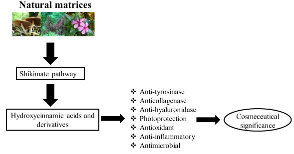
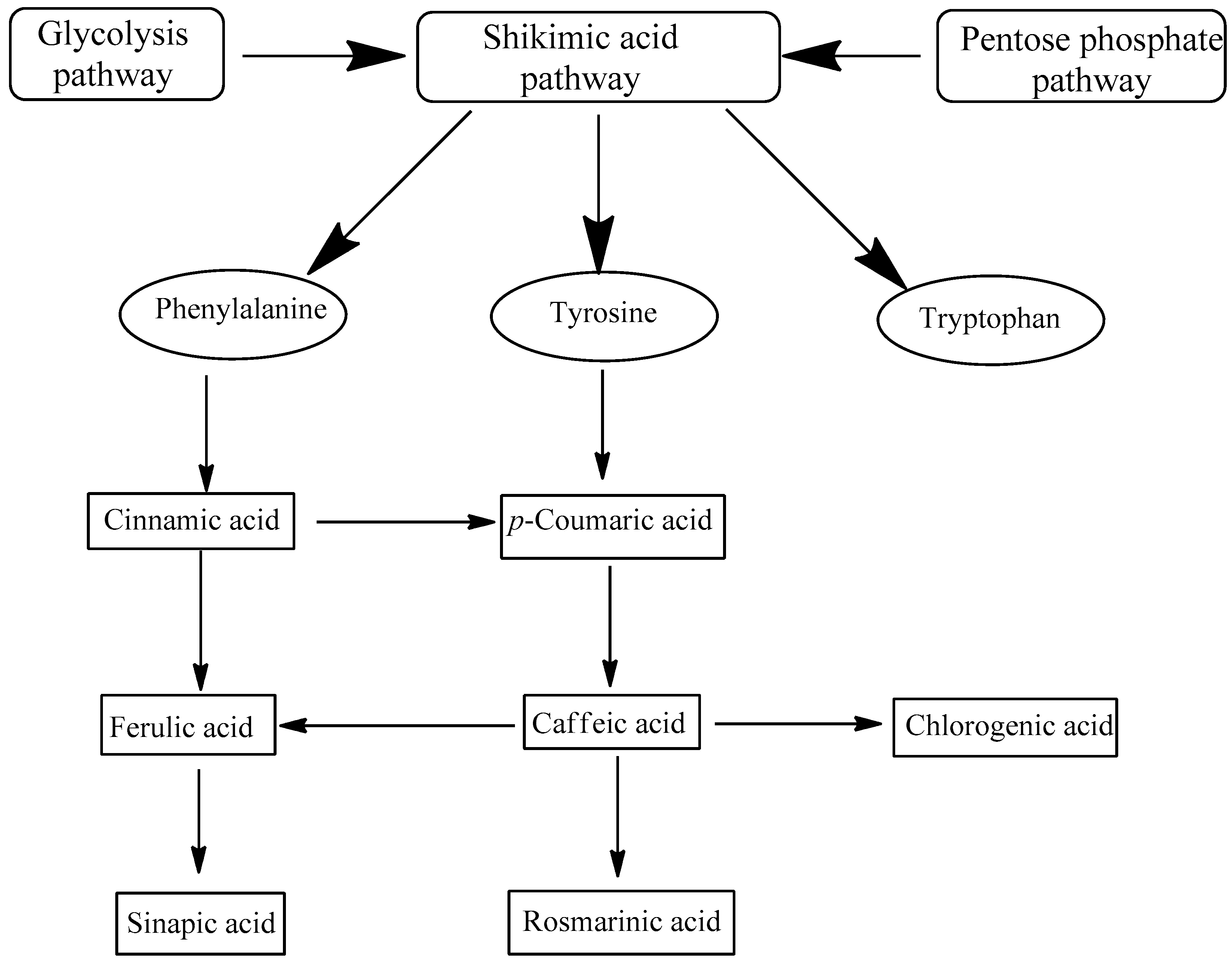
1. Introduction
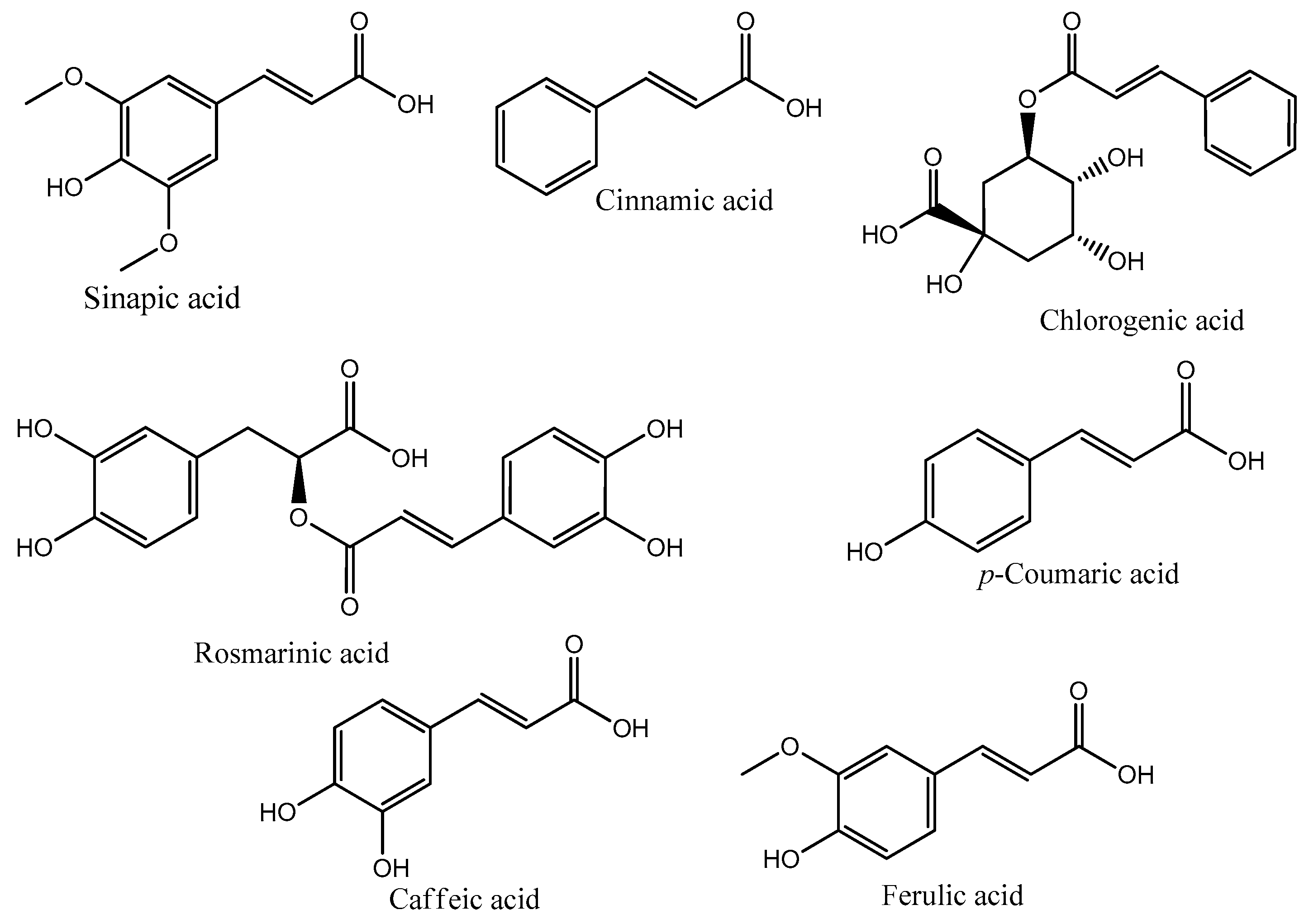
2. Hydroxycinnamic Acids
2.1. Anti-Aging and Depigmentation Properties
2.2. Anti-Inflammatory Potential
2.3. Antimicrobial Activity
3. Challenges in the Use of Hydroxycinnamic Acids in Topical Formulations
3.1. Microencapsulation
3.2. Skin Permeation Studies
4. Concluding Remarks
5. Future Perspectives
Acknowledgments
Author Contributions
Conflicts of Interest
References
- European Commission. Regulation (EC) No 1223/2009 of the European Parliament and of the Council of 30 November 2009 on cosmetic products. Off. J. Eur. Union 2009, 342–359. [Google Scholar]
- Secchi, M.; Castellani, V.; Collina, E.; Mirabella, N.; Sala, S. Assessing eco-innovations in green chemistry: Life Cycle Assessment (LCA) of a cosmetic product with a bio-based ingredient. J. Clean. Prod. 2016, 129, 269–281. [Google Scholar] [CrossRef]
- Carvalho, I.T.; Estevinho, B.N.; Santos, L. Application of microencapsulated essential oils in cosmetic and personal healthcare products—A review. Int. J. Cosmet. Sci. 2016, 38, 109–119. [Google Scholar] [CrossRef] [PubMed]
- Ramli, N.S. Immigrant Entrepreneurs on the World’s Successful Global Brands in the Cosmetic Industry. Procedia Soc. Behav. Sci. 2015, 195, 113–122. [Google Scholar] [CrossRef]
- Taofiq, O.; González-Paramás, A.M.; Martins, A.; Barreiro, M.F.; Ferreira, I.C.F.R. Mushrooms extracts and compounds in cosmetics, cosmeceuticals and nutricosmetics—A review. Ind. Crops Prod. 2016, 90, 38–48. [Google Scholar] [CrossRef]
- Barbulova, A.; Colucci, G.; Apone, F. New Trends in Cosmetics: By-Products of Plant Origin and Their Potential Use as Cosmetic Active Ingredients. Cosmetics 2015, 2, 82–92. [Google Scholar] [CrossRef]
- Rodrigues, F.; Pimentel, F.B.; Oliveira, M.B.P.P. Olive by-products: Challenge application in cosmetic industry. Ind. Crops Prod. 2015, 70, 116–124. [Google Scholar] [CrossRef]
- Kornhauser, A.; Coelho, S.G.; Hearing, V.J. Effects of cosmetic formulations containing hydroxyacids on sun-exposed skin: Current applications and future developments. Dermatol. Res. Pract. 2012, 2012, 710893. [Google Scholar] [CrossRef] [PubMed]
- Taofiq, O.; Heleno, S.; Calhelha, R.; Alves, M.; Barros, L.; Barreiro, M.; González-Paramás, A.; Ferreira, I. Development of Mushroom-Based Cosmeceutical Formulations with Anti-Inflammatory, Anti-Tyrosinase, Antioxidant, and Antibacterial Properties. Molecules 2016, 21, 1372. [Google Scholar] [CrossRef] [PubMed]
- Wang, H.M.D.; Chen, C.C.; Huynh, P.; Chang, J.S. Exploring the potential of using algae in cosmetics. Bioresour. Technol. 2015, 184, 355–362. [Google Scholar] [CrossRef] [PubMed]
- Ferraro, V.; Anton, M.; Santé-Lhoutellier, V. The “sisters” α-helices of collagen, elastin and keratin recovered from animal by-products: Functionality, bioactivity and trends of application. Trends Food Sci. Technol. 2016, 51, 65–75. [Google Scholar] [CrossRef]
- Lee, S.M.; Lee, Y.R.; Cho, K.S.; Cho, Y.N.; Lee, H.A.; Hwang, D.Y.; Jung, Y.J.; Son, H.J. Stalked sea squirt (Styela clava) tunic waste as a valuable bioresource: Cosmetic and antioxidant activities. Process Biochem. 2015, 50, 1977–1984. [Google Scholar] [CrossRef]
- Azmi, N.; Hashim, P.; Hashim, D.M.; Halimoon, N.; Majid, N.M.N. Anti-elastase, anti-tyrosinase and matrix metalloproteinase-1 inhibitory activity of earthworm extracts as potential new anti-aging agent. Asian Pac. J. Trop. Biomed. 2014, 4, S348–S352. [Google Scholar] [CrossRef] [PubMed]
- Lephart, E.D. Skin aging and oxidative stress: Equol’s anti-aging effects via biochemical and molecular mechanisms. Ageing Res. Rev. 2016, 31, 36–54. [Google Scholar] [CrossRef] [PubMed]
- Dey, T.B.; Chakraborty, S.; Jain, K.K.; Sharma, A.; Kuhad, R.C. Antioxidant phenolics and their microbial production by submerged and solid state fermentation process: A review. Trends Food Sci. Technol. 2016, 53, 60–74. [Google Scholar]
- Dias, M.I.; Sousa, M.J.; Alves, R.C.; Ferreira, I.C.F.R. Exploring plant tissue culture to improve the production of phenolic compounds: A review. Ind. Crops Prod. 2016, 82, 9–22. [Google Scholar] [CrossRef]
- Levin, J.; del Rosso, J.Q.; Momin, S.B. How much do we really know about our favorite cosmeceutical ingredients? J. Clin. Aesthet. Dermatol. 2010, 3, 22–41. [Google Scholar] [PubMed]
- Heleno, S.A.; Martins, A.; Queiroz, M.J.R.P.; Ferreira, I.C.F.R. Bioactivity of phenolic acids: Metabolites versus parent compounds: A review. Food Chem. 2015, 173, 501–513. [Google Scholar] [CrossRef] [PubMed] [Green Version]
- Pereira, D.M.; Valentão, P.; Pereira, J.A.; Andrade, P.B. Phenolics: From chemistry to biology. Molecules 2009, 14, 2202–2211. [Google Scholar] [CrossRef]
- Teixeira, J.; Gaspar, A.; Garrido, E.M.; Garrido, J.; Borges, F. Hydroxycinnamic acid antioxidants: An electrochemical overview. BioMed Res. Int. 2013, 2013, 251754. [Google Scholar] [CrossRef] [PubMed]
- Zhang, H.; Tsao, R. Dietary polyphenols, oxidative stress and antioxidant and anti-inflammatory effects. Curr. Opin. Food Sci. 2016, 8, 33–42. [Google Scholar] [CrossRef]
- Jiang, K.; Li, L.; Long, L.; Ding, S. Comparison of alkali treatments for efficient release of p-coumaric acid and enzymatic saccharification of sorghum pith. Bioresour. Technol. 2016, 207, 1–10. [Google Scholar] [CrossRef] [PubMed]
- Pragasam, S.J.; Murunikkara, V.; Sabina, E.P.; Rasool, M. Ameliorative effect of p-coumaric acid, a common dietary phenol, on adjuvant-induced arthritis in rats. Rheumatol. Int. 2013, 33, 325–334. [Google Scholar] [CrossRef] [PubMed]
- Pei, K.; Ou, J.; Huang, J.; Ou, S. p-Coumaric acid and its conjugates: Dietary sources, pharmacokinetic properties and biological activities. J. Sci. Food Agric. 2016, 96, 2952–2962. [Google Scholar] [CrossRef] [PubMed]
- Kwak, J.Y.; Park, S.; Seok, J.K.; Liu, K.H.; Boo, Y.C. Ascorbyl coumarates as multifunctional cosmeceutical agents that inhibit melanogenesis and enhance collagen synthesis. Arch. Dermatol. Res. 2015, 307, 635–643. [Google Scholar] [CrossRef] [PubMed]
- Yang, W.S.; Jeong, D.; Yi, Y.; Park, J.G.; Seo, H.; Moh, S.H.; Hong, S.; Cho, J.Y. IRAK1/4-Targeted Anti-Inflammatory Action of Caffeic Acid. Mediat. Inflamm. 2013, 2013, 518183. [Google Scholar] [CrossRef] [PubMed]
- Kumar, N.; Pruthi, V. Potential applications of ferulic acid from natural sources. Biotechnol. Rep. 2014, 4, 86–93. [Google Scholar] [CrossRef]
- Ouimet, M.A.; Faig, J.J.; Yu, W.; Uhrich, K.E. Ferulic Acid-Based Polymers with Glycol Functionality as a Versatile Platform for Topical Applications. Biomacromolecules 2015, 16, 2911–2919. [Google Scholar] [CrossRef] [PubMed]
- Casanova, F.; Estevinho, B.N.; Santos, L. Preliminary studies of rosmarinic acid microencapsulation with chitosan and modified chitosan for topical delivery. Powder Technol. 2016, 297, 44–49. [Google Scholar] [CrossRef]
- Kim, G.-D.; Park, Y.S.; Jin, Y.-H.; Park, C.-S. Production and applications of rosmarinic acid and structurally related compounds. Appl. Microbiol. Biotechnol. 2015, 2083–2092. [Google Scholar] [CrossRef] [PubMed]
- Al Danaf, N.; Melhem, R.A.; Assaf, K.I.; Nau, W.M.; Patra, D. Photophysical properties of neutral and dissociated forms of rosmarinic acid. J. Lumin. 2016, 175, 50–56. [Google Scholar] [CrossRef]
- Li, H.R.; Habasi, M.; Xie, L.Z.; Aisa, H.A. Effect of chlorogenic acid on melanogenesis of B16 melanoma cells. Molecules 2014, 19, 12940–12948. [Google Scholar] [CrossRef] [PubMed]
- Ruifeng, G.; Yunhe, F.; Zhengkai, W.; Ershun, Z.; Yimeng, L.; Minjun, Y.; Xiaojing, S.; Zhengtao, Y.; Naisheng, Z. Chlorogenic acid attenuates lipopolysaccharide-induced mice mastitis by suppressing TLR4-mediated NF-κB signaling pathway. Eur. J. Pharmacol. 2014, 729, 54–58. [Google Scholar] [CrossRef] [PubMed]
- Silambarasan, T.; Manivannan, J.; Raja, B.; Chatterjee, S. Prevention of cardiac dysfunction, kidney fibrosis and lipid metabolic alterations in l-NAME hypertensive rats by sinapic acid—Role of HMG-CoA reductase. Eur. J. Pharmacol. 2016, 777, 113–123. [Google Scholar] [CrossRef] [PubMed]
- Govindan, S.; Johnson, E.E.R.; Christopher, J.; Shanmugam, J.; Thirumalairaj, V.; Gopalan, J. Antioxidant and anti-aging activities of polysaccharides from Calocybe indica var. APK2. Exp. Toxicol. Pathol. 2016, 68, 329–334. [Google Scholar] [CrossRef] [PubMed]
- Shindo, Y.; Witt, E.; Han, D.; Epstein, W.; Packer, L. Enzymic and non-enzymic antioxidants in epidermis and dermis of human skin. J. Investig. Dermatol. 1994, 102, 122–124. [Google Scholar] [CrossRef] [PubMed]
- Carocho, M.; Ferreira, I.C.F.R. A review on antioxidants, prooxidants and related controversy: Natural and synthetic compounds, screening and analysis methodologies and future perspectives. Food Chem. Toxicol. 2013, 51, 15–25. [Google Scholar] [CrossRef] [PubMed]
- Theocharis, A.D.; Skandalis, S.S.; Gialeli, C.; Karamanos, N.K. Extracellular matrix structure. Adv. Drug Deliv. Rev. 2016, 97, 4–27. [Google Scholar] [CrossRef] [PubMed]
- Tobin, D.J. Introduction to skin aging. J. Tissue Viability 2016. [Google Scholar] [CrossRef] [PubMed]
- Matsui, M.S.; Hsia, A.; Miller, J.D.; Hanneman, K.; Scull, H.; Cooper, K.D.; Baron, E. Non-Sunscreen Photoprotection: Antioxidants Add Value to a Sunscreen. J. Investig. Dermatol. Symp. Proc. 2009, 14, 56–59. [Google Scholar] [CrossRef] [PubMed]
- Yang, C.-H.; Chang, N.-F.; Chen, Y.-S.; Lee, S.-M.; Lin, P.-J.; Lin, C.-C. Comparative study on the photostability of arbutin and deoxy arbutin: Sensitivity to ultraviolet radiation and enhanced photostability by the water-soluble sunscreen, benzophenone-4. Biosci. Biotechnol. Biochem. 2013, 77, 1127–1130. [Google Scholar] [CrossRef] [PubMed]
- Desmedt, B.; Courselle, P.; De Beer, J.O.; Rogiers, V.; Grosber, M.; Deconinck, E.; De Paepe, K. Overview of skin whitening agents with an insight into the illegal cosmetic market in Europe. J. Eur. Acad. Dermatol. Venereol. 2016, 30, 943–950. [Google Scholar] [CrossRef] [PubMed]
- Kwak, S.Y.; Lee, S.; Choi, H.R.; Park, K.C.; Lee, Y.S. Dual effects of caffeoyl-amino acidyl-hydroxamic acid as an antioxidant and depigmenting agent. Bioorg. Med. Chem. Lett. 2011, 21, 5155–5158. [Google Scholar] [CrossRef] [PubMed]
- Aoshima, H.; Miyase, T.; Warashina, T. Caffeic acid oligomers with hyaluronidase inhibitory activity from Clinopodium gracile. Chem. Pharm. Bull. 2012, 60, 499–507. [Google Scholar] [CrossRef] [PubMed]
- Thangboonjit, W.; Limsaeng-u-rai, S.; Panich, U. their Activities in Melanoma Cells Exposed to. Siriraj Med. J. 2014, 66, 5–10. [Google Scholar]
- Chaiprasongsuk, A.; Onkoksoong, T.; Pluemsamran, T.; Limsaengurai, S.; Panich, U. Photoprotection by dietary phenolics against melanogenesis induced by UVA through Nrf2-dependent antioxidant responses. Redox Biol. 2016, 8, 79–90. [Google Scholar] [CrossRef] [PubMed]
- Pluemsamran, T.; Onkoksoong, T.; Panich, U. Caffeic acid and ferulic acid inhibit UVA-induced matrix metalloproteinase-1 through regulation of antioxidant defense system in keratinocyte HaCaT cells. Photochem. Photobiol. 2012, 88, 961–968. [Google Scholar] [CrossRef] [PubMed]
- Saija, A.; Tomaino, A.; Trombetta, D.; de Pasquale, A.; Uccella, N.; Barbuzzi, T.; Paolino, D.; Bonina, F. In vitro and in vivo evaluation of caffeic and ferulic acids as topical photoprotective agents. Int. J. Pharm. 2000, 199, 39–47. [Google Scholar] [CrossRef]
- Staniforth, V.; Huang, W.C.; Aravindaram, K.; Yang, N.S. Ferulic acid, a phenolic phytochemical, inhibits UVB-induced matrix metalloproteinases in mouse skin via posttranslational mechanisms. J. Nutr. Biochem. 2012, 23, 443–451. [Google Scholar] [CrossRef] [PubMed]
- Fan, Q.; Jiang, H.; Yuan, E.D.; Zhang, J.X.; Ning, Z.X.; Qi, S.J.; Wei, Q.Y. Tyrosinase inhibitory effects and antioxidative activities of novel cinnamoyl amides with amino acid ester moiety. Food Chem. 2012, 134, 1081–1087. [Google Scholar] [CrossRef] [PubMed]
- Chochkova, M.; Stoykova, B.; Ivanova, G.; Ranz, A.; Guo, X.; Lankmayr, E.; Milkova, T. N-Hydroxycinnamoyl amides of fluorinated amino acids: Synthesis, anti-tyrosinase and DPPH scavenging activities. J. Fluor. Chem. 2013, 156, 203–208. [Google Scholar] [CrossRef]
- An, S.M.; Koh, J.S.; Boo, Y.C. p-Coumaric acid not only inhibits human tyrosinase activity in vitro but also melanogenesis in cells exposed to UVB. Phyther. Res. 2010, 24, 1175–1180. [Google Scholar]
- Jun, H.; Lee, J.H.; Cho, B.R.; Seo, W.D.; Kim, D.W.; Cho, K.J.; Lee, S.J. p-Coumaric acid inhibition of CREB phosphorylation reduces cellular melanogenesis. Eur. Food Res. Technol. 2012, 235, 1207–1211. [Google Scholar] [CrossRef]
- Seo, Y.K.; Kim, S.J.; Boo, Y.C.; Baek, J.H.; Lee, S.H.; Koh, J.S. Effects of p-Coumaric acid on erythema and pigmentation of human skin exposed to ultraviolet radiation. Clin. Exp. Dermatol. 2011, 36, 260–266. [Google Scholar] [CrossRef] [PubMed]
- Seok, J.K.; Boo, Y.C. p-Coumaric acid attenuates UVB-induced release of stratifin from keratinocytes and indirectly regulates matrix metalloproteinase 1 release from fibroblasts. Korean J. Physiol. Pharmacol. 2015, 19, 241–247. [Google Scholar] [CrossRef] [PubMed]
- Song, K.; An, S.M.; Kim, M.; Koh, J.S.; Boo, Y.C. Comparison of the antimelanogenic effects of p-coumaric acid and its methyl ester and their skin permeabilities. J. Dermatol. Sci. 2011, 63, 17–22. [Google Scholar] [CrossRef] [PubMed]
- Oliveira, K.B.; Palú, É.; Weffort-Santos, A.M.; Oliveira, B.H. Infl uence of rosmarinic acid and Salvia offi cinalis extracts on melanogenesis of B16f10 cells. Braz. J. Pharmacogn. 2013, 23, 249–258. [Google Scholar] [CrossRef]
- Psotova, J.; Svobodova, A.; Kolarova, H.; Walterova, D. Photoprotective properties of Prunella vulgaris and rosmarinic acid on human keratinocytes. J. Photochem. Photobiol. B Biol. 2006, 84, 167–174. [Google Scholar] [CrossRef] [PubMed]
- Sánchez-Campillo, M.; Gabaldon, J.A.; Castillo, J.; Benavente-García, O.; Del Baño, M.J.; Alcaraz, M.; Vicente, V.; Alvarez, N.; Lozano, J.A. Rosmarinic acid, a photo-protective agent against UV and other ionizing radiations. Food Chem. Toxicol. 2009, 47, 386–392. [Google Scholar] [CrossRef] [PubMed]
- Murata, T.; Miyase, T.; Yoshizaki, F. Hyaluronidase inhibitory rosmarinic acid derivatives from Meehania urticifolia. Chem. Pharm. Bull. 2011, 59, 88–95. [Google Scholar] [CrossRef] [PubMed]
- Lin, L.; Dong, Y.; Zhao, H.; Wen, L.; Yang, B.; Zhao, M. Comparative evaluation of rosmarinic acid, methyl rosmarinate and pedalitin isolated from Rabdosia serra (MAXIM.) HARA as inhibitors of tyrosinase and α-glucosidase. Food Chem. 2011, 129, 884–889. [Google Scholar] [CrossRef] [PubMed]
- Ding, H.Y.; Chou, T.H.; Liang, C.H. Antioxidant and antimelanogenic properties of rosmarinic acid methyl ester from Origanum vulgare. Food Chem. 2010, 123, 254–262. [Google Scholar] [CrossRef]
- Freitas, J.V.; Gaspar, L.R. In vitro photosafety and efficacy screening of apigenin, chrysin and beta-carotene for UVA and VIS protection. Eur. J. Pharm. Sci. 2016, 89, 146–153. [Google Scholar] [CrossRef] [PubMed]
- Kwak, S.Y.; Yang, J.K.; Choi, H.R.; Park, K.C.; Kim, Y.B.; Lee, Y.S. Synthesis and dual biological effects of hydroxycinnamoyl phenylalanyl/prolyl hydroxamic acid derivatives as tyrosinase inhibitor and antioxidant. Bioorg. Med. Chem. Lett. 2013, 23, 1136–1142. [Google Scholar] [CrossRef] [PubMed]
- Georgiev, L. Anti-tyrosinase, antioxidant and antimicrobial activities of hydroxycinnamoylamides. Med. Chem. Res. 2013, 22, 4173–4182. [Google Scholar] [CrossRef]
- Działo, M.; Mierziak, J.; Korzun, U.; Preisner, M.; Szopa, J.; Kulma, A. The potential of plant phenolics in prevention and therapy of skin disorders. Int. J. Mol. Sci. 2016, 17, 160. [Google Scholar] [CrossRef] [PubMed]
- Balupillai, A.; Prasad, R.N.; Ramasamy, K.; Muthusamy, G.; Shanmugham, M.; Govindasamy, K.; Gunaseelan, S. Caffeic Acid Inhibits UVB-induced Inflammation and Photocarcinogenesis Through Activation of Peroxisome Proliferator-activated Receptor-γ in Mouse Skin. Photochem. Photobiol. 2015, 91, 1458–1468. [Google Scholar] [CrossRef] [PubMed]
- Rivelli, D.P.; Filho, C.A.H.; Almeida, R.L.; Ropke, C.D.; Sawada, T.C.H.; Barros, S.B.M. Chlorogenic acid UVA-UVB photostability. Photochem. Photobiol. 2010, 86, 1005–1007. [Google Scholar] [CrossRef] [PubMed]
- Taofiq, O.; Calhelha, R.C.; Heleno, S.; Barros, L.; Martins, A.; Santos-Buelga, C.; Queiroz, M.J.R.P.; Ferreira, I.C.F.R. The contribution of phenolic acids to the anti-inflammatory activity of mushrooms: Screening in phenolic extracts, individual parent molecules and synthesized glucuronated and methylated derivatives. Food Res. Int. 2015, 76, 821–827. [Google Scholar] [CrossRef] [Green Version]
- Taofiq, O.; Martins, A.; Barreiro, M.F.; Ferreira, I.C.F.R. Anti-inflammatory potential of mushroom extracts and isolated metabolites. Trends Food Sci. Technol. 2016, 50, 193–210. [Google Scholar] [CrossRef]
- Pillai, S.; Oresajo, C.; Hayward, J. Ultraviolet radiation and skin aging: Roles of reactive oxygen species, inflammation and protease activation, and strategies for prevention of inflammation induced matrix degradation—A review. Int. J. Cosmet. Sci. 2005, 27, 17–34. [Google Scholar] [CrossRef] [PubMed]
- Lee, H.J.; Kim, M.H.; Choi, Y.Y.; Kim, E.H.; Hong, J.; Kim, K.; Yang, W.M. Improvement of atopic dermatitis with topical application of Spirodela polyrhiza. J. Ethnopharmacol. 2016, 180, 12–17. [Google Scholar] [CrossRef] [PubMed]
- Ukawa, Y.; Izumi, Y.; Ohbuchi, T.; Takahashi, T.; Ikemizu, S.; Kojima, Y. Oral administration of the extract from Hatakeshimeji (Lyophyllum decastes sing.) mushroom inhibits the development of atopic dermatitis-like skin lesions in NC/Nga mice. J. Nutr. Sci. Vitaminol. 2007, 53, 293–296. [Google Scholar] [CrossRef] [PubMed]
- Park, H.S.; Hwang, Y.H.; Kim, M.K.; Hong, G.E.; Lee, H.J.; Nagappan, A.; Yumnam, S.; Kim, E.H.; Heo, J.D.; Lee, S.J.; et al. Functional polysaccharides from Grifola frondosa aqueous extract inhibit atopic dermatitis-like skin lesions in NC/Nga mice. Biosci. Biotechnol. Biochem. 2015, 79, 147–154. [Google Scholar] [CrossRef] [PubMed]
- Wu, G.; Li, L.; Sung, G.H.; Kim, T.W.; Byeon, S.E.; Cho, J.Y.; Park, C.W.; Park, H.J. Inhibition of 2,4-dinitrofluorobenzene-induced atopic dermatitis by topical application of the butanol extract of Cordyceps bassiana in NC/Nga mice. J. Ethnopharmacol. 2011, 134, 504–509. [Google Scholar] [CrossRef] [PubMed]
- Choi, J.H.; Kim, H.G.; Jin, S.W.; Han, E.H.; Khanal, T.; Do, M.T.; Hwang, Y.P.; Choi, J.M.; Chun, S.S.; Chung, Y.C.; et al. Topical application of Pleurotus eryngii extracts inhibits 2,4-dinitrochlorobenzene-induced atopic dermatitis in NC/Nga mice by the regulation of Th1/Th2 balance. Food Chem. Toxicol. 2013, 53, 38–45. [Google Scholar] [CrossRef] [PubMed]
- Lee, J.; Jung, E.; Koh, J.; Kim, Y.S.; Park, D. Effect of rosmarinic acid on atopic dermatitis. J. Dermatol. 2008, 35, 768–771. [Google Scholar] [CrossRef] [PubMed]
- Jang, A.H.; Kim, T.H.; Kim, G.D.; Kim, J.E.; Kim, H.J.; Kim, S.S.; Jin, Y.H.; Park, Y.S.; Park, C.S. Rosmarinic acid attenuates 2,4-dinitrofluorobenzene-induced atopic dermatitis in NC/Nga mice. Int. Immunopharmacol. 2011, 11, 1271–1277. [Google Scholar] [CrossRef] [PubMed]
- Tsang, M.S.M.; Jiao, D.; Chan, B.C.L.; Hon, K.L.; Leung, P.C.; Lau, C.B.S.; Wong, E.C.W.; Cheng, L.; Chan, C.K.M.; Lam, C.W.K.; et al. Anti-Inflammatory Activities of Pentaherbs Formula, Berberine, Gallic Acid and Chlorogenic Acid in Atopic Dermatitis-Like Skin Inflammation. Molecules 2016, 21, 519. [Google Scholar] [CrossRef] [PubMed]
- Kim, M. The Effect of Prunella on Anti-Inflammatory Activity in RAW264.7 Mouse Macrophage Cells. Food Nutr. Sci. 2012, 3, 1290–1295. [Google Scholar] [CrossRef]
- Búfalo, M.C.; Ferreira, I.; Costa, G.; Francisco, V.; Liberal, J.; Cruz, M.T.; Lopes, M.C.; Batista, M.T.; Sforcin, J.M. Propolis and its constituent caffeic acid suppress LPS-stimulated pro-inflammatory response by blocking NF-κB and MAPK activation in macrophages. J. Ethnopharmacol. 2013, 149, 84–92. [Google Scholar] [CrossRef] [PubMed]
- Zhang, M.; Zhou, J.; Wang, L.; Li, B.; Guo, J.; Guan, X.; Han, Q.; Zhang, H. Caffeic acid reduces cutaneous tumor necrosis factor alpha (TNF-α), IL-6 and IL-1β levels and ameliorates skin edema in acute and chronic model of cutaneous inflammation in mice. Biol. Pharm. Bull. 2014, 37, 347–354. [Google Scholar] [CrossRef] [PubMed]
- Saibabu, V.; Fatima, Z.; Khan, L.A.; Hameed, S. Therapeutic potential of dietary phenolic acids. Adv. Pharmacol. Sci. 2015, 2015, 823539. [Google Scholar] [CrossRef] [PubMed]
- Alam, M.A.; Subhan, N.; Hossain, H.; Hossain, M.; Reza, H.M.; Rahman, M.M.; Ullah, M.O. Hydroxycinnamic acid derivatives: A potential class of natural compounds for the management of lipid metabolism and obesity. Nutr. Metab. 2016, 13, 27. [Google Scholar] [CrossRef] [PubMed]
- Vo, V.A.; Lee, J.W.; Kim, J.Y.; Park, J.H.; Lee, H.J.; Kim, S.S.; Kwon, Y.S.; Chun, W. Phosphorylation of Akt mediates anti-inflammatory activity of 1-p-coumaroyl β-d-glucoside against lipopolys accharide-induced inflammation in RAW264.7 cells. Korean J. Physiol. Pharmacol. 2014, 18, 79–86. [Google Scholar] [CrossRef] [PubMed]
- Lee, J.W.; Choi, Y.J.; Park, J.H.; Sim, J.Y.; Kwon, Y.S.; Lee, H.J.; Kim, S.S.; Chun, W. 3,4,5-Trihydroxycinnamic acid inhibits lipopolysaccharide-induced inflammatory response through the activation of Nrf2 pathway in BV2 microglial cells. Biomol. Ther. 2013, 21, 60–65. [Google Scholar] [CrossRef] [PubMed]
- Park, S.Y.; Hwang, J.S.; Jang, M.; Lee, S.H.; Park, J.H.; Han, I.O. A novel caffeic acid-1-piperonylpiperazine hybridization compound HBU-47 inhibits LPS-mediated inflammation in RAW 264.7 macrophage cells. Int. Immunopharmacol. 2014, 19, 60–65. [Google Scholar] [CrossRef] [PubMed]
- Liu, M.; Song, S.; Li, H.; Jiang, X.; Yin, P.; Wan, C.; Liu, X.; Liu, F.; Xu, J. The protective effect of caffeic acid against inflammation injury of primary bovine mammary epithelial cells induced by lipopolysaccharide. J. Dairy Sci. 2014, 97, 2856–2865. [Google Scholar] [CrossRef] [PubMed]
- Nagasaka, R.; Chotimarkorn, C.; Shafiqul, I.M.; Hori, M.; Ozaki, H.; Ushio, H. Anti-inflammatory effects of hydroxycinnamic acid derivatives. Biochem. Biophys. Res. Commun. 2007, 358, 615–619. [Google Scholar] [CrossRef] [PubMed]
- da Cunha, F.M.; Duma, D.; Assreuy, J.; Buzzi, F.C.; Niero, R.; Campos, M.M.; Calixto, J.B. Caffeic acid derivatives: In vitro and in vivo anti-inflammatory properties. Free Radic. Res. 2004, 38, 1241–1253. [Google Scholar] [CrossRef] [PubMed]
- Bose, J.S.; Gangan, V.; Jain, S.K.; Manna, S.K. Downregulation of inflammatory responses by novel caffeic acid ester derivative by inhibiting NF-κB. J. Clin. Immunol. 2009, 29, 90–98. [Google Scholar] [CrossRef] [PubMed]
- Lou, L.; Zhou, J.; Liu, Y.; Wei, Y.I.; Zhao, J.; Deng, J. Chlorogenic acid induces apoptosis to inhibit inflammatory proliferation of IL-6-induced fibroblast-like synoviocytes through modulating the activation of JAK/STAT and NF-κB signaling pathways. Exp. Ther. Med. 2016, 11, 2054–2060. [Google Scholar] [CrossRef] [PubMed]
- Hwang, S.J.; Kim, Y.-W.; Park, Y.; Lee, H.-J.; Kim, K.-W. Anti-inflammatory effects of chlorogenic acid in lipopolysaccharide-stimulated RAW 264.7 cells. Inflamm. Res. 2014, 63, 81–90. [Google Scholar] [CrossRef] [PubMed]
- Francisco, V.; Costa, G.; Figueirinha, A.; Marques, C.; Pereira, P.; Miguel Neves, B.; Celeste Lopes, M.; García-Rodríguez, C.; Teresa Cruz, M.; Teresa Batista, M. Anti-inflammatory activity of Cymbopogon citratus leaves infusion via proteasome and nuclear factor-κB pathway inhibition: Contribution of chlorogenic acid. J. Ethnopharmacol. 2013, 148, 126–134. [Google Scholar] [CrossRef] [PubMed] [Green Version]
- Chen, W.-P.; Wu, L.-D. Chlorogenic acid suppresses interleukin-1β-induced inflammatory mediators in human chondrocytes. Int. J. Clin. Exp. Pathol. 2014, 7, 8797–8801. [Google Scholar] [PubMed]
- Ambothi, K.; Prasad, N.R.; Balupillai, A. Ferulic acid inhibits UVB-radiation induced photocarcinogenesis through modulating inflammatory and apoptotic signaling in Swiss albino mice. Food Chem. Toxicol. 2015, 82, 72–78. [Google Scholar] [CrossRef] [PubMed]
- Kim, E.O.; Min, K.J.; Kwon, T.K.; Um, B.H.; Moreau, R.A.; Choi, S.W. Anti-inflammatory activity of hydroxycinnamic acid derivatives isolated from corn bran in lipopolysaccharide-stimulated Raw264.7 macrophages. Food Chem. Toxicol. 2012, 50, 1309–1316. [Google Scholar] [CrossRef] [PubMed]
- Vo, V.A.; Lee, J.-W.; Park, J.-H.; Kwon, J.-H.; Lee, H.J.; Kim, S.-S.; Kwon, Y.-S.; Chun, W. N-(p-Coumaryol)-Tryptamine Suppresses the Activation of JNK/c-Jun Signaling Pathway in LPS-Challenged RAW 264.7 Cells. Biomol. Ther.. 2014, 22, 200–206. [Google Scholar] [CrossRef] [PubMed]
- Pragasam, S.J.; Venkatesan, V.; Rasool, M. Immunomodulatory and anti-inflammatory effect of p-coumaric acid, a common dietary polyphenol on experimental inflammation in rats. Inflammation 2013, 36, 169–176. [Google Scholar] [CrossRef] [PubMed]
- Geller, F.; Schmidt, C.; Göttert, M.; Fronza, M.; Schattel, V.; Heinzmann, B.; Werz, O.; Flores, E.M.M.; Merfort, I.; Laufer, S. Identification of rosmarinic acid as the major active constituent in Cordia americana. J. Ethnopharmacol. 2010, 128, 561–566. [Google Scholar] [CrossRef] [PubMed]
- Lembo, S.; Balato, A.; Di Caprio, R.; Cirillo, T.; Giannini, V.; Gasparri, F.; Monfrecola, G. The Modulatory Effect of Ellagic Acid and Rosmarinic Acid on Ultraviolet-B-Induced Cytokine/Chemokine Gene Expression in Skin Keratinocyte (HaCaT) Cells. Biomed Res. Int. 2014, 2014, 346793. [Google Scholar]
- Zdařilová, A.; Svobodová, A.; Šimánek, V.; Ulrichová, J. Prunella vulgaris extract and rosmarinic acid suppress lipopolysaccharide-induced alteration in human gingival fibroblasts. Toxicol. Vitr. 2009, 23, 386–392. [Google Scholar] [CrossRef] [PubMed]
- Huang, N.; Hauck, C.; Yum, M.Y.; Rizshsky, L.; Widrlechner, M.P.; McCoy, J.A.; Murphy, P.A.; Dixon, P.M.; Nikolau, B.J.; Birt, D.F. Rosmarinic acid in Prunella vulgaris ethanol extract inhibits lipopolysaccharide-Lnduced prostaglandin E2 and nitric oxide in RAW 264.7 mouse macrophages. J. Agric. Food Chem. 2009, 57, 10579–10589. [Google Scholar] [CrossRef] [PubMed]
- Chu, X.; Ci, X.; He, J.; Jiang, L.; Wei, M.; Cao, Q.; Guan, M.; Xie, X.; Deng, X. Effects of a natural prolyl oligopeptidase inhibitor, rosmarinic acid, on lipopolysaccharide-induced acute lung injury in mice. Molecules 2012, 17, 3586–3598. [Google Scholar] [CrossRef] [PubMed]
- Al-Musayeib, N.; Perveen, S.; Fatima, I.; Nasir, M.; Hussain, A. Antioxidant, anti-glycation and anti-inflammatory activities of phenolic constituents from cordia sinensis. Molecules 2011, 16, 10214–10226. [Google Scholar] [CrossRef] [PubMed]
- Heffernan, A.L.; Baduel, C.; Toms, L.M.L.; Calafat, A.M.; Ye, X.; Hobson, P.; Broomhall, S.; Mueller, J.F. Use of pooled samples to assess human exposure to parabens, benzophenone-3 and triclosan in Queensland, Australia. Environ. Int. 2015, 85, 77–83. [Google Scholar] [CrossRef] [PubMed]
- Velegraki, T.; Hapeshi, E.; Fatta-Kassinos, D.; Poulios, I. Solar-induced heterogeneous photocatalytic degradation of methyl-paraben. Appl. Catal. B Environ. 2015, 178, 2–11. [Google Scholar] [CrossRef]
- Teodoro, G.R.; Ellepola, K.; Seneviratne, C.J.; Koga-Ito, C.Y. Potential use of phenolic acids as anti-Candida agents: A review. Front. Microbiol. 2015, 6, 1–11. [Google Scholar] [CrossRef] [PubMed]
- Martins, N.; Barros, L.; Henriques, M.; Silva, S.; Ferreira, I.C.F.R. Activity of phenolic compounds from plant origin against Candida species. Ind. Crops Prod. 2015, 74, 648–670. [Google Scholar] [CrossRef]
- Khatkar, A.; Nanda, A.; Kumar, P.; Narasimhan, B. Synthesis, antimicrobial evaluation and QSAR studies of p-coumaric acid derivatives. Arab. J. Chem. 2014. [Google Scholar] [CrossRef]
- Karunanidhi, A.; Thomas, R.; Van Belkum, A.; Neela, V. In Vitro Antibacterial and Antibiofilm Activities of Chlorogenic Acid against Clinical Isolates of Stenotrophomonas maltophilia including the Trimethoprim/Sulfamethoxazole Resistant Strain. BioMed Res. Int. 2013, 2013, 392058. [Google Scholar] [CrossRef] [PubMed]
- Jeong, J.M.; Lee, K.I.; Kim, S.M. Simultaneous determination of benzoic Acid, Caffeic acid and Chlorogenic acid in seeds of Eriobotrya japonica and their antibacterial Effect. J. Appl. Biol. Chem. 2014, 57, 89–93. [Google Scholar]
- Lou, Z.; Wang, H.; Zhu, S.; Ma, C.; Wang, Z. Antibacterial activity and mechanism of action of chlorogenic acid. J. Food Sci. 2011, 76, M398–M403. [Google Scholar] [CrossRef] [PubMed]
- Guzman, J.D.; Mortazavi, P.N.; Munshi, T.; Evangelopoulos, D.; McHugh, T.D.; Gibbons, S.; Malkinson, J.; Bhakta, S. 2-Hydroxy-substituted cinnamic acids and acetanilides are selective growth inhibitors of Mycobacterium tuberculosis. Medchemcomm 2014, 5, 47. [Google Scholar] [CrossRef]
- Gangan, V.D.; Jazly, L.; Chakraborty, C.T.; Bhatia, D.S.; Dubey, R.S.; Sankhe, S.S. Synthesis and Antibacterial Activity of Novel Caffeic Acid Hybrid Derivatives. J. Pharm. Res. 2014, 2, 28–34. [Google Scholar]
- Daglia, M.; Papetti, A.; Grisoli, P.; Aceti, C.; Spini, V.; Dacarro, C.; Gazzani, G. Isolation, identification, and quantification of roasted coffee antibacterial compounds. J. Agric. Food Chem. 2007, 55, 10208–10213. [Google Scholar] [CrossRef] [PubMed]
- Bajko, E.; Kalinowska, M.; Borowski, P.; Siergiejczyk, L.; Lewandowski, W. 5-O-Caffeoylquinic acid: A spectroscopic study and biological screening for antimicrobial activity. LWT Food Sci. Technol. 2016, 65, 471–479. [Google Scholar] [CrossRef]
- De Vita, D.; Friggeri, L.; D’Auria, F.D.; Pandolfi, F.; Piccoli, F.; Panella, S.; Palamara, A.T.; Simonetti, G.; Scipione, L.; Di Santo, R.; et al. Activity of caffeic acid derivatives against Candida albicans biofilm. Bioorg. Med. Chem. Lett. 2014, 24, 1502–1505. [Google Scholar] [CrossRef] [PubMed]
- Parkar, S.G.; Stevenson, D.E.; Skinner, M.A. The potential influence of fruit polyphenols on colonic microflora and human gut health. Int. J. Food Microbiol. 2008, 124, 295–298. [Google Scholar] [CrossRef] [PubMed]
- Fiamegos, Y.C.; Kastritis, P.L.; Exarchou, V.; Han, H.; Bonvin, A.M.J.J.; Vervoort, J.; Lewis, K.; Hamblin, M.R.; Tegos, G.P. Antimicrobial and efflux pump inhibitory activity of caffeoylquinic acids from Artemisia absinthium against gram-positive pathogenic bacteria. PLoS ONE 2011, 6, e18127. [Google Scholar] [CrossRef] [PubMed] [Green Version]
- Xia, D.; Wu, X.; Shi, J.; Yang, Q.; Zhang, Y. Phenolic compounds from the edible seeds extract of Chinese Mei (Prunus mume Sieb. et Zucc) and their antimicrobial activity. LWT Food Sci. Technol. 2011, 44, 347–349. [Google Scholar] [CrossRef]
- Klančnik, A.; Možina, S.S.; Zhang, Q. Anti-Campylobacter Activities and Resistance Mechanisms of Natural Phenolic Compounds in Campylobacter. PLoS ONE 2012, 7, e51800. [Google Scholar] [CrossRef] [PubMed]
- Wang, J.; Lou, J.; Luo, C.; Zhou, L.; Wang, M.; Wang, L. Phenolic compounds from Halimodendron halodendron (Pall.) voss and their antimicrobial and antioxidant activities. Int. J. Mol. Sci. 2012, 13, 11349–11364. [Google Scholar] [CrossRef] [PubMed]
- Borges, A.; Saavedra, M.J.; Simões, M. The activity of ferulic and gallic acids in biofilm prevention and control of pathogenic bacteria. Biofouling 2012, 28, 755–767. [Google Scholar] [CrossRef] [PubMed]
- Lemos, M.; Borges, A.; Teodósio, J.; Araújo, P.; Mergulhão, F.; Melo, L.; Simões, M. The effects of ferulic and salicylic acids on Bacillus cereus and Pseudomonas fluorescens single- and dual-species biofilms. Int. Biodeterior. Biodegrad. 2014, 86, 42–51. [Google Scholar] [CrossRef]
- Ergün, B.Ç.; Çoban, T.; Onurdag, F.K.; Banoglu, E. Synthesis, antioxidant and antimicrobial evaluation of simple aromatic esters of ferulic acid. Arch. Pharm. Res. 2011, 34, 1251–1261. [Google Scholar] [CrossRef] [PubMed]
- Šiler, B.; Živković, S.; Banjanac, T.; Cvetković, J.; Nestorović Živković, J.; Ćirić, A.; Soković, M.; Mišić, D. Centauries as underestimated food additives: Antioxidant and antimicrobial potential. Food Chem. 2014, 147, 367–376. [Google Scholar] [CrossRef] [PubMed]
- Faria, N.C.G.; Kim, J.H.; Gonçalves, L.A.P.; Martins, M.D.L.; Chan, K.L.; Campbell, B.C. Enhanced activity of antifungal drugs using natural phenolics against yeast strains of Candida and Cryptococcus. Lett. Appl. Microbiol. 2011, 52, 506–513. [Google Scholar] [CrossRef] [PubMed]
- Lou, Z.; Wang, H.; Rao, S.; Sun, J.; Ma, C.; Li, J. P-Coumaric acid kills bacteria through dual damage mechanisms. Food Control 2012, 25, 550–554. [Google Scholar] [CrossRef]
- Wang, J.; Pan, X.; Han, Y.; Guo, D.; Guo, Q.; Li, R. Rosmarinic acid from eelgrass shows nematicidal and antibacterial activities against pine wood nematode and its carrying bacteria. Mar. Drugs 2012, 10, 2729–2740. [Google Scholar] [CrossRef] [PubMed]
- Moreno, S.; Scheyer, T.; Romano, C.S.; Vojnov, A. A antioxidant and antimicrobial activities of rosemary extracts linked to their polyphenol composition. Free Radic. Res. 2006, 40, 223–231. [Google Scholar] [CrossRef] [PubMed]
- Abedini, A.; Roumy, V.; Mahieux, S.; Biabiany, M.; Standaert-Vitse, A.; Rivière, C.; Sahpaz, S.; Bailleul, F.; Neut, C.; Hennebelle, T. Rosmarinic acid and its methyl ester as antimicrobial components of the hydromethanolic extract of Hyptis atrorubens Poit. (Lamiaceae). Evid. Based Complement. Altern. Med. 2013, 2013, 604536. [Google Scholar] [CrossRef] [PubMed]
- Engels, C.; Schieber, A.; Gänzle, M.G. Sinapic acid derivatives in defatted Oriental mustard (Brassica juncea L.) seed meal extracts using UHPLC-DAD-ESI-MS n and identification of compounds with antibacterial activity. Eur. Food Res. Technol. 2012, 234, 535–542. [Google Scholar] [CrossRef]
- Kot, B.; Wicha, J.; Piechota, M.; Wolska, K.; Grużewska, A. Antibiofilm activity of trans-cinnamaldehyde, p-coumaric, and ferulic acids on uropathogenic Escherichia coli. Turk. J. Med. Sci. 2015, 45, 919–924. [Google Scholar] [CrossRef] [PubMed]
- Biswick, T.; Park, D.H.; Choy, J.H. Enhancing the UV A1 screening ability of caffeic acid by encapsulation in layered basic zinc hydroxide matrix. J. Phys. Chem. Solids 2012, 73, 1510–1513. [Google Scholar] [CrossRef]
- Lee, D.S.; Woo, J.Y.; Ahn, C.B.; Je, J.Y. Chitosan-hydroxycinnamic acid conjugates: Preparation, antioxidant and antimicrobial activity. Food Chem. 2014, 148, 97–104. [Google Scholar] [CrossRef] [PubMed]
- Panwar, R.; Pemmaraju, S.C.; Sharma, A.K.; Pruthi, V. Efficacy of ferulic acid encapsulated chitosan nanoparticles against Candida albicans biofilm. Microb. Pathog. 2016, 95, 21–31. [Google Scholar] [CrossRef] [PubMed]
- Dias, M.I.; Ferreira, I.C.F.R.; Barreiro, M.F. Microencapsulation of bioactives for food applications. Food Funct. 2015, 6, 1035–1052. [Google Scholar] [CrossRef] [PubMed]
- Ignatova, M.G.; Manolova, N.E.; Rashkov, I.B.; Markova, N.D.; Toshkova, R.A.; Georgieva, A.K.; Nikolova, E.B. Poly(3-hydroxybutyrate)/caffeic acid electrospun fibrous materials coated with polyelectrolyte complex and their antibacterial activity and in vitro antitumor effect against HeLa cells. Mater. Sci. Eng. C 2016, 65, 379–392. [Google Scholar] [CrossRef] [PubMed]
- Vashisth, P.; Kumar, N.; Sharma, M.; Pruthi, V. Biomedical applications of ferulic acid encapsulated electrospun nanofibers. Biotechnol. Rep. 2015, 8, 36–44. [Google Scholar] [CrossRef]
- Casanova, F.; Santos, L. Encapsulation of cosmetic active ingredients for topical application—A review. J. Microencapsul. 2016, 33, 1–17. [Google Scholar] [CrossRef]
- Kubota, K.; Shibata, A.; Yamaguchi, T. The molecular assembly of the ionic liquid/aliphatic carboxylic acid/aliphatic amine as effective and safety transdermal permeation enhancers. Eur. J. Pharm. Sci. 2016, 86, 75–83. [Google Scholar] [CrossRef] [PubMed]
- Uchida, T.; Kadhum, W.R.; Kanai, S.; Todo, H.; Oshizaka, T.; Sugibayashi, K. Prediction of skin permeation by chemical compounds using the artificial membrane, Strat-M™. Eur. J. Pharm. Sci. 2015, 67, 113–118. [Google Scholar]
- Uchida, T.; Yakumaru, M.; Nishioka, K.; Higashi, Y.; Sano, T.; Todo, H.; Sugibayashi, K. Evaluation of a Silicone Membrane as an Alternative to Human Skin for Determining Skin Permeation Parameters of Chemical Compounds. Chem. Pharm. Bull 2016, 64, 1338–1346. [Google Scholar] [CrossRef]
- Thitilertdecha, P.; Guy, R.H.; Rowan, M.G. Characterisation of polyphenolic compounds in Clerodendrum petasites S. Moore and their potential for topical delivery through the skin. J. Ethnopharmacol. 2014, 154, 400–407. [Google Scholar] [CrossRef] [Green Version]
- Mujica Ascencio, S.; Choe, C.S.; Meinke, M.C.; Müller, R.H.; Maksimov, G.V.; Wigger-Alberti, W.; Lademann, J.; Darvin, M.E. Confocal Raman microscopy and multivariate statistical analysis for determination of different penetration abilities of caffeine and propylene glycol applied simultaneously in a mixture on porcine skin ex vivo. Eur. J. Pharm. Biopharm. 2016, 104, 51–58. [Google Scholar] [CrossRef]
- Žilius, M.; Ramanauskiene, K.; Briedis, V. Release of propolis phenolic acids from semisolid formulations and their penetration into the human skin in vitro. Evid. Based Complement. Altern. Med. 2013, 2013, 958717. [Google Scholar] [CrossRef] [PubMed]
- Zhang, L.W.; Al-Suwayeh, S.A.; Hsieh, P.W.; Fang, J.Y. A comparison of skin delivery of ferulic acid and its derivatives: Evaluation of their efficacy and safety. Int. J. Pharm. 2010, 399, 44–51. [Google Scholar] [CrossRef] [PubMed]
- Harwansh, R.K.; Mukherjee, P.K.; Bahadur, S.; Biswas, R. Enhanced permeability of ferulic acid loaded nanoemulsion based gel through skin against UVA mediated oxidative stress. Life Sci. 2015, 141, 202–211. [Google Scholar] [CrossRef] [PubMed]
- Nazaré, A.C.; De Faria, C.M.Q.G.; Chiari, B.G.; Petrônio, M.S.; Regasini, L.O.; Silva, D.H.S.; Corrêa, M.A.; Isaac, V.L.B.; Da Fonseca, L.M.; Ximenes, V.F. Ethyl ferulate, a component with anti-inflammatory properties for emulsion-based creams. Molecules 2014, 19, 8124–8139. [Google Scholar] [CrossRef] [PubMed]
- Monti, D.; Tampucci, S.; Chetoni, P.; Burgalassi, S.; Saino, V.; Centini, M.; Staltari, L.; Anselmi, C. Permeation and distribution of ferulic acid and its α-cyclodextrin complex from different formulations in hairless rat skin. AAPS PharmSciTech 2011, 12, 514–520. [Google Scholar] [CrossRef] [PubMed]
- Bai, J.; Lu, Y.; Li, P.Y.; Liu, C.M.; Wu, H.C.; Wen, R.; Du, S.Y. Development and in vitro evaluation of a transdermal hydrogel patch for ferulic acid. Pak. J. Pharm. Sci. 2014, 27, 369–375. [Google Scholar] [PubMed]


| Compound | Source | Bioactivity | Effects | Reference |
|---|---|---|---|---|
| Ascorbyl-3-p-coumarate; Ascorbyl-2-p-coumarate | Commercial | Antityrosinase | At 100 µM, decreased melanin content by 65% and 59%, respectively. | [25] |
| Ascorbyl-3-p-coumarate; Ascorbyl-2-p-coumarate | Commercial | Anti-collagenase | At 100–300 µM, they promoted collagen release by 120%–144% and 125%–191%, respectively. | [25] |
| Caffeoyl-amino acidyl-hydroxamic acid | Synthesized | Antityrosinase | At 100 μM, displayed anti-tyrosinase activity. | [43] |
| Caffeic acid oligomers | Clinopodium gracile (Benth.) Kuntze | Anti-hyaluronidase | From 19–1000 μM, compounds showed up to 50% anti-hyaluronidase activity. | [44] |
| Chlorogenic acid | Commercial | Antityrosinase | At 500 μM after 48-h exposure to B16 melanoma cells, melanin levels were suppressed. | [32] |
| Dietary phenolic acids | Commercial | Antityrosinase | p-Coumaric acid 22.86 ± 2.1, caffeic acid 43.09 ± 2.3 and ferulic acid 51.85 ± 1.7 μM were responsible for 30% inhibition of tyrosinase activity. | [45] |
| Dietary phenolic acids | Commercial | Antityrosinase | Caffeic acid 24.1 + 6.2 and ferulic acid >30 μM caused 30% inhibition f tyrosinase. | [46] |
| Ferulic and caffeic acids | Commercial | Anti-collagenase | Ferulic (15–30 µM) and caffeic (3.75–30 µM) suppressed UVA-induced MMP-1 activity. | [47] |
| Ferulic and caffeic acids | Commercial | Photoprotection | Both compounds at 200 µL offered protective activity to UVB-induced skin erythema. | [48] |
| Ferulic acid | Commercial | Anti-collagenase | FA applied topically at 0.01, 0.05–1 mg/site/mouse, significantly suppressed the expression of MMP-2 and MMP-9. | [49] |
| Hydroxycinnamic amides | Synthesized | Antityrosinase | At 0.185–475 µM, all nine derivatives significantly inhibited tyrosinase activity up to 50%. | [50] |
| N-Hydroxycinnamoyl amides | Synthesized | Antityrosinase | All investigated amides significantly inhibited tyrosinase activity. | [51] |
| p-Coumaric acid | Commercial | Antityrosinase | At 10 µg/mL, showed a higher tyrosinase activity inhibition than arbutin, but comparable to kojic acid. | [52] |
| Oryza sativa L. | Antityrosinase | Reduced MITF and tyrosinase mRNA expression by 73% and 82%, respectively. | [53] | |
| Commercial | Antityrosinase | Inhibited hyperpigmentation up to 77% in human skin. | [54] | |
| Commercial | Anti-collagenase | At 30 µg/mL, inhibited MMP-1 expression from dermal fibroblasts. | [55] | |
| p-Coumaric acid, methyl p-coumarate | Commercial | Antityrosinase | p-Coumaric acid, methyl p-coumarate at 3 µM and 30 µM caused 50% tyrosinase inhibition. | [56] |
| Rosmarinic acid | Salvia officinalis L. | Antityrosinase | At 10 μM, tyrosinase activity was inhibited by 20%. | [57] |
| Rosmarinic acid | Prunella vulgaris L. | Photoprotection | At 0.9–18 mg/L, UVA-induced changes in human keratinocytes cells were suppressed. | [58] |
| Rosmarinus officinalis L. | Photoprotection | Oral administration of rosmarinic acid suppressed cutaneous alterations in vivo due to UVA exposure. | [59] | |
| Rosmarinic acid derivatives | Meehania urticifolia (Miq.) Makino | Anti-hyaluronidase | Between 183 and 1049 μM, compounds showed up to 50% anti-hyaluronidase activity. | [60] |
| Rosmarinic acid, methyl rosmarinate | Rabdosia serra (Maxim.) Y.N. Lee | Antityrosinase | At 0.4 mM, rosmarinic acid and methyl rosmarinate inhibited tyrosinase activity by 19.80% and 37.10% respectively. | [61] |
| Rosmarinic acid methyl ester | Origanum vulgare L. | Antityrosinase | At 20 μg/mL, the expression of MITF, tyrosinase, TRP-2 and TRP-1 was downregulated. | [62] |
| Compound | Source | Effect | Reference |
|---|---|---|---|
| 1-p-Coumaroyl β-d-glucoside | Salix hulteni L. | Up to 400 µM suppressed TNF-α and IL-1β levels, reduced iNOS and COX-2 expression and inhibited Iκβ degradation. | [85] |
| 3,4,5-Trihydroxycinnamic acid | Commercial | At 100 µM, it suppressed NO production up to 70% and reduced Iκβ degradation. | [86] |
| Acetyl-caffeic acid–1-piperonylpiperazine | Synthesized | At 20 μM, up to 60%–70% of NO was suppressed and NF-κβ activation inhibited. | [87] |
| Caffeic acid | Commercial | At 10–200 μg/mL, IL-8, IL-1β, IL-6 and TNF-α levels were suppressed, IκBα degradation and p65 phosphorylation inhibited. | [88] |
| Commercial | At 10 μg/mL, it suppressed NO levels, blocked NF-κβ translocation and prevented IκB-α degradation. | [81] | |
| Caffeic acid phenethyl ester (CAPE) | Commercial | At 1 µM, COX-1 and IL-1β expression was suppressed. | [89] |
| Caffeic acid derivatives (methyl, ethyl, butyl) | Commercial | At 21.4, 11.9 and 8.4 µM, the derivatives inhibited NO levels up to 50%. | [90] |
| Caffeic acid methyl vanillate ester | Synthesized | At 15 μM, it suppressed NO levels and inhibited TNF-α, COX-2 and ICAM-1 expression. | [91] |
| Chlorogenic acid | Commercial | 0.5–100 μmol/L of CGA suppressed the expression of NF‑κB, p50 and IKKα/β. | [92] |
| Commercial | Intraperitoneally at 2.5–50 mg/kg, it suppressed TNF-α, IL-1β and IL-6 release by inhibiting the TLR4-mediated NF-κβ signaling pathway. | [33] | |
| Commercial | At 20 μM, levels NO, IL-1β, TNF-α and IL-6 were suppressed and the expression of COX-2 and iNOS reduced. | [93] | |
| Cymbopogon citratus (DC.) Stapf | At 140 μg/mL, the level of NO was significantly suppressed. | [94] | |
| Commercial | Up to 20 μM of CGA reduced the expression of IL-1β and COX-2. | [95] | |
| Cinnamic acid, glucuronated and methylated derivatives | Synthesized | NO levels were suppressed significantly at 224 ± 16 μM. | [69] |
| Ferulic | Commercial | FA topically and intraperitoneally inhibited the expression of TNF-α and IL-6. | [96] |
| Hydroxycinnamic amides | Corn bran | All four amides evaluated inhibited NO level and dose-dependently suppressed iNOS expression. | [97] |
| p-Coumaric acid, glucuronated and methylated derivative | Synthesized | NO levels were suppressed significantly at 442 ± 33 μM. | [69] |
| N-(p-Coumaroyl) tryptamine | Zea mays L. | Up to 40 µM suppressed TNF-α, NO, PGE2, IL-1β, iNOS and COX-2 expression and prevented JNK/c-Jun and Akt phosphorylation. | [98] |
| p-Coumaric | Commercial | Suppressed TNF-α levels in vivo at 100 mg/kg body weight in arthritis-induced rats. | [99] |
| Rosmarinic acid | Cordia Americana (L.) Gottschling & J.S.Mill. | At 36.03 µg/mL, TNF-α levels were inhibited up to 36.75% ± 1.55%, and MAPK was inhibited up to 50% at 1.16 ± 0.13 µg/mL. | [100] |
| Commercial | At 2.75 μM expression of IL-6 and IL-8 was suppressed. | [101] | |
| Commercial | At 1 µg/mL, TNF-α levels were reduced and iNOS expression suppressed. | [102] | |
| Prunella vulgaris L. | At 2.67 μM, PGE2 and NO production was inhibited by 15% and 17%, respectively. | [103] | |
| Commercial | TNF- α, IL-6 and IL-1β levels were suppressed after administration of 5, 10 and 20 mg/kg of rosmarinic acid/mice weight. | [104] | |
| Prunella vulgaris L. | At 66 μg/mL, PGE2 production was suppressed by 72% | [80] | |
| Trans-caffeic acid | Cordia sinensis Lam. | At 100 mg/kg, it suppressed carrageen-induced paw edema in rat by 50% | [105] |
| Compound | Source | Microorganism | Effect | Reference |
|---|---|---|---|---|
| 2-Coumaric acid | Synthesized | Mycobacterium tuberculosis. | MIC value of 122 µM | [114] |
| 3,4-Dialkoxy caffeic acids | Synthesized | Staphylococcus aureus, Corynebacterium diphtheria, Escherichia coli, Klebsiella pneumonia, Salmonella typhi. | GI 100 μg/mL | [115] |
| 5-O-caffeoylquinic acid | Coffea robusta L.Linden | S. aureus, Streptococcus mutans. | 2.7–6.3 mg/mL | [116] |
| Commercial | Escherichia coli, Staphylococcus aureus, Enterococcus faecium, Proteus vulgaris, Pseudomonas aeruginosa, Klebsiella pneumoniae and Candida albicans. | MIC 5–10 mg/mL | [117] | |
| Caffeic and cinnamic acid ester | Synthesized | Candida albicans biofilm. | MIC 32 µg/mL | [118] |
| Caffeic, chlorogenic, o-coumaric, p-coumaric acid | Commercial | E. coli, S. aureus, Salmonella typhimurium, Lactobacillus rhamnosus. | MIC 125–1000 μg/mL | [119] |
| Caffeoylquinic acids | Artemisia absinthium L. | S. aureus, E. faecalis, E. coli, C. albicans, Methicillin-resistant S. aureus, Bacillus cereus. | MIC 32–256 μg/mL | [120] |
| Caffeoylquinic acids | Prunus mume seeds | S. aureus, E. coli, Salmonella enterica, Vibrio parahaemolyticus, C. albicans, Saccharomyces cerevisiae, Aspergillus niger. | MIC 10–250 μg/mL | [121] |
| Chlorogenic acid | Synthesized | S. aureus, Streptococcus pneumoniae, Bacillus subtilis, E. coli, Shigella dysenteriae Salmonella Typhimurium. | MIC 20–80 μg/mL | [113] |
| Chlorogenic, rosmarinic, sinapic and ferulic acid | Commercial | Campylobacter jejuni, Campylobacter coli. | MIC 4.9–313 μg/mL | [122] |
| Ferulic acid | Halimodendron halodendron (Pall.) | Agrobacterium tumefaciens, E. coli, Pseudomonas lachrymans, Xanthomonas vesicatoria, B. subtilis, S. aureus, Staphylococcus haemolyticus. C. albicans and Magnaporthe oryzae. | MIC 28.1–149.7 μg/mL | [123] |
| Commercial | P. aeruginosa, E. coli, L. monocytogenes, S. aureus biofilm formation. | MBC 500–5000 μg/mL | [124] | |
| Commercial | Bacillus cereus and Pseudomonas fluorescens single- and dual-species biofilms. | MIC 500 μg/mL | [125] | |
| Ferulic acid esters | Synthesized | Escherichia coli, Klebsiella pneumoniae, Staphylococcus aureus, Enterococcus faecalis, Candida albicans, Candida krusei, Candida parapsilosis. | MIC 8–1024 μg/mL | [126] |
| Ferulic acid, p- coumaric acid | Commercial | Bacillus cereus, Micrococcus flavus, S. aureus, Listeria monocytogenes, E. coli, Enterobacter cloacae, P. aeruginosa, S. typhimurium, C. albicans. | MIC 0.01–0.04 mg/mL | [127] |
| o-Coumaric, m-coumaric, p-coumaric acid | Commercial | C. albicans, Candida parapsilosis, Candida glabrata, Candida tropicalis, Candida krusei, Candida lusitaniae, Cryptococcus neoformans. | GI 5.9%–99.9% | [128] |
| p-Coumaric acid | Commercial | S. aureus, Streptococcus pneumoniae, B. subtilis, E. coli, Shigella dysenteriae, S. typhimurium. | MIC 10–80 μg/mL | [129] |
| p-Coumaric acid derivatives | Synthesized | S. aureus, B. subtilis, E. coli, C. albicans, Aspergillus niger. | MIC 0.68–1.93 μM/mL | [110] |
| Rosmarinic acid | Zostera marina L. | Pantoea agglomerans, Stenotrophomonas maltophilia, Klebsiella sp., Streptomyces sp. | MIC 1 mg/mL | [130] |
| Rosmarinus officinalis L. | S. aureus. | MIC 5 µg/mL | [131] | |
| Rosmarinic acid, methyl rosmarinate | Hyptis atrorubens Poit. | Staphylococcus epidermidis, Stenotrophomonas maltophilia, Enterococcus faecalis, Staphylococcus lugdunensis, P. aeruginosa, Corynebacterium, Mycobacterium smegmatis, Staphylococcus warneri. | MIC 0.3–2.5 mg/mL | [132] |
| Sinapic acid | Brassica juncea L. | B. subtilis, E. coli, Listeria innocua, Listeria monocytogenes, Pseudomonas fluorescens, S. aureus, Lactobacillus plantarum. | MIC 0.2–0.7 g/L | [133] |
| Trans-cinnamaldehyde, p-coumaric, ferulic acid | Commercial | E. coli biofilm. | 0.25%–0.5% concentration | [134] |
© 2017 by the authors. Licensee MDPI, Basel, Switzerland. This article is an open access article distributed under the terms and conditions of the Creative Commons Attribution (CC BY) license ( http://creativecommons.org/licenses/by/4.0/).
Share and Cite
Taofiq, O.; González-Paramás, A.M.; Barreiro, M.F.; Ferreira, I.C.F.R. Hydroxycinnamic Acids and Their Derivatives: Cosmeceutical Significance, Challenges and Future Perspectives, a Review. Molecules 2017, 22, 281. https://doi.org/10.3390/molecules22020281
Taofiq O, González-Paramás AM, Barreiro MF, Ferreira ICFR. Hydroxycinnamic Acids and Their Derivatives: Cosmeceutical Significance, Challenges and Future Perspectives, a Review. Molecules. 2017; 22(2):281. https://doi.org/10.3390/molecules22020281
Chicago/Turabian StyleTaofiq, Oludemi, Ana M. González-Paramás, Maria Filomena Barreiro, and Isabel C. F. R. Ferreira. 2017. "Hydroxycinnamic Acids and Their Derivatives: Cosmeceutical Significance, Challenges and Future Perspectives, a Review" Molecules 22, no. 2: 281. https://doi.org/10.3390/molecules22020281