Antibacterial Activities of Pyrenylated Coumarins from the Roots of Prangos hulusii

1
Department of Pharmacognosy, Faculty of Pharmacy, Istanbul University, Istanbul 34116, Turkey
2
Department of Pharmaceutical Microbiology, Faculty of Pharmacy, Istanbul Yeni Yuzyil University, Istanbul 34110, Turkey
*
Authors to whom correspondence should be addressed.
Molecules 2017, 22(7), 1098; https://doi.org/10.3390/molecules22071098
Submission received: 9 June 2017
/
Revised: 25 June 2017
/
Accepted: 28 June 2017
/
Published: 1 July 2017
(This article belongs to the Collection Bioactive Compounds)
Abstract
:The dichloromethane extract of the roots of Prangos hulusii, a recently described endemic species from Turkey, has yielded nine known and one new prenylated coumarins. The structures were elucidated by spectroscopic methods and direct comparison with the reference compounds where available. The root extract and its prenylated coumarins exhibit antimicrobial activity against nine standard and six clinically isolated strains at a concentration between 5 and 125 µg/mL. In particular, the new coumarin, 4′-senecioiloxyosthol (1), displayed 5 µg/mL MIC (Minimum Inhibitory Concentration) value against Bacillus subtilis ATCC 9372, murraol (4) and auraptenol (5) showed 63 µg/mL MIC value against Klebsiella pneumoniae ATCC 4352 and Bacillus subtilis ATCC 9372, and isoimperatorin (9) exhibited 16 µg/mL MIC value.
1. Introduction
Prangos is an important genus of Apiaceae family, with 43 species known worldwide [1]. There are 17 species of Prangos in Turkey; nine of them are endemic [2]. Members of this genus have carminative, laxative, stomachic, stimulant, emmenagogue, antienflammatuar, antimicrobial, and antidiabetic properties, and are used for the treatment of burns, hemorrhoids, and wounds [3,4,5,6]. Many coumarin, alkaloid, flavonoid, and terpenoid derivatives were isolated from the roots, aerial parts, and fruits of Prangos species [7,8,9,10]. Prangos hulusii (S. G. Şenol, H. Yıldırım & Ö. Seçmen) is a newly identified endemic species from Flora of Turkey [11]. Preliminary biological activity studies on the extracts of the roots of P. hulusii showed the presence of antimicrobial and cytotoxic activities [12].
The dichloromethane extract of the roots of P. hulusii was subjected to a series of chromatographic separations to yield a new coumarin, 4′-senecioiloxyosthol (1), along with nine known coumarins; osthol (3) [13], murraol (4) [14], auraptenol (5) [15], meranzin (6) [16], hydroxyosthol-epoxide (7) [17], meranzin hydrate (8) [16], isoimperatorin (9) [18], oxypeucedanin (10) [18], psoralen (11) [19], and two phytosterols; stigmasterol and β-sitosterol [20]. Structures of the isolated compounds (Figure 1) were elucidated using spectroscopic techniques and chemical transformations as well as by direct comparison with the reference standards where available. Antimicrobial activities of the dichloromethane extract of the roots of P. hulusii and prenylated coumarins isolated from this extract were investigated against the standard and clinically isolated 15 bacterial strains.
2. Results and Discussion
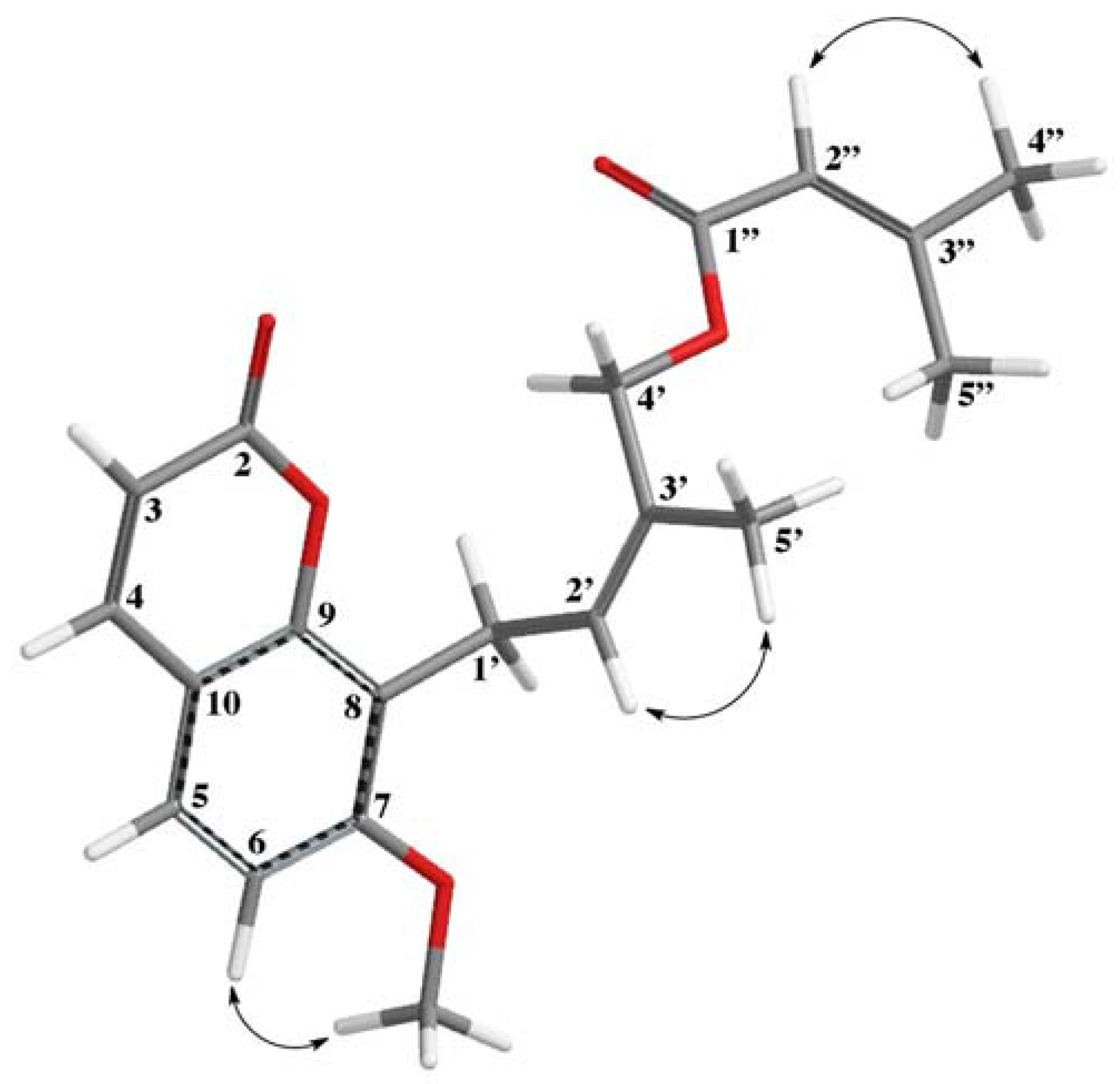
4′-Senecioiloxyosthol (1) was obtained as a colorless gum. The HRESIMS spectrum of 1 suggests a molecular formula of C20H22O5 with 10 degrees of unsaturation based on the [M + H]+ molecular peak at m/z 343.1544 (calcd m/z 343.1545). The 1H-NMR spectrum (Table 1) of 1 was very similar to that of osthol [13,16], with the exception of a missing vinylic methyl group signal of the prenyl side chain of osthol. Instead of two vinylic methyl signals, the 1H-NMR spectrum of 1 displayed a vinylic methyl group signal at δH 1.72 (3H) and a methylene singlet at δH 4.87 (2H), indicating that the second vinylic methyl group of osthol side chain was replaced with an acyloxy bearing methylene group. Furthermore, the typical vinylic narrow quintet proton signal observed at δH 5.72 (J = 1.3 Hz) along with the two vinylic methyl group doublets at δH 2.19 and 1.90 (each 3H, J = 1.3 Hz) suggest the presence of a senecioil group as the acyl group. The 2D-ROESY spectrum of 1 exhibited interactions between C-5′ methyl group protons and H-2′ proton of the prenylated side chain of osthol (Figure 2) as well as displayed interactions between H-6 and the methoxy group protons at C-7, and H-2″ proton and H-4″ methyl protons of the senecioiloxy acyl group, which clearly confirms the presence of a senecioiloxy acyl group at the C-4′ methyl group of osthol in 1. Furthermore, 13C-NMR (Table 1), 2D COSY, UV and IR spectroscopic data (see experimental section and supplemental data) of 1 corroborated the structure as 4′-senecioiloxyosthol. Previously, 4′-angeloiloxy derivative of osthol (2) (i.e., macrocarpin) was reported from Lomatium macrocarpum (Hook. & Arn.) C. & R., another Apiaceaen plant [21]. The 1H-NMR spectroscopic data reported for macrocarpin (2) were similar to that of 1 with the exception of the presence of angeloiloxy acyl group signals [i.e., δH 6.04 (1H, br t), 1.97 (3H) and 1.88 (3H)] instead of a senecioiloxy acyl group signals.
The antimicrobial activity of extracts and isolated coumarins of Prangos hulusii was evaluated against Gram-positive and Gram-negative nine reference standards and six clinically isolated microorganism strains. The results of minimum inhibition concentration (MIC, in µg/mL) values are summarized in Table 2. The best antimicrobial activity was observed against Escherichia coli with the dichlormethane (DCM) extract (i.e., MIC at 156 µg/mL), followed by the petroleum ether (PE) and methanol (MeOH) extracts (i.e., each MIC at 313 µg/mL). All three extracts showed good activity against Enterococcus faecalis (MIC at 313 µg/mL). Similar activities were detected with the DCM extract against Proteus mirabilis, with the PE extract against Staphylococcus aureus and with the MeOH extract against Klebsiella pneumoniae ATCC 4352. No activity was observed with all of the tested extracts against clinical isolates K. pneumoniae, Acinetobacter baumannii, and E. coli, and only a weak activity was detected against other reference and clinical isolate bacteria.
The new coumarin, 4′-senecioiloxyosthol (1), showed the best activity against Bacillus subtilis (Table 2) (MIC at 5 µg/mL), whereas the structurally related osthol (3) and isoimperatorin (9) displayed very good activity against clinical isolate Methicillin-resistant Staphylococcus aureus (MRSA) (MIC at 16 µg/mL), similar to the reference antibiotic Cefotaxime (CEF) (see Table 2). In contrast, auraptenol (5) and murraol (4) exhibited good activity against K. pneumoniae ATCC 4352 and Bacillus subtilis ATCC 9372 (MIC at 63 µg/mL) and poor activity against Methicillin Resistant Coagulase-Negative Staphylococci (MRCNS) (MIC at 125 µg/mL). Furthermore, auraptenol (5) displayed a good activity against Staphylococcus epidermidis ATCC 12228 and MRCNS (MICs at 63 and 125 µg/mL, respectively), osthol (3) against B. subtilis ATCC 9372, S. aureus ATCC 25923, K. pneumoniae ATCC 4352 and Methicillin-sensitive Staphylococcus aureus (MSSA) (all MICs at 125 µg/mL).
The antimycobacterial activity of prenylated coumarins and prenylated furanocoumarins [22,23] as well as the antimicrobial activity of furanocoumarins and prenylated furanocoumarins were reported previously [24]. In the latter publication, xanthotoxin (8-methoxyfuranocoumarin) was described as the most potent compound against B. subtilis ATCC 6633 strain with an MIC value at 30 µg/mL, whereas the new prenylated coumarin 4′-senecioiloxyosthol (1) was 6-fold more active against B. subtilis ATCC 9372 than that of xanthotoxin, with an MIC value at 5 µg/mL.
3. Materials and Methods
3.1. General Experimental Procedures
UV spectra were recorded on a UV-1700 PharmaSpec Shimadzu spectrophotometer (Shimadzu Corp., Kyoto, Japan) in MeOH. IR spectra were measured with a PerkinElmer Spectrum 2000 FT-IR spectrometer (PerkinElmer Corp., Waltham, MA, USA). NMR experiments were conducted on a Varian Mercury FT-NMR 400 MHz spectrometer (Agilent Corp., Santa Clara, CA, USA) using tetramethylsilane (TMS) as an internal standard. High Resolution Electrospray Ionization Mass Spectra (HRESIMS) and Electrospray Ionization Mass Spectra (ESIMS) were determined on Waters SYNAPT G1 mass spectrometer (Waters Corp., Milford, MA, USA).
3.2. Plant Material
The roots of Prangos hulusii were collected from Ödemiş by Hulusi Kütük, İzmir on March 2012, in Turkey. The plant was identified by Professor Emine Akalin Uruşak and a voucher specimen was deposited in the Herbarium of Istanbul University, Faculty of Pharmacy (ISTE 99676).
3.3. Isolation of Compounds
The dried and coarsely powdered roots (835 g) were exhaustively extracted with PE, DCM and MeOH, sequentially, using a Soxhlet Apparatus. The solvents were evaporated under reduced pressure in a rotary evaporator. The dichloromethane extract (55.6 g) was dissolved in acetone (2000 mL), and kept in a refrigerator overnight. Following the removal of precipitated hydrocarbon mixtures by filtration, the solvent was removed in vacuo to yield 45.8 g viscous oil. A portion of the defatted extract (5 g) was chromatographed on a Sephadex LH-20 (5 cm × 60 cm) packed in hexane-dichloromethane-methanol (7:4.5:0.5) and Preparative Thin Layer Chromatography (prep. TLC) (1-2 mm thickness, silica gel developed with cyclohexane-EtOAc mixtures, 4:1, 3:2, 1:1) was used for the final purification of compounds. 4′-senecioiloxyosthol (1) (4.7 mg), osthol (3) (39 mg), murraol (4) (5 mg), auraptenol (5) (8.2 mg), meranzin (6) (7 mg), hydroxyosthol-epoxide (7) (4 mg), meranzin hydrate (8) (12 mg), isoimperatorin (9) (23 mg), oxypeucedanin (10) (60 mg), psoralen (11) (7 mg), stigmasterol (3 mg), and β-sitosterol (2.8 mg) were isolated. Furthermore, meranzin (6) (11.3 mg), meranzin hydrate (8) (10.2 mg), and auraptenol (5) (7.6 mg) were prepared from osthol (3) semi-synthetically [16] as a reference material.
3.4. 4′-Senecioiloxyosthol (1)
IR (KBr) νmax: 2970, 2915, 2842, 1732, 1717, 1651, 1608, 1281, 1251, 1226, 1145, 1118, 1090 and 832 cm−1. UV (MeOH) λmax (log ε) 321 (4.06), 258(sh) (3.82), 247 (3.26) and 224(sh) (3.81) nm. For 1H (CDCl3, 400 MHz) and 13C-NMR (CDCl3, 100 MHz) spectroscopic data, see Table 1; HRESIMS m/z: 343.1544 (calcd for C20H23O5 343.1545).
3.5. Antimicrobial Activity
The antimicrobial activity of the extracts and isolated coumarins of Prangos hulusii was evaluated against nine reference standard microorganisms, both Gram-positive and Gram-negative; S. aureus ATCC 25923, S. epidermidis ATCC 12228, E. faecalis ATCC 29212, Pseudomonas aeruginosa ATCC 27853, E. coli ATCC 10799, K. pneumoniae ATCC 4352, Salmonella choleraesuis ATCC 14028, P. mirabilis ATCC 7002, B. subtilis ATCC 9372, and six clinical isolates; (MSSA), (MRSA), (MRCNS), K. pneumoniae, A. baumannii, E. coli, by using a standard microbroth dilution method modified with rezasurin [25,26]. The experiments were performed with two replications and the results were expressed as average values.
3.6. Determination of Antibacterial Activity
The MIC values of extracts and isolated compounds were determined using microbroth dilution method in 96-well microtitre plates. The bacterial cultures were prepared from overnight cultures on Tryptic Soy Agar (TSA) at 37 °C for 24 h by diluting in Mueller Hinton Broth (MHB) from approx. 108 CFU/mL to 2 × 10⁶ CFU/mL. Then, 50 μL Mueller Hinton Broth (MHB) was added to the wells starting from the first well and continuing up to the twelfth. The extracts and isolated compounds were prepared 1/10 (v/v) in DMSO and 50 μg/mL of these were added to the first wells. Two-fold serial dilutions were made, achieving a final concentration ranging from 5000 to 10 μg/mL. The positive controls for Ciprofloxacin (CPR), Tetracycline (TTR), Cefotaxime (CEF), and Oxacillin (OXA) were determined with the final concentrations from 64 to 0.1 µg/mL. In addition, an extra row of DMSO was used as a vehicle control to determine its possible inhibitory activity. Finally, 25 μL of bacterial suspensions and % 0.001 resazurin solution were added to each well.
After incubating the bacteria at 37 °C for 24 h, the microtitre plates were examined visually for microbial growth which appeared as pink, colored by resazurin dye. In each row, the well containing the least concentration that showed no visible growth was considered the MIC. The bacterial samples were inoculated on TSA plates and incubated at 37 °C for 24 h.
4. Conclusions
Investigation of the dichloromethane extract of the roots of P. hulusii yielded several pyrenylated coumarins and furanocoumarins with antimicrobial activities. Prangos species frequently used for the treatment of burns and wounds in traditional folk medicine [3,4,5,6], perhaps the presence of pyrenylated coumarins with antimicrobial activity may play an important role for the aforementioned folkloric use of Prangos species.
Supplementary Materials
Supplementary materials containing spectroscopic data of the new coumarin are available online.
Acknowledgments
We thank Hulusi Kütük for the collection and Emine Akalın Uruşak for the identification of plant material. This work was supported by grant from the Scientific Research Projects Coordination Unit of Istanbul University, grant No. 39751.
Author Contributions
N.T. suggested the idea of the investigations; N.T. and M.M. designed the experiments obtained; N.T., S.Y.-T. and M.M. purified, and characterized all compounds for biological assays; N.T., M.M. and E.T. contributed to the discussion of results, and wrote the paper. M.B. and E.T. measured the antimicrobial activity. All authors read and approved the final manuscript.
Conflicts of Interest
The authors declare no conflict of interest.
References
- Pimenov, M.G.; Leonov, M.V. The Asian Umbelliferae biodiversity database (ASIUM) with particular feference to South-West Asian taxa. Turk. J. Bot. 2004, 28, 139–145. [Google Scholar]
- Güner, A.; Aslan, S.; Ekim, T.; Vural, M.; Babac, M.T. A Checklist of the Flora of Turkey (Vascular Plants); Nezahat Gokyigit Botanic Garden Publications, Flora Series I: İstanbul, Turky, 2012. [Google Scholar]
- Sharma, N.; Ashok, P.K.; Negi, A.; Lakshmayya, B. A review on ethnobotany, phytochemical and pharmacological dynamics of Prangos pabularia Lindl. J. Nat. Rem. 2013, 13, 68–75. [Google Scholar]
- Kafash-Farkhad, N.; Asadi-Samani, M.; Rafieian-Kopaei, M. A review on phytochemistry and pharmacological effects of Prangos ferulacea (L.) Lindl. Life Sci. J. 2013, 10, 360–367. [Google Scholar]
- Doković, D.D.; Bulatović, V.M.; Božić, B.D.; Kataranovski, M.V.; Zrakić, T.M.; Kovačević, N.N. 3,5-Nonadiyne isolated from the rhizome of Cachrys ferulacea inhibits endogenous nitric oxide release by rat peritoneal macrophages. Chem. Pharm. Bull. 2004, 52, 853–854. [Google Scholar]
- Ulubelen, A.; Topcu, G.; Tan, N.; Olcal, S.; Johansson, C.; Ucer, M.; Birman, H.; Tamer, S. Biological activities of a Turkish medicinal plant, Prangos platychlaena. J. Ethnopharmacol. 1995, 45, 193–197. [Google Scholar] [CrossRef]
- Shikishima, Y.; Takaishi, Y.; Honda, G.; Ito, M.; Takeda, Y.; Kodzhimatov, O.K.; Ashurmetov, O. Terpenoids and γ-pyrone derivatives from Prangos tschimganica. Phytochemistry 2001, 57, 135–141. [Google Scholar] [CrossRef]
- Shikishima, Y.; Takaishi, Y.; Honda, G.; Ito, M.; Takeda, Y.; Kodzhimatov, O.K.; Ashurmetov, O.; Lee, K.H. Chemical constituents of Prangos tschimganica; structure elucidation and absolute configuration of coumarin and furanocoumarin derivatives with anti-HIV activity. Chem. Pharm. Bull. 2001, 49, 877–880. [Google Scholar] [CrossRef] [PubMed]
- Tada, Y.; Shikishima, Y.; Takaishi, Y.; Shibata, H.; Higuti, T.; Honda, G.; Ito, M.; Takeda, Y.; Kodzhimatov, O.K.; Ashurmetov, O.; et al. Coumarins and γ-pyrone derivatives from Prangos pabularia: Antibacterial activity and inhibition of cytokine release. Phytochemistry 2002, 59, 649–654. [Google Scholar] [CrossRef]
- Sajjadi, S.E.; Mehregan, I. Chemical composition of the essential oil of Prangos asperula Boiss. Subsp. haussknechtii (Boiss.) Herrnst. Etheyn fruits. DARU J. Fac. Pharm. Sci. 2003, 11, 79–81. [Google Scholar]
- Şenol, G.S.; Hasan, Y.; Özcan, S. Prangos hulusii sp. nov. (Apiaceae) from West Anatolia, Turkey. Nord. J. Bot. 2011, 29, 402–407. [Google Scholar]
- Tütüniş-Yazıcı, S.; Tan, N.; Meriçli, F.; Özsoy, N.; Tan, E. Biological activities of endemic Prangos hulusii. Planta Med. 2013, 79, PN113. [Google Scholar]
- Wei, Y.; Zhang, T.; Ito, Y. Preparative isolation of osthol and xanthotoxol from Common Cnidium Fruit (Chinese traditional herb) using stepwise elution by high-speed counter-current chromatography. J. Chromatogr. A 2004, 1033, 373–377. [Google Scholar] [CrossRef] [PubMed]
- Ito, C.; Furukawa, H. Constituents of Murraya exotica L. structure elucidation of new coumarins. Chem. Pharm. Bull. 1987, 35, 4277–4285. [Google Scholar] [CrossRef]
- Nakatani, N.; Yamada, Y.; Fuwa, H. 7-Geranyloxycoumarin from juice oil of Hassaku (Citrus hassaku) and antimicrobial effects of related coumarins. Agric. Biol. Chem. 1987, 51, 419–423. [Google Scholar]
- Riviere, C.; Goossens, L.; Pommery, N.; Fourneau, C.; Delelis, A.; Henichart, J.P. Antiproliferative effects of isopentenylated coumarins isolated from Phellolophium madagascariense Baker. Nat. Prod. Res. 2006, 20, 909–916. [Google Scholar] [CrossRef] [PubMed]
- Zhao, J.; Zhou, M.; Liu, Y.; Zhang, G.; Luo, Y. Chromones and coumarins from the dried fructus of Cnidium monnieri. Fitoterapia 2011, 82, 767–771. [Google Scholar] [CrossRef] [PubMed]
- Seo, W.D.; Kim, Y.J.; Ryu, H.W.; Kim, J.H.; Han, S.; Ra, J.; Seo, K.H.; Jang, K.C.; Leeb, J.H. Identification and characterisation of coumarins from the roots of Angelica dahurica and their inhibitory effects against cholinesterase. J. Func. Foods 2013, 5, 1421–1431. [Google Scholar] [CrossRef]
- Chunyan, C.; Bo, S.; Ping, L.; Jingmei, L.; Ito, Y. Isolation and purification of psoralen and bergapten from Ficus carica L leaves by high-speed countercurrent chromatography. J. Liq. Chromatogr. Relat. Technol. 2009, 32, 136–143. [Google Scholar] [CrossRef] [PubMed]
- Pierre, L.L.; Moses, M.N. Isolation and Characterisation of Stigmasterol and β-Sitosterol from Odontonema strictum (Acanthaceae). J. Innov. Pharm. Biol. Sci. 2015, 2, 88–96. [Google Scholar]
- Steck, W. Coumarins and chromones from Lomatium macrocarpum. Phytochemistry 1973, 12, 2283–2286. [Google Scholar] [CrossRef]
- Schinkovitz, A.; Gibbons, S.; Stavri, M.; Cocksedge, M.J.; Bucar, F. Ostruthin: An antimycobacterial coumarin from the roots of Peucedanum ostruthium. Planta Med. 2003, 69, 369–371. [Google Scholar] [CrossRef] [PubMed]
- Stavri, M.; Gibbons, S. The antimycobacterial constituents of dill (Anethum graveolens). Phytother. Res. 2005, 19, 938–941. [Google Scholar] [CrossRef] [PubMed]
- Walasek, M.; Grzegorczyk, A.; Malm, A.; Skalicka-Wozniak, K. Bioactivity-guided isolation of antimicrobial coumarins from Heracleum mantegazzianum Sommier & Levier (Apiaceae) fruits by high-performance counter-current chromatography. Food Chem. 2015, 186, 133–138. [Google Scholar] [PubMed]
- Sarker, S.D.; Nahar, L.; Kumarasamy, Y. Microtitre plate-based antibacterial assay incorporating resazurin as an indicator of cell growth, and its application in the In Vitro antibacterial screening of phytochemicals. Methods 2007, 42, 321–324. [Google Scholar] [CrossRef] [PubMed]
- Clinical and Laboratory Standards Institute (CLSI (NCCLS)). M7-A7, Methods for Dilution Antimicrobial Susceptibility Tests for Bacteria That Grow Aerobically; Approved Standard, Seventh Edition 1-56238-587-9; Clinical and Laboratory Standards Institute: Wayne, PA, USA, 2006. [Google Scholar]
Sample Availability: Samples of the compounds are not available from the authors. |
Figure 1.
Structures of prenylated coumarins 1–10.
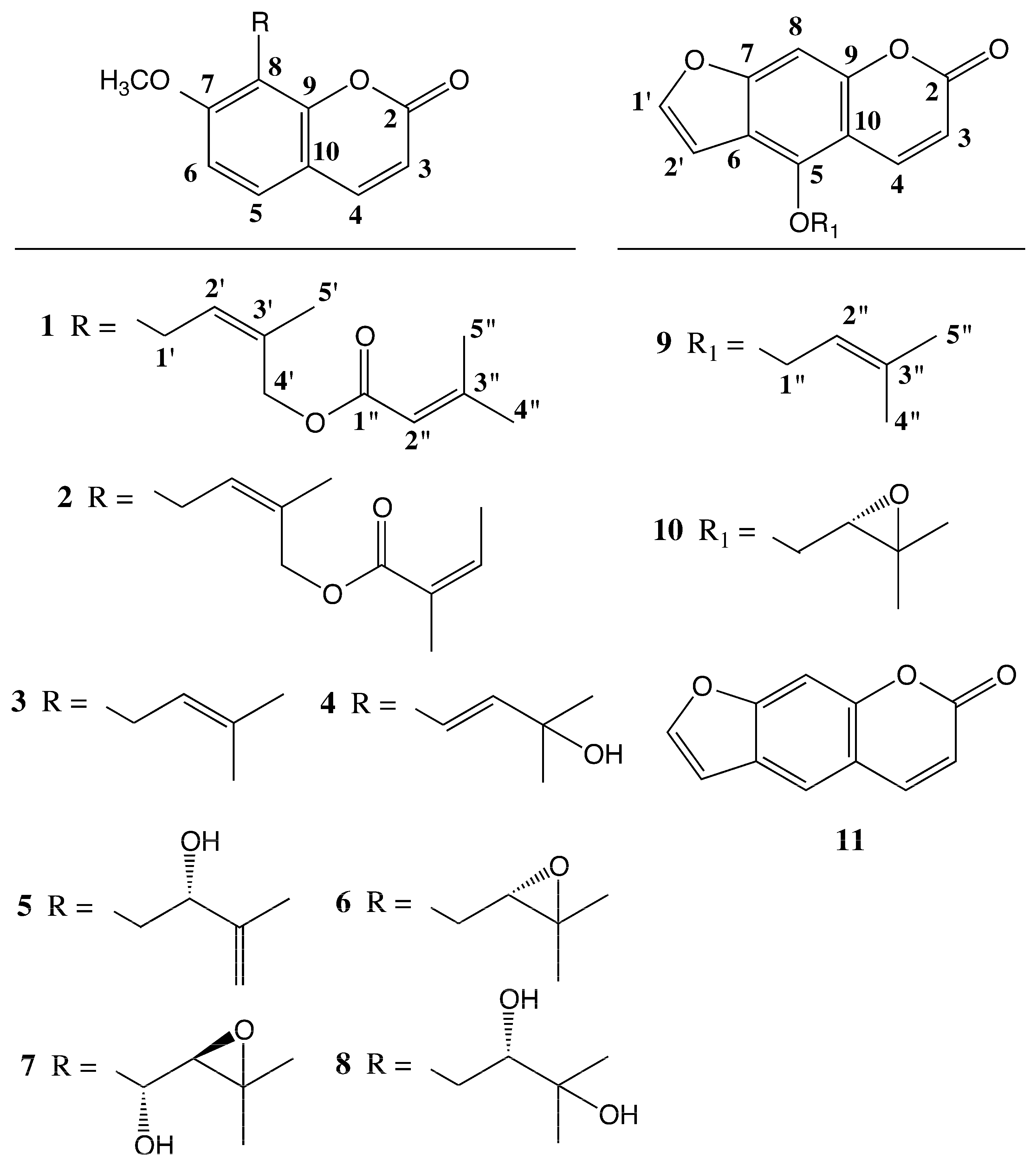
Figure 2.
Interactions observed in the ROESY spectrum of 4′-Senecioiloxyosthol.


Table 1.
1H-NMR and 13C-NMR data of Compound 1.
| Positions | ΔH (J in Hz) | ΔC, Type |
|---|---|---|
| 2 | 161.14, C | |
| 3 | 6.23 d (9.8), 1H | 116.08, CH |
| 4 | 7.60 d (9.8), 1H | 143.65, CH |
| 5 | 7.30 d (8.4), 1H | 126.46, CH |
| 6 | 6.82 d (8.4), 1H | 107.3, CH |
| 7 | 160.09, C | |
| 8 | 116.66, C | |
| 9 | 152.86, C | |
| 10 | 112.92, C | |
| -OCH3 | 3.89 s, 3H | 56.01, CH3 |
| 1′ | 3.62 d (7.9), 2H | 21.45, CH2 |
| 2′ | 5.50 br t (7.9), 1H | 113.04, CH |
| 3′ | 131.24, C | |
| 4′ | 4.87 s, 2H | 62.40, CH2 |
| 5′ | 1.72 br s, 3H | 21.61, CH3 |
| 1″ | 166.8, C | |
| 2″ | 5.72 quint (1.3), 1H | 126.51, CH |
| 3″ | 156.41, C | |
| 4″ | 1.90 br d (1.3), 3H | 27.39, CH3 |
| 5″ | 2.17 br d (1.3), 3H | 20.2, CH3 |
Table 2.
Antimicrobial activity of the prenylated coumarins and root extracts of P. hulusii.
| Test Strains | Prenylated Coumarins (μg/mL) ** | Extracts (μg/mL) *** | References **** | ||||||||||||
|---|---|---|---|---|---|---|---|---|---|---|---|---|---|---|---|
| 1 * | 3 | 4 | 5 | 6 | 8 | 9 | 10 | DCM | PE | MeOH | TTR | OXA | CEF | CPR | |
| Minimum Inhibitory Concentration (MIC) μg/mL | |||||||||||||||
| S. epidermidis ATCC 12228 | 625 | >125 | >125 | 63 | >125 | 125 | >125 | 125 | 625 | 313 | 625 | 64 | 0.5 | 4 | 0.5 |
| S. aureus ATCC 25923 | 625 | 125 | >250 | >250 | >250 | 250 | >250 | 250 | 1250 | 625 | 1250 | 0.5 | 0.5 | 2 | 1 |
| E. faecalis ATCC 29212 | 313 | >250 | >250 | >250 | >250 | 250 | >250 | 250 | 313 | 313 | 313 | 16 | 8 | 2 | 1 |
| K. pneumoniae ATCC 4352 | 313 | 125 | 63 | 63 | 125 | 250 | 125 | 250 | 625 | 625 | 313 | 8 | 4 | 2 | 0.5 |
| B. subtilis ATCC 9372 | 5 | 125 | 63 | 63 | >250 | 250 | >250 | 125 | 1250 | 1250 | 1250 | 0.3 | 0.1 | 0.5 | 0.5 |
| E. coli ATCC 10799 | 313 | >125 | >125 | >125 | >125 | 250 | >125 | 250 | 156 | 313 | 313 | 2 | 32 | 1 | 0.3 |
| P. aeruginosa ATCC 27853 | 156 | >125 | >125 | >125 | >125 | 250 | >125 | 125 | 313 | 313 | 625 | 32 | >64 | 16 | 0.5 |
| S. choleraesuis ATCC 14028 | 313 | >125 | >125 | >125 | >125 | 125 | >125 | 125 | 625 | 625 | 1250 | 4 | >64 | 0.3 | 0.3 |
| P. mirabilis ATCC 7002 | 313 | >125 | >125 | >125 | >125 | 250 | >125 | 250 | 313 | 625 | 625 | 64 | 32 | 1 | 1 |
| Methicillin-sensitive Staphylococcus aureus (MSSA) | 625 | 125 | >250 | >250 | >250 | 250 | >250 | 250 | 1250 | 1250 | 2500 | 64 | 64 | 2 | 0.1 |
| Methicillin-resistant Staphylococcus aureus (MRSA) | 625 | 16 | >250 | >250 | >250 | 250 | 16 | 125 | 1250 | 1250 | 2500 | 8 | 8 | 16 | 0.5 |
| Methicillin Resistant Coagulase-Negative Staphylococci (MRCNS) | 313 | >250 | 125 | 125 | 125 | 125 | >250 | 125 | 1250 | 1250 | 1250 | 8 | 8 | 16 | 4 |
| K. pneumoniae | 313 | >125 | >125 | >125 | >125 | 250 | >125 | 250 | 2500 | 2500 | 2500 | 2 | >64 | >64 | 2 |
| A. baumannii | n.t. | >125 | >125 | >125 | >125 | 125 | >125 | 125 | 2500 | 1250 | 2500 | n.t. | n.t. | n.t. | >16 |
| E. coli | 156 | >125 | >125 | >125 | >125 | 250 | >125 | 250 | 2500 | 2500 | 2500 | >64 | >64 | >64 | 0.5 |
Starting concentrations: * 1250 μg/mL; ** 250 μg/mL; *** 5000 μg/mL; **** 64 μg/mL. TTR = Tetracycline, OXA = Oxacilline, CEF = Cefotaxime, CPR = Ciprofloxacin. n.t.: not tested PE: petroleum ether; DCM: dichlormethane; MeOH: methanol.
© 2017 by the authors. Licensee MDPI, Basel, Switzerland. This article is an open access article distributed under the terms and conditions of the Creative Commons Attribution (CC BY) license (http://creativecommons.org/licenses/by/4.0/).
Share and Cite
MDPI and ACS Style
Tan, N.; Yazıcı-Tütüniş, S.; Bilgin, M.; Tan, E.; Miski, M. Antibacterial Activities of Pyrenylated Coumarins from the Roots of Prangos hulusii. Molecules 2017, 22, 1098. https://doi.org/10.3390/molecules22071098
AMA Style
Tan N, Yazıcı-Tütüniş S, Bilgin M, Tan E, Miski M. Antibacterial Activities of Pyrenylated Coumarins from the Roots of Prangos hulusii. Molecules. 2017; 22(7):1098. https://doi.org/10.3390/molecules22071098
Chicago/Turabian StyleTan, Nur, Seçil Yazıcı-Tütüniş, Merve Bilgin, Emir Tan, and Mahmut Miski. 2017. "Antibacterial Activities of Pyrenylated Coumarins from the Roots of Prangos hulusii" Molecules 22, no. 7: 1098. https://doi.org/10.3390/molecules22071098