Article

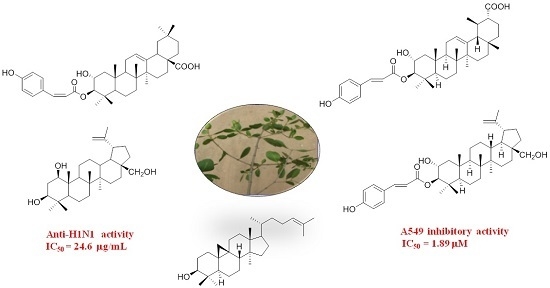
Cytotoxic and Antiviral Triterpenoids from the Mangrove Plant Sonneratia paracaseolaris

1
Key Laboratory of Marine Drugs, Chinese Ministry of Education, School of Medicine and Pharmacy, Ocean University of China, Qingdao 266003, China
2
College of Chemistry and Chemical Engineering, Ocean University of China, Songling Road 238, Qingdao 266100, China
3
Cancer Research institute, Binzhou Medical University Hospital, Yellow river second Road 661, Binzhou 256603, China
*
Authors to whom correspondence should be addressed.
These authors contributed equally to this work.
Molecules 2017, 22(8), 1319; https://doi.org/10.3390/molecules22081319
Submission received: 18 July 2017 / Revised: 2 August 2017 / Accepted: 7 August 2017 / Published: 9 August 2017
(This article belongs to the Section Natural Products Chemistry)

Abstract

:
A chemical investigation was conducted on the aerial parts of the mangrove plant Sonneratia paracaseolaris, yielding five new triterpenoid paracaseolins A–E (14, and 11) together with twelve known analogues (510, 1217). Their structures were established by extensive spectroscopic methods and comparisons their spectroscopic data with those of the known related compounds. The cytotoxicities against P388, HeLa, A549, and K562 tumor cell lines and anti-H1N1 (Influenza A virus) activities for the isolates were evaluated. Compound 4 showed potent cytotoxicity against the A549 cell line with an IC50 value of 1.89 µM, and compound 1 exhibited significant anti-H1N1 virus activity with an IC50 value of 28.4 µg/mL. A preliminary structure activity relationship was discussed.

Graphical Abstract

1. Introduction

Mangrove plants of the genus Sonneratia (family Sonneratiaceae) consist of nine species and are generally distributed in the tropical and subtropical regions in the world [1]. The fruit, bark, and leaves of several Sonneratia plants have been used as folk medicines for treatment of various diseases, such as asthma, febris, ulcers, hepatitis, piles, sprains, and hemorrhages [2]. So far, almost half of the species of the genus have been studied for their chemical constituents, yielding various types of compounds, such as triterpenoids, steroids, alkaloids, flavonoids, aromatics, and tannins, some of which exhibited cytotoxic [3,4,5], anti-HIV [5], and antioxidant [6] activities.
Sonneratia paracaseolaris is one of the six Sonneratia species in China [7] which was reported to be a natural hybrid of Sonneratia alba and Sonneratia caseolaris [8,9]. At present, only one report regarding the chemical study of Sonneratia paracaseolaris provided a novel α-alkylbutenolide dimer [10], triggering our interest to study the chemical profiles of Sonneratia paracaseolaris. In the previous chemical and bioassay experiments, we found that the methanol extract showed moderate cytotoxicities against the A549 cell line and was rich in triterpenoids. In order to find bioactive triterpenoids from this plant, combined chemistry and bioassay-guided quick isolation of triterpenoid-rich portions of the methanol extract yielded the five new triterpenoid paracaseolins A–E (14 and 11), together with twelve known ones (510, 1217) (Figure 1). These triterpenoid compounds could be divided into four carbon skeletons: lupane-, ursane-, oleanane-, and cycloartane-types. The major structure isolated from the species was characterized by lupane-type triterpenoids (110). All the isolates (117) were evaluated for cytotoxic activities against the selected P388, HeLa, A549, and K562 tumor cell lines, as well as anti-influenza A H1N1 virus activities. Herein, we describe the isolation, structural elucidation, and the cytotoxic and anti-H1N1 activities of all the isolates.

2. Results and Discussion

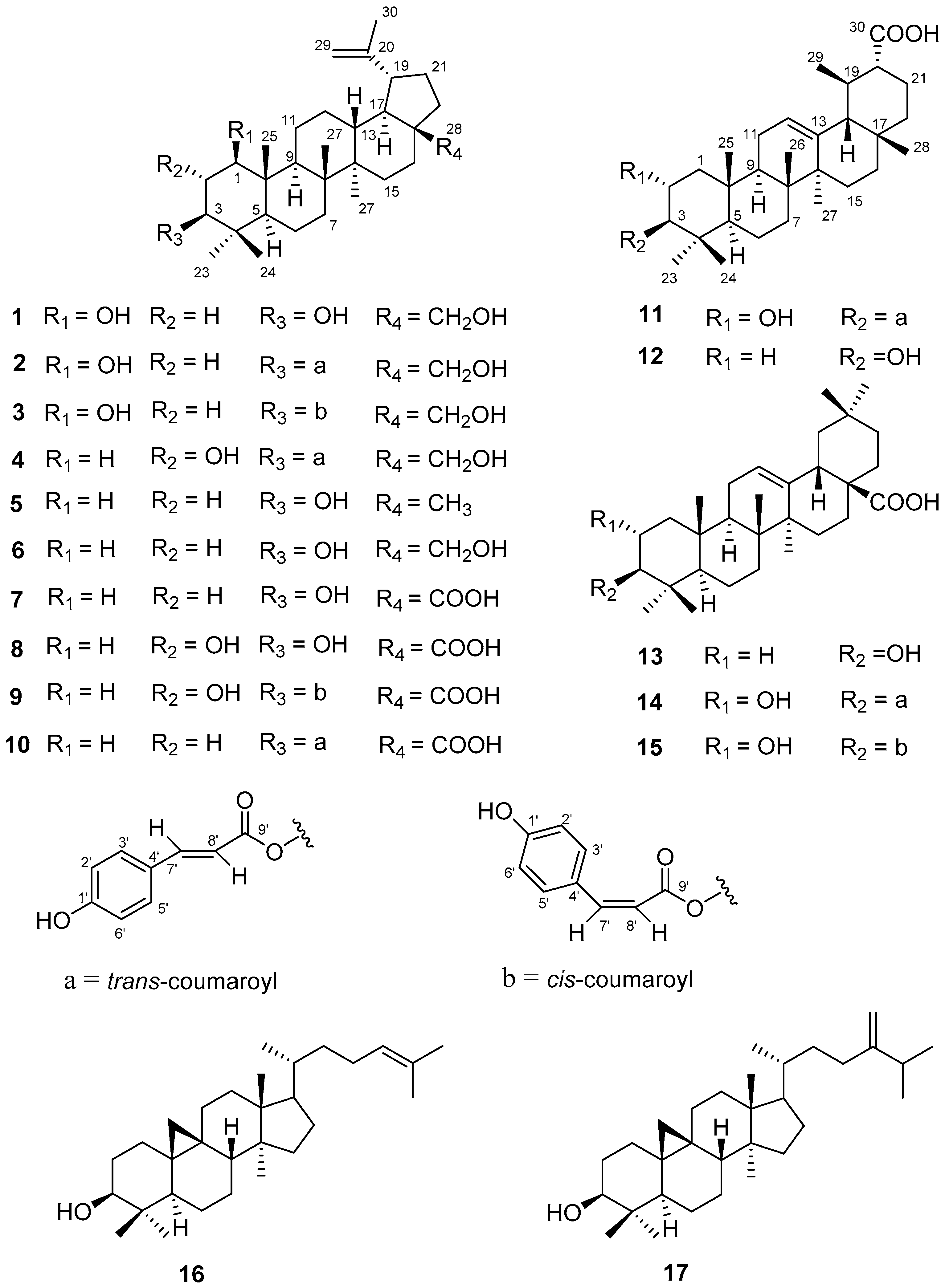
2.1. Structure Elucidation

Paracaseolin A (1) was isolated as irregular plates with a molecular formula C30H50O3 established by HR-ESI-MS (high resolution electrospray ionization mass spectrometry) ([M − H] at m/z = 457.3688; calcd. for 457.3676). The IR absorption bands at 3359, 1681, and 881 cm−1 were assigned to hydroxyl and olefinic groups. 13C-NMR (Table 1) and DEPT (distortionless enhancement by polarization transfer) spectra exhibited a total of 30 carbon signals, which were classified into six methyls, eleven methylenes (including one oxymethylene and one olefinic carbon), seven methines (including two oxymethines), and six quaternary carbons (including one olefinic carbon). The 1H-NMR spectrum (Table 1) showed five singlet methyls at δH 0.61 (3H, s, H-24), 0.76 (3H, s, H-25), 0.83 (3H, s, H-23), 0.93 (3H, s, H-27), and 0.98 (3H, s, H-26), indicating the presence of a triterpenoidal skeleton of compound 1. Additionally, two terminal olefinic proton signals at δH 4.53 (1H, s, H-29) and 4.66 (1H, s, H-29), along with an olefinic methyl singlet at δH 1.63 (3H, s, H-30), indicated the presence of an isopropenyl residue possibly belonging to a lupane skeleton [11]. In fact, the NMR data of 1 were closely similar to the reported lup-20(29)-en-2α,3β,28-triol, except for those of C1–C3 in the A ring [12]. Connective 1H-1H COSY (hydrogen-hydrogen correlation spectroscopy) correlations from H-1 to H-3 and the HMBC (heteronuclear multiple bond correlation) cross peaks of H3-25 with C-1 (δC 77.9), C-5 (δC 52.6), and C-10 (δC 42.9), of H3-23 and H3-24 with C-3 (δC 74.0), C-4 (δC 38.0), and C-5 further suggesting compound 1 to be lup-20(29)-en-2,3,28-triol. The relative configurations of H-1 and H-3 were assigned as axial α-orientation by the half-peak-width data, 22.1 Hz for H-1 and 22.8 Hz for H-3 [13], together with the NOESY (nuclear overhauser effect spectroscopy) correlations between H-3 (δH 2.96, m) and H3-23 (δH 0.83, s) and between H-1 (δH 3.14, m) and H-5 (δH 0.46, d, J = 11.0 Hz), which was consistent with the stable cyclohexane conformation. Thus, compound 1 was determined as 1β,3β-dihydroxy betulin, named as paracaseolin A (1).
Paracaseolin B (2), isolated as fine needles, had the molecular formula C39H56O5 established by HR-ESI-MS data ([M − H] at m/z 603.4058; calcd. for 603.4044). The UV spectrum exhibited absorption maxima at 228 and 312 nm, suggesting the presence of aromatic rings in the molecule. The IR spectrum showed absorption bands for hydroxyl (3376 cm−1), α,β-unsaturated carbonyl (1682 cm−1), and aromatic (1604, 1513, 1451, and 831 cm−1) functionalities. In the 1H-NMR spectrum (Table 1) of 2, the signals at δH 7.53 (2H, d, J = 8.6 Hz, H-2′, H-6′), 7.51 (1H, d, J = 16.0 Hz, H-7′), 6.77 (2H, d, J = 8.6 Hz, H-3′, H-5′), and 6.33 (1H, d, J = 16.0 Hz, H-8′) implied the presence of the partial structure, a trans-p-coumaroyl functionality. Additional 1D NMR data for 2 were almost identical to 1, except for a distinct downfield shift of H-3 at δH 4.47 (1H, dd, J = 11.9, 4.7 Hz, H-3) in 2, indicating the position of the O-trans-p-coumaroyl functionality at C-3. This speculation was further confirmed by HMBC correlations of H-3, H-7′ and H-8′ with C-9′ (δC 166.2). The large coupling constants of H-3 (J = 11.9 Hz) and H-1 (J = 10.8 Hz), as well as the NOESY correlations of both H-3 and H-1 with H-5 could assign their axial α-orientations. Compound 2 was then identified as 1β-hydroxy-3β-O-trans-p-coumaroyl betulin.
Paracaseolin C (3), a white amorphous powder, had the same molecular formula of C39H56O5 as compound 2 from the HR-ESI-MS data ([M − H] at m/z 603.4056; calcd. for 603.4044). A comparable study of 1D NMR data (Table 2) of 3 with 2 (Table 1) showed their closely structural similarity except for the small coupling constant of H-7′ and H-8′ (JH-7′/H-8′ = 13.0 Hz) in 3, indicating the presence of a cis-p-coumaroyl group. Accordingly, compound 3 was determined as 1β-hydroxy-3β-O-cis-p-coumaroyl betulin.
Paracaseolin D (4), a white amorphous powder, had the molecular formula C39H56O5, as determined by HR-ESI-MS data ([M − H] at m/z 603.4056; calcd. for 603.4044). 1D NMR spectra of compound 4 were very similar to those of 2 apart from a slight difference in ring A. The split pattern of H-3 (δH 4.48, d, J = 10.0 Hz) in 4 showed double peak instead of multiple peak in 2 and the carbon signal at C-3 shifted from δC 76.7 in 2 to δC 83.4 in 4 (Table 2), suggesting the ring A of compound 4 to be 2, 3-dioxyged structure which was supported by 1H-1H COSY correlation between H-2 (δH 3.67) and H-3. The O-trans-p-coumaroyl group was also assigned at C-3 on the basis of the HMBC correlation of H-3 with C-9′ (δC 166.7). The half-peak-width value (26.6 Hz) of H-2 further indicated an axial β-orientation. Compound 4 was finally determined as 2α-hydroxy-3β-O-trans-p-coumaroyl betulin.
HR-ESI-MS data ([M − H] at m/z 617.3850; calcd. for 617.3837) showed the molecular formula of paracaseolin E (11) as C39H54O6. The IR spectrum displayed hydroxyl (3485 cm−1), carboxylic acid (1710 cm−1), α,β-unsaturated carbonyl (1682 cm−1) and phenyl (1603, 1510, 1457, and 832 cm−1) absorption bands. A trans-coumaroyl moiety was observed by comparison the 1D NMR data with those of 2 (Table 1). Apart from the trans-coumaroyl moiety, 30 skeleton carbon signals were observed in 13C-NMR spectrum. In its 1H-NMR spectrum, seven characteristic methyl signals including six singlet methyls (δH 0.76, 0.80, 0.85, 0.92, 0.97, 1.07) and one doublet (δH 0.83, d, J = 6.1 Hz) further suggested compound 11 could be a ursonic acid type triterpenoid similar to the known dulcioic acid, the co-isolated compound 12 (Figures S37–S40, Supporting Information) [14]. The two proton signals at δH 3.69 (1H, m, W1/2) = 22.9 Hz, H-2) and 4.51 (1H, d, J = 9.9 Hz, H-3), as well as two carbon resonances at δc 64.8 (C-2) and 83.6 (C-3), and the additional trans-coumaroyl signals, indicated the ring A of 11 identical with 4. This speculation was further confirmed by the combined analysis of 1H-1H COSY and HMBC spectra (Figure 2). The doublet methyl was assigned at C-19 rather than C-20 due to the 1H-1H COSY correlations of H-19 (δH 1.32, m) with H-18 (δH 2.12, d, J = 11.0 Hz) and H3-29 (δH 0.83, d, J = 6.1 Hz), and the HMBC correlations of H3-29 with C-18 (δc 52.4) and C-20 (δc 38.5). The substituted pattern of 2-hydroxy-3-O-trans-p-coumaroyl was also supported by the HMBC correlation of H-3 and C-9′ (δc 166.7), and the 1H-1H COSY correlation between H-3 and H-2. The relative configuration of 2α-hydroxy-3β-O-trans-p-coumaroyl was in agreement with compound 4 dependent on the half-peak-width value (26.9 Hz) for H-2 and the NOESY correlation of H-2 with H3-25, together with the large coupling constant of H-3 (JH-3/H-2 = 9.9 Hz). Accordingly, compound 11 was identified as 2α-hydroxy-3β-O-trans-p-coumaroyl dulcioic acid.
In addition to the new compounds, a spectroscopic data comparison with the literature allowed the known compounds to be identified as lupeol (5) [15,16], betulin (6) [11,17], betulinic acid (7) [11,18], alphitolic acid (8) [19], 3β-O-cis-p-coumaroyl alphitolic acid (9) [20,21], 3β-O-trans-p-coumaroyl betulinic acid (10) [22], dulcioic acid (12) [14], oleanolic acid (13) [23], 3β-O-trans-p-coumaroyl maslinic acid (14) [24], 3β-O-cis-p-coumaroyl maslinic acid (15) [25], cycloartenol (16) [26], and 24-methylenecycloartenol (17) [26], respectively.

2.2. Biological Evaluations

The cytotoxicities of the isolates were evaluated against the selected K562, P388, HeLa, and A549 tumor cell lines in vitro with adriamycin (ADM) as a positive control, and the results are summarized in Table 3. Almost all of the compounds were active against the P388 cell line and exhibited different levels of cytotoxicities against HeLa and A549 cell lines, but were not sensitive toward the K562 cell line. In particular, new compound 4 showed the strongest cytotoxicity against A549 cells with an IC50 value of 1.89 µM, and compounds 3 and 14 displayed low IC50 values, ranging from 11.04 to 13.10 µM, against A549, HeLa, and P388 cell lines, whereas only 15 showed cytotoxic activity against the K562 cell line with an IC50 value of 16.28 µM. Furthermore, a preliminary structure activity relationships were analyzed. Comparing the cytotoxic data of 1 and 3, 8 and 9, as well as 13 and 14 against HeLa and P388 cell lines, it is suggested that the introduction of an O-p-coumaroyl group at C-3 could increase the cytotoxicities no matter the cis or trans configuration, which is consistent with previous reports [21]. Another comparison between 2 and 4, and 7 and 8 implied that 2-OH played significant roles on the cytotoxicities, whereas the OH group at C-1 and the substituent patterns at C-28 showed no effects, which could be predicted from a comparison between compounds 1 and 57.
Antiviral activities of all the isolates against influenza A H1N1 virus (IAV) were also evaluated by CPE (the cytopathic effects) assay [27]. Only new compound 1 exhibited significant anti-H1N1 virus activity with an IC50 value of 28.4 µg/mL, very close to the positive control of Ribavirin with an IC50 value of 24.6 µg/mL. The other compounds showed no activity with inhibition rates less than 50% at 50 µg/mL (Table S2, Supporting Information).

3. Materials and Methods

3.1. General Experimental Procedures

Optical rotations were measured with a JASCO P-1020 polarimeter (JASCO, Tokyo, Japan). UV spectra were taken on a Beckman DU640 spectrophotometer (Beckman Coulter Inc., Brea, CA, USA). IR spectra were recorded on a NICOLET NEXUS 470 spectrophotometer (International Equipment Trading Ltd., Vernon Hills, IL, USA) in KBr discs, with υ in cm−1. 1H-, 13C-NMR, DEPT, and 2D NMR spectra were recorded on a JEOL JNM-ECP 600 (JEOL Ltd., Tokoyo, Japan) and Varian 500 spectrophotometers (Varian, Palo Alto, CA, USA), with δ in ppm with solvent residual signals as internal standards (DMSO: δH 2.50 ppm, δC 39.5 ppm), and J in Hz. Semi-preparative HPLC was performed using an ODS column (YMC-Pack ODS-A, 10 × 250 mm, 5 µm) (YMC Co. Ltd., Kyoto, Japan).; The ratios of solvent were described as a mixture by v/v. HR-ESI-MS was measured on a Thermo Scientific LTQ Orbitrap XL mass spectrometer (Thermo Fisher Scientific Inc., Waltham, MA, USA) in m/z. A Sephadex LH-20 (GE Healthcare, Uppsala, Sweden) and silica gel (SiO2; 100–200 mesh, 200–300 mesh, and 300–400 mesh; Qingdao Marine Chemical Inc., Qingdao, China) were used for column chromatography (CC), and TLC was carried out on silica gel GF254 (10–40 mm; Qingdao Marine Chemical Inc., Qingdao, China) plates; spots were visualized under UV light and by spraying with 5% H2SO4 in C2H5OH (v/v) followed by heating.

3.2. Plant Material

The aerial parts of Sonneratia paracaseolaris were collected in Wenchang, Hainan Province, China, in October 2007, and was identified by Associate Prof. Cairong Zhong (Dongzhai Mangrove Forest National Nature Reserve). A voucher specimen (NO. WC-2007-10) was deposited at the State Key Laboratory of Marine Drugs, Ocean University of China, Qingdao, China.

3.3. Extraction and Isolation

The dried and powered aerial parts of Sonneratia paracaseolaris (11.5 kg) were extracted with methanol (30 L × 4 times, each, three days) at room temperature. The solvent was concentrated under reduced pressure to yield crude extract (490 g). After desalting with anhydrous methanol, the left residue (246 g) was subjected to vacuum liquid chromatography (VLC) on a silica gel column (100–200 mesh, 10 cm × 20 cm, 500 g) and eluted with a gradient of petroleum ether/acetone (v/v 100:1, 50:1, 25:1, 10:1, 5:1, 2:1, 1:1, 0:1, each 10 L) to yield ten fractions (Fr.1–Fr.10). Each fraction was detected by HPLC and was preliminarily bioassayed for cytotoxicities, and Fr. 4, 7–9 were found to be the most active fractions containing the main triterpenoids of this species. Thus, Fr.4 (1.017 g) was separated by silica gel CC (300–400 mesh, 2 cm × 20 cm, 20 g) eluted with petroleum ether/atone (v/v 25:1, 4 L) to afford three sub-fractions (Fr.4.1–Fr.4.3). Fr.4.2 (570.4 mg) was purified by reversed-phase silica gel CC (2 cm × 9 cm, 10 g) with a gradient of MeOH/H2O (v/v 60:40, 65:35, 70:30, 75:25, 80:20, 85:15, 90:10, 95:5, 100:0, each 200 mL) to yield Fr.4.2.1–Fr.4.2.8. Fr.4.2.8 (203 mg) was further separated by semi-preparative RP-HPLC (C18, MeOH, 100%, 1.5 mL/min) to obtain compounds 5 (15.3 mg, tR 46.8 min), 16 (4.4 mg, tR 75.6 min) and 17 (6.4 mg, tR 80.0 min). Fr.7 (1.637 g) was subjected to silica gel CC (200–300 mesh, 3 cm × 20 cm, 50 g) using petroleum ether/acetone (v/v 20:1, 15:1, 10:1, 5:1, 2:1, each 1 L) as an eluent to yield four fractions (Fr.7.1–Fr.7.4). Fr.7.4 (1.264 g) was further chromatographed on silica gel CC (200–300 mesh, 2 cm × 20 cm, 30 g) eluted with CH2Cl2/EtOAc (v/v 50:1, 4 L) to afford compound 6 (200.3 mg, between 3 L and 4 L). Fr.8 (1.525 g) was chromatographed by a Sephadex LH-20 (3 cm × 75 cm, 500 g) eluted with MeOH to afford six sub-fractions (Fr.8.1–Fr.8.6). Fr.8.2 (125.3 mg) was further purified by semi-preparative RP-HPLC (C18, MeOH/H2O, 85:15, 1.5 mL/min) to yield compounds 7 (10.8 mg, tR 40.0 min), 12 (3.4 mg, tR 47.1 min,) and 13 (10.8 mg, tR 45.1 min). Fr.9 (6.398 g) was submitted to Sephadex LH-20 column chromatography (3 cm × 75 cm, 500 g) with MeOH to remove the pigments and carbohydrates, and further purified by silica gel column (200–300 mesh, 4 cm × 20 cm, 130 g) eluted with petroleum ether/acetone (v/v 20:1, 10:1, 5:1, 2:1, each 2 L) to afford nine sub-fractions (Fr.9.1–Fr.9.9). Fr.9.1 (1.041 g) was further separated to six fractions (Fr.9.1.1–Fr.9.1.6) by silica gel CC (300–400 mesh, 2 cm × 20 cm, 25 g) with CH2Cl2/EtOAc (v/v 50:1, 40:1, 30:1, 20:1, 10:1, 5:1, 2:1, each 500 mL). Fr.9.1.3 was further purified by semi-preparative RP-HPLC (C18, MeOH/H2O, 85:15, 1.5 mL/min) to yield compound 11 (4.2 mg, tR 69.5 min). Fr.9.1.5 (200 mg) was successively separated by reversed-phase silica gel CC (RP-18, 2 cm × 9 cm, 30 g) eluted with a gradient of MeOH/H2O (40:60, 50:50, 60:40, 70:30. 80:20, 90:10, 100:0, each 200 mL) to give Fr.9.1.5.1–Fr.9.1.5.8. Fr.9.1.5.6 (57.4 mg) was further purified by RP-HPLC (C18, MeOH/H2O, 80:20, 1.5 mL/min) to afford compounds 8 (9.8 mg, tR 51.5 min) and 10 (1.3 mg, tR 45.4 min), while Fr.9.1.5.7 (101.3 mg) was dealt with the same way by RP-HPLC (C8, MeOH/H2O, 84:16, 1.5 mL/min) to obtain compounds 9 (tR 8.2 mg, 59.8 min,), 14 (6.4 mg, tR 92.1 min), and 15 (20.9 mg, tR 69.5 min). Fr.9.4 (664 mg) was chromatographed on silica gel CC (200–300 mesh, 1.5 cm × 20 cm, 13 g) with petroleum ether/acetone (v/v 60:1, 50:1, 40:1, 20:1, 10:1, 5:1, each 100 mL) as the eluent to give Fr.9.4.1–Fr.9.4.6. Fr.9.4.3 (244.6 mg) was further separated by reversed-phase silica gel CC (2 cm × 9 cm, 10 g) eluted with a gradient of MeOH/H2O (v/v 50:50, 60:40, 70:30, 80:20, 90:10, 100:0, each 100 mL) and then purified by HPLC (C8, MeOH/H2O, 79:21, 1.5 mL/min) to yield compounds 1 (8.4 mg, tR 25.0 min), 2 (16.8 mg, tR 52.3 min), 3 (2.1 mg, tR 46.2 min), and 4 (5.5 mg, tR 43.5 min).
Paracaseolin A (1β,3β-dihydroxy betulin (1): Irregular plates; mp 248–250 °C; [ α ] D 25 = 1.45 (c 0.46, CH3OH); IR (KBr): νmax = 3359, 2925, 1681, 1457, 1373, 1180, 986, 881 cm−1; for 1H-NMR (DMSO, 500 MHz) and 13C-NMR (DMSO, 125 MHz) spectroscopic data, see Table 1; HR-ESI-MS (negative ion mode): m/z = 457.3688 [M − H] (calcd. for C30H49O3: 457.3676).
Paracaseolin B (1β-hydroxy-3β-O-trans-p-coumaroyl betulin (2): Fine needles (CHCl3-MeOH, 1:1); mp 264–266 °C; [ α ] D 25 = 28.03 (c 0.40, CH3OH); UV (CH3OH): λmax (log ε) = 228 (2.58), 312 (2.85) nm; IR (KBr): νmax = 3376, 2947, 1682, 1604, 1513, 1451, 1373, 1264, 1168, 1024, 831, 736 cm−1; for 1H-NMR (DMSO, 500 MHz) and 13C-NMR (DMSO, 125 MHz) spectroscopic data, see Table 1; HR-ESI-MS (negative ion mode): m/z = 603.4058 [M − H] (calcd. for C39H55O5: 603.4044).
Paracaseolin C (1β-hydroxy-3β-O-cis-p-coumaroyl betulin (3): White amorphous powder; mp 272–273 °C; [ α ] D 25 = 5.18 (c 0.09, CH3OH); UV (CH3OH): λmax (log ε) = 228 (2.68), 310 (2.63) nm; IR (KBr): νmax = 3287, 2927, 1695, 1602, 1512, 1452, 1372, 1165, 1023, 978, 881, 833, 736 cm−1; for 1H-NMR (DMSO, 500 MHz) and 13C-NMR (DMSO, 125 MHz) spectroscopic data, see Table 2; HR-ESI-MS (negative ion mode): m/z = 603.4056 [M − H] (calcd. for C39H55O5: 603.4044).
Paracaseolin D (2α-hydroxy-3β-O-trans-p-coumaroyl betulin (4): White amorphous powder; mp 280–281 °C; [ α ] D 25 = 4.00 (c 0.11, CH3OH); UV (CH3OH): λmax (log ε) = 227 (2.70), 313 (2.78) nm; IR (KBr): νmax = 3443, 2927, 1699, 1603, 1512, 1454, 1380, 1283, 1166, 1023, 960, 881, 831, 760 cm−1; for 1H-NMR (DMSO, 500 MHz) and 13C-NMR (DMSO, 125 MHz) spectroscopic data, see Table 2; HR-ESI-MS (negative ion mode): m/z = 603.4056 [M − H] (calcd. for C39H55O5: 603.4044).
Paracaseolin E (2α-hydroxy-3β-O-trans-p-coumaroyl dulcioic acid (11): White amorphous powder; mp 282–284 °C; [ α ] D 25 = 14.77 (c 0.16, CH3OH); UV (CH3OH): λmax (log ε) = 225 (2.60), 311 (2.74) nm; IR (KBr): νmax = 3485, 2927, 1710, 1686, 1603, 1510, 1457, 1282, 1167, 1025, 962, 880, 832, 745 cm−1; for 1H-NMR (DMSO, 600 MHz) and 13C-NMR (DMSO, 150 MHz) spectroscopic data, see Table 2; HR-ESI-MS (negative ion mode): m/z = 617.3850 [M − H] (calcd. for C39H55O5: 617.3837).

3.4. Cytotoxicity Assay

All the cell lines were purchased from Shanghai Institute of Cell Biology (Shanghai, China). A549, P388, and K562 cell lines were grown in RPMI 1640 while HeLa cell line was maintained in DMEM. Each medium contained 10% fetal bovine serum (FBS) and 1% penicillin-streptomycin. These cell lines were incubated in a humidified atmosphere with 5% CO2 at 37 °C.
Well-growing carcinoma cells were collected and seeded in 96-well plates at 1 × 10⁵/mL density used for each sample. Samples diluted to a gradient concentration with DMSO were added into the cells at final concentrations of 0, 1.5625, 3.125, 6.25, 12.5, and 50 µM, respectively, with three duplicate wells for each group. To the control group was added DMSO. After incubation at 37 °C, 5% CO2 for 48 h, cytotoxicities were determined by MTT (3-(4,5-dimethylthiazol-2-yl)-2,5-diphenyltetrazolium bromide) colorimetric assay [28] against K562 (human leukemia cells) and P388 (mouse leukemia cells), and SRB (Sulforhodamine B) assay [29] against HeLa (human cervical carcinoma cells) and A549 (human lung carcinoma cells). Adriamycin (doxorubicin, ADM, purity >98%, LuKang Cisen, Jining, China) was used as a positive control, and IC50 values >50 µM were considered to be inactive in cytotoxic assays.

3.5. Anti-H1N1 Virus Assay

The antiviral activity against H1N1 was evaluated by the CPE inhibition assay [27]. Confluent MDCK cell monolayers were firstly incubated with influenza virus (A/Puerto Rico/8/34 (H1N1), PR/8) at 37 °C for 1 h. After removing the virus dilution, cells were maintained in infecting media (RPMI 1640, 4 µg/mL of trypsin) containing different concentrations of test compounds at 37 °C. After 48 h incubation at 37 °C, cells were fixed with 100 µL of 4% formaldehyde for 20 min at room temperature. After removal of the formaldehyde, the cells were stained with 0.1% crystal violet for 30 min. The plates were washed and dried, and the intensity of crystal violet staining for each well was measured in a microplate reader (Bio-Rad, Hercules, CA, USA) at 570 nm. The IC50 was calculated as the compound concentration required inhibiting CPE production at 48 h post-infection by 50% Ribavirin (LuKang Cisen, Jining, China, purity > 98%) was used as positive control, and compounds with an inhibition rate of >70%, >50%, and <30% at 50 µg/mL were, respectively, regarded as having strong, moderate, and weak activities.

4. Conclusions

Seventeen triterpenoids, including four new lupane derivatives (14), one new ursane derivative (11), as well as twelve known oleanane and cycloartane derivatives (510, 1217), were isolated from the aerial parts of Sonneratia paracaseolaris for the first time. Compound 4 showed potent cytotoxicity against the A549 cell line, and compound 1 exhibited significant anti-H1N1 virus activity. Furthermore, the preliminary structure activity relationship analysis suggested the importance of the O-p-coumaroyl group at C-3 with respect to cytotoxicities. This study indicates that the plant Sonneratia paracaseolaris is rich in structurally-diverse triterpenoids with potential activities and is worthy of further investigation.

Supplementary Materials

The 1D, 2D NMR, and MS spectra for compounds 14 and 11 (Figures S1–S49) are available online.

Acknowledgments

This work was supported by the National Natural Science Foundation of China (nos. 41476107 and 41376142), Public Science and Technology Research Funds Projects of Ocean (no. 201405038), and the Science and Technology Development Foundation of Binzhou (2015ZC0304), Research Foundation of Binzhou Medical University (BY2015KYQD31). Special thanks are given to J. Li and W. Wang (School of Medicine and Pharmacy, Ocean University of China, Qingdao, China) for the cytotoxicity and antivirus tests.

Author Contributions

G.-Q. Li and X.-L. Tang conceived and designed the experiments; K.-K. Gong and P.-L. Li performed the experiments; K.-K. Gong, D.Q, and X.-W. Zhang analyzed the data; M.-J. Chu and G.-F. Qin contributed biological evaluations; and K.-K. Gong wrote the paper.

Conflicts of Interest

There are no conflict of interest and no controversy among all authors concerning the publication of the manuscript.

References

  1. Qin, H.N.; Graham, S.; Gilbert, M.G. Sonneratia linnaeus f., Suppl. Pl. 38, 252. 1782, nom. cons. In Flora of China; Wu, Z.Y., Ed.; Science Publishers: Beijing, China, 2007; Volume 13, pp. 286–288. [Google Scholar]
  2. Bandaranayake, W.M. Traditional and medicinal uses of mangroves. Mangroves Salt Marshes 1998, 2, 133–148. [Google Scholar] [CrossRef]
  3. Wu, S.B.; Wen, Y.; Li, X.W.; Zhao, Y.; Zhao, Z.; Hu, J.F. Chemical constituents from the fruits of Sonneratia caseolaris and Sonneratia ovate (Sonneratiaceae). Biochem. Syst. Ecol. 2009, 37, 1–5. [Google Scholar] [CrossRef]
  4. Tian, M.Q.; Dai, H.F.; Li, X.M.; Wang, B.G. Chemical constituents of marine medicinal mangrove plant Sonneratia caseolaris. Chin. J. Oceanol. Limnol. 2009, 27, 288–296. [Google Scholar] [CrossRef]
  5. Liu, H.L.; Huang, X.Y.; Li, J.; Xin, G.R.; Guo, Y.W. Absolute configurations of Integracins A, B and 15′-Dehydroxy-Integracin B. Chirality 2012, 24, 459–462. [Google Scholar] [CrossRef] [PubMed]
  6. Sadhu, S.K.; Ahmed, F.; Ohtsuki, T.; Ishibashi, M. Flavonoids from Sonneratia caseolaris. J. Nat. Med. 2006, 60, 264–265. [Google Scholar] [CrossRef]
  7. Wang, R.J.; Chen, Z.Y. Systematics and biogeography study on the family Sonneratiaceae. Guihaia 2002, 22, 214–219. [Google Scholar]
  8. Li, H.S.; Chen, G.Z. The distribution status and conservation of mangrove species of Sonneratia paracaseolaris. J. Guangdong Educ. Inst. 2006, 26, 81–83. [Google Scholar]
  9. Wang, R.J.; Chen, Z.Y.; Chen, E.Y.; Zheng, X.R. Two hybrids of the genus Sonneratia (Sonneratiaceae) from China. Guihaia 1999, 19, 199–204. [Google Scholar]
  10. Chen, X.L.; Liu, H.L.; Li, J.; Xin, G.R.; Guo, Y.W. Paracaseolide A, first α-alkylbutenolide dimer with an unusual tetraquinane oxacage bislactone skeleton from Chinese mangrove Sonneratia paracaseolaris. Org. Lett. 2011, 13, 5032–5035. [Google Scholar] [CrossRef] [PubMed]
  11. Siddiqui, S.; Hafeez, F.; Begum, S.; Siddiqui, B.S. Oleanderol, a new pentacyclic triterpene from the leaves of Nerium oleander. J. Nat. Prod. 1988, 51, 229–233. [Google Scholar] [CrossRef]
  12. Schmidt, J.; Himmelreich, U.; Adam, G. Brassinosteroids, sterols and Lup-20(29)-en-2α, 3β, 28-triol from Rheum rhabarbarum. Phytochemistry 1995, 40, 527–531. [Google Scholar] [CrossRef]
  13. Gong, K.K.; Tang, X.L.; Zhang, G.; Cheng, C.L.; Zhang, X.W.; Li, P.L.; Li, G.Q. Polyhydroxylated steroids from the south China sea soft coral Sarcophyton sp. and their cytotoxic and antiviral activities. Mar. Drugs. 2013, 11, 4788–4798. [Google Scholar] [PubMed]
  14. Giang, P.M.; Son, P.T. A pentacyclic triterpenoid acid from Lonicera Japonica Thunb Caprifoliaceae of Vietnam. J. Chem. 2003, 41, 108–109. [Google Scholar]
  15. Sholichin, M.; Yamasaki, K.; Kasai, R.; Tanaka, O. 13C nuclear magnetic resonance of lupine-type triterpenes, lupeol, betulin and betulinic acid. Chem. Pharm. Bull. 1980, 28, 1006–1008. [Google Scholar] [CrossRef]
  16. Fotie, J.; Bohle, D.S.; Leimanis, M.L.; Georges, E.; Rukunga, G.; Nkengfack, A.E. Lupeol long-chain fatty acid esters with antimalarial activity from Holarrhena floribunda. J. Nat. Prod. 2005, 69, 62–67. [Google Scholar] [CrossRef] [PubMed]
  17. Patra, A.; Chaudhuri, S.K.; Panda, S.K. Betulin-3-caffeate from Quercus suber, 13C-nmr spectra of some lupenes. J. Nat. Prod. 1988, 51, 217–220. [Google Scholar] [CrossRef]
  18. Reher, G.; Budesinsky, M. Triterpenoids from plants of the sanguisorbeae. Phytochemistry 1992, 31, 3909–3914. [Google Scholar] [CrossRef]
  19. Aguirre, M.C.; Delporte, C.; Backhouse, N.; Erazo, S.; Letelier, M.E.; Cassels, B.K.; Silva, X.; Alegria, S.; Negrete, R. Topical anti-inflammatory activity of 2α- hydroxyl pentacyclic triterpene acids from the leaves of Ugni molinae. Bioorg. Med. Chem. 2006, 14, 5673–5677. [Google Scholar] [CrossRef] [PubMed]
  20. Yagi, A.; Okamura, N.; Haraguchi, Y.; Noda, K.; Nishioka, I. Studies on the constituents of Zizyphi fructus. I. structure of three new p-coumaroylates of alphitolic acid. Chem. Pharm. Bull. 1978, 26, 1798–1802. [Google Scholar] [CrossRef]
  21. Lee, S.M.; Min, B.S.; Lee, C.G.; Kim, K.S.; Kho, Y.H. Cytotoxic triterpenoids from the fruit of Zizyphus jujuba. Planta Med. 2003, 69, 1051–1054. [Google Scholar] [PubMed]
  22. David, J.P.; Meira, M.; David, J.M.; Guedes, M.L.S. Triterpenos and alkyl ferulates from Maprounea guianensis. Quimca Nova 2004, 27, 62–65. [Google Scholar] [CrossRef]
  23. Mahato, S.B.; Kundu, A.P. 13C-NMR spectra of pentacyclic triterpenoids—A compilation and some salient features. Phytochemistry 1994, 37, 1517–1575. [Google Scholar] [CrossRef]
  24. Yagi, A.; Okamura, N.; Haraguchi, Y.; Noda, K.; Nishioka, I. Studies on the constituents of zizyphi fructus. II. Structure of new p-coumaroylates of maslinic acid. Chem. Pharm. Bull. 1978, 26, 3075–3079. [Google Scholar] [CrossRef]
  25. Lee, S.M.; Park, J.G.; Lee, Y.H.; Lee, C.G.; Byung, S.M.; Kim, J.H.; Lee, H.K. Anti-complementary activity of triterpenoides from fruits of Zizyphus jujuba. Biol. Pharm. Bull. 2004, 27, 1883–1886. [Google Scholar] [CrossRef] [PubMed]
  26. Teresa, J.P.; Urones, J.G.; Marcos, I.S.; Basabe, P.; Cuadrado, M.J.S.; Moro, R.F. Triterpenes from Euphorbia Broteri. Phytochemistry 1987, 26, 1767–1776. [Google Scholar]
  27. Wang, W.; Zhang, P.; Yu, G.L.; Li, C.X.; Hao, C.; Qi, X.; Zhang, L.J.; Guan, H.S. Preparation and anti-influenza A virus activity of κ-carrageenan oligosaccharide and its sulphated derivatives. Food. Chem. 2012, 133, 880–888. [Google Scholar] [CrossRef]
  28. Alley, M.C.; Scudiero, D.A.; Monks, A. Feasibility of drug screening with panels of human tumor cell lines using a microculture tetrazolium assay. Cancer Res. 1998, 48, 589–601. [Google Scholar]
  29. Skehan, P.; Storeng, R.; Scudiero, D.; Monks, A.; McMahon, J.; Vistica, D.; Warren, J.T.; Bokesch, H.; Kenney, S.; Boyd, M.R. New colorimetric cytotoxicity assay for anticancer-drug screening. J. Natl. Cancer 1990, 82, 1107–1112. [Google Scholar] [CrossRef]
Sample Availability: Samples of the compounds are not available from the authors.
Figure 1. Structures of compounds 117.
Molecules 22 01319 g001
Figure 2. 1H-1H COSY ( Molecules 22 01319 i001
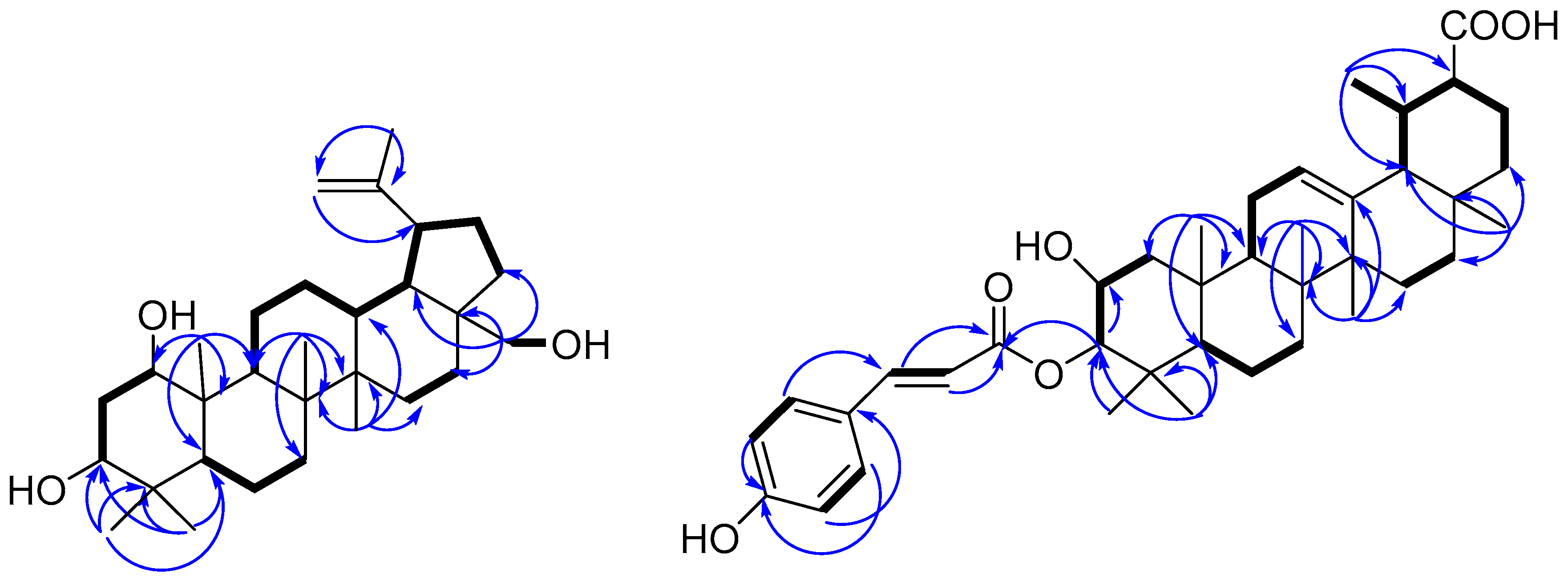
), and selected HMBC ( Molecules 22 01319 i002) correlations of compounds 1 and 11.
Molecules 22 01319 g002
Table 1. 1H- and 13C-NMR spectroscopic data for compounds 14 a (δ in ppm, DMSO, J in Hz).
Compounds1 b,c2 b3 b4 b
No.δHδC δHδC δHδC δHδC
13.14 m (W1/2 22.1)77.9d3.30 dd (10.8, 4.7)77.2d3.27 d77.2d1.87 m 0.84m47.7d
21.47 m38.5t1.68 m34.0t1.68 m33.8t3.67 m (W1/2 26.6 )64.9t
32.96 m (W1/2 22.8)74.0d4.47 dd (11.9, 4.7)76.7d4.40 dd (12.5, 4.5)76.9d4.48 d (10.0 )83.4d
4 38.0s 37.6s 37.4s 39.1s
50.46 d (11.0)52.6d0.68 m52.2d0.65 m52.2d0.86 m54.5d
61.38 m17.6t1.43 m17.4t1.43 m17.4t1.28 m17.8t
71.28 m33.9t1.28 m33.8t1.28 m33.8t1.40 m 1.29 m33.6t
8 41.0t 41.0t 41.0t 40.5t
91.36 m51.0d1.44 m50.7d1.43 m50.7d1.32 m49.6d
10 42.9s 42.8s 42.8s 37.7s
111.13 m 2.35 m23.1t1.15 m 2.35 m23.0t1.11 m 2.32 m23.0t1.29 m 2.34 m20.5t
120.87 m 1.48 m25.1d0.90 m 1.49 m25.0d0.97 m 1.49 m25.1d0.93 m 1.60 m24.7d
131.59 m36.5d1.59 m36.5d1.60 m36.5d1.60 m36.7d
14 42.3s 42.3s 42.3s 42.3s
151.57 m26.7t1.61 m26.8t1.61 m26.7t1.62 m26.6t
161.01 m 1.88 m29.1t1.04 m 1.88 m29.1t1.02 m 1.88 m29.1t1.01 m 1.88 m29.0t
17 47.3s 47.3s 47.3s 47.3s
181.45 m48.2d1.44 m48.2d1.46 m48.2d1.46 m48.1d
192.36 m47.3d2.36 m47.3d2.36 m47.3d2.35 m47.4d
20 150.4s 150.4s 150.4s 150.3s
211.22 m 1.83 m29.3t1.22 m 1.83 m29.3t1.24 m 1.84 m29.2t1.24 m 1.84 m29.3t
220.87 m 1.85 m33.8t0.86 m 1.85 m33.7t0.83 m1.85 m33.7t0.84 m 1.85 m33.8t
230.83 s28.0q0.77 s27.6q0.76 s27.5q0.78 s28.3q
240.61 s15.4q0.84 s16.0q0.71 s16.0q0.83 s17.6q
250.76 s12.2q0.84 s12.2q0.81 s12.1q0.87 s17.0q
260.98 s16.0q1.00 s16.2q0.99 s16.1q0.99 s15.7q
270.93 s14.5q0.96 s14.5q0.95 s14.5q0.96 s14.4q
283.07 d (10.8)58.0t3.07 d (10.7)58.0t3.07 d (10.8)57.9t3.07 d (10.8)57.9t
3.51 d (10.8)3.51 d (10.7)3.51 d (10.8)3.51 d (10.8)
294.53 s 4.66 s109.6t4.54 s 4.67 s109.5t4.54 s 4.67 s109.6t4.55 s 4.68 s109.6t
301.63 s18.7q1.64 s18.7q1.64 s18.7q1.65 s18.8q
1′ 160.0s 159.6s 159.9s
2′,6′ 7.53 d (8.6)130.3d7.60 d (8.6)132.4d7.49 d (8.6)130.1d
3′,5′ 6.77 d (8.6)115.8d6.72 d (8.6)115.0d6.74 d (8.6)116.0d
4′ 124.9s 125.1s 124.3s
7′ 7.51 d (16.0)144.5d6.82 d (13.0)143.1d7.51 d (16.2)144.2d
8′ 6.33 d (16.0)114.4d5.73 d (13.0)115.0d6.32 d (16.2)114.4d
9′ 166.2s 165.8s 166.7s
a Assignments were based on 1D and 2D NMR experiments (COSY, HMBC, HSQC, and NOESY) and recorded in DMSO; b 1H-, 13C-NMR, DEPT and 2D NMR spectra were recorded on a Varian 500 NMR; c 1-OH 4.02 d (5.3); 3-OH 4.28 d (5.0); 28-OH 4.20 br s; d overlapped.
Table 2. 1H- and 13C-NMR spectroscopic data for compound 11 a (δ in ppm, DMSO, J in Hz).
Compound11 b
No.δHδC
11.88 m 0.95 m47.6 t
23.69 m (W1/2 22.9)64.8 d
34.51 d (9.9)83.6 d
4 39.8 s
50.93 m 54.4 d
61.47 m 1.88 m 17.9 t
71.54 m 32.5 t
8 41.7t
91.56 m46.8 d
10 37.5 s
111.89 m23.9 t
125.15 br s 125.1 d
13 138.4 s
14 41.7 s
151.80 m27.5 t
161.53 m 23.0 t
17 30.3s
182.12 d (11.0)52.4 d
191.32 m 38.5 d
201.56 m 38.5 d
211.91 m 36.4 t
221.80 m 39.6t
230.80 s 28.5q
240.85 s 17.1 q
250.97 s17.1 q
260.76 s16.4 q
271.07 s23.2q
280.92 s21.1q
290.83 d (6.1)23.2 q
30 181.4 s
1′ 125.1s
2′,6′7.55 d (8.8)130.2 d
3′,5′6.79 d (8.8)115.8 d
4′ 159.8 s
7′7.52 d (16.4)144.1 d
8′6.38 d (16.4)115.0 d
9′ 166.7 s
a Assignments were based on 1D and 2D NMR experiments (COSY, HMBC, HSQC, and NOESY) and recorded in DMSO; b 1H-, 13C-NMR, DEPT and 2D NMR spectra were recorded on a JEOL JNM-ECP 600 NMR.
Table 3. Cytotoxic activities of compounds 117 (IC50, µM).
Compounds P388 aHeLa bA549 bK562 a
1>50>50>50>50
227.25>50>50>50
322.3933.2014.43>50
410.5619.131.89>50
5>50>50>50>50
644.4042.46>50>50
741.97>50>50>50
834.38>5027.49>50
922.3627.1515.43>50
1039.03>5037.32>50
11>5030.41>50>50
1239.77>5018>50
13>50>5023.90>50
1411.0413.10>50>50
1523.0424.90>5016.28
16>50>50>50>50
17>50>50>50>50
ADM (Adriamycin) c0.30.60.20.2
a By MTT method. b By SRB method. c Positive control.

