Preclinical Assessment of a 68Ga-DOTA-Functionalized Depsipeptide as a Radiodiagnostic Infection Imaging Agent

 ,
,  ,
,
Abstract
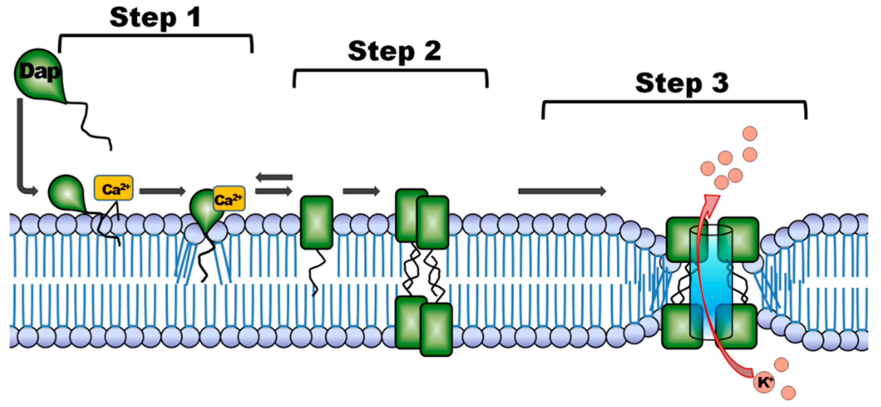
:1. Introduction
2. Results
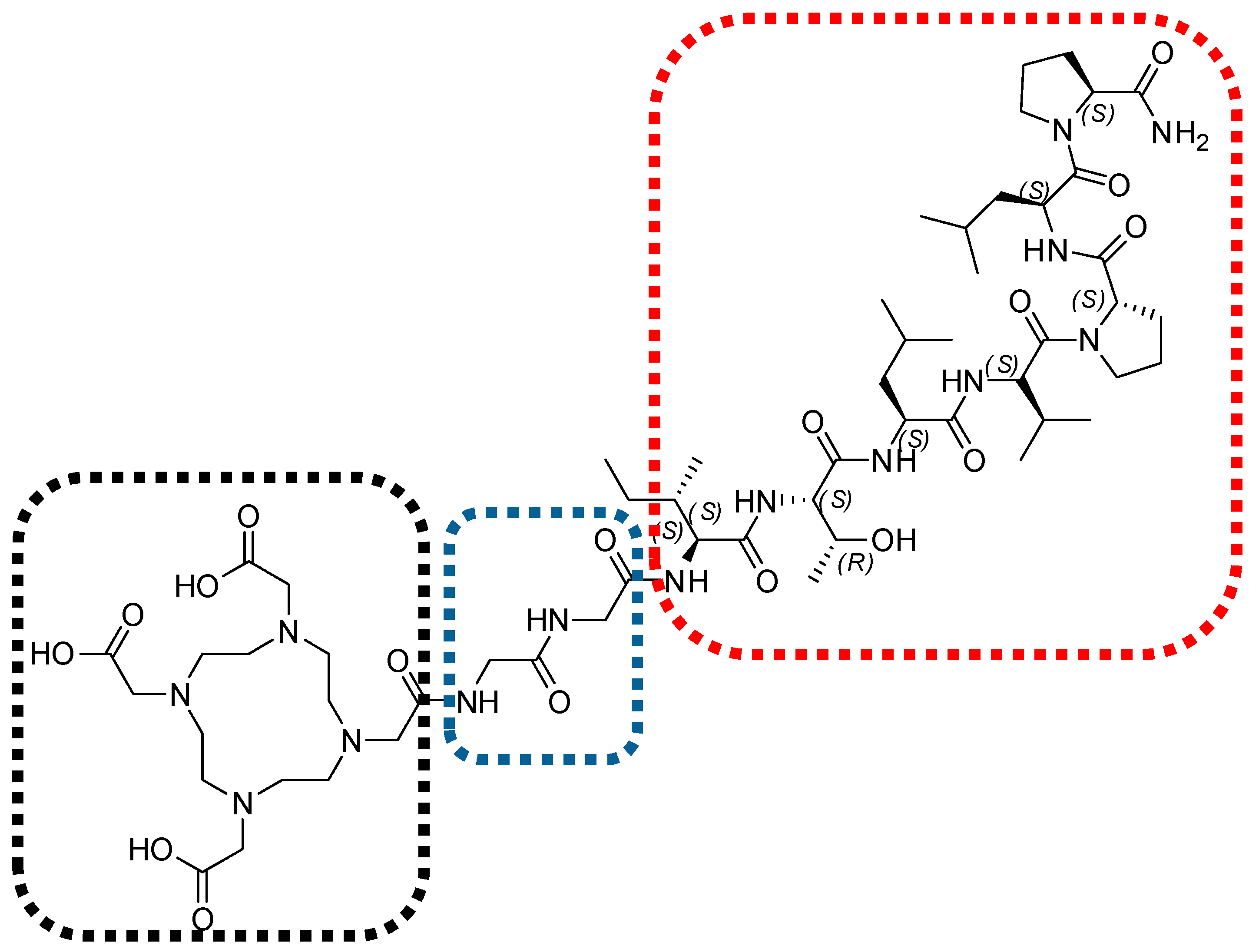
2.1. 68Ga-Radiolabeling and Tracer Formulation
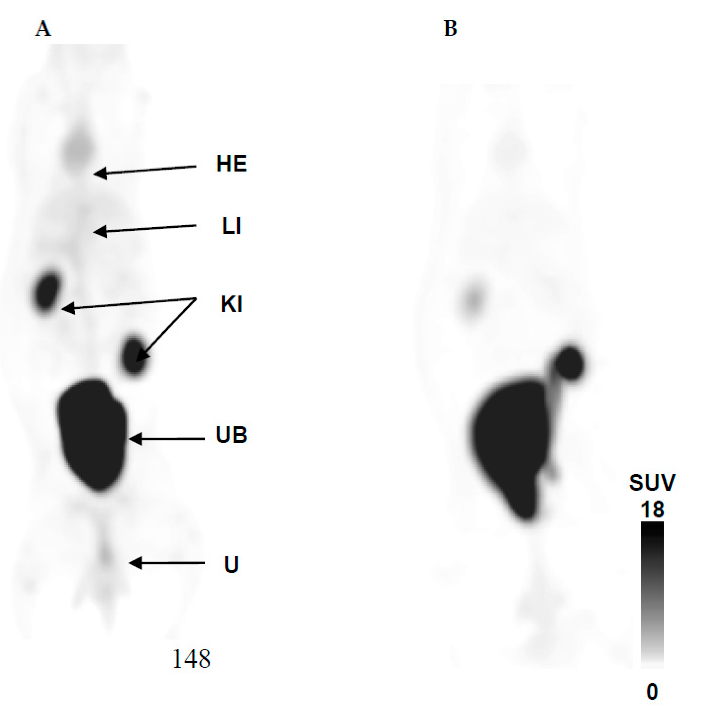
2.2. 68Ga-DOTA-TBIA101 Biodistribution in Healthy Animals
2.3. 68Ga-DOTA-TBIA101 Selectivity
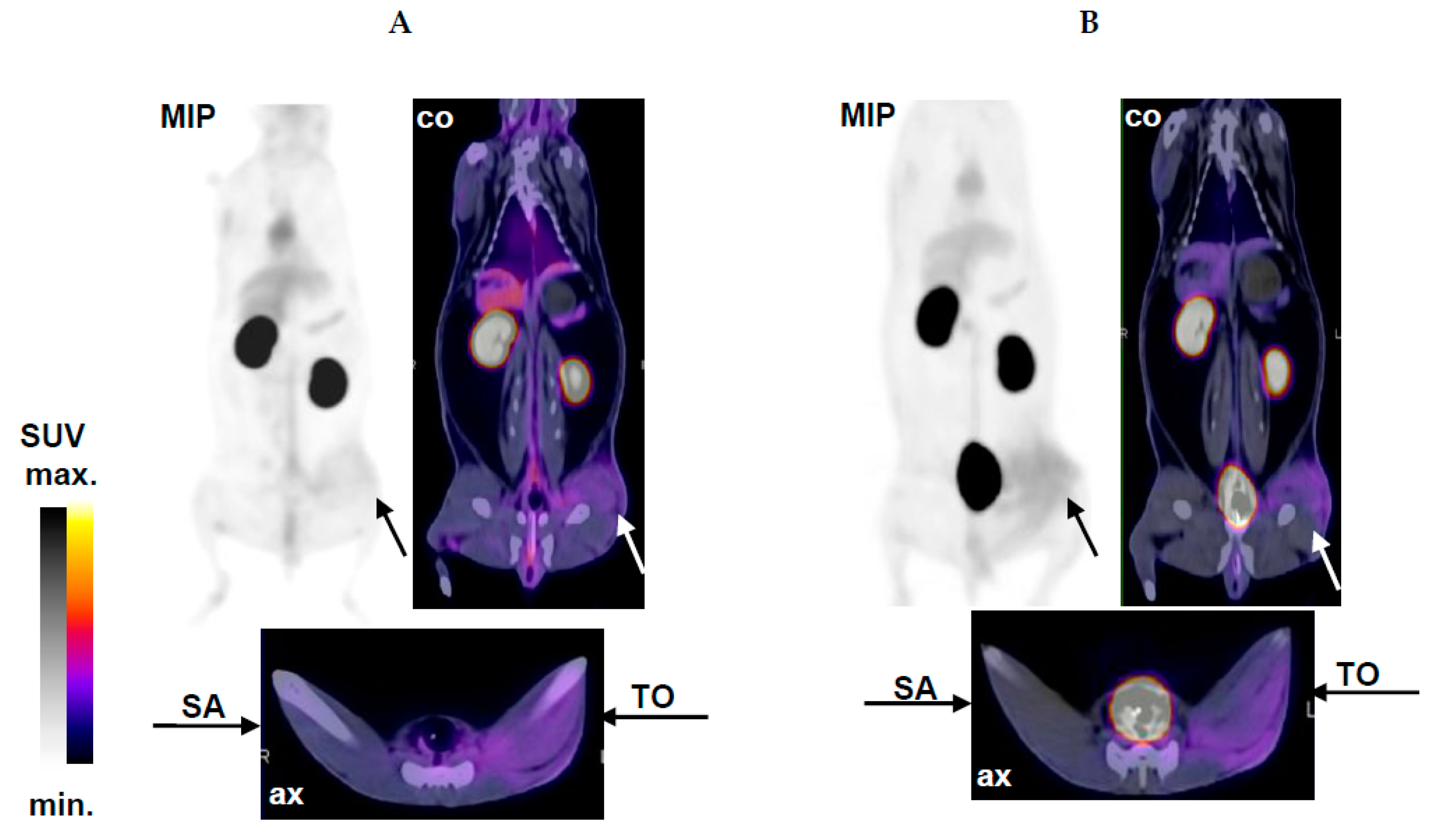
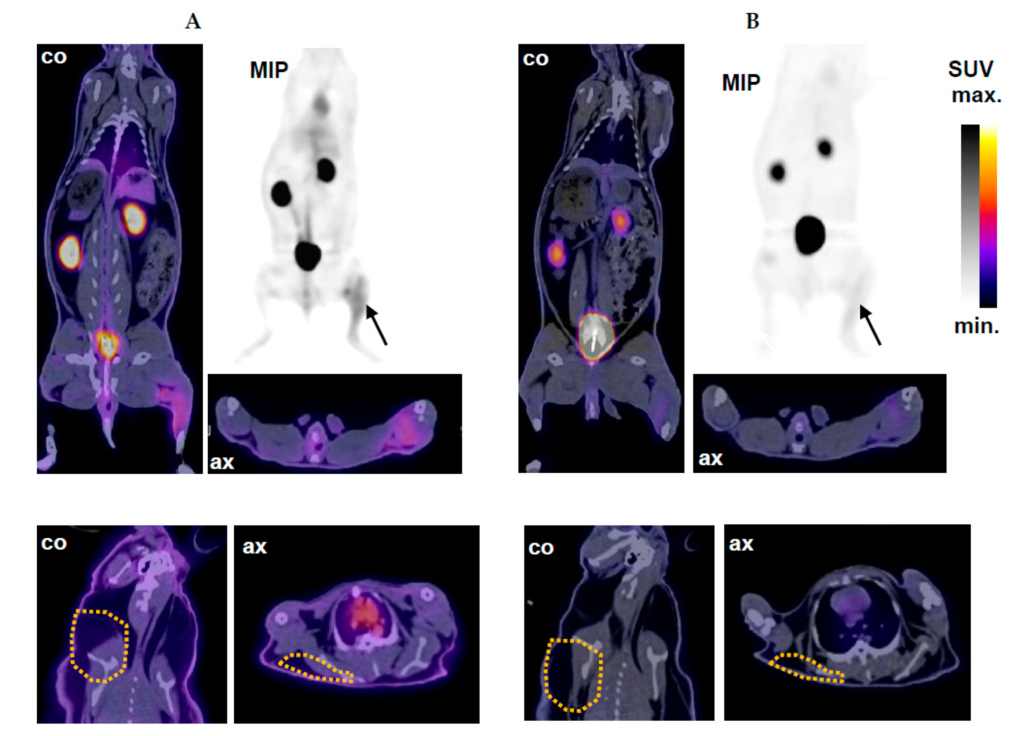
2.4. PET/CT Imaging of Mycobacterial Infection with 68Ga-DOTA-TBIA101
2.5. PET/CT Image Quantification
2.6. Histopathological Examination
3. Discussion
3.1. Biodistribution
3.2. Selectivity
3.3. Mycobacterial Imaging
3.4. Clinical Relevance and Limitations
4. Materials and Methods
4.1. Material
4.2. 68Ga-DOTA-TBIA101 Radiolabeling and Formulation
4.3. Study Design, Animal Preparation, and Tracer Administration
4.4. PET/CT Imaging
4.5. Bacteriology and Histopathology
4.6. Biocontaminant Prevention
4.7. Statistical Analysis
5. Conclusions
Acknowledgments
Author Contributions
Conflicts of Interest
References
- Namavari, M.; Gowrishankar, G.; Hoehne, A.; Jouannot, E.; Gambhir, S. Synthesis of [18F]-labelled maltose derivatives as PET tracers for imaging bacterial infection. Mol. Imaging Biol. 2015, 17, 168–176. [Google Scholar] [CrossRef] [PubMed]
- Wang, Z.; Ning, X. Clinical diagnosis of bacterial infection via FDG-PET imaging. Can. Chem. Trans. 2013, 1, 85–104. [Google Scholar]
- Boerman, O.C.; Dams, E.T.; Oyen, W.J.; Corstens, F.H.; Storm, G. Radiopharmaceuticals for scintigraphic imaging of infection and inflammation. Inflamm. Res. 2001, 50, 55–64. [Google Scholar] [CrossRef] [PubMed]
- Bakheet, S.M.; Saleem, M.; Powe, J.; Al Amro, A.; Larsson, S.G.; Mahassin, Z. F-18 fluorodeoxyglucose chest uptake in lung inflammation and infection. Clin. Nucl. Med. 2000, 25, 273–278. [Google Scholar] [CrossRef] [PubMed]
- Zhuang, H.; Alavi, A. 18-Fluorodeoxyglucose positron emission tomographic imaging in the detection and monitoring of infection and inflammation. Semin. Nucl. Med. 2002, 32, 47–59. [Google Scholar]
- Signore, A.; Chianelli, M.; D’Alessandria, C.; Annovazzi, A. Receptor targeting agents for imaging inflammation/infection: where are we now? Q. J. Nucl. Med. 2006, 50, 236–242. [Google Scholar]
- Lupetti, A.; Nibbering, P.H.; Welling, M.M.; Pauwels, E.K. Radiopharmaceuticals: new antimicrobial agents. Trends Biotechnol. 2003, 21, 70–73. [Google Scholar] [CrossRef]
- Basu, S.; Chryssikos, T.; Moghadam-Kia, S.; Zhuang, H.; Torigian, D.A.; Alavi, A. Positron emission tomography as a diagnostic tool in infection: Present role and future possibilities. Semin. Nucl. Med. 2009, 39, 36–51. [Google Scholar] [CrossRef] [PubMed]
- Kumar, V.; Boddeti, D.K. 68Ga-radiopharmaceuticals for PET imaging of infection and inflammation. Recent Results Cancer Res. 2013, 194, 189–219. [Google Scholar] [PubMed]
- Sathekge, M. The potential role of 68Ga-labeled peptides in PET imaging of infection. Nucl. Med. Commun. 2008, 29, 663–665. [Google Scholar] [CrossRef] [PubMed]
- Akhtar, M.S.; Iqbal, J.; Khan, M.A.; Irfanullah, J.; Jehangir, M.; Khan, B.; Muhammad, G.; Nadeem, M.A.; Afzal, M.S.; Imran, M.B. 99mTc-labeled antimicrobial peptide ubiquicidin (29–41) accumulates less in Escherichia coli infection than in Staphlococcus aureus infection. J. Nucl. Med. 2004, 45, 849–856. [Google Scholar] [PubMed]
- Nibbering, P.H.; Welling, M.M.; Paulusma-Annema, A.; Brouwer, C.P.; Lupetti, A.; Pauwels, E.K. 99mTc-Labeled UBI 29–41 peptide for monitoring the efficacy of antibacterial agents in mice infected with Staphylococcus aureus. J. Nucl. Med. 2004, 45, 321–326. [Google Scholar] [PubMed]
- Fani, M.; Andre, J.P.; Maecke, H.R. 68Ga-PET: A powerful generator-based alternative to cyclotron-based PET radiopharmaceuticals. Contrast Media Mol. Imaging 2008, 3, 67–77. [Google Scholar] [CrossRef] [PubMed]
- Velikyan, I.; Beyer, G.J.; Bergström-Pettermann, E.; Johansen, P.; Bergström, M.; Långström, B. The importance of high specific radioactivity in the performance of 68Ga-labeled peptide. Nucl. Med. Biol. 2008, 35, 529–536. [Google Scholar] [CrossRef] [PubMed]
- Velikyan, I. Prospective of 68Ga-radiopharmaceutical development. Theranostics 2014, 4, 47–80. [Google Scholar] [CrossRef] [PubMed]
- Velikyan, I. Positron emitting [68Ga]Ga-based imaging agents: Chemistry and diversity. Med. Chem. 2011, 7, 345–379. [Google Scholar] [CrossRef] [PubMed]
- Ebenhan, T.; Chadwick, N.; Sathekge, M.M.; Govender, P.; Govender, T.; Kruger, H.G.; Marjanovic-Painter, B.; Zeevaart, J.R. Peptide synthesis, characterization and 68Ga-radiolabeling of NOTA-conjugated ubiquicidin fragments for prospective infection imaging with PET/CT. Nucl. Med. Biol. 2014, 41, 390–400. [Google Scholar] [CrossRef] [PubMed]
- Ebenhan, T.; Govender, T.; Kruger, G.; Pulker, T.; Zeevaart, J.R.; Sathekge, M. Synthesis of 68Ga-NOTA-UBI30–41 and in vivo biodistribution in vervet monkeys towards potential PET/CT imaging of infection. J. Nucl. Med. 2012, 53, 1520. [Google Scholar]
- Ujula, T.; Salomäki, S.; Virsu, P.; Lankinen, P.; Mäkinen, T.J.; Autio, A.; Yegutkin, G.G.; Knuuti, J.; Jalkanen, S.; Roivainen, A. Synthesis, 68Ga labeling and preliminary evaluation of DOTA peptide binding vascular adhesion protein-1: A potential PET imaging agent for diagnosing osteomyelitis. Nucl. Med. Biol. 2009, 36, 631–641. [Google Scholar] [CrossRef] [PubMed]
- Ebenhan, T.; Zeevaart, J.R.; Venter, J.D.; Govender, T.; Kruger, G.H.; Jarvis, N.V.; Sathekge, M.M. Preclinical evaluation of 68Ga-labeled 1,4,7-triazacyclononane-1,4,7-triacetic acid-ubiquicidin as a radioligand for PET infection imaging. J. Nucl. Med. 2014, 55, 308–314. [Google Scholar] [CrossRef] [PubMed]
- Bionda, N.; Stawikowski, M.; Stawikowska, R.; Cudic, M.; López-Vallejo, F.; Treitl, D.; Medina-Franco, J.; Cudic, P. Effects of cyclic lipodepsipeptide structural modulation on stability, antibacterial activity, and human cell toxicity. Chem. Med. Chem. 2012, 7, 871–882. [Google Scholar] [CrossRef] [PubMed]
- Bionda, N.; Fleeman, R.M.; Shaw, L.N.; Cudic, P. Effect of ester to amide or N-methylamide substitution on bacterial membrane depolarization and antibacterial activity of novel cyclic lipopeptides. Chem. Med. Chem. 2013, 8, 1394–1402. [Google Scholar] [CrossRef] [PubMed]
- Trevisi, L.; Bova, S.; Cargnelli, G.; Danieli-Betto, D.; Floreani, M.; Germinario, E.; D'Auria, M.V.; Luciani, S. Callipeltin A, a cyclic depsipeptide inhibitor of the cardiac sodium–calcium exchanger and positive inotropic agent. BioChem. Biophys. Res. Commun. 2000, 279, 219–222. [Google Scholar] [CrossRef] [PubMed]
- Steenbergen, J.N.; Alder, J.; Thorne, G.M.; Tally, F.P. Daptomycin: A lipopeptide antibiotic for the treatment of serious Gram-positive infections. J. Antimicrob. Chemother. 2005, 55, 283–288. [Google Scholar] [CrossRef] [PubMed]
- Xie, Y.; Yang, L. Calcium and magnesium ions are membrane-active against stationary-phase staphylococcus aureus with high specificity. Sci. Rep. 2016, 6, 20628. [Google Scholar] [CrossRef] [PubMed]
- Jung, D.; Powers, J.P.; Straus, S.K.; Hancock, R.E. Lipid-specific binding of the calcium-dependent antibiotic daptomycin leads to changes in lipid polymorphism of model membranes. Chem. Phys. Lipids 2008, 154, 120–128. [Google Scholar] [CrossRef] [PubMed]
- Mokaleng, B.B.; Ebenhan, T.; Ramesh, S.; Govender, T.; Kruger, H.G.; Parboosing, R.; Hazari, P.P.; Mishra, A.K.; Marjanovic-Painter, B.; Zeevaart, J.R.; et al. Synthesis, 68Ga-radiolabeling, and preliminary in vivo assessment of a depsipeptide-derived compound as a potential PET/CT infection imaging agent. BioMed Res. Int. 2015, 2015, 284354. [Google Scholar] [CrossRef] [PubMed]
- Nyuyki, F.; Plotkin, M.; Graf, R.; Michel, R.; Steffen, I.; Denecke, T.; Geworski, L.; Fahdt, D.; Brenner, W.; Wurm, R. Potential impact of 68Ga-DOTATOC PET/CT on stereotactic radiotherapy planning of meningiomas. Eur J. Nucl. Med. Mol. Imaging 2010, 37, 310–318. [Google Scholar] [CrossRef] [PubMed]
- Poeppel, T.D.; Binse, I.; Petersenn, S.; Lahner, H.; Schott, M.; Antoch, G.; Brandau, W.; Bockisch, A.; Boy, C. 68Ga-DOTATOC versus 68Ga-DOTATATE PET/CT in functional imaging of neuroendocrine tumors. J. Nucl. Med. 2011, 52, 1864–1870. [Google Scholar] [CrossRef] [PubMed]
- Breeman, W.A.; de Jong, M.; de Blois, E.; Bernard, B.F.; Konijnenberg, M.; Krenning, E.P. Radiolabelling DOTA-peptides with 68Ga. Eur. J. Nucl. Med. Mol. Imaging 2005, 32, 478–485. [Google Scholar] [CrossRef] [PubMed]
- Rossouw, D.D.; Breeman, W.A.P. Scaled-up radiolabelling of DOTATATE with 68Ga eluted from a SnO2-based 68Ge/68Ga generator. Appl. Radiat. Isot. 2012, 70, 171–175. [Google Scholar] [CrossRef] [PubMed]
- Silvola, J.; Autio, A.; Luoto, P.; Jalkanen, S.; Roivainen, A. Preliminary evaluation of novel 68Ga-DOTAVAP-PEG-P2 peptide targeting vascular adhesion protein-1. Clin. Physiol. Funct. Imaging 2010, 30, 75–78. [Google Scholar] [CrossRef] [PubMed]
- Hoigebazar, L.; Jeong, J.M.; Hong, M.K.; Kim, Y.J.; Lee, J.Y.; Shetty, D.; Lee, Y.-S.; Lee, D.S.; Chung, J.-K.; Lee, M.C. Synthesis of 68Ga-labeled DOTA-nitroimidazole derivatives and their feasibilities as hypoxia imaging PET tracers. Bioorg. Med. Chem. 2011, 19, 2176–2181. [Google Scholar] [CrossRef] [PubMed]
- Autio, A.; Ujula, T.; Luoto, P.; Salomäki, S.; Jalkanen, S.; Roivainen, A. PET imaging of inflammation and adenocarcinoma xenografts using vascular adhesion protein 1 targeting peptide 68Ga-DOTAVAP-P1: comparison with 18F-FDG. Eur. J. Nucl. Med. Mol. Imaging 2010, 37, 1918–1925. [Google Scholar] [CrossRef] [PubMed]
- Kumar, V.; Boddeti, D.K. 68Ga-Radiopharmaceuticals for PET Imaging of Infection and Inflammation. In Theranostics, Gallium-68, and Other Radionuclides; Springer: Berlin, Heidelberg, 2013; pp. 189–219. [Google Scholar]
- Hancock, R.E.; Sahl, H.G. Antimicrobial and host-defense peptides as new anti-infective therapeutic strategies. Nat. Biotechnol. 2006, 24, 1551–1557. [Google Scholar] [CrossRef] [PubMed]
- Brotz-Oesterhelt, H.; Bandow, J.E.; Labischinski, H. Bacterial proteomics and its role in antibacterial drug discovery. Mass Spectrom. Rev. 2005, 24, 549–565. [Google Scholar] [CrossRef] [PubMed]
- Bionda, N.; Pitteloud, J.P.; Cudic, P. Cyclic lipodepsipeptides: A new class of antibacterial agents in the battle against resistant bacteria. Future Med. Chem. 2013, 5, 1311–1330. [Google Scholar] [CrossRef] [PubMed]
- Ebenhan, T.; Venter, J.D.; Maguire, G.E.M.; Kruger, H.G.; Mkhize, T.; Wagener, J.; Zeevaart, J.R.; Sathekge, M.M. Novel radiopharmaceutical and preclinical aspects of Ga-68-UBI: A selective PET tracer for imaging of infection In:Third Theragnostics World Congress on Gallium-68 and PRRT: Abstracts. J. Nucl. Med. 2015, 56 (Suppl. 2), 2–30. [Google Scholar] [CrossRef]
- Nanni, C.; Marangoni, A.; Quarta, C.; Di Pierro, D.; Rizzello, A.; Trespidi, S.; D′Ambrosio, D.; Ambrosini, V.; Donati, M.; Aldini, R. Small animal PET for the evaluation of an animal model of genital infection. Clin. Physiol. Funct. Imaging 2009, 29, 187–192. [Google Scholar] [CrossRef] [PubMed]
- Lankinen, P.; Makinen, T.J.; Poyhonen, T.A.; Virsu, P.; Salomaki, S.; Hakanen, A.J.; Jalkanen, S.; Aro, H.T.; Roivainen, A. 68Ga-DOTAVAP-P1 PET imaging capable of demonstrating the phase of inflammation in healing bones and the progress of infection in osteomyelitic bones. Eur. J. Nucl. Med. Mol. Imaging 2008, 35, 352–364. [Google Scholar] [CrossRef] [PubMed]
Sample Availability: Samples of the compounds TBIA101 and NOTA-TBIA101 are available from the authors. |


| Activity Biodistribution (%ID/g) # | Activity Concentration (SUV) # | |||
|---|---|---|---|---|
| Organ or Tissue | 5 min | 60 min | 5 min | 60 min |
| Heart/Blood Pool | 0.053 ± 0.003 | 0.028 ± 0.003 *** | 0.35 ± 0.15 | 0.22 ± 0.06 |
| Liver | 0.057 ± 0.007 | 0.017 ± 0.002 *** | 0.35 ± 0.03 | 0.37 ± 0.01 |
| Spleen | 0.021 ± 0.0.03 | 0.014 ± 0.001 ** | 0.63 ± 0.01 | 0.30 ± 0.01 *** |
| Urinary Bladder | 1.32 ± 0.15 | 1.24 ± 0.002 | 9.2 ± 7.7 | 14.2 ± 1.9 |
| Kidney (L/R) | 0.16 ± 0.05 | 0.096 ± 0.035 ** | 2.5 ± 2.4 | 2.8 ± 3.1 |
| Lung (L/R) | 0.028 ± 0.002 | 0.014 ± 0.003 *** | 0.26 ± 0.04 | 0.13 ± 0.02 ** |
| Stomach | 0.004 ± 0.001 | 0.003 ± 0.001 | 0.09 ± 0.01 | 0.09 ± 0.04 |
| Quadriceps (R) | 0.011 ± 0.002 | 0.003 ± 0.001 *** | 0.24 ± 0.10 | 0.09 ± 0.02 |
| Triceps (L) | 0.009 ± 0.002 | 0.003 ± 0.001 ** | 0.24 ± 0.04 | 0.11 ± 0.01 * |
| T/NT Ratio (SUV Based) | Image Acquisition Postinjection $ | |
|---|---|---|
| 5 min | 60 min | |
| NZR1: Thigh (L/R)/Triceps (L/R) | 1.2 ± 0.11 | 1.2 ± 0.24 |
| NZR2: Thigh (SA)/Triceps (L/R) | 1.0 ± 0.02 | 1.2 ± 0.15 |
| NZR2: Thigh (TO)/Triceps (L/R) | 2.6 ± 0.37 *** | 2.8 ± 2.3 ** |
| NZR3: Thigh (MTB)/Triceps (L/R) | 2.6 ± 0.35 *** | 2.8 ± 0.16 *** |
| NZR3: Thigh (MTB)/Thigh (L) | 2.1 ± 0.26 *** | 2.0 ± 0.31 *** |
| NZR3: Scruff (SA)/Scruff (R) | 1.1 ± 0.12 | 1.3 ± 0.35 |
© 2017 by the authors. Licensee MDPI, Basel, Switzerland. This article is an open access article distributed under the terms and conditions of the Creative Commons Attribution (CC BY) license (http://creativecommons.org/licenses/by/4.0/).
Share and Cite
Ebenhan, T.; Mokaleng, B.B.; Venter, J.D.; Kruger, H.G.; Zeevaart, J.R.; Sathekge, M. Preclinical Assessment of a 68Ga-DOTA-Functionalized Depsipeptide as a Radiodiagnostic Infection Imaging Agent. Molecules 2017, 22, 1403. https://doi.org/10.3390/molecules22091403
Ebenhan T, Mokaleng BB, Venter JD, Kruger HG, Zeevaart JR, Sathekge M. Preclinical Assessment of a 68Ga-DOTA-Functionalized Depsipeptide as a Radiodiagnostic Infection Imaging Agent. Molecules. 2017; 22(9):1403. https://doi.org/10.3390/molecules22091403
Chicago/Turabian StyleEbenhan, Thomas, Botshelo Brenda Mokaleng, Jacobus Daniel Venter, Hendrik Gert Kruger, Jan Rijn Zeevaart, and Mike Sathekge. 2017. "Preclinical Assessment of a 68Ga-DOTA-Functionalized Depsipeptide as a Radiodiagnostic Infection Imaging Agent" Molecules 22, no. 9: 1403. https://doi.org/10.3390/molecules22091403