Article

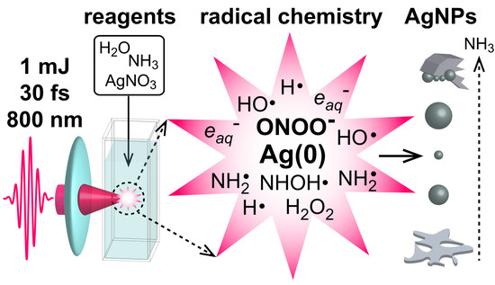
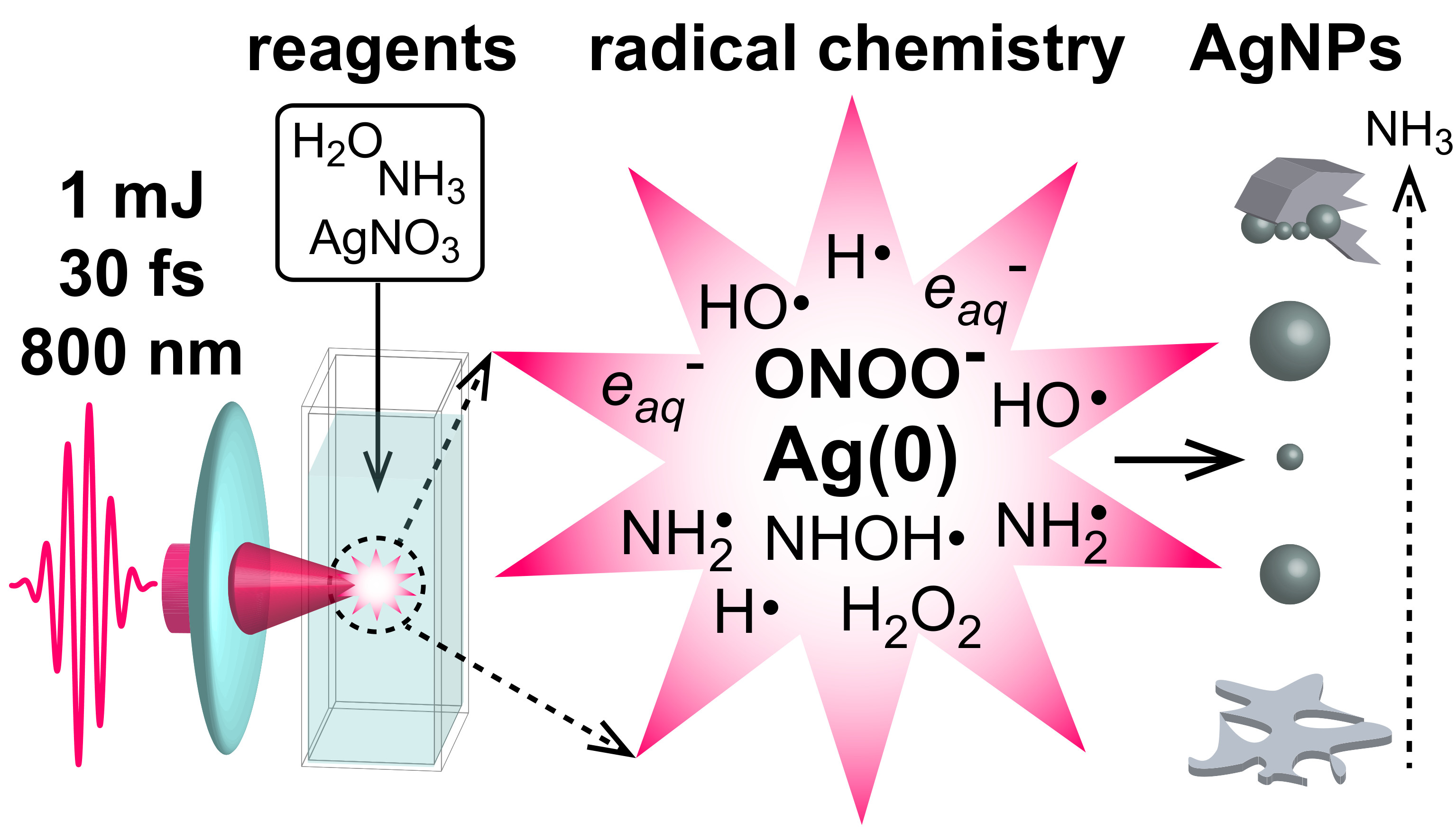
Radical Chemistry in a Femtosecond Laser Plasma: Photochemical Reduction of Ag+ in Liquid Ammonia Solution

by
Victoria Kathryn Meader
,
Mallory G. John
,
Laysa M. Frias Batista
,
Syeda Ahsan
and
Katharine Moore Tibbetts
*
Department of Chemistry, Virginia Commonwealth University, Richmond, VA 23220, USA
*
Author to whom correspondence should be addressed.
Molecules 2018, 23(3), 532; https://doi.org/10.3390/molecules23030532
Submission received: 9 February 2018 / Revised: 23 February 2018 / Accepted: 25 February 2018 / Published: 27 February 2018
(This article belongs to the Special Issue Radical Chemistry)

Abstract

:
Plasmas with dense concentrations of reactive species such as hydrated electrons and hydroxyl radicals are generated from focusing intense femtosecond laser pulses into aqueous media. These radical species can reduce metal ions such as Au3+ to form metal nanoparticles (NPs). However, the formation of H2O2 by the recombination of hydroxyl radicals inhibits the reduction of Ag+ through back-oxidation. This work has explored the control of hydroxyl radical chemistry in a femtosecond laser-generated plasma through the addition of liquid ammonia. The irradiation of liquid ammonia solutions resulted in a reaction between NH3 and OH·, forming peroxynitrite and ONOO, and significantly reducing the amount of H2O2 generated. Varying the liquid ammonia concentration controlled the Ag+ reduction rate, forming 12.7 ± 4.9 nm silver nanoparticles at the optimal ammonia concentration. The photochemical mechanisms underlying peroxynitrite formation and Ag+ reduction are discussed.

Graphical Abstract

1. Introduction

The chemistry of short-lived radical species such as e a q and OH· in aqueous solutions has a wide applicative scope. This radical chemistry is integral to the understanding of radiation-induced damage in biological systems [1,2], the development of radiation-based strategies for the removal of environmental pollutants [3,4,5], and the design of environmentally friendly synthesis routes to metal nanoparticles (NPs) [6,7,8,9,10]. Most aqueous radical chemistry studies use ionizing radiation to induce radical formation [1,2,3,6,7,8,9], but the same radical species can be generated by focusing ultrashort laser pulses into aqueous media. Sufficiently intense pulses induce multiphoton ionization of water molecules, forming a dense, localized plasma, in a process called optical breakdown (OB) [11,12]. The formation of OB plasma in water includes several reactions, not limited to the following [13,14,15,16,17,18]:
H 2 O n h ν e + H + + OH ·
e e aq
H 2 O n h ν H · + OH ·
2 OH · H 2 O 2
H · + H 2 O H 3 O + + e aq
These reactions enable the photochemical reduction of metal ions in solution, making metal NPs without chemical reducing agents [18,19,20,21,22,23,24,25,26,27,28,29]. In particular, high concentrations of both immediately formed free electrons (1020 to 1022 cm−1) [12] and subsequently formed hydrated electrons (up to 0.1 M) [17] in OB plasmas enable efficient metal ion reduction, even in air-saturated solutions, where O2 acts as a scavenger [30]. Both hydrated electrons and H2O2 are well suited to the photochemical conversion of Au3+ ions to gold nanoparticles (AuNPs), because of their predisposition towards autocatalytic reduction [27,28,29]. However, H2O2 formation hinders the application of this technique to other metals, because H2O2 is a strong oxidant. For instance, Ag0 back-oxidizes to Ag+ in the presence of H2O2 [6,31,32], inhibiting silver nanoparticle (AgNP) formation by Ag+ photochemical reduction in an OB plasma [20,24].
The back-oxidation of Ag0 to Ag+ in the presence of H2O2 is well known; many γ radiation methods prepare AgNPs from Ag+ ions by using secondary alcohols as hydroxyl radical scavengers [9] or stabilizers [6,9]. Ethylenediaminetetraacetic acid (EDTA) has also been reported to help Ag+ ion reduction under γ irradiation, where Ag+ ions complex to the carboxylate groups in EDTA [7,33]. The preparation of AgNPs in OB plasma is made possible by adding other species to the Ag+ precursor solution, including TiO2 [34], sodium citrate [35], and sodium dodecylsulfate (SDS) surfactant [36]. The laser ablation of silver targets immersed in electrolyte solutions has also yielded colloidal AgNPs [37], although the AgNPs formed by laser ablation can also be back-oxidized in OB plasma [31]. An additional challenge to forming AgNPs from AgNO3 with radiation-based methods may lie in the activity of the nitrate ion as a hydrated-electron scavenger [30,38]. For this reason, a number of studies used AgClO4 as their Ag+ AgNP precursor in radiolysis experiments [39,40,41]. Recently, colloidal AgNPs were synthesized from aqueous AgNO3 solutions containing polyvinylpyrrolidone (PVP) in OB plasma, but only when ammonia was present in the solutions [24]. Earlier studies have reported complexation of the Ag+ ion with ammonia to form Ag(NH3)2+ [40,42], which the authors of [24] attributed as the ability to form AgNPs. The lowered reduction potential of Ag(NH3)2+ compared to free Ag+ [40] suggests that manipulation of the NH3 concentration may enable control over both the formation rate and the resulting properties of AgNPs.
Silver’s microbial properties have motivated studies into silver nanostructure preparation and the incorporation of silver nanostructures into medical devices [43,44,45]. The unique optical properties of colloidal silver also make it a cost-effective surface-enhanced Raman spectroscopy (SERS) substrate option as compared to AuNPs [35,46]. In this article, we aim to understand the radical chemistry leading to AgNP formation when aqueous solutions of ammonia and AgNO3 are irradiated by strong-field, ultrashort laser pulses. We provide evidence of the major radical-mediated reactions and products formed in OB plasma, including peroxynitrite and ONOO, from the irradiation of different concentrations of aqueous ammonia solution in an OB plasma. Neither AgNO3 nor AgClO4 solutions produced AgNPs in the absence of ammonia. In the presence of ammonia, the irradiated solutions of both AgNO3 and AgClO4 showed that the growth kinetics and final sizes and morphologies of the AgNPs depended on the ammonia concentration. Understanding the radical-mediated reactions involved will further the development of other laser and γ radiation-based synthesis techniques for metal NPs.

2. Experimental Methods

2.1. Materials

Silver nitrate, AgNO3 (Acros, Fair Lawn, NJ, USA), silver perchlorate, AgClO4 (Fisher Scientific, Fair Lawn, NJ, USA), sodium nitrite, NaNO2 (Fisher), potassium nitrate, KNO3 (Fisher), potassium hydroxide, KOH (Fisher), hydrogen peroxide, H2O2 (30%; Fisher), and ammonia solution, NH3 (32%; Emplura, Billerica, MA, USA) were used without further purification and were made into stock solutions using Milli-Q filtered water (18 MΩ cm−1). Titanium dioxide powder, TiO2 (Sigma Aldrich, St. Louis, MO, USA) and sulfuric acid, H2SO4 (Fisher) were used without further purification.

2.2. Sample Preparation

AgNPs were prepared by irradiating precursor solutions containing 0.1 mM Ag+ in the form of either AgNO3 or AgClO4, in the presence of ammonia (0–20 mM). The Ag+ and ammonia working solutions were prepared from stock solutions directly in the cuvettes immediately before irradiation.
Experiments testing the amount of H2O2 produced in the OB plasma were carried out by irradiating water or water with different concentrations of ammonia (0–600 mM). The ammonia was added from a stock solution directly into the cuvette; this was immediately followed by irradiation (60–600 s). Once the irradiation was complete, 400 µL of titanium(IV) sulfate (25 mM) was added to the cuvette, and an absorption spectrum was recorded. More details on the quantification of H2O2 by titanium(IV) sulfate are provided in Section 2.4.
The synthesis of peroxynitrite was adapted from [47], in which NaNO2 (0.58 M) was added to a stirring solution of H2O2 (0.31 M) and H2SO4 (0.15 M), followed by the immediate addition of KOH (2.5 M).

2.3. Instrumentation

The experimental setup has been described previously [29]. Briefly, a titanium–sapphire chirped-pulse amplifier (Astrella, Coherent, Inc., Santa Clara, CA, USA), delivering 5 mJ, 30 fs pulses, with the bandwidth centered at 800 nm and a repetition rate of 1 kHz, was used. The pulse energy was adjusted with a zero-order λ /2 waveplate (ThorLabs, Inc., Newton, NJ, USA) and a broadband thin-film polarizer (Altechna, Vilnius, Lithuania) to 1 mJ. The laser beam was expanded from 11 to 29 mm prior to focusing with an f = 5 cm aspheric lens, to produce a focal beam waist of 6.5 µm and a peak intensity of 2.5 × 10¹⁶ W cm−2. The solutions were irradiated for times ranging from 60 to 600 s, depending on the experiment. To monitor the reaction kinetics during irradiation, the experiments were performed in a home-built in situ UV-visible spectrometer, consisting of a stabilized deuterium–tungsten light source (Ocean Optics, DH2000-BAL, Winter Park, FL, USA), optical fibers, two pairs of off-axis parabolic mirrors, and a compact spectrometer (Ocean Optics, HR4000).

2.4. Characterization

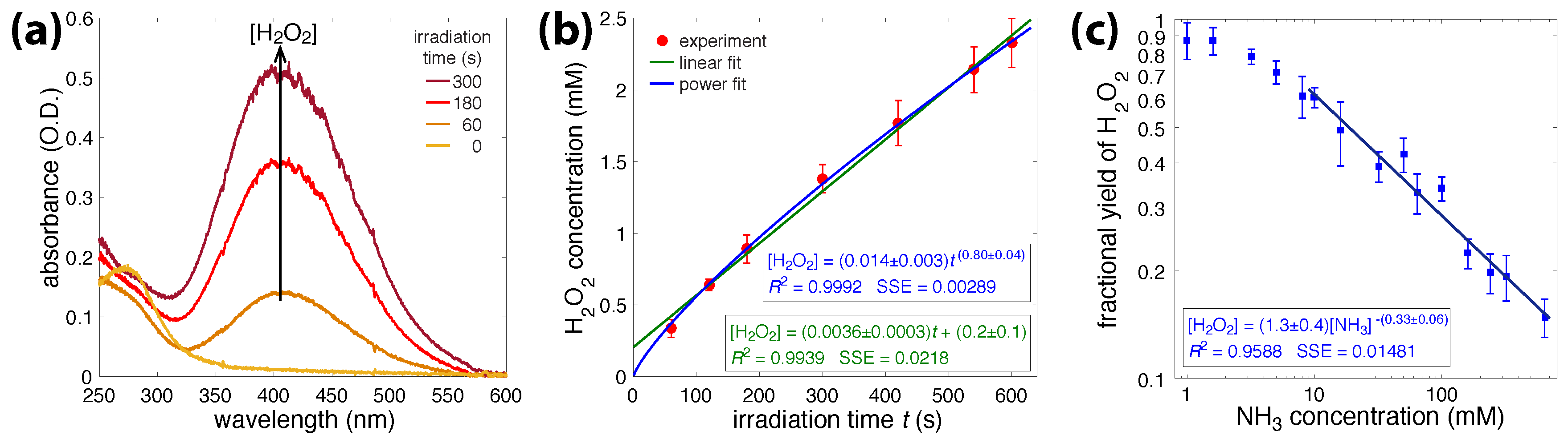
2.4.1. Quantification of H2O2

Hydrogen peroxide concentrations were quantified following the method in [48]. A solution of titanium(IV) sulfate (25 mM) was prepared by digesting a weighed amount of TiO2 in concentrated H2SO4 for 16 h at 170° and diluting with water once cooled to room temperature. Titanium(IV) sulfate (Ti4+) reacts with H2O2 according to
Ti 4 + + H 2 O 2 + 2 H 2 O Ti O 2 H 2 O 2 + 4 H +
to form pertitanic acid, TiO2H2O2, which absorbs at 407 nm with the intensity directly related to its concentration [29,48]. The H2O2 formed in the irradiated solutions was quantified against a calibration curve, which was constructed by adding titanium(IV) sulfate to different concentrations of standardized H2O2 solutions; all solutions contained 3.3 mM titanium(IV) sulfate. The H2O2 solutions were standardized by titrating with KMnO4 [49], which itself was standardized by titrating weighed amounts of sodium oxalate, following the procedure from [50]. Further details of this calibration step can be found in [29].

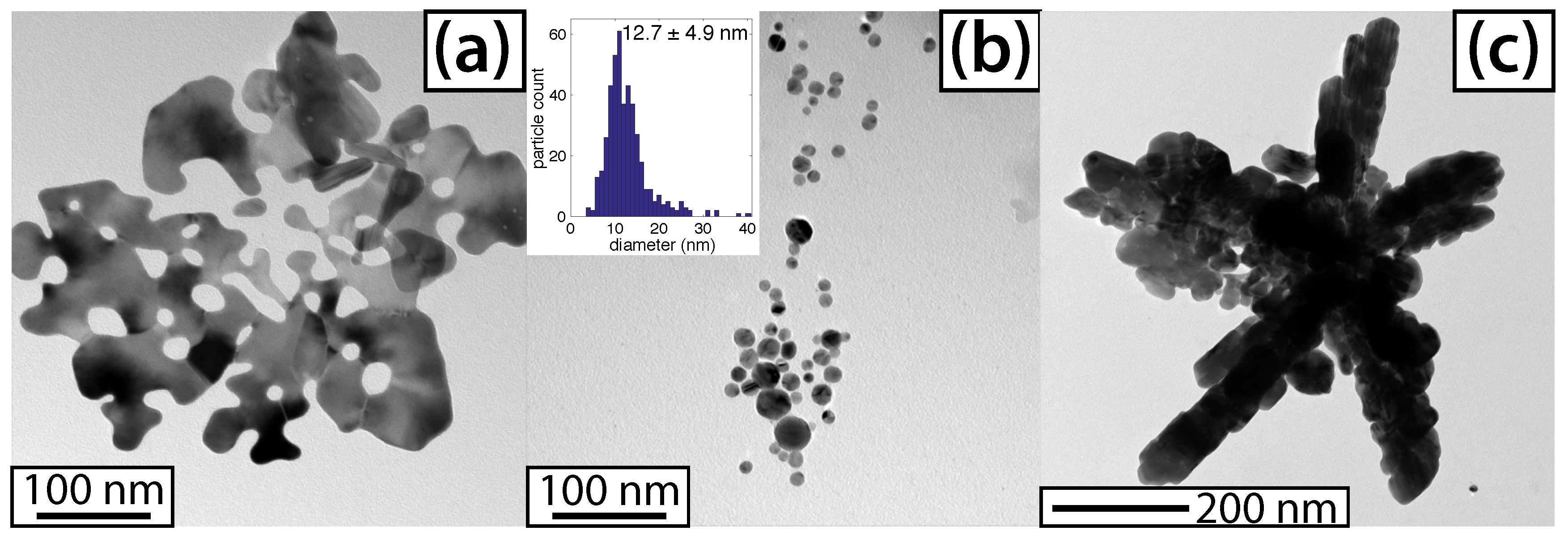
2.4.2. Transmission Electron Microscopy (TEM)

AgNP images were collected using TEM (JEOL JEM-1230). Colloidal AgNPs were drop-casted onto a carbon-coated grid (Ted Pella, Inc., Redding, CA, USA) and left to dry for 24 h or longer. ImageJ software was used to determine particle sizes.

2.4.3. Ion Chromatography (IC)

The chromatographic equipment consisted of a DIONEX ICS-1000 ion chromatogram coupled to a mass spectrometer (ThermoFisher Scientific, Fair Lawn, NJ, USA). Chromatographic separations were carried out using an AS14A column (maintained at 30 °C) and an AS14 guard column running through an AERS 500 4 mm suppressor. The eluent was 8 mM aqueous sodium bicarbonate and 1 mM aqueous sodium carbonate, the flow rate was 1.2 L/min, and the injection volume was 0.5 µL. All measurements were performed at room temperature.

3. Results

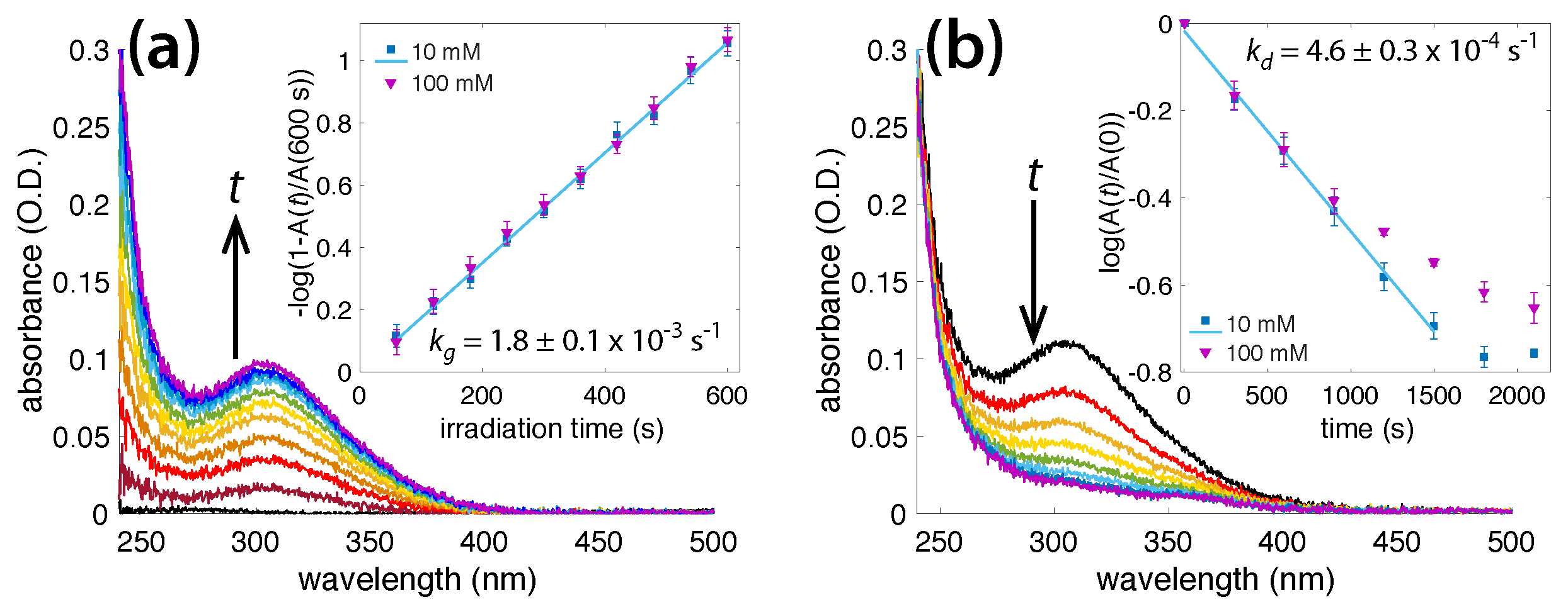
3.1. Irradiation of Aqueous Ammonia Solutions

To obtain the baseline rate of H2O2 formation under our experimental conditions, water samples were irradiated for times ranging from 60 to 600 s, and titanium(IV) sulfate was added to the samples afterwards. Figure 1a shows the absorption spectrum of TiO2H2O2 formed under the specified times (0–300 s). The increased absorbance at 400 nm with irradiation time reflected the production of H2O2 [29,48], which was produced in higher quantities as the irradiation continued. Figure 1b shows the H2O2 concentration as a function of the irradiation time. The experimental data were fit to both linear (green) and power (blue) functions, by nonlinear least-squares methods. The power law gave a closer fit, demonstrating that the H2O2 concentration grew relative to time as t 0.8 . The sublinear growth in the H2O2 concentration may have resulted from H2O2 molecule fragmentation in the laser plasma or the reaction of hydrogen peroxide with additional OH· radicals, or possibly both.
To quantify radical-scavenging properties of ammonia, NH3 solutions (1–600 mM) were irradiated for 300 s, and the H2O2 concentrations were quantified with titanium(IV) sulfate. The fractional yields of H2O2 produced at different NH3 concentrations, relative to the irradiation of pure water for 300 s, are shown in Figure 1c, and the associated numerical values are reported in Table S1 of the supporting information. A linear fit to the log–log data, in the range of 10 to 600 mM NH3, resulted in the relationship [ H 2 O 2 ] [ NH 3 ] 0.33 . This result shows that ammonia reacts with OH· radicals or H2O2, or both, and that the quantity of H2O2 produced shrinks as the ammonia concentrations increase.
In solutions containing at least 10 mM NH3, a peak centered around 302 nm appeared and increased in intensity with irradiation time, and subsequently disappeared once irradiation was terminated. Figure 2a shows the absorbance spectra collected every 60 s during the irradiation of a 10 mM NH3 solution. A growth rate constant, k g , could be calculated using the slope
log 10 1 A ( t ) A ( 600 s ) = k g t ,
where A(t) is the 302 nm absorbance at time t (in seconds), and A(600) is the 302 nm absorbance after 600 s. To prevent the quantity on the left side of Equation (7) from approaching infinity as t 600 s, the reference quantity A(600) was taken to be the mean value plus the standard deviation obtained over four experiments. The inset in Figure 2a shows a plot of Equation (7) over time, for 10 and 100 mM NH3 solutions, with a growth rate constant value of k g = 1.8 ± 0.1 × 10⁻³ s−1. Raising the NH3 concentration above 10 mM did not affect the growth rate; the absorbance values in the Figure 2a inset overlap for both the 10 and 100 mM ammonia solutions. This suggests that the growth of the 302 nm peak was zeroth order with respect to NH3, within the concentration range of 10–100 mM.
When the laser irradiation stopped, the 302 nm absorbance peak disappeared slowly. Figure 2b shows the spectra of the 10 mM ammonia solution, collected every 300 s after the initial 600 s irradiation. The decay rate constant, k d , was calculated by extracting the slope of
log 10 A ( t ) A ( 0 ) = k d t ,
where A(t) A(0) is the 302 nm absorbance immediately following irradiation. The inset in Figure 2b shows the decaying 302 nm peak for 10 and 100 mM NH3 solutions. The linear region (1000 s for 100 mM and 1500 s for 10 mM NH3) shows a decay rate constant value of k d = 4.6 ± 0.3 × 10⁻⁴ s−1. The most likely chemical species behind this 302 nm peak was peroxynitrite, ONOO, which is known to absorb at 302 nm [51,52]. A further discussion of the radical reactions leading to the formation of this transient product is presented in Section 4.

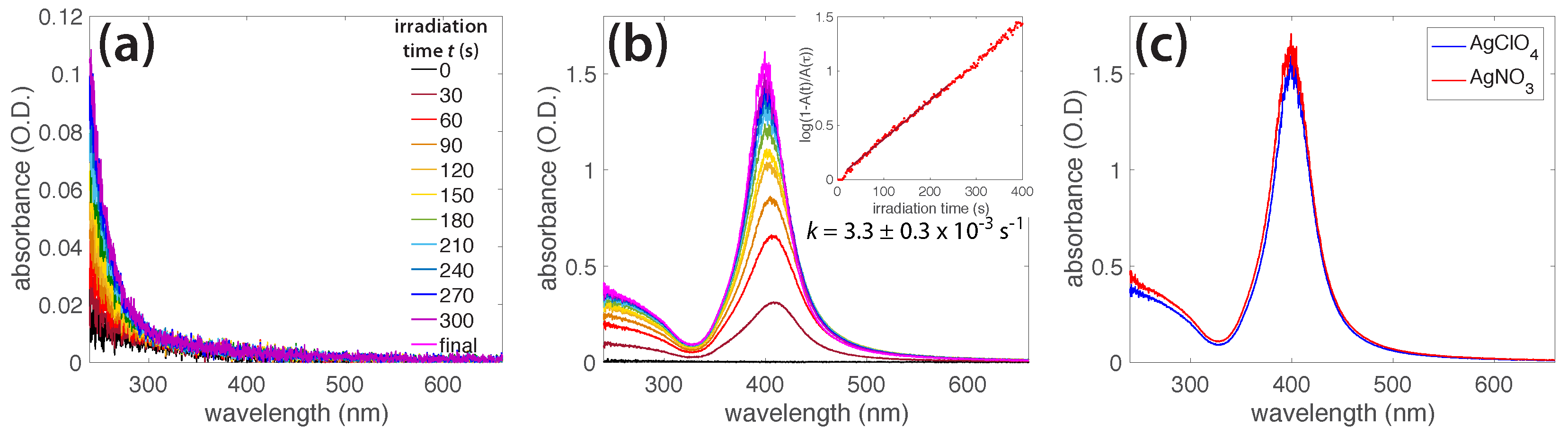
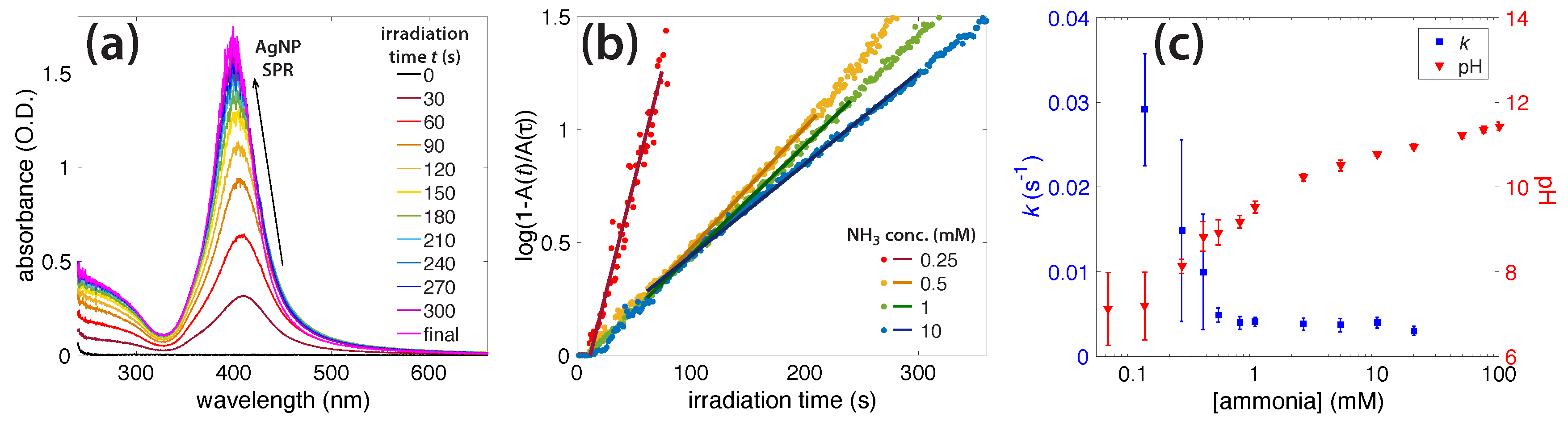
3.2. Photochemical AgNO3 Reduction in Liquid Ammonia Solutions

In the absence of NH3, AgNP formation in the femtosecond laser-irradiated AgNO3 solutions was unreliable, and generally no AgNPs were formed at all (Supporting Information, Figure S1a), as was consistent with previous results [24]. Any particles that did form quickly agglomerated and precipitated out of the solution. Hydrogen peroxide oxidized Ag0 back to Ag+ [6,31,32], disrupting photochemical AgNP synthesis [20,24]. This process was evident from the decreased amount of H2O2 produced by the irradiation of a 0.1 mM AgNO3 solution, as compared to pure water (Supporting Information, Figure S1b). Adding NH3 to the AgNO3 solution enabled AgNP formation, for which both the growth rate and AgNP properties were highly dependent on the NH3 concentration.
Figure 3a shows the growth of the AgNP surface plasmon resonance (SPR) peak at around 400 nm with the irradiation time for a solution containing 1 mM NH3. The cessation of the peak growth at 300 s of irradiation indicated the complete conversion of Ag+ to AgNPs. Figure 3b shows the rate of AgNP SPR growth in different NH3 concentrations as a semi-log plot, according to Equation (7). The growth rate constant k was extracted from the slope of the least-squares fit line, according to Equation (7). The fastest growth occurred with the lower concentrations of NH3, and, as more was added, the formation kinetics slowed. Figure 3c shows both the AgNP growth rate k and initial solution pH as functions of the NH3 concentration; numerical values are given in the supporting information in Table S2.

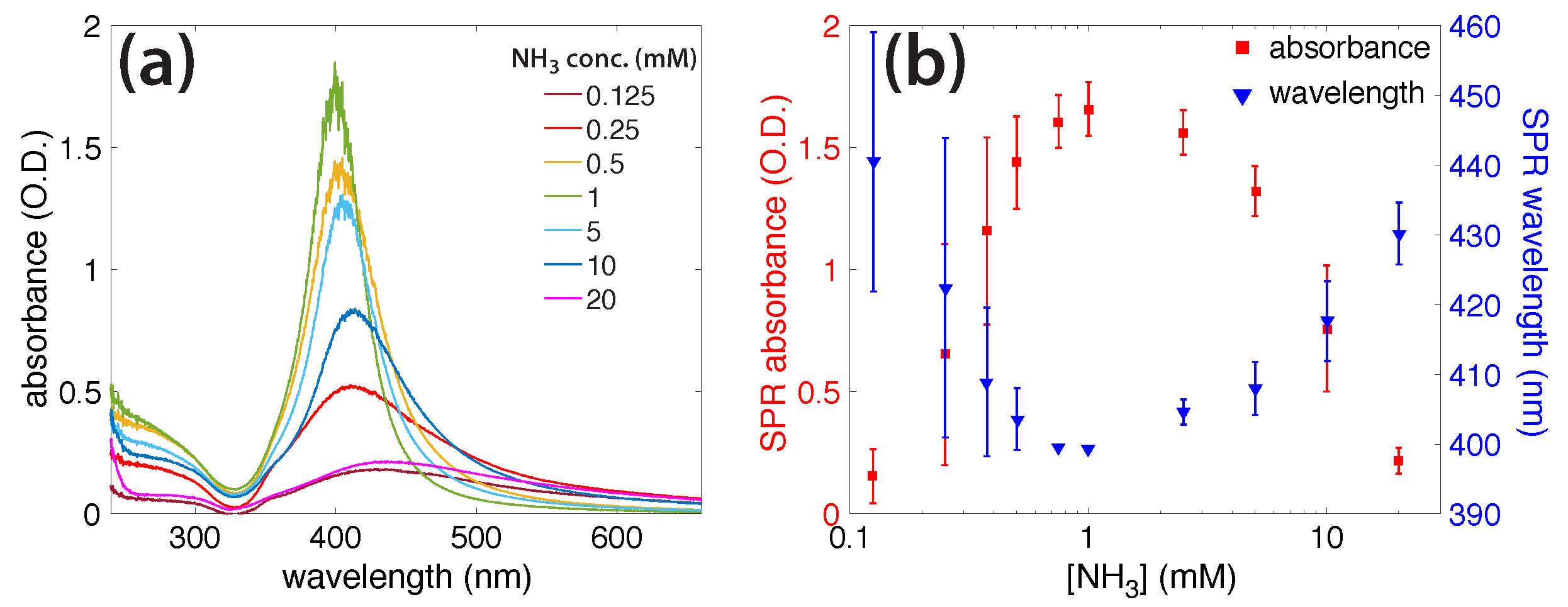
3.3. Characterization of AgNPs

Figure 4a shows the final AgNP absorbance spectra, synthesized in different concentrations of NH3. At 0.25 and 10 mM, the formation kinetics were respectively fast and slow with respect to the kinetics of the 1 mM solution (Figure 3b). The SPR absorbance at both 0.25 and 10 mM NH3 concentrations were less intense, red-shifted, and broader, as compared to the SPR absorbance at 1 mM, suggesting that larger NPs were formed and suggesting possible particle agglomeration. Figure 4b shows the relationship between the AgNP SPR absorbance and wavelength for each of the NH3 concentrations. The SPR peak is most intense and blue-shifted at 1 mM NH3, implying that this concentration was optimal for producing small, monodisperse AgNPs.
The TEM analysis of the AgNP products was consistent with the absorption spectra. Figure 5 shows representative TEM images of AgNPs prepared with (a) 0.25, (b) 1, and (c) 10 mM NH3. At the optimal 1 mM concentration, the AgNPs formed had a mean size of 12.7 ± 4.9 nm. This size distribution was more monodisperse than a previous report of 10.3 ± 8.5 nm AgNPs synthesized by the femtosecond laser-irradiation of solutions containing AgNO3, NH3, and PVP as a capping agent [24], despite that we used no capping agent. Lower NH3 concentrations, for which the kinetics are faster, gave large amorphous, plate-like particles. Higher concentrations yielded agglomerations of particles, forming a variety of shapes, including star-like structures. Additional TEM images are presented in the supporting information, in Figures S3–S5.

4. Discussion

We have demonstrated in Section 3.1 that adding liquid ammonia to water reduces the amount of H2O2 produced in OB plasma (Figure 1c). This result indicates that ammonia acts as an effective OH· and H2O2 scavenger, which is in agreement with previous literature [3,4,5]. For ammonia solutions of ≥10 mM, an absorbance peak centered at 302 nm appeared during irradiation and slowly disappeared once the laser was turned off (Figure 2a,b). The 302 nm absorbance led us to believe that this species was peroxynitrite, ONOO [4,5,30,47,51,52,53,54]. In this section, we aim to understand the reactions involved between NH3 and OH· or H2O2 that occur during multiphoton absorption in water. The relevant reactions taken from the literature are summarized in Table 1.
Reactions involving ammonia in laser plasma are most likely initiated by hydroxyl radicals (Reaction 1 in Table 1), on the basis of previous reports that NH3 reacts with H2O2 only when a solution is irradiated with UV light to produce OH· [5]. Subsequent reactions of the NH2· radical can yield hydroxylamine and hydrazine (Reactions 2 and 3 in Table 1), which are known to reduce Ag+ ions and form AgNPs [55,56]. Although hydroxylamine and hydrazine were not detected in our experiments, they may be formed in small quantities and would be expected to contribute to Ag+ reduction. The NH2· radicals can also react with H2O2 and O2, eventually forming HNO2 and NO3 (Reactions 4–8 in Table 1). NO3 can scavenge hydrated electrons (Reaction 9), and both NO2 and NO3 form ONOOH or ONOO (Reactions 10–12). In the pH range in which we see what we believe is ONOO (pH 10.62–11.42; Supporting Information, Table S1), any ONOOH formed via Reaction 10 would quickly deprotonate to form ONOO. Once formed, peroxynitrite is somewhat stable in pH values above its pKa of 6.7 [52,57,58], and it decays spontaneously according to Reactions 13–19. To determine which reactions led to ONOO formation in the OB plasma, and to confirm that the species absorbing at 302 nm was ONOO, additional experiments were conducted.
First, NaNO2 (1 mM) and KNO3 (0.05 mM) solutions, both with pH 10 (adjusted by adding KOH), were irradiated for 600 s under the same conditions as for our previous experiments. Figure 6a shows the initial and final absorbance spectra of the KNO3 solution (light and dark green) and NaNO2 solution (light and dark blue), along with the final absorption spectrum of the irradiated NH3 (red). While the irradiated KNO3 gave a broad increase in absorption below 350 nm, the irradiated NaNO2 produced an absorption peak nearly identical to that of the irradiated NH3 solution. This result indicated that NO2, not NO3, was the dominant ONOO precursor in our experiments. The presence of both OH· and H2O2 in the OB plasma was consistent with both Reactions 10 and 11 in Table 1, being possible pathways to ONOO formation.
Next, to verify that the species absorbing at 302 nm was peroxynitrite, we chemically synthesized ONOO by following the procedure in [47], involving a reaction between acidic H2O2 and alkaline nitrite. Figure 6b compares the absorption spectrum of our chemically synthesized ONOO to that of a 10 mM NH3 solution irradiated for 600 s. The spectra overlap at around 302 nm suggests that the unknown species was indeed peroxynitrite. The shoulder-peak in the chemically synthesized ONOO spectrum (Figure 6b), centered around 370 nm, was due to remaining NO2 in the solution, and it matched the absorbance of the initial 1 mM NaNO2 solution in Figure 6a (light-blue spectrum).
Finally, to determine whether NO2 and NO3 were produced as the final products in our experiments, ion chromatography of the irradiated NH3 solutions was performed. Figure 6c shows overlaid ion chromatograms for the three irradiated NH3 solutions, normalized to the NO3 peak height. The inset in Figure 6c shows the NO 2 / NO 3 peak-height ratio as a function of the ammonia concentration, with a dramatic increase in the amount of NO2 produced in the 100 mM NH3 solution. This result was consistent with previous reports of increased NO2 production relative to NO3 at high pH when NH3 and H2O2 solutions are irradiated with UV light [5,30]. This result may be explained by the solution pH (11.42; Table S1) approaching a pKa of 11.6 for H2O2 [5,66]. Reactions 15–19 in Table 1 show the radical-mediated reactions that occur near the pKa of H2O2, for which HNO2 is one of the products (Reaction 19).
Our observed ONOO decay rate, k d = 4.6 × 10⁻⁴ s−1, was significantly higher than the reported thermal decay rate of 1.3 × 10⁻⁵ s−1 and 3.2 × 10⁻⁵ s−1 at pH values of 13 and 12, respectively [63,65]. While these differences may have been caused by the lower pH range of 10.62–11.42 used in our experiments, it is also possible that the long-lived photolysis products in our experiments accelerated ONOO decomposition. The presence of H2O2 is known to speed up the degradation of peroxynitrite [47], which makes it the likely cause of the fast decay. We note that our ONOO decay experiments, reported in Figure 2b, were carried out in darkness, except for periodic UV-vis measurements of the absorption spectra every 300 s; thus photochemical decomposition according to Reaction 12 [52] was unlikely to have caused accelerated ONOO decay. In any case, the ultimate products of nitrite and nitrate (Figure 6c) suggested that our experiments followed similar mechanisms to those observed in previous reports on the conversion of ammonia to nitrite and nitrate through UV irradiation, in the presence of H2O2 [4,5,69]. It is difficult to interpret experimental observations related to the formation and decay mechanisms of peroxynitrite; thus findings are controversial [4,5,52,53,60,64,65,70]. A full discussion of the step-by-step mechanisms under various irradiation conditions is beyond the scope of this article and may be found elsewhere [5,52,60,70]. Our results demonstrate a new way to form this species and open avenues to its further investigation on ultrafast timescales.
The reactive species produced from ammonia photolysis, summarized in Table 1, assist the photochemical reduction of AgNO3 to generate AgNPs; the final AgNP morphology is determined in part by the ammonia concentration (Figure 5). No AgNP formation was observed when ammonia was not present (Supporting Information, Figure S1). Because nitrate is known to readily accept hydrated electrons (Reaction 9, Table 1) [30,38], the nitrate group of the AgNO3 precursor could hinder AgNP formation. To test this contention, we irradiated solutions containing AgClO4 (0.1 mM), a salt used in earlier radiolysis experiments because the ClO4 ion does not scavenge hydrated electrons [39,40,41]. Figure 7a shows the absorption spectra of a AgClO4 solution irradiated for 600 s, with no AgNP formation. When ammonia (1 mM) was added to the AgClO4 solution and irradiated for 420 s, the 400 nm SPR absorption feature of the AgNPs grew with the irradiation time, at a rate constant of k = 3.3 ± 0.3 × 10⁻³ s−1 (Figure 7b), which was comparable to the rate constant of k = 4.1 ± 0.6 × 10⁻³ s−1 for AgNO3. Finally, the absorption spectra of the AgNPs prepared from the irradiation of both individual precursors, AgClO4 and AgNO3, displayed strong similarity (Figure 7c). These experiments supported the role of ammonia in driving the full reduction of Ag+ to form AgNPs and the negligible role that the nitrate group on AgNO3 plays in hindering AgNP formation in the absence of ammonia.
The similar AgNP formation rates and spectral properties observed for the 1 mM ammonia solutions of both Ag+ precursors suggest that the counterion has little effect on the Ag+ reduction rate. Instead, the reduction is controlled by reducing species produced from the photolysis of water and NH3 in OB plasma. On the basis of the high concentration of water (55 M) compared to NH3, the most likely reducing species is the hydrated electron, which can be formed at up to decimolar concentrations in OB plasma [17]. We are now in a position to explain the dependence of the observed Ag+ reduction kinetics on the NH3 concentration. Reduction kinetics are fast in low NH3 concentrations and slow as the NH3 molarity is increased (Figure 3).
At NH3 concentrations below 1 mM, the solution pH (5.71–8.42; Figure 3c and Table S1) is below the pKa of NH4+ of 9.26, too low to support Ag+ complexation with ammonia to form Ag(NH3)2+ [42]. The fast reduction kinetics may therefore be attributed to the high reduction potential of free Ag+ as compared to the complex Ag(NH3)2+ [40,42]. These conditions result in the rapid formation of amorphous plate-like particles, as we observed at 0.25 mM (Figure 5a and Figure S3). At 1 mM NH3, the solution pH rose to 9.47, at which a small amount of NH4+ was present and most Ag+ was found as the Ag(NH3)2+ complex. Such complexation slows the reduction kinetics sufficiently to produce uniform spherical AgNPs (Figure 5b and Figure S4). As the NH3 concentration is further increased, a competing reaction mechanism can occur, in which the excess NH3 forms significant amounts of ONOO. The observed decrease in the Ag+ reduction rate could be explained by the back-oxidation of Ag0, because ONOO acts as a strong oxidant [52,71]. Slow reduction led to the formation of large agglomerated AgNPs (Figure 5c and Figure S5), and no AgNPs were formed at all at NH3 concentrations above 20 mM. The absence of AgNP formation at high NH3 concentrations was consistent with increased ONOO production during irradiation and its accelerated decay after irradiation, in the presence of AgNO3 in 100 mM ammonia solution (Figure S2b,c). Collectively, these results indicate that for the optimal reduction of Ag+, finding a concentration of ammonia high enough to react with enough hydroxyl radicals and form the Ag(NH3)2 complex, but low enough to avoid excess peroxynitrite production, is key.

5. Conclusions

This work investigated the radical-mediated chemistry induced by a femtosecond laser plasma in aqueous solution. Adding liquid ammonia decreased the amount of H2O2 produced during water photolysis and formed a species that was determined to be peroxynitrite, ONOO. The addition of NH3 to the aqueous solution was necessary to reduce Ag+ ions to form AgNPs, and 1 mM NH3 was determined to be the optimal concentration for making spherical AgNPs with mean diameters of 12.7 ± 4.9 nm. The extreme sensitivity of Ag+ reduction kinetics and the AgNP morphology demonstrated that understanding the reactions of the radical species produced in the laser plasma is crucial for NP synthesis using femtosecond laser irradiation methods. We anticipate that controlling the numbers of radical species in femtosecond laser plasma by varying the amount of NH3 added to aqueous solutions will help to better formation techniques for other metal NPs or alloyed metal NPs in the future.

Supplementary Materials

The following are available online.

Acknowledgments

The authors acknowledge the support of the American Chemical Society Petroleum Research Fund through Grant No. 57799-DNI10 and of Virginia Commonwealth University. Microscopy was performed at the VCU Microscopy Facility, supported, in part, by funding from the NIH-NCI Cancer Center Support Grant No. P30 CA016059.

Author Contributions

K.M.T., V.K.M., and M.G.J. designed the research. V.K.M., M.G.J., L.M.F.B., and S.A. performed the experiments and analyzed the data. V.K.M., M.G.J., L.M.F.B., and K.M.T. wrote the manuscript.

Conflicts of Interest

The authors declare no conflict of interest.

References

  1. LaVerne, J.A.; Pimblott, S.M. Yields of Hydroxyl Radical and Hydrated Electron Scavenging Reactions in Aqueous Solutions of Biological Interest. Radiat. Res. 1993, 135, 16–23. [Google Scholar] [CrossRef] [PubMed]
  2. Breen, A.P.; Murphy, J.A. Reactions of oxyl radicals with DNA. Free Radic. Biol. Med. 1995, 18, 1033–1077. [Google Scholar] [CrossRef]
  3. Getoff, N. Radiation-induced degradation of water pollutants—state of the art. Radiat. Phys. Chem. 1996, 47, 581–593. [Google Scholar] [CrossRef]
  4. Huang, L.; Li, L.; Dong, W.; Liu, Y.; Hou, H. Removal of Ammonia by OH Radical in Aqueous Phase. Environ. Sci. Technol. 2008, 42, 8070–8075. [Google Scholar] [CrossRef] [PubMed]
  5. Wang, J.; Song, M.; Chen, B.; Wang, L.; Zhu, R. Effects of pH and H2O2 on ammonia, nitrite, and nitrate transformations during UV254nm irradiation: Implications to nitrogen removal and analysis. Chemosphere 2017, 184, 1003–1011. [Google Scholar] [CrossRef] [PubMed]
  6. Wu, H.; Xu, X.; Ge, X.; Zhang, Z. Preparation of silver nanocrystals in microemulsion by the γ-radiation method. Radiat. Phys. Chem. 1997, 50, 585–588. [Google Scholar]
  7. Belloni, J.; Mostafavi, M.; Remita, H.; Marignier, J.L.; Delcourt, M.O. Radiation-induced synthesis of mono- and multi-metallic clusters and nanocolloids. New J. Chem. 1998, 22, 1239–1255. [Google Scholar] [CrossRef]
  8. Li, T.; Park, H.G.; Choi, S.H. γ-Irradiation-induced preparation of Ag and Au nanoparticles and their characterizations. Mater. Chem. Phys. 2007, 105, 325–330. [Google Scholar] [CrossRef]
  9. Du, B.D.; Phu, D.V.; Duy, N.N.; Lan, N.T.K.; Lang, V.T.K.; Thanh, N.V.K.; Phong, N.T.P.; Hien, N.Q. Preparation of colloidal silver nanoparticles in poly(N-vinylpyrrolidone) by γ-irradiation. J. Exp. Nanosci. 2008, 3, 207–213. [Google Scholar] [CrossRef]
  10. Wang, C.H.; Liu, C.J.; Wang, C.L.; Chien, C.C.; Hwu, Y.; Liu, R.S.; Yang, C.S.; Je, J.H.; Lin, H.M.; Margaritondo, G. Intense X-ray induced formation of silver nanoparticles stabilized by biocompatible polymers. Appl. Phys. A 2009, 97, 295–300. [Google Scholar] [CrossRef]
  11. Noack, J.; Vogel, A. Laser-Induced Plasma Formation in Water at Nanosecond to Femtosecond Time Scales: Calculation of Thresholds, Absorption Coefficients, and Energy Density. IEEE J. Quantum Electron. 1999, 35, 1156–1167. [Google Scholar] [CrossRef]
  12. Vogel, A.; Noack, J.; Hüttman, G.; Paltauf, G. Mechanisms of femtosecond laser nanosurgery of cells and tissues. Appl. Phys. B 2005, 81, 1015–1047. [Google Scholar] [CrossRef]
  13. Kurihara, K.; Kizling, J.; Stenius, P.; Fendler, J.H. Laser and Pulse Radiolytically Induced Colloidal Gold Formation in Water and in Water-in-Oil Microemulsions. J. Am. Chem. Soc. 1983, 105, 2574–2579. [Google Scholar] [CrossRef]
  14. Crowell, R.A.; Bartels, D.M. Multiphoton Ionization of Liquid Water with 3.0–5.0 eV Photons. J. Phys. Chem. 1996, 100, 17940–17949. [Google Scholar] [CrossRef]
  15. Chin, S.L.; Lagacé, S. Generation of H2, O2, and H2O2 from Water by the Use of Intense Femtosecond Laser Pulses and the Possibility of Laser Sterilization. Appl. Opt. 1996, 35, 907–911. [Google Scholar] [CrossRef] [PubMed]
  16. Reuther, A.; Laubereau, A.; Nikogosyan, D.N. Primary Photochemical Processes in Water. J. Phys. Chem. 1996, 100, 16794–16800. [Google Scholar] [CrossRef]
  17. Pommeret, S.; Gobert, F.; Mostafavi, M.; Lampre, I.; Mialocq, J.C. Femtochemistry of the Hydrated Electron at Decimolar Concentration. J. Phys. Chem. A 2001, 105, 11400–11406. [Google Scholar] [CrossRef]
  18. Nakashima, N.; Yamanaka, K.; Saeki, M.; Ohba, H.; Taniguchi, S.; Yatsuhashi, T. Metal Ion Reductions by Femtosecond Laser Pulses with Micro-Joule Energy and their Efficiencies. J. Photochem. Photobiol. A 2016, 319–320, 70–77. [Google Scholar] [CrossRef]
  19. Nakamura, T.; Mochidzuki, Y.; Sato, S. Fabrication of Gold Nanoparticles in Intense Optical field by femtosecond laser irradiation of aqueous solution. J. Mater. Res. 2008, 23, 968–974. [Google Scholar] [CrossRef]
  20. Nakamura, T.; Magara, H.; Herbani, Y.; Sato, S. Fabrication of silver nanoparticles by highly intense laser irradiation of aqueous solution. Appl. Phys. A 2011, 104, 1021. [Google Scholar] [CrossRef]
  21. Islam Sarker, M.S.; Nakamura, T.; Sato, S. Composition-controlled ternary Rh–Pd–Pt solid-solution alloy nanoparticles by laser irradiation of mixed solution of metallic ions. J. Mater. Res. 2014, 29, 856–864. [Google Scholar] [CrossRef]
  22. Belmouaddine, H.; Shi, M.; Karsenti, P.L.; Meesat, R.; Sanche, L.; Houde, D. Dense Ionization and Subsequent Non-Homogeneous Radical-Mediated Chemistry of Femtosecond Laser-Induced Low Density Plasma in Aqueous Solutions: Synthesis of Colloidal Gold. Phys. Chem. Chem. Phys. 2017, 19, 7897–7909. [Google Scholar] [CrossRef] [PubMed]
  23. Herbani, Y.; Nakamura, T.; Sato, S. Spectroscopic monitoring on irradiation-induced formation of AuAg alloy nanoparticles by femtosecond laser. AIP Conf. Proc. 2016, 1711, 030005. [Google Scholar]
  24. Herbani, Y.; Nakamura, T.; Sato, S. Silver nanoparticle formation by femtosecond laser induced reduction of ammonia-containing AgNO3 solution. J. Phys. Conf. Ser. 2017, 817, 012048. [Google Scholar] [CrossRef]
  25. Uwada, T.; Wang, S.F.; Liu, T.H.; Masuhara, H. Preparation and micropatterning of gold nanoparticles by femtosecond laser-induced optical breakdown. J. Photochem. Photobiol. A 2017, 346, 177–186. [Google Scholar] [CrossRef]
  26. Odhner, J.H.; Moore Tibbetts, K.; Tangeysh, B.; Wayland, B.B.; Levis, R.J. Mechanism of Improved Au Nanoparticle Size Distributions Using Simultaneous Spatial and Temporal Focusing for Femtosecond Laser Irradiation of Aqueous KAuCl4. J. Phys. Chem. C 2014, 118, 23986–23995. [Google Scholar] [CrossRef]
  27. Tangeysh, B.; Moore Tibbetts, K.; Odhner, J.H.; Wayland, B.B.; Levis, R.J. Gold Nanoparticle Synthesis Using Spatially and Temporally Shaped Femtosecond Laser Pulses: Post-Irradiation Auto-Reduction of Aqueous [AuCl4]. J. Phys. Chem. C 2013, 117, 18719–18727. [Google Scholar] [CrossRef]
  28. Tibbetts, K.M.; Tangeysh, B.; Odhner, J.H.; Levis, R.J. Elucidating Strong Field Photochemical Reduction Mechanisms of Aqueous [AuCl4]: Kinetics of Multiphoton Photolysis and Radical-Mediated Reduction. J. Phys. Chem. A 2016, 120, 3562–3569. [Google Scholar] [CrossRef] [PubMed]
  29. Meader, V.K.; John, M.G.; Rodrigues, C.J.; Tibbetts, K.M. Roles of Free Electrons and H2O2 in the Optical Breakdown-Induced Photochemical Reduction of Aqueous [AuCl4]. J. Phys. Chem. A 2017, 121, 6742–6754. [Google Scholar] [CrossRef] [PubMed]
  30. Gonzalez, M.C.; Braun, A.M. VUV photolysis of aqueous solutions of nitrate and nitrite. Res. Chem. Intermed. 1995, 21, 837–859. [Google Scholar] [CrossRef]
  31. Besner, S.; Meunier, M. Femtosecond Laser Synthesis of AuAg Nanoalloys: Photoinduced Oxidation and Ions Release. J. Phys. Chem. C 2010, 114, 10403–10409. [Google Scholar] [CrossRef]
  32. He, D.; Garg, S.; Waite, T.D. H2O2-Mediated Oxidation of Zero-Valent Silver and Resultant Interactions among Silver Nanoparticles, Silver Ions, and Reactive Oxygen Species. Langmuir 2012, 28, 10266–10275. [Google Scholar] [CrossRef] [PubMed]
  33. Remita, S.; Mostafavi, M.; Delcourt, M.O. Stabilization, growth, and reactivity of silver aggregates produced by radiolysis in the presence of EDTA. New J. Chem. 1994, 18, 581–588. [Google Scholar]
  34. Zeng, H.; Zhao, C.; Qiu, J.; Yang, Y.; Chen, G. Preparation and optical properties of silver nanoparticles induced by a femtosecond laser irradiation. J. Cryst. Growth 2007, 300, 519–522. [Google Scholar] [CrossRef]
  35. Xu, B.B.; Ma, Z.C.; Wang, L.; Zhang, R.; Niu, L.G.; Yang, Z.; Zhang, Y.L.; Zheng, W.H.; Zhao, B.; Xu, Y.; et al. Localized flexible integration of high-efficiency surface enhanced Raman scattering (SERS) monitors into microfluidic channels. Lab Chip 2011, 11, 3347–3351. [Google Scholar] [CrossRef] [PubMed]
  36. Abid, J.P.; Wark, A.W.; Brevet, P.F.; Girault, H.H. Preparation of silver nanoparticles in solution from a silver salt by laser irradiation. Chem. Commun. 2002, 792–793. [Google Scholar] [CrossRef]
  37. Šišková, K.; Vlčková, B.; Turpin, P.Y.; Fayet, C. Ion-Specific Effects on Laser Ablation of Silver in Aqueous Electrolyte Solutions. J. Phys. Chem. C 2008, 112, 4435–4443. [Google Scholar] [CrossRef]
  38. Zepp, R.G.; Hoigne, J.; Bader, H. Nitrate-induced photooxidation of trace organic chemicals in water. Environ. Sci. Technol. 1987, 21, 443–450. [Google Scholar] [CrossRef] [PubMed]
  39. Rémita, S.; Mostafavi, M.; Delcourt, M.O. EDTA and CN- Complexing Effect on the Kinetics, Spectral Properties, and Redox Properties of Ag10 and Ag12+ in Aqueous Solution. J. Phys. Chem. 1996, 100, 10187–10193. [Google Scholar] [CrossRef]
  40. Texier, I.; Rémita, S.; Archirel, P.; Mostafavi, M. Reduction of AgI1(NH3)2+ to Ag01(NH3)2 in Solution. Redox Potential and Spectral Study. J. Phys. Chem. 1996, 100, 12472–12476. [Google Scholar] [CrossRef]
  41. Ershov, B.G.; Henglein, A. Reduction of Ag+ on Polyacrylate Chains in Aqueous Solution. J. Phys. Chem. B 1998, 102, 10663–10666. [Google Scholar] [CrossRef]
  42. Goia, D.V. Preparation and formation mechanisms of uniform metallic particles in homogeneous solutions. J. Mater. Chem. 2004, 14, 451–458. [Google Scholar] [CrossRef]
  43. Chopra, I. The increasing use of silver-based products as antimicrobial agents: A useful development or a cause for concern? J. Antimicrob. Chemother. 2007, 59, 587–590. [Google Scholar] [CrossRef] [PubMed]
  44. Rai, M.; Yadav, A.; Gade, A. Silver nanoparticles as a new generation of antimicrobials. Biotechnol. Adv. 2009, 27, 76–83. [Google Scholar] [CrossRef] [PubMed]
  45. Morones, J.R.; Elechiguerra, J.L.; Camacho, A.; Holt, K.; Kouri, J.B.; Ramírez, J.T.; Yacaman, M.J. The bactericidal effect of silver nanoparticles. Nanotechnology 2005, 16, 2346. [Google Scholar] [CrossRef] [PubMed]
  46. Shin, Y.; Lee, C.; Yang, M.S.; Jeong, S.; Kim, D.; Kang, T. Two-dimensional Hyper-branched Gold Nanoparticles Synthesized on a Two-dimensional Oil/Water Interface. Sci. Rep. 2014, 4, 6119. [Google Scholar] [CrossRef] [PubMed]
  47. Papée, H.M.; Petriconi, G.L. Formation and Decomposition of Alkaline ‘Pernitrite’. Nature 1964, 204, 142. [Google Scholar] [CrossRef]
  48. Eisenberg, G. Colorimetric Determination of Hydrogen Peroxide. Ind. Eng. Chem. Anal. Ed. 1943, 15, 327–328. [Google Scholar] [CrossRef]
  49. Huckaba, C.E.; Keyes, F.G. The Accuracy of Estimation of Hydrogen Peroxide by Potassium Permanganate Titration. J. Am. Chem. Soc. 1948, 70, 1640–1644. [Google Scholar] [CrossRef] [PubMed]
  50. McBride, R.S. The Standardization of Potassium Permanganate Solution by Sodium oxalate. J. Am. Chem. Soc. 1912, 34, 393–416. [Google Scholar] [CrossRef]
  51. Hughes, M.N.; Nicklin, H.G. The chemistry of pernitrites. Part I. Kinetics of decomposition of pernitrous acid. J. Chem. Soc. A 1968, 450–452. [Google Scholar] [CrossRef]
  52. Edwards, J.O.; Plumb, R.C. Progress in Inorganic Chemistry; John Wiley & Sons, Inc.: Hoboken, NJ, USA, 1994; Chapter The Chemistry of Peroxonitrites; pp. 599–635. [Google Scholar]
  53. Plumb, R.C.; Edwards, J.O. Color centers in UV-irradiated nitrates. J. Phys. Chem. 1992, 96, 3245–3247. [Google Scholar] [CrossRef]
  54. Mahoney, L.R. Evidence for the formation of hydroxyl radicals in the isomerization of pernitrous acid to nitric acid in aqueous solution. J. Am. Chem. Soc. 1970, 92, 5262–5263. [Google Scholar] [CrossRef]
  55. Leopold, N.; Lendl, B. A New Method for Fast Preparation of Highly Surface-Enhanced Raman Scattering (SERS) Active Silver Colloids at Room Temperature by Reduction of Silver Nitrate with Hydroxylamine Hydrochloride. J. Phys. Chem. B 2003, 107, 5723–5727. [Google Scholar] [CrossRef]
  56. Li, Y.; Wu, Y.; Ong, B.S. Facile Synthesis of Silver Nanoparticles Useful for Fabrication of High-Conductivity Elements for Printed Electronics. J. Am. Chem. Soc. 2005, 127, 3266–3267. [Google Scholar] [CrossRef] [PubMed]
  57. Radi, R.; Beckman, J.S.; Bush, K.M.; Freeman, B.A. Peroxynitrite oxidation of sulfhydryls. The cytotoxic potential of superoxide and nitric oxide. J. Biol. Chem. 1991, 266, 4244. [Google Scholar] [PubMed]
  58. Goldstein, S.; Lind, J.; Merényi, G. Chemistry of Peroxynitrites as Compared to Peroxynitrates. Chem. Rev. 2005, 105, 2457–2470. [Google Scholar] [CrossRef] [PubMed]
  59. Pagsberg, P. Investigation of the NH2 radical produced by pulse radiolysis of ammonia in aqueous solution. In Aspects of Research at Risø; Aspects of Research at Risø: Roskilde, Denmark, 1972; pp. 209–222. [Google Scholar]
  60. Dey, G. Nitrogen compounds’ formation in aqueous solutions under high ionizing radiation: An overview. Radiat. Phys. Chem. 2011, 80, 394–402. [Google Scholar] [CrossRef]
  61. Rigg, T.; Scholes, G.; Weiss, J. 580. Chemical actions of ionising radiations in solutions. Part X. The action of X-rays on ammonia in aqueous solution. J. Chem. Soc. 1952, 3034–3038. [Google Scholar] [CrossRef]
  62. Maurer, P.; Thomas, C.F.; Kissner, R.; Rüegger, H.; Greter, O.; Röthlisberger, U.; Koppenol, W.H. Oxidation of Nitrite by Peroxynitrous Acid. J. Phys. Chem. A 2003, 107, 1763–1769. [Google Scholar] [CrossRef]
  63. Merényi, G.; Lind, J.; Goldstein, S.; Czapski, G. Mechanism and Thermochemistry of Peroxynitrite Decomposition in Water. J. Phys. Chem. A 1999, 103, 5685–5691. [Google Scholar] [CrossRef]
  64. Thøgersen, J.; Kissner, R.; Nauser, T.; Koppenol, W.H.; Richter, B.; Jensen, F.; Keiding, S.R.; Jensen, S.J.K. Primary photochemistry of peroxynitrite in aqueous solution. Chem. Phys. Lett. 2015, 641, 187–192. [Google Scholar] [CrossRef]
  65. Goldstein, S.; Rabani, J. Mechanism of Nitrite Formation by Nitrate Photolysis in Aqueous Solutions: The Role of Peroxynitrite, Nitrogen Dioxide, and Hydroxyl Radical. J. Am. Chem. Soc. 2007, 129, 10597–10601. [Google Scholar] [CrossRef] [PubMed]
  66. Staehelin, J.; Hoigne, J. Decomposition of ozone in water: Rate of initiation by hydroxide ions and hydrogen peroxide. Environ. Sci. Technol. 1982, 16, 676–681. [Google Scholar] [CrossRef]
  67. Crittenden, J.C.; Hu, S.; Hand, D.W.; Green, S.A. A kinetic model for H2O2/UV process in a completely mixed batch reactor. Water Res. 1999, 33, 2315–2328. [Google Scholar] [CrossRef]
  68. Barker, G.C.; Fowles, P.; Stringer, B. Pulse radiolytic induced transient electrical conductance in liquid solutions. Part 2.-Radiolysis of aqueous solutions of NO, NO and Fe(CN). Trans. Faraday Soc. 1970, 66, 1509–1519. [Google Scholar] [CrossRef]
  69. Zhu, X.; Castleberry, S.R.; Nanny, M.A.; Butler, E.C. Effects of pH and Catalyst Concentration on Photocatalytic Oxidation of Aqueous Ammonia and Nitrite in Titanium Dioxide Suspensions. Environ. Sci. Technol. 2005, 39, 3784–3791. [Google Scholar] [CrossRef] [PubMed]
  70. Mack, J.; Bolton, J.R. Photochemistry of nitrite and nitrate in aqueous solution: A review. J. Photochem. Photobiol. A Chem. 1999, 128, 1–13. [Google Scholar] [CrossRef]
  71. Hughes, M.N.; Nicklin, H.G.; Sackrule, W.A.C. The chemistry of peroxonitrites. Part III. The reaction of peroxonitrite with nucleophiles in alkali, and other nitrite producing reactions. J. Chem. Soc. A 1971, 3722–3725. [Google Scholar] [CrossRef]
Sample Availability: Samples of the compounds are not available from the authors.
Figure 1. (a) Absorption spectrum of TiO2H2O2 complex formed after adding titanium(IV) sulfate to irradiated water; (b) H2O2 concentration vs irradiation time. Experimental data points (red) fit with with linear (green) and power (blue) functions; (c) Log–log plot of fractional H2O2 yield vs. NH3 concentration with linear fit a 10–600 mM NH3 range. Error bars in (b,c) denote standard deviation over four independent experiments.
Molecules 23 00532 g001
Figure 2. (a) Absorbance spectra taken every 60 s during irradiation of 10 mM NH3 solution. Inset: Equation (7) plotted for 10 and 100 mM NH3 solutions vs irradiation time, with linear least-squares fit; (b) Absorbance spectra taken every 300 s following termination of initial 600 s laser irradiation of 10 mM NH3 solution. Inset: Equation (8) plotted for 10 and 100 mM NH3 with linear least-squares fit.
Molecules 23 00532 g002
Figure 3. (a) Absorbance peak of silver nanoparticles (AgNPs) in 1 mM NH3 as it grew during irradiation; (b) AgNP formation rates in different concentrations of ammonia; (c) Rate constant as a function of NH3 concentration, overlaid with the initial pH of Ag–NH3 solutions.
Molecules 23 00532 g003
Figure 4. (a) Absorbance spectra of silver nanoparticles prepared with different amounts of NH3, labeled; (b) Surface plasmon resonance (SPR) absorbance (red, left) and SPR wavelength (blue, right) as function of NH3 concentration.
Molecules 23 00532 g004
Figure 5. Transmission electron microscopy (TEM) images of silver nanoparticles, with (a) 0.25; (b) 1; and (c) 10 mM NH3. Histogram overlay corresponds to (b).
Molecules 23 00532 g005
Figure 6. (a) Absorption spectra of initial and irradiated KNO3 and NaNO2 solutions, with irradiated NH3 for comparison; (b) Absorption spectra of irradiated NH3 and chemically synthesized ONOO; (c) Ion chromatograms overlaid for 1, 10, and 100 mM NH3 solutions irradiated for 600 s. Peak heights normalized to NO3 peak (labeled). Inset shows NO 2 / NO 3 peak-height ratio as a function of NH3 concentration.
Molecules 23 00532 g006
Figure 7. (a) Absorption spectra of AgClO4 in water irradiated for 600 s, showing no silver nanoparticle (AgNP) formation in the absence of ammonia; (b) Absorbance peak of AgNPs from AgClO4 (0.1 mM) in NH3 (1 mM) as it grew during irradiation; (c) Absorbance spectra of AgNPs formed from irradiating AgClO4 (red) and AgNO3 (blue) in 1 mM NH3.
Molecules 23 00532 g007
Table 1. Proposed reactions, rate constants, and references for photolysis of water and ammonia.
Reaction No.EquationRate Constant (M−1 s−1)Ref.
1NH3 + OH· → NH2· + H2O1 × 108[59]
2NH2· + OH· → NH2OH9.5 × 109[59]
32NH2· → N2H4[60]
4NH2· + H2O2 → ·NHOH + H2O9 × 107[59]
5NH2· + O2 → NH2O2·[61]
6NH2O2· + OH· → HNO2 + H2O[61]
7NO2 + OH· → NO2· + OH[62]
8NO2· + OH· → NO3 + H+1 × 10¹⁰[62]
9NO3 + eaq → NO32−[30]
10NO2 + OH· → ONOOH4.5 × 109[63]
11NO2 + H2O2 → ONOO + H2O[47]
12NO3 + h ν → ONOO[64]
13ONOO + h ν → NO30.9 s−1[65]
13’ONOO → NO3~10−5 s−1[63]
14ONOO → NO2 + 1 2 O2[52]
15H2O2 → HO2 + H+pKa = 11.6[66]
16HO2 + OH· → OH + O27.5 × 109[67]
17HO2 + NH2· → NH2O2· + H+[4]
18NH2O2· → NO· + H2O[60]
19NO· + OH· → HNO28.9 × 109[68]

