Modification and Characterization of Fe3O4 Nanoparticles for Use in Adsorption of Alkaloids
by
Linyan Yang
1,2, 
Jing Tian
1,
Jiali Meng
1,
Ruili Zhao
1,
Cun Li
1,*,
Jifei Ma
1,* and
Tianming Jin
1,* 1
College of Animal Science and Veterinary Medicine, Tianjin Agricultural University, Tianjin 300384, China
2
Guangxi Key Laboratory for the Chemistry and Molecular Engineering of Medicinal Resources, Chemical and Pharmaceutical College of Guangxi Normal University, Guilin 541004, China
*
Authors to whom correspondence should be addressed.
Molecules 2018, 23(3), 562; https://doi.org/10.3390/molecules23030562
Submission received: 27 January 2018
/
Revised: 23 February 2018
/
Accepted: 27 February 2018
/
Published: 2 March 2018
(This article belongs to the Special Issue Innovative Extraction Techniques and Hyphenated Instrument Configuration for Complex Matrices Analysis)
Abstract
:Magnetite (Fe3O4) is a ferromagnetic iron oxide of both Fe(II) and Fe(III), prepared by FeCl2 and FeCl3. XRD was used for the confirmation of Fe3O4. Via the modification of Tetraethyl orthosilicate (TEOS), (3-Aminopropyl)trimethoxysilane (APTMS), and Alginate (AA), Fe3O4@SiO2, Fe3O4@SiO2-NH2, and Fe3O4@SiO2-NH2-AA nanoparticles could be obtained, and IR and SEM were used for the characterizations. Alkaloid adsorption experiments exhibited that, as for Palmatine and Berberine, the most adsorption could be obtained at pH 8 when the adsorption time was 6 min. The adsorption percentage of Palmatine was 22.2%, and the adsorption percentage of Berberine was 23.6% at pH 8. Considering the effect of adsorption time on liquid phase system, the adsorption conditions of 8 min has been chosen when pH 7 was used. The adsorption percentage of Palmatine was 8.67%, and the adsorption percentage of Berberine was 7.25%. Considering the above conditions, pH 8 and the adsorption time of 8min could be chosen for further uses.
1. Introduction
Although there are many pure phases of iron oxide in nature, the most popular magnetic nanoparticles (MNPs) are the nanoscale zero-valent iron (nZVI), Fe3O4 and γ-Fe2O3. Magnetite (Fe3O4) is a ferromagnetic black color iron oxide of both Fe(II) and Fe(III), which has been the most extensively studied [1]. In 2001, Asher reported co-precipitation method using oleic acid as the surface modification agent to obtain Fe3O4 nanoparticles (2–15 nm) [2]. NaOH and diethylene glycol could also be used as the catalyst and reducing agent to fabricate Fe3O4 nanoparticles of 80–180 nm in size [3,4,5]. However, Fe3O4 nanoparticles could easily aggregate due to the nanoscale effect and magnetic gravitational effect. It is an effective method of preventing the aggregate of these nanoparticles to wrap the surface of Fe3O4 nanoparticles. Fe3O4@SiO2 composite nanoparticles have the desirable properties of magnetic nanoparticles while also benefiting from the SiO2 shell, such as good hydrophilicity, stability, and biocompatibility [6,7,8]. In 2016, Tang reported that (3-aminopropyl)-triethoxysilane (APTES) was used as surface modification reagents to get Fe3O4@SiO2-NH2, which could be used for selective removal of Zn(II) ions from wastewater [9]. While Fe3O4@SiO2-NH2 nanoparticles could also be modified to obtain mercaptoamine-functionalised silica-coated magnetic nanoparticles for the removal of mercury and lead ions from wastewater [10]. As for the removal of ions, arsenate removal could be achieved by calcium alginate-encapsulated magnetic sorbent, which was prepared by physical method [11]. Superparamagnetic sodium alginate-coated Fe3O4 nanoparticles (Alg-Fe3O4) were used for removal of malachite green (MG) from aqueous solutions using batch adsorption technique, and the Alg-Fe3O4 nanoparticles were synthesized using in situ coprecipitation of FeCl2 and FeCl3 in alkaline solution in the presence of sodium alginate [12]. While multifunctional alginate microspheres could also be used for biosensing, drug delivery, and magnetic resonance imaging [13]. To obtain the good biocompatibility, Fe3O4 nanoparticles need to be modified. Fe3O4@SiO2 composite nanoparticles have the desirable properties of good hydrophilicity. (3-Aminopropyl)trimethoxysilane (APTMS) was used as surface modification reagents to get Fe3O4@SiO2-NH2nanoparticles. While calcium alginate-encapsulated magnetic sorbent could be prepared by physical method. Superparamagnetic sodium alginate-coated Fe3O4 nanoparticles (Alg-Fe3O4) could also be synthesized using in situ coprecipitation of FeCl2 and FeCl3 in alkaline solution in the presence of sodium alginate. Covalent modification methods via alginate have been rarely seen. In order to investigate the effects of the covalent alginate-modified method, alkaloid adsorption experiments were designed to study the properties of alginate-modified Fe3O4@SiO2-NH2 nanoparticles.
2. Experimental Section
2.1. Materials and Physical Measurements
(3-Aminopropyl)trimethoxysilane (APTMS), N-Hydroxysuccinimide (NHS) and 1-(3-Dimethylaminopropyl)-3-ethylcarbodiimide hydrochloride (EDC) were purchased from Shanghai source Biological Technology Co., Ltd. (Shanghai, China). Alginate (AA) was purchased from Solarbio Life Science (Beijing Solarbio Biological Technology Co., Ltd., Beijing, China). All commercially available chemicals and solvents were of reagent grade and used without further purification. X-ray powder diffraction (XRD) intensities were measured on a Rigaku D/max-IIIA diffractometer (Cu-Kα, λ = 1.54056 Å). Changes in morphology and size could be characterized by Scanning Electronic Microscopy (SEM) (KAI MEIKE CHEMICAL Co., Ltd., Liaocheng, China).
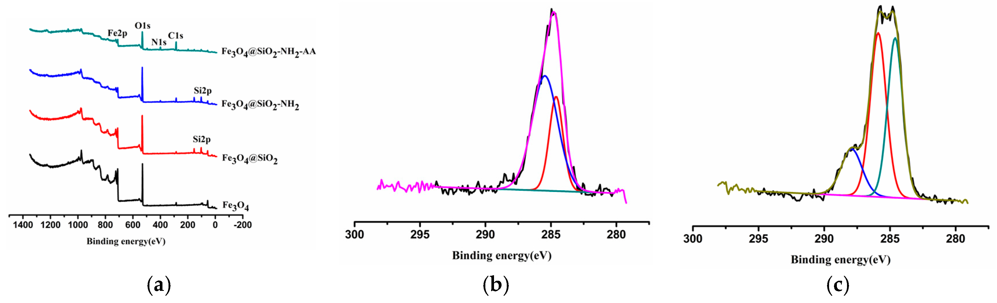
XPS spectra were recorded using a Kratos Axis Ultra DLD spectrometer (KAI MEIKE CHEMICAL Co., Ltd.) employing a monochromated Al-Kα X-ray source (hv = 1486.6 eV). The vacuum in the main chamber was kept above 3 × 10⁻⁶ Pa during XPS data acquisitions. General survey scans (binding energy range: 0–1200 eV; pass energy: 160 eV) and high-resolution spectra (pass energy: 40 eV) in the regions of N1s were recorded. Binding energies were referenced to the C1s binding energy at 284.60 eV.
The adsorption data were obtained by RP-HPLC (Reversed phase high performance liquid chromatography). The HPLC system was from Agilent Technologies 1260 Infinity (Agilent Technologies, SantaClara, CA, USA), and was equipped with a quaternary pump and UV-Vis detector (Agilent Technologies). The chromatographic separation was carried out on an ACE Super C18 column (250 × 4.6 mm i.d., 5 μm, FLM, Guangzhou, China). Mobile phase consisted of 50% solution (v/v) of acetonitrile in water (0.1% H3PO4 and 0.1% SDS). The flow rate was 1 mL/min and the column temperature was set to 40 °C. The effluent was monitored at 265 nm and the injection volume was 20 μL.
2.2. Preparation and Modification of Fe3O4 Nanoparticles
Magnetite nanoparticles were prepared and modified with TEOS, APTMS, and AA to get Fe3O4@SiO2, Fe3O4@SiO2-NH2, and Fe3O4@SiO2-NH2-AA nanoparticles, respectively (Figure 1).
2.2.1. Preparation of Fe3O4 Nanoparticles
Briefly, 7.5 mL of 0.12 M FeCl2 and 7.5 mL of 0.2 M FeCl3 solutions were mixed in a 100-mL flask. The whole reaction system was completed under nitrogen protection. After the magnetic stirring was uniform, the reaction system was heated to 55 °C, which maintained for 15 min. 7.2 mL of 3 M NaOH solution was then added to the reaction system. The reaction system was kept at 55 °C for 40 min. Then the reaction system was stirred at 90 °C for 30 min and cooled to room temperature. The black precipitate was collected by magnetic decantation and washed with deionized water repeatedly until the washings were neutral. The obtained black precipitate was then dried over vacuum at 40 °C overnight, which could be used for XRD measurement [14,15].
2.2.2. Preparation of Fe3O4@SiO2
Fe3O4 (10 mg) was acidized by HCl (0.1 mol/L) under the 100 W of ultrasound for 20 min. The supernatant was discarded after adsorption by the magnet. The residue was washed with ultrapure water for twice, and resuspended in ethanol/ultrapure water (20 mL:5 mL). NH3⋅H2O (250 μL) was added to the samples of Fe3O4, and the mixture was reacted for 20 min under the 100 W of ultrasound. TEOS (32 μL) was added into the samples. And then the samples were oscillated at 37 °C and 140 r/min for 6 h, followed by adsorption by the magnet. The supernatant was discarded, and the residue was washed with ethanol for twice to yield Fe3O4@SiO2, which was resuspended in ethanol (4 mL) [16].
2.2.3. Preparation of Fe3O4@SiO2-NH2
APTMS (50 μL) was dropwise added to the samples of Fe3O4@SiO2 obtained previously, and the mixture was reacted for 24 h. After rinsing with ethanol for twice, the samples named as Fe3O4@SiO2-NH2 were vacuum-dried at 80 °C overnight [17].
2.2.4. Preparation of Fe3O4@SiO2-NH2-AA
An AA solution (5 mg/mL in MES buffer, pH 6.0) was mixed with N,N-dimethylformamide (DMF; 3:1, v/v). Then the AA solution (3.75 mg/mL) was converted to N-hydroxysuccinimide esters by sequential reaction with EDC (36.3 mg/mL in MES buffer, pH 6.0) for 15 min and NHS (10.95 mg/mL in MES buffer, pH 6.0) for 60 min. The solution was finally introduced to the freshly Fe3O4@SiO2-NH2 nanoparticles and reacted overnight at room temperature. After washing by ethanol, the samples of Fe3O4@SiO2-NH2-AA could be obtained by vacuum-dried process [18].
2.3. Alkaloid Adsorption Test
2.3.1. Preparation of Calibration Standards
100 µg/mL standard solutions in methanol of Palmatine and Berberine were obtained from Solarbio (Beijing, China), and then further diluted in pattern of 1:2 to produce the working solutions with a series of concentrations. The concentration range of calibration standards for Palmatine were 50 µg/mL, 25 µg/mL, 12.5 µg/mL, 6.25 µg/mL, 3.125 µg/mL, 1.5625 µg/mL, 0.78125 µg/mL, while the concentration range of calibration standards for Berberine were 25 µg/mL, 12.5 µg/mL, 6.25 µg/mL, 3.125 µg/mL, 1.5625 µg/mL, 0.78125 µg/mL.
2.3.2. Influence from pH
Approximate 8 mL of mixed standard stock solution (0.5 μg/mL, in methanol, pH 5, 6, 7, 8, 9), 10 mg of Fe3O4@SiO2-NH2-AA nanoparticles was ultrasonic shocked for 6 min, and then the supernatant and magnetic nanoparticles were obtained by magnetic separation. The magnetic nanoparticles were washed by deionized water (1 mL × 2). The supernatant and detergent were combined. 1.5 mL of the mixture was dried by nitrogen blower at 80 °C. The residue was redissolved in 400 μL of methanol, which was filtered (0.22 μm) for subsequent HPLC analysis.
2.3.3. Influence from Adsorption Time
Approximate 8 mL of mixed standard stock solution (0.5 μg/mL, in methanol), 10 mg of Fe3O4@SiO2-NH2-AA nanoparticles was ultrasonic shocked for a certain time (2 min, 4 min, 6 min, 8 min, 10 min), and then the supernatant and magnetic particles were obtained by magnetic separation. The magnetic nanoparticles were washed by deionized water (1 mL × 2). The supernatant and detergent were combined. The mixture was dried by nitrogen blower at 80 °C. The residue was redissolved in 400 μL of methanol, which was filtered (0.22 μm) for subsequent HPLC analysis [19,20,21].
3. Results and Discussion
3.1. XRD Analysis of Fe3O4 Nanoparticles
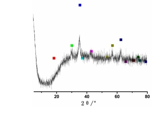
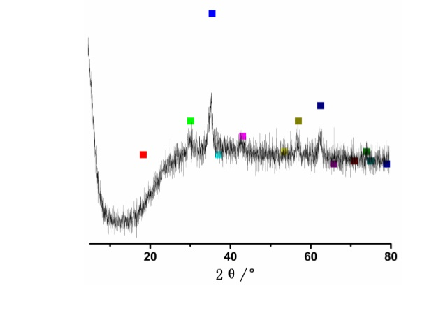
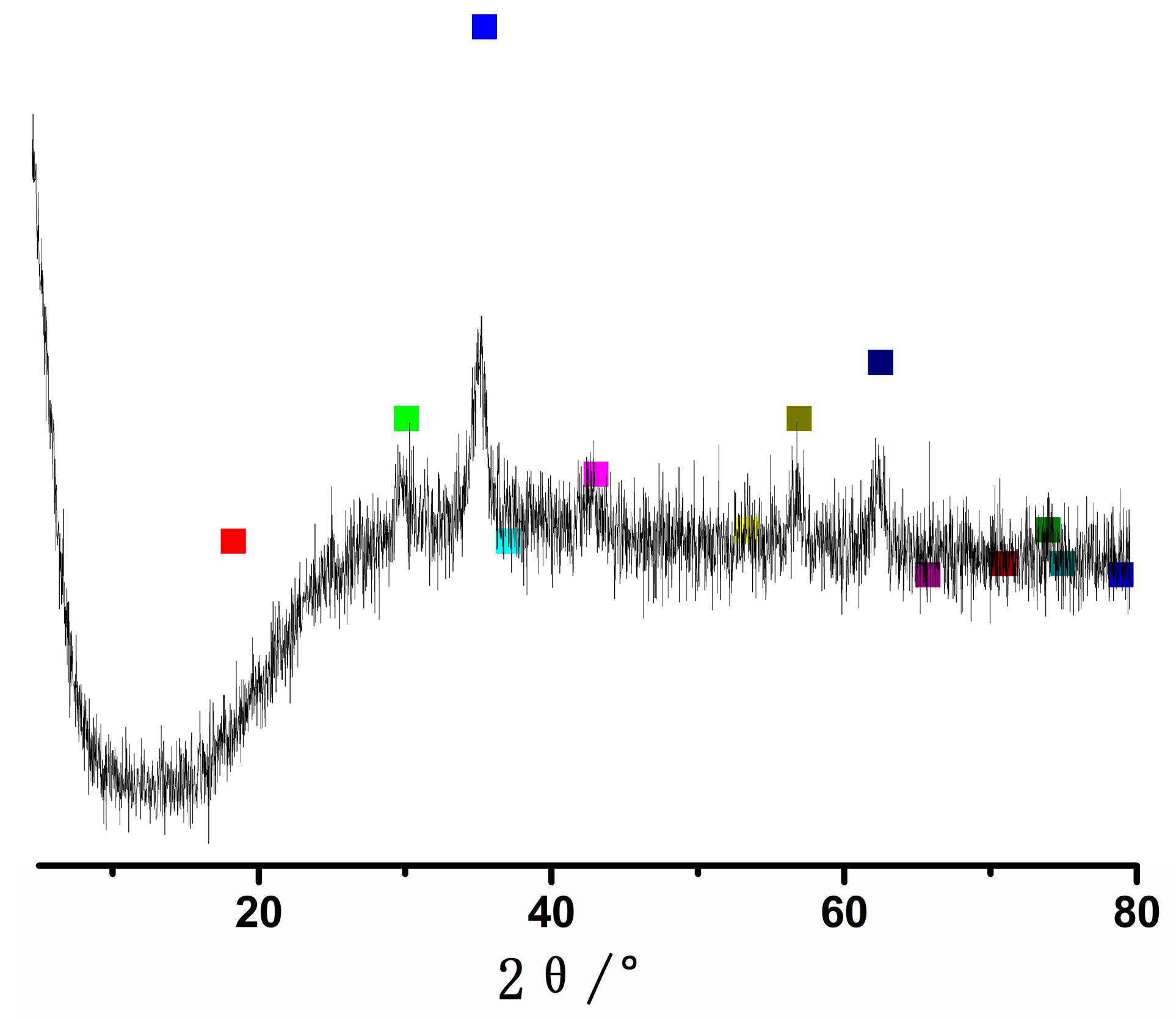
The XRD pattern of Fe3O4 nanoparticles is shown in the Figure 2. The peaks at 2θ values of 30.1°, 35.4°, 43.1°, 53.4°, 56.9° and 62.5° are indexed as the diffractions of (220), (311), (222), (422), (511) and (440) respectively, which resembles the standard diffraction spectrum of Fe3O4 (JCPDSPDF#19-0629) with respect to its reflection peaks positions [5].
3.2. FTIR Spectra Analysis of Nanoparticles
The Fe3O4@SiO2-NH2 and Fe3O4@SiO2-NH2-AA nanoparticles were obtained after the surface modification steps. It is apparent that the IR spectra contains not only the peaks in spectra of Fe3O4 nanoparticles (Fe-O, 567 cm−1) [15]. 1560 cm−1 (C-N vibration) reflected that APTMS was successfully modified onto Fe3O4@SiO2nanoparticles [22]. A strong IR peak appears at 1648 cm−1, corresponding to the strong bending vibration of the amide I group, which showed that the modification was successful and Fe3O4@SiO2-NH2nanoparticles were indeed coated with AA (Figure 3) [17,23,24].
3.3. XPS Analysis of Nanoparticles
Figure 4a shows the low-resolution XPS survey spectra of Fe3O4, Fe3O4@SiO2, Fe3O4@SiO2-NH2 and Fe3O4@SiO2-NH2-FA samples, all of which are semiquantitative. The low-resolution XPS survey spectra (Figure 4a) of Fe3O4@SiO2-NH2 have peaks of N1s, which showed that APTMS have been modified successfully. High-resolution C1s XPS spectra of the Fe3O4@SiO2-NH2 samples have peaks at 284.603 eV (C-H/C-C) and 285.459 eV (C-O/C-N) (Figure 4b). High-resolution C1s XPS spectra of the Fe3O4@SiO2-NH2-AA samples have peaks at 284.605 eV (C-H/C-C), 285.891 eV (C-O/C-N), and 287.916 eV (O-C=O/O=C-NH) (Figure 4c), which showed that amide reaction was successful [25].
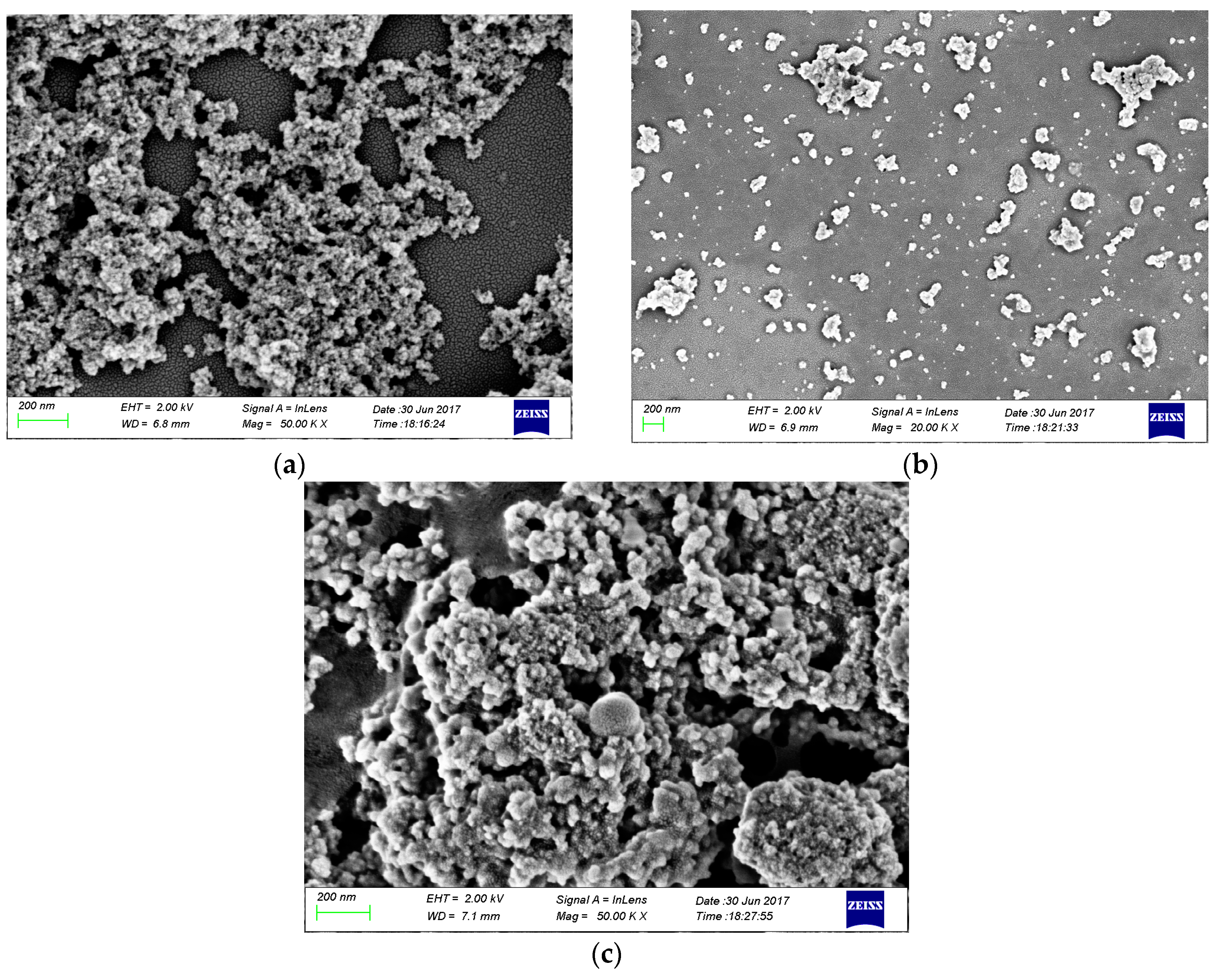
3.4. SEM Analysis of Nanoparticles
Figure 5a–c show SEM images of Fe3O4, Fe3O4@SiO2-NH2, and Fe3O4@SiO2-NH2-AA nanoparticles. Small particle size of Fe3O4 particles is obvious, while a good dispersion effect could be achieved by Fe3O4@SiO2-NH2 nanoparticles. As for Fe3O4@SiO2-NH2-AA nanoparticles, no good dispersion could be achieved, while better morphology could be achieved, which showed that AA was successfully modified onto Fe3O4@SiO2-NH2 nanoparticles [22]. Almost all particle size of Fe3O4 particlesis below 100 nm, as for Fe3O4@SiO2-NH2 nanoparticles and Fe3O4@SiO2-NH2-AA nanoparticles, particle size is becoming larger and larger, which could also prove that the modification is successful.
3.5. Analysis of Alkaloid Adsorption Test
Electrostatic interactions between alkaloids and charged surfaces, therefore, often play a major role in the adsorption behavior of alkaloids. Therefore, Palmatine and Berberine were selected for alkaloid adsorption assay in the current study.
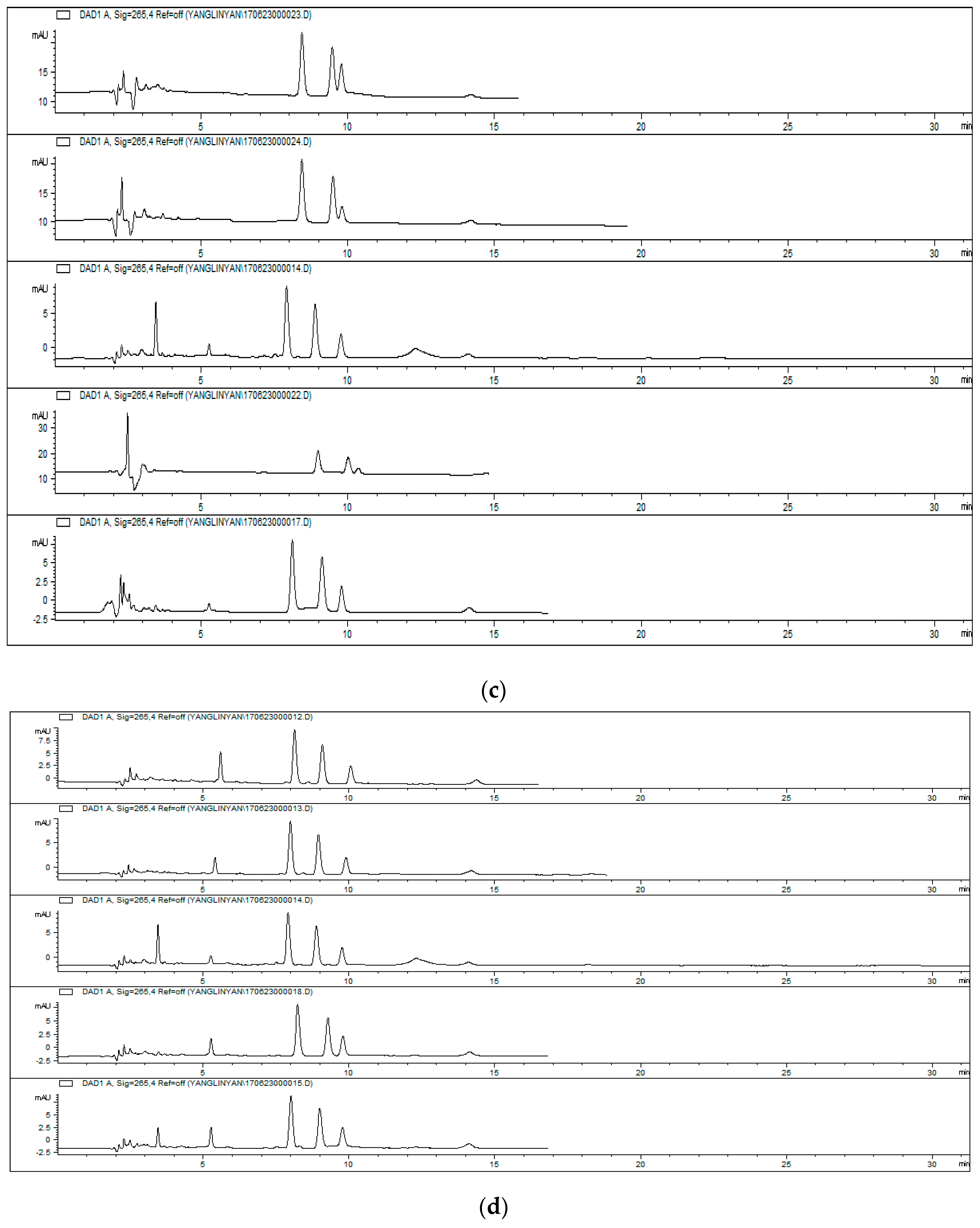
Figure 6a is the chromatogram associated with the concentrations of the standard curve, which belongs to Palmatine. Figure 6b is the chromatogram associated with the concentrations of the standard curve, which belongs to Berberine. The Equation process is as follows:
V0 = 10 mL, V1 = 1.5 mL, V2 = 0.4 mL, m is the capacity of alkaloid in the supernatant and detergent, C is the concentration of the supernatant and detergent, which could be obtained by the standard curve.
C0 = 0.5 μg/mL, V = 8 mL, Ap is the adsorption percentage of alkaloid.
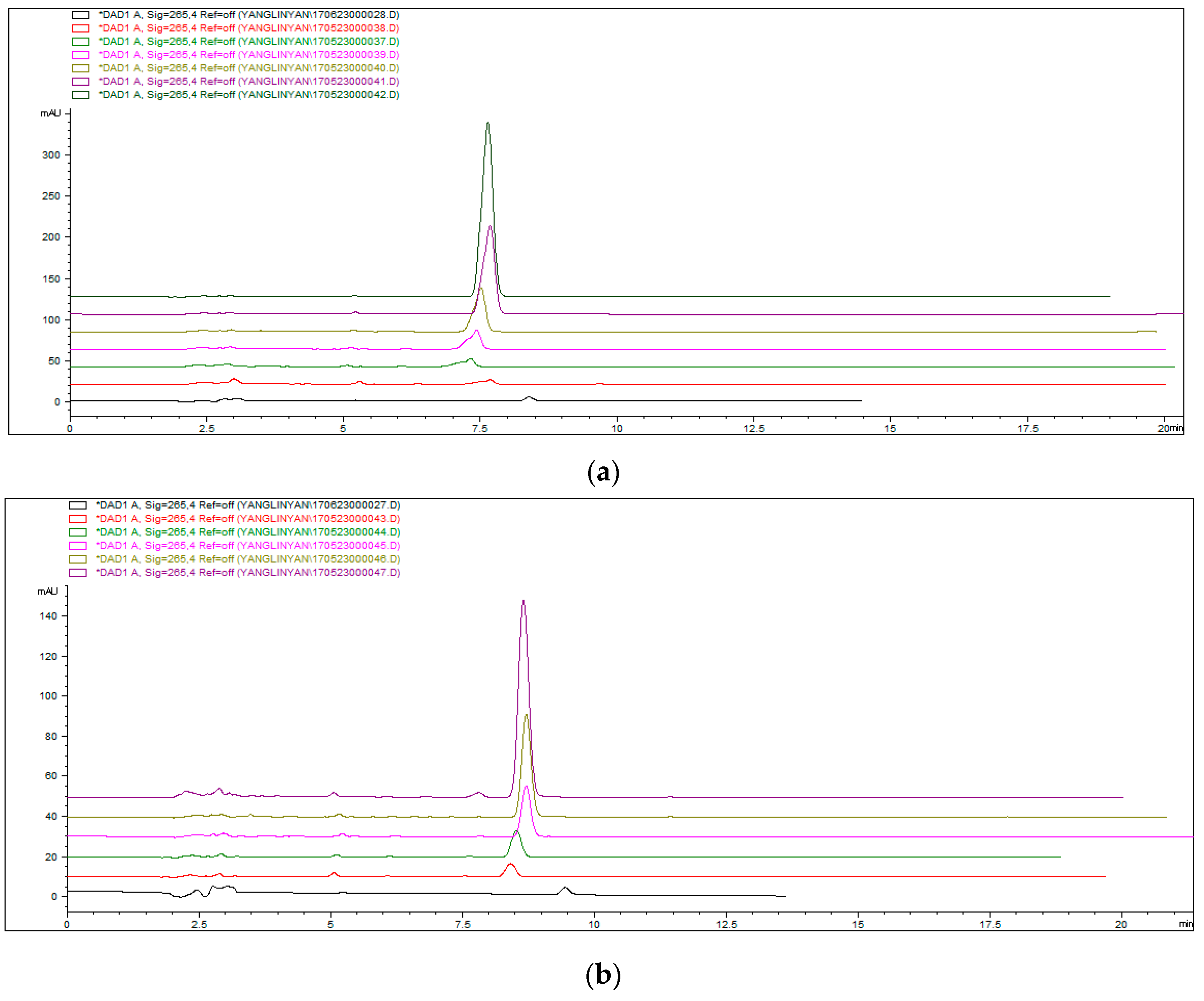
From Table S1, as for Palmatine and Berberine, the most adsorption could be obtained at pH 8. Considering the effect of alkaline on liquid phase system, the adsorption conditions of pH 8 has been chosen. The adsorption percentage of Palmatine was 22.2%, and the adsorption percentage of Berberine was 23.6%. At pH 8, the carboxylic acid of Fe3O4@SiO2-NH2-AA nanoparticles was converted to a negatively-charged carboxylate ion. Therefore, quaternary ammonium alkaloids were significantly adsorbed onto the carboxylic acid-rich surface, possibly due to electrostatic interactions. The results from this study seem to fit well with a previous report on the study of the charge interaction of alkaloids and polyelectrolyte films.
From Table S2, as for Berberine, the most adsorption could be obtained at 8 min. While the most adsorption could be obtained at 10 min for Palmatine. Considering the effect of adsorption time on liquid phase system, the adsorption conditions of 8 min has been chosen. The adsorption percentage of Palmatine was 8.67%, and the adsorption percentage of Berberine was 7.25%.
The effect of pH was greater than that of adsorption time. Considering the above conditions, pH 8 and the adsorption time of 8 min could be chosen for further uses.
4. Conclusions
In conclusion, magnetite (Fe3O4) could be prepared by FeCl2 and FeCl3, which is a ferromagnetic black color iron oxide of both Fe(II) and Fe(III). XRD was used for the determination of Fe3O4 nanoparticles. The peaks at 2θ values of 30.1°, 35.4°, 43.1°, 53.4°, 56.9° and 62.5° resemble the standard diffraction spectrum of Fe3O4 (JCPDSPDF#19-0629) with respect to its reflection peaks positions. Fe3O4 could be used for modification at the subsequent trials. Fe3O4@SiO2 nanoparticles were successfully obtained by TEOS. Fe3O4@SiO2-NH2 nanoparticles were prepared by APTMS, while Fe3O4@SiO2-NH2-AA nanoparticles were obtained by activated AA via amidation reaction. IR, XPS and SEM analysis were used for the characterizations of Fe3O4@SiO2-NH2 and Fe3O4@SiO2-NH2-AA nanoparticles. Alkaloid adsorption experiments implied that Fe3O4@SiO2-NH2-AA nanoparticles as a absorbent could be used for the adsorption of the alkaloids. At pH 8, the carboxylic acid of Fe3O4@SiO2-NH2-AA nanoparticleswas converted to a negatively-charged carboxylate ion. Therefore, quaternary ammonium alkaloids were significantly adsorbed onto the carboxylic acid-rich surface, possibly due to electrostatic interactions. As for Palmatine and Berberine, the most adsorption could be obtained at pH 8 when the adsorption time was 6 min. The adsorption percentage of Palmatine was 22.2%, while the adsorption percentage of Berberine was 23.6% at pH 8. As for the effect of adsorption time on liquid phase system, the adsorption conditions of 8 min has been chosen when pH 7 was used. Considering the above conditions, pH 8 and the adsorption time of 8 min could be chosen for further uses. This work demonstrates the potential of AA modification in a Fe3O4-based alkaloid adsorption study. In further experiments, when the amidation reaction is performed, residual carboxyl groups from AA on the modified Fe3O4@SiO2-NH2-AA nanoparticles may be used for bio-molecule immobilization.
Supplementary Materials
The supplementary materials are available online.
Acknowledgments
This work was supported by the Research Project of Tianjin Education Commission (2017KJ190), the Open Topic of Guangxi Key Laboratory for the Chemistry and Molecular Engineering of Medicinal Resources (CMEMR2016-B12), the National Natural Science Foundation of China (No. 31572492, No. 31072109, No. 31372482), the Innovative and Entrepreneurial Training Plan for Tianjin College Students (201710061035), the Veterinary Biotechnology Scientific Research Innovation Team of Tianjin, China (Grant No. TD12-5019), the General Fund of Application Foundation&Advanced Technology Program of Tianjin (14JCYBJC30000).
Author Contributions
Linyan Yang conceived and designed the experiments; Jing Tian and Jiali Meng performed the experiments; Ruili Zhao, Cun Li, and Jifei Ma analyzed the data; Tianming Jin contributed reagents/materials/analysis tools, revised and finalized the paper; Linyan Yang wrote the paper.
Conflicts of Interest
The authors declare no conflict of interest.
References
- Mohammed, L.; Gomaa, H.G.; Ragab, D.; Zhu, J. Magnetic nanoparticles for environmental and biomedical applications: A review. Particuology 2017, 30, 1–4. [Google Scholar] [CrossRef]
- Xu, X.; Friedman, G.; Humfeld, K.D.; Majetich, S.A.; Asher, S.A. Synthesis and utilization of monodisperse superparamagnetic colloidal particles for magnetically controllable photonic crystals. Chem. Mater. 2002, 14, 1249–1256. [Google Scholar] [CrossRef]
- Ge, J.; Hu, Y.; Biasini, M.; Beyermann, W.P.; Yin, Y. Superparamagnetic magnetite colloidal nanocrystal clusters. Angew. Chem. Int. Ed. 2007, 46, 4342–4345. [Google Scholar] [CrossRef] [PubMed]
- Ge, J.; Hu, Y.; Yin, Y. Highly tunable superparamagnetic colloidal photonic crystals. Angew. Chem. Int. Ed. 2007, 46, 7428–7431. [Google Scholar] [CrossRef] [PubMed]
- Wang, W.; Zheng, L.L.; Lu, F.H.; Hong, R.J.; Chen, M.Z.Q.; Zhuang, L. Facile synthesis and characterization of magnetochromatic Fe3O4 nanoparticles. AIP Adv. 2017, 7, 056317. [Google Scholar] [CrossRef]
- Ahmed, S.A.; Soliman, E.M. Silica coated magnetic particles using microwave synthesis for removal of dyes from natural water samples: Synthesis, characterization, equilibrium, isotherm and kinetics studies. Appl. Surf. Sci. 2013, 284, 23–32. [Google Scholar] [CrossRef]
- Yan, H.; Zhang, J.C.; You, C.X.; Song, Z.W.; Yu, B.W.; Shen, Y. Surface modification of Fe3O4 nanoparticles and their magnetic properties. Int. J. Miner. Metall. Mater. 2009, 16, 226–229. [Google Scholar] [CrossRef]
- Zhang, L.; Shao, H.P.; Zheng, H.; Lin, T.; Guo, Z.M. Synthesis and characterization of Fe3O4@SiO2 magnetic compositenanoparticles by a one-pot process. Int. J. Miner. Metall. Mater. 2016, 23, 1112–1118. [Google Scholar] [CrossRef]
- Bao, S.G.; Tang, L.H.; Li, K.; Ning, P.; Peng, J.H.; Guo, H.B.; Zhu, T.T.; Liu, Y. Highly selective removal of Zn(II) ion from hot-dip galvanizing pickling waste with amino-functionalized Fe3O4@SiO2 magnetic nano-adsorbent. J. Colloid Interface Sci. 2016, 462, 235–242. [Google Scholar] [CrossRef] [PubMed]
- Bao, S.G.; Li, K.; Ning, P.; Peng, J.H.; Jin, X.; Tang, L.H. Highly selective removal of mercury and lead ions from wastewater by mercaptoamine-functionalised silica-coated magnetic nano-adsorbents: Behaviours and mechanisms. Appl. Surf. Sci. 2017, 393, 457–466. [Google Scholar] [CrossRef]
- Lim, S.F.; Zheng, Y.M.; Zou, S.W.; Chen, J.P. Uptake of arsenate by an alginate-encapsulated magnetic sorbent: Process performance and characterization of adsorption chemistry. J. Colloid Interface Sci. 2009, 333, 33–39. [Google Scholar] [CrossRef] [PubMed]
- Mohammadi, A.; Daemi, H.; Barikani, M. Fast removal of malachite green dye using novel superparamagnetic sodium alginate-coated Fe3O4 nanoparticles. Int. J. Biol. Macromol. 2014, 69, 447–455. [Google Scholar] [CrossRef] [PubMed]
- Joshi, A.; Solanki, S.; Chaudhari, R.; Bahadur, D.; Aslam, M.; Srivastava, R. Multifunctional alginate microspheres for biosensing, drug delivery and magnetic resonance imaging. Acta Biomater. 2011, 7, 3955–3963. [Google Scholar] [CrossRef] [PubMed]
- Wang, J.Y.; Ren, L.; Wang, X.Q.; Wang, Q.; Wan, Z.F.; Li, L.; Liu, W.M.; Wang, X.M.; Li, M.L.; Tong, D.W.; et al. Superparamagnetic microsphere-assisted fluoroimmunoassay for rapid assessment of acute myocardial infarction. Biosens. Bioelectr. 2009, 24, 3097–3102. [Google Scholar] [CrossRef] [PubMed]
- Rezayan, A.H.; Mousavi, M.; Kheirjou, S.; Amoabediny, G.; Ardestani, M.S.; Mohammadnejad, J. Monodisperse magnetite (Fe3O4) nanoparticles modified with water soluble polymers for the diagnosis of breast cancer by MRI method. J. Magn. Magn. Mater. 2016, 420, 210–217. [Google Scholar] [CrossRef]
- Biradar, A.V.; Patil, V.S.; Chandra, P.; Doke, D.S.; Asefa, T. A trifunctional mesoporous silica-based, highly active catalyst for one-pot, three-step cascade reactions. Chem. Commun. 2015, 51, 8496–8499. [Google Scholar] [CrossRef] [PubMed]
- Guo, L.; Ding, W.; Meng, F. Fabrication and in vitro evaluation of folate-modified iron ferrite nanoparticles with high doxorubicin loading for receptors-magnetic-guided drug delivery. Nano Brief Rep. Rev. 2014, 9, 1450021–1450028. [Google Scholar] [CrossRef]
- Cai, Y.; Yuan, F.; Wang, X.; Sun, Z.; Chen, Y.; Liu, Z.; Wang, X.; Yang, S.; Wang, S. Synthesis of core-shell structured Fe3O4@carboxymethylcellulose magnetic composite for highly efficient removal of Eu(III). Cellulose 2017, 24, 175–190. [Google Scholar] [CrossRef]
- Liu, L.; Wang, Z.B.; Song, Y.; Yang, J.; Wu, L.J.; Yang, B.Y.; Wang, Q.H.; Wang, L.Q.; Wang, R.X.; Yang, C.J. Simultaneous determination of eight alkaloids in rat plasma by UHPLC-MS/MS after oral administration of Coptisdeltoidea C.Y. Cheng et Hsiao and Coptischinensis Franch. Molecules 2016, 21, 913–915. [Google Scholar] [CrossRef] [PubMed]
- Wang, Q.; Long, Y.; Yao, L.; Ye, M.; Xu, L. C18-COOH silica: preparation, characterization and its application in purification of quaternary ammonium alkaloids from Coptischinensis. Phytochem. Anal. 2017, 28, 332–343. [Google Scholar] [CrossRef] [PubMed]
- Ye, L.H.; Liu, X.D.; Chang, Y.X.; An, M.R.; Wang, S.L.; Xu, J.J.; Peng, L.Q. Analysis of isoquinoline alkaloids using chitosan-assisted liquid-solid extration followed by microemulsion liquid chromatography employing a sub-2-micron particles stationary phase. Electrophoresis 2016, 37, 3118–3125. [Google Scholar] [CrossRef] [PubMed]
- Jiang, W.; Wu, J.; Shen, Y.; Tian, R.; Zhou, S.; Jiang, W. Synthesis and characterization of doxorubicin loaded pH-sensitive magnetic core-shell nanocomposites for targeted drug delivery applications. Nano Brief Rep. Rev. 2016, 11, 1650127–1650140. [Google Scholar] [CrossRef]
- Jung, Y.C.; Muramatsu, H.; Fujisawa, K.; Kim, J.H.; Hayashi, T.; Kim, Y.A.; Endo, M.; Terrones, M.; Dresselhaus, M.S. Optically and biologically active mussel protein-coated double-walled carbon nanotubes. Small 2011, 7, 3292–3297. [Google Scholar] [CrossRef] [PubMed]
- Andhariya, N.; Upadhyay, R.; Mehta, R.; Chudasama, B. Folic acid conjugated magnetic drug delivery system for controlled release of doxorubicin. J. Nanopart. Res. 2013, 15, 1–12. [Google Scholar] [CrossRef]
- Chen, X.L.; Zhao, T.T.; Zou, J.L. A novel mimetic peroxidase catalyst by using magnetite-containing silica nanoparticles as carriers. Microchim. Acta 2009, 164, 93–99. [Google Scholar] [CrossRef]
Sample Availability: Samples of the compounds are not available from the authors. |
Figure 1.
The diagram of surface modification stages.
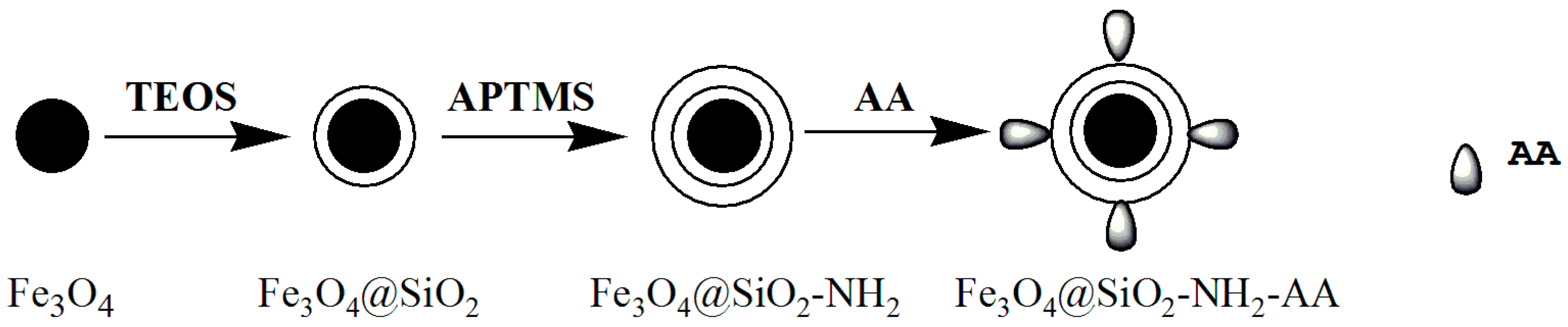
Figure 2.
XRD pattern of Fe3O4 nanoparticles. (Color squares are the standard diffraction spectrum of Fe3O4).
Figure 2.
XRD pattern of Fe3O4 nanoparticles. (Color squares are the standard diffraction spectrum of Fe3O4).

Figure 3.
FTIR spectra of: as-prepared Fe3O4 nanoparticles (black); Fe3O4@SiO2-NH2 (red); Fe3O4@SiO2-NH2-AA (blue).
Figure 3.
FTIR spectra of: as-prepared Fe3O4 nanoparticles (black); Fe3O4@SiO2-NH2 (red); Fe3O4@SiO2-NH2-AA (blue).
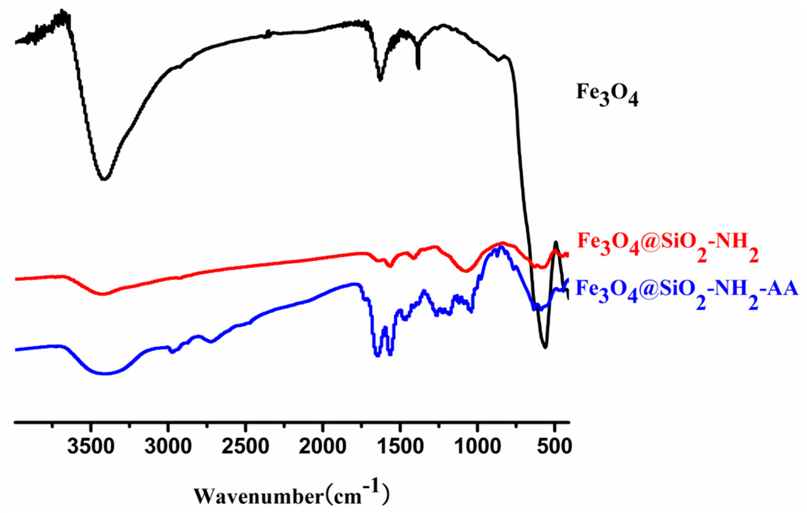
Figure 4.
(a) XPS wide scan spectra of Fe3O4, Fe3O4@SiO2, Fe3O4@SiO2-NH2, Fe3O4@SiO2-NH2-AA nanoparticles; (b) High-resolution XPS C1s spectra of Fe3O4@SiO2-NH2; (c) High-resolution XPS C1s spectra of Fe3O4@SiO2-NH2-AA.
Figure 4.
(a) XPS wide scan spectra of Fe3O4, Fe3O4@SiO2, Fe3O4@SiO2-NH2, Fe3O4@SiO2-NH2-AA nanoparticles; (b) High-resolution XPS C1s spectra of Fe3O4@SiO2-NH2; (c) High-resolution XPS C1s spectra of Fe3O4@SiO2-NH2-AA.

Figure 5.
The SEM images of nanoparticles: (a) Fe3O4, (b) Fe3O4@SiO2-NH2, (c) Fe3O4@SiO2-NH2-AA.

Figure 6.
(a) Concentration gradient chromatogram for Palmatine. (Standrad curve: y = 5.41437 + 61.51865x, R = 0.99958, linear range: 0.78125–50 μg/mL); (b) Concentration gradient chromatogram for Berberine. (Standard curve: y = −3.38806 + 53.63054x, R = 0.99899, linear range: 0.78125–25 μg/mL); (c) HPLC charomatograms of the supernatant after adsorption. Conditions: pH adjustment was as follows: 5, 6, 7, 8, 9; adsorption time was 6 min; (d) HPLC charomatograms of the supernatant after adsorption. Conditions: adsorption time adjustment was as follows: 2 min, 4 min, 6 min, 8 min, 10 min, while pH 7 was used.

© 2018 by the authors. Licensee MDPI, Basel, Switzerland. This article is an open access article distributed under the terms and conditions of the Creative Commons Attribution (CC BY) license (http://creativecommons.org/licenses/by/4.0/).
Share and Cite
MDPI and ACS Style
Yang, L.; Tian, J.; Meng, J.; Zhao, R.; Li, C.; Ma, J.; Jin, T. Modification and Characterization of Fe3O4 Nanoparticles for Use in Adsorption of Alkaloids. Molecules 2018, 23, 562. https://doi.org/10.3390/molecules23030562
AMA Style
Yang L, Tian J, Meng J, Zhao R, Li C, Ma J, Jin T. Modification and Characterization of Fe3O4 Nanoparticles for Use in Adsorption of Alkaloids. Molecules. 2018; 23(3):562. https://doi.org/10.3390/molecules23030562
Chicago/Turabian StyleYang, Linyan, Jing Tian, Jiali Meng, Ruili Zhao, Cun Li, Jifei Ma, and Tianming Jin. 2018. "Modification and Characterization of Fe3O4 Nanoparticles for Use in Adsorption of Alkaloids" Molecules 23, no. 3: 562. https://doi.org/10.3390/molecules23030562