Steroidal Saponins from Vernonia amygdalina Del. and Their Biological Activity
by
Jing Wang
†, 
Hua Song
†,
Xiaoxue Wu
,
Shuyi Zhang
,
Xuemin Gao
,
Funan Li
,
Xuan Zhu
* and
Qing Chen
* Department of Pharmacy, School of Pharmaceutical Science, Xiamen University, Xiamen 361102, China
*
Authors to whom correspondence should be addressed.
†
These authors contributed equally to this work and should be considered co-first authors.
Molecules 2018, 23(3), 579; https://doi.org/10.3390/molecules23030579
Submission received: 22 January 2018
/
Revised: 1 March 2018
/
Accepted: 2 March 2018
/
Published: 5 March 2018
Abstract
:In the present study, four new steroidal saponins, namely vernoniamyoside A–D (1–4), together with the two known steroidal saponins vernoamyoside D (5) and vernonioside B2 (6) were isolated from the ethanol extract of leaves of the African medicinal plant Vernonia amygdalina Del. (Asteraceae). Their structures were demonstrated by spectral analyses along with 1D and 2D nuclear magnetic resonance (NMR) techniques and mass spectrometry (MS). The cytotoxicity of the compounds was also tested by the 3-(4,5-dimethylthiazol-2-yl)-2,5-diphenyltetrazolium bromide (MTT) method on the cell lines Hela, MCF-7, BT-549 and MDA-MB-231. Vernoniamyoside A, vernoniamyoside B, and vernonioside B2 showed cytotoxicity towards BT-549 cell lines. Vernoniamyoside C, vernoniamyoside D and vernoamyoside D showed different levels of cytotoxic activities.

1. Introduction
Vernonia amygdalina Del. from the family Asteraceae is distributed throughout tropical Africa, especially in West Africa. It has received considerable scientific interest due to the observation that adult chimpanzees with malaria returned to normal activity after chewing the extract of the bitter juice of this species [1]. Over the years, several studies on the chemical components of this species, including flavonoids [2], sesquiterpene lactones [3], steroidal saponins [4,5,6], and fatty acids [7], have been performed. Previous studies have indicated different bioactivities of this species, including anti-inflammation [8], anti-malaria [9], anti-obesity [10,11], antioxidant [12,13], anti-tumor [14], and other activities [15]. Water and chloroform extracts of V. amygdalina (VA) interfere with the DNA synthesis of MCF-7 cells in breast cancer, affecting the activity of ERKs in vitro and inhibiting human breast cancer cells [16,17]. Via the MTT method, it has been verified that MCF-7 cells are inhibited in vitro and the DNA synthesis of BT-549 cells in breast cancer is disturbed by the addition of VA extract [18,19]. Moreover, three active fractions extracted from ethanol extracts of V. amygdalina have an inhibiting effect on tumor cells, the essential component being steroidal saponins [14]. So far, a certain amount of steroidal saponins has been obtained from V. amygdalina; however, studies on the anti-tumor activity are rare. It is therefore especially important to investigate the separation and biological activity of steroidal saponins in V. amygdalina.
In the present study, four new steroidal saponins, vernoniamyoside A–D (1–4) along with two known steroidal saponins vernoamyoside D (5) and vernonioside B2 (6) were obtained. It is expected that these steroidal saponins also demonstrate anti-tumor activity, especially anti-breast cancer activity. Therefore, all compounds were evaluated for their cytotoxicity toward human Hela, MCF-7, BT-549, and MDA-MB-231 cell lines by means of the MTT method. The isolation, structure identification, and biological activities of the aforementioned compounds are described.
2. Results and Discussion
2.1. Isolation, Characterization, and Structure Elucidation of Compounds
Compound 1 was obtained as a white powder, and its molecular formula C35H50O11, determined by high resolution electronspray ionization-mass spectrum (HR-ESI-MS) at m/z 669.3240 [M + Na]+ (calcd for C35H50NaO11, 669.3206), has 11 degrees of unsaturation. In the 1H NMR spectrum of compound 1, two olefinic proton signals at δH 5.40 (1H, br s, H-7) and 5.62 (1H, d, H-11), two angular methyl singlets at δH 0.52 (3H, s, CH3-18) and 0.87 (3H, s, CH3-19), two methyl doublets at δH 0.83 (3H, d, CH3-26), 0.91 (3H, d, CH3-27), and one methyl singlet 2.15 (3H, s, CH3-29) were observed. The 13C-NMR and distortionless enhancement by polarization transfer (DEPT) spectra indicated that compound 1 contained 35 carbon signals, including five methyls (at δC 13.8 (C-18), 19.4 (C-19), 17.1 (C-26), 17.0 (C-27) and 29.2 (C-29)), eight methines (including cyclic olefinic carbon signals at δC 121.9 (C-7), 133.1 (C-8), 143.9 (C-9), and 117.6 (C-11)), eight quaternary carbons (including two carbonyl carbon signals at δC 213.0 (C-28) and 214.2 (C-16) and a lactone carbon signal at δC 177.3 (C-21)), and 14 methylenes. Particularly, an obvious carbonyl signal at δC 214.2, corresponding to δH 2.63 (1H, H-17), 2.98 (H-20), 2.02 and 2.34 (H-15), in 1H-detected heteronuclear multiple-bond correlation (HMBC) was observed, suggesting a carbonyl group is connected to C-16. The 1H and 13C NMR analysis (see Table 1) supported the hypothesis that compound 1 might be Δ7, 9 (11) stigmastane-type steroid derivative, with the same skeleton as vernonioside A3 [5]. Moreover, two carbon signals at δC 83.0 (C-24), 33.5 (C-25) and obvious HMBC correlations of δH 0.83 (3H, d, CH3-26) with δC 83.0, 33.5, 17.0 and δC 0.91 (3H, d, CH3-27) with δC 83.0, 33.5, 17.1 were observed. According to the NMR data and analysis, we suggest that compound 1 contains a-HOCCH (CH3)2 group. A carbonyl signal at δC 213.0 (C-28) also showed HMBC correlations with a methyl signal at δH 2.15 (3H, s, CH3-29). In the HMBC spectrum, δH 2.63 (H-17) has correlations of δC 26.0 (C-22), 36.8 (C-20), δH 4.63 (H-23) has correlations of δC 26.0 (C-22), 36.8 (C-20), 177.3 (C-21).
The typical carbon signal δC 100.9 has a 1H-detected heteronuclear single quantum correlation (HSQC) correlation of δH 4.24 (1H, d, H-1′) and in the HMBC spectrum, obvious correlations from δH 4.24 to δC 76.3 and δH 3.57 to δC 100.9 suggest a glucose moiety in compound 1. The signal δC 100.9 is caused by anomeric carbon and the glucose moiety connected to C-3. The other carbon signals δC 61.1 and a series of signals between δC 70.0 and 80.0, along with proton signals between δH 2.90 and 3.90, are a better validation of the previous assumption. Through acid hydrolysis and a comparison of the retention time to standard d-glucose, the glucose moiety was confirmed to be d-glucose. Compared to the known compounds, compound 1 has the same side chain as vernoamyoside D [6].
First, the planar structure of compound 1 was determined. The nuclear overhauser effect spectroscopy (NOESY) correlation between H-3 (δH 3.57) and H-5 (δH 1.35), H-5 (δH 1.35) and H-14 (δH 2.65), Me-18 (δH 0.52) and Me-19 (δH 0.87), as well as Me-18 (δH 0.52) and H-20 (δH 2.98) indicated that compound 1 contains trans rings of nuclear parent and H-3, H-5 and H-14 were α-configurated, while Me-18, Me-19, and H-20 were β-configurated. A NOESY correlation from H-17 (δH 2.63) to H-14 (δH 2.65) was observed, which determined that H-17 has an α-orientation. Peaks from H-23 (δH 4.63) to H-20 (δH 2.98) indicated that the proton H-23 was β-configurated. The configuration of C-24 could not be determined by NOESY; thus, we compared the carbon signals between compound 1 and vernoamyoside D [6] with the same fragment. Thus, compound 1 was determined and named vernoniamyoside A (see Figure 1, Figure 2 and Figure 3).
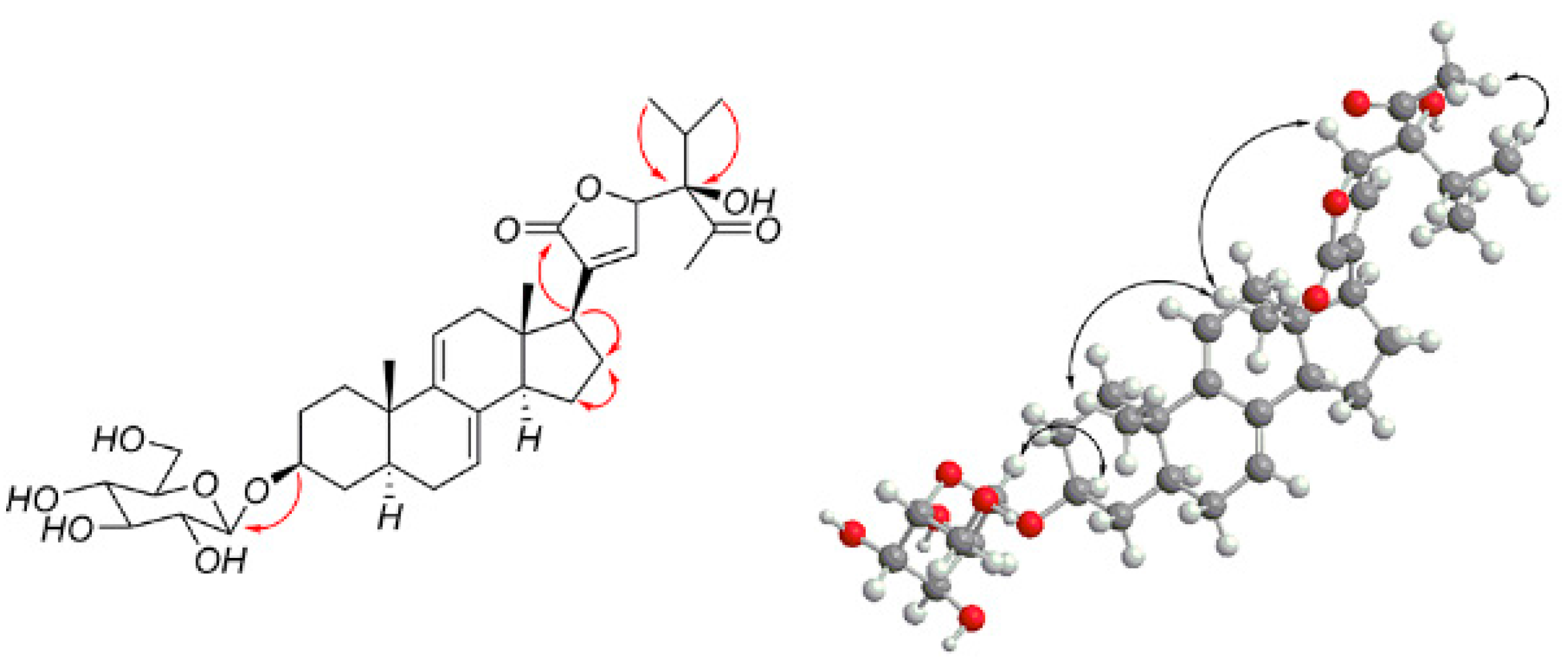
Compound 2 was obtained as a white powder, and its molecular formula C36H54O11, determined by HR-ESI-MS at m/z 685.3554 [M + Na]+, (calcd for C36H54NaO11, 685.3553), has 10 degrees of unsaturation. The 1H NMR spectrum of compound 2 showed the following specific signals: two methyl doublets at δH 0.87 (3H, d, CH3-26), 1.03 (3H, d, CH3-27), and two methyl singlets at 1.26 (3H, s, CH3-29) and 3.15 (3H, s, OCH3). The 13C NMR spectra indicated 36 carbon signals and showed the following typical five methyl signals: (at δC 11.8 (C-18), 19.4 (C-19), 16.5 (C-26), 17.6 (C-27), and 15.0 (C-29)). In the HMBC spectrum, we observed correlations between δH 1.39 and 1.76 with δC 22.8 (C-16), δH 1.39 with δC 28.0 (C-15), 50.9 (C-17), 135.8 (C-8), and δH 1.76 with δC 41.8 (C-13), 50.9 (C-17), indicating that the signal δC 22.8 is connected to C-16. We therefore infer that compound 2 is Δ7, 9 (11), a stigmastane-type steroid derivative with the same skeleton as vernonioside B1 [1] by comparison with 1H-NMR and 13C-NMR (see Table 1).
A large number of 13C-NMR signals were between δC 70.0 and 90.0, indicating that there is a highly oxidized side chain. In the HMBC spectrum, the correlations of H-25 (δH 1.91), H-26 (δH 0.87), and H-27 (δH 1.03) to the carbon signals at δC 82.6 (C-24) and H-22 (δH 4.58), H-23 (δH 4.62), H-29 (δH 1.26) to δC 106.9 (C-28), and H-22 (δH 4.58), H-23 (δH 4.62), H-20 (δH 2.76) to δC 175.4 (C-21) indicate that the side chain is similar compared to that of vernoamyoside C [6]. The difference between compound 2 and vernoamyoside C is C-21, the absence of signals at δH 5.45 and δC 100.0, and the presence of signals at δC 175.4, indicating that the side chain is highly oxidized. This helped us to determine the planar structure of compound 2. The correlation between H-3 (δH 3.56) and H-5 (δH 1.29), H-5 (δH 1.29) and H-14 (δH 1.94), Me-18 (δH 0.54) and Me-19 (δH 0.85), and Me-18 (δH 0.54) and H-20 (δH 2.76) indicated that compound 2 has the trans rings of the nuclear parent and H-3, H-5 and H-14 adopted an α-configuration, while Me-18, Me-19 and H-20 were β-configurated. This is similar to compound 1. The NOESY spectrum from H-17 (δH 2.19) to H-14 (δH 1.94) was observed, which determined that the H-17 has α-orientation. Peaks from H-23 (δH 4.62) to H-20 (δH 2.76) indicated that proton H-23 was β-configurated. The significant signals from H-20 (δH 2.76) to H-18 (δH 0.54), H-22 (δH 4.58) and H-23 (δH 4.62) indicated that H-22 (δH 4.58) and H-23 (δH 4.62) were β-configurated. The correlation between H-29 (δH 1.26) and H-14 (δH 1.94) suggested that CH3-29 has an α-configuration [6]. Thus, compound 2 was determined and named vernoniamyoside B (see Figure 1 and Figure 4).
Compound 3 was obtained as a white powder, and its molecular formula C35H50O11, determined by HR-ESI-MS at m/z 669.3250 [M + Na]+ (calcd for C35H50NaO11, 669.3206), has 11 degrees of unsaturation. The proton signal is at δH 4.40 (H-16), which has correlation with δC 48.7 (C-14) in the HMBC spectrum. The correlations between H-17 (δH 2.41) and the carbon signal at δC 74.3 in the HMBC spectrum confirm that the δC 74.3 is connected to C-16. The 1H NMR and 13C NMR signals (see Table 1) of compound 3 indicated that it is a Δ7, 9 (11) stigmastane-type steroid derivative with the same skeleton as vernoamyoside D [6]. According to the HMBC, the signal at δH 7.49 has a correlation with δC 56.6 (C-17), 173.3 (C-21), 82.8 (C-23), while δH 2.41 (H-17) and 5.24 (H-23) have correlations with δC 133.9. Therefore, there is a double bond between C-20 (δC 133.9) and C-22 (δC 146.8). Thus, the planar structure of compound 3 could be determined. Compared to the 1H NMR and 13C NMR signals, the side chain is the same as in vernonioside A4 [20]. The significant nuclear overhauser effect (NOE) correlations between H-16 (δH 4.40) and Me-18 (δH 0.30) suggested that OH-16 has an β-configuration. Therefore, compound 3 was determined and named vernoniamyoside C (see Figure 1 and Figure 5).
Compound 4 was obtained as a white powder, and its molecular formula C35H50O10, determined by HR-ESI-MS at m/z 653.3303 [M + Na]+ (calcd for C35H50NaO10, 653.3257), has 11 degrees of unsaturation. The 13C NMR spectra indicated 35 carbon signals. Compared with the 1H NMR and 13C NMR signals (see Table 1) of compound 2, compound 4 is also a Δ7, 9 (11) stigmastane-type steroid derivative with a glycoside. As for the side chain, δH 5.22 (H-23) has correlations with δC 135.7 in the HMBC spectrum. Compound 4 had similar 1H, 13C NMR, and NOESY data when compared with compound 3. Thus, compound 4 was determined and named vernoniamyoside D (see Figure 1 and Figure 6).
2.2. Results of the Cytotoxicity Test
According to a previous study, MCF-7 cells are inhibited in vitro, and the DNA synthesis of BT-549 cells in breast cancer is disturbed by the application of V. amygdalina extract [18,19]. A series of steroidal saponins exhibited anti-tumor activity, especially anti-breast tumor activity [14]. In this study, the cytotoxicity of compounds 1–6 was tested on BT-549, MDA-MB-231, MCF-7, and Hela cell lines. The anti-tumor activity of Δ7, 9 (11) stigmastane-type steroidal saponins was introduced for the first time. As seen in Table 2, the inhibition against the BT-549 cell line of compound 1 could reach up to 63.61%, while compound 2 and 6 also showed cytotoxicity towards the BT-549 cell line (inhibition = 62.17 and 51.14%). This leads us to infer that compounds 1, 2, and 6 are highly toxic towards BT-549 cell lines, while they showed a general cytotoxicity to cell lines MDA-MB-231, MCF-7, and Hela. Based on this, these compounds might play a certain role in the treatment of breast cancer. The cytotoxicity activities of compound 3, 4, and 5 showed different levels against the tested cell lines. Moreover, further studies are necessary to confirm whether these compounds are also toxic towards other tumor cell lines. Both compounds have the same sugar chains; therefore, the different activities might be due to the side chain and the nuclear parent. Furthermore, it should be highlighted that compounds 1-6 was had a different selectivity for tumor cell lines.
3. Materials and Methods
3.1. General Experimental Procedures
Optical rotations were measured with an Automatic polarimeter (Hackettstown, NJ, USA). UV spectra were recorded by a Shimadzu UV-2600 PC spectrophotometer (Suzhou, Jiangsu, China). IR (KBr-disks) spectra were measured using a Bruker Alpha (Karlsruhe, Germany). The HR-ESI-MS spectra were recorded by a Thermo Scientific Q Exactive Plus Orbitrap LC-MS/MS system (Waltham, MA, USA); NMR spectra were recorded by a Bruker AVANCE III 600 MHz spectrometer (Zurich, Switzerland) in CD3COCD3, with TMS as internal standard. The preparative high-performance liquid chromatography (HPLC) system consisted of an LC-6AD intelligent prep. pump (Kyoto, Japan), an SPD-20A intelligent UV/VIS detector (Kyoto, Japan), and a YMC-Park ODS-A column (5 μm, 250 × 10 mm I.D., YMC Co. Ltd., Ishikawa, Japan). Silica gel GF254 for thin-layer chromatography (TLC) and silica gel (200–300 mesh) for column chromatography (CC) were obtained from Qingdao Marine Chemical Factory (Qingdao, Shandong, China). Sephadex LH-20 (Merck, Darmstadt, Germany) was suited for size-exclusion chromatography. The cell lines BT-549 Hela, MCF-7, and MDA-MB-231 were obtained from China Center for Type Culture Collection (Wuhan, Hubei, China); MTT were obtained from Sigma Company. All solvents were purchased from Sinopharm Chemical Reagents (Shanghai, China). Methyl alcohol used for HPLC analysis was of chromatographic grade (Sigma, St. Louis, MO, USA). All aqueous solutions were prepared with double-distilled water.
3.2. Plant Material
Dried leaves of V. amygdalina were collected in Xiamen, Fujian Province, China, and identified by Qing Chen, Assistant Professor from the School of Pharmaceutical Sciences, Xiamen University.
3.3. Extraction and Isolation
The dried leaves (7.5 kg) were extracted with 95% ethanol and concentrated under vacuum to obtain crude extract. Subsequently, the crude extract was suspended in H2O and partitioned in petroleum ether, dichloromethane (CH2Cl2), ethyl acetate, and n-butanol in sequence to obtain the petroleum ether fraction (170 g), the dichloromethane fraction (224 g), the ethyl acetate fraction (80 g), and the n-butanol fraction (60 g), respectively. The dichloromethane fraction (224 g), a dark green syrup, was subjected to macroporous resin column chromatography (CC) (4 kg, D101) and eluted with gradient methyl alcohol (MeOH)-H2O (8:2) and MeOH. After removing the solvents under vacuum, the MeOH-H2O extract (110 g) was subjected to silica gel column chromatography (CC) (1 kg, 200–300 mesh) and eluted with gradient CH2Cl2-MeOH (50:1 to 1:1 v/v) and MeOH to obtain fractions A (9.8 g), B (14.2 g), C (21.7 g), D (22.2 g), E (15.7 g), F (14.7 g), and G (8.5 g) after deducting the solvents. Fraction C was re-chromatographed on a silica gel CC eluted with CH2Cl2-MeOH, using a gradient (15:1 to 5:1 v/v) to obtain five fractions (Ca-Ce). Fraction Cb was further purified by Sephadex LH-20 eluted with MeOH to obtain 32 fractions, which were pooled together using TLC to obtain the three sub-fractions Cba-Cbc. The fraction Cbb (922 mg) was separated by preparative HPLC, using a YMC-Park ODS-A column (5 μm, 250 × 10 mm I.D.) with a flow rate of 5 mL/min and a mobile phase of MeOH-H2O (75:25) to obtain the five sub-fractions Cbba-Cbbe. Sub-fraction Cbba (100 mg) was purified by preparative HPLC, using a YMC-Park ODS-A column (5 μm, 250 × 10 mm I.D.) with a flow rate of 5 mL/min and a mobile phase of MeOH-H2O (58:42) to yield compound 1 (49 mg). Sub-fraction Cbbd (18 mg) was purified by preparative HPLC, using a YMC-Park ODS-A column (5 μm, 250 × 10 mm I.D.) with a flow rate of 5 mL/min and a mobile phase of MeOH-H2O (70:30) to yield compound 4 (5.1 mg). Sub-fraction Cbbe (40 mg) was purified by preparative HPLC, using a YMC-Park ODS-A column (5 μm, 250 × 10 mm I.D.) with a flow rate of 5 mL/min and a mobile phase of MeOH-H2O (75:25) to yield compound 2 (21 mg). Fraction D was re-chromatographed on silica gel CC eluted with CH2Cl2-MeOH, using a gradient (12:1 to 1:1 v/v) which was pooled together using TLC to obtain six fractions (Da-Df). Fraction Dd (828 mg) was purified by preparative HPLC, using a YMC-Park ODS-A column (5 μm, 250 × 10 mm I.D.) with a flow rate of 5 mL/min and a mobile phase of MeOH-H2O (57:43) to yield compounds 3 (22 mg) and 5 (16 mg). Fraction Db was further purified by preparative HPLC, using a YMC-Park ODS-A column (5 μm, 250 × 10 mm I.D.) with a flow rate of 5 mL/min and a mobile phase of MeOH-H2O (71:29) to obtain two sub-fractions. Sub-fraction Dbb (40 mg) was further purified by preparative HPLC, using a YMC-Park ODS-A column (5 μm, 250 × 10 mm I.D.) with a flow rate of 5 mL/min and a mobile phase of MeOH-H2O (73:27) to obtain compound 6 (27 mg).
3.3.1. Vernoniamyoside A (1)
White amorphous powder; HR-ESI-MS m/z 669.3240 [M + Na]+ (calcd for C35H50NaO11, 669.3206); [α: −0.32 (c 0.05, CH3OH); UV (DMSO): λmax: 252.0, 208.8 nm; IR (KBr) vmax: 3392, 2933, 2361, 1770, 1704, 1354, 1025 cm−1; 1H and 13C NMR data: see Table 1.
3.3.2. Vernoniamyoside B (2)
White amorphous powder; HR-ESI-MS m/z 685.3554 [M + Na]+ (calcd for C36H54NaO11, 685.3553); [α: +0.40 (c 0.10, CH3OH); UV (DMSO): λmax: 252.0, 207.2 nm; IR (KBr) vmax: 3395, 2937, 2360, 1775, 1382, 1025 cm−1; 1H and 13C NMR data: see Table 1.
3.3.3. Vernoniamyoside C (3)
White amorphous powder; HR-ESI-MS m/z 669.3250 [M + Na]+ (calcd for C35H50NaO11, 669.3206); [α: −0.20 (c 0.05, CH3OH); UV (DMSO): λmax: 252.0, 208.8 nm; IR (KBr) vmax: 3735, 3421, 2933, 2361, 1750, 1716, 1507, 1024 cm−1; 1H and 13C NMR data: see Table 1.
3.3.4. Vernoniamyoside D (4)
White amorphous powder; HR-ESI-MS m/z 653.3303 [M + Na]+ (calcd for C35H50NaO10, 653.3257); [α: −0.24 (c 0.05, CH3OH); UV (DMSO): λmax: 252.0, 208.8 nm; IR (KBr) vmax: 3735, 3421, 2933, 2361, 1750, 1716, 1558, 1507, 1024 cm−1; 1H and 13C NMR data: see Table 1.
3.3.5. Vernoamyoside D (5)
White amorphous powder; 1H and 13C NMR data comparable to published data [6]. 13H NMR (600Hz, CD3COCD3): δH 5.33 (1H, br s, H-7) and 5.51 (1H, d, H-11),0.44 (3H, s, CH3-18), 0.83 (3H, s, CH3-19), 0.84 (3H, d, CH3-26), 0.91 (3H, d, CH3-27) and 2.16 (3H, s, CH3-29). 13C NMR (150Hz, CD3COCD3): δC 33.7 (C-1),29.3 (C-2), 76.3 (C-3), 34.2 (C-4), 38.7 (C-5), 29.8 (C-6), 121.9 (C-7), 135.1(C-8), 143.8 (C-9), 35.6 (C-10), 118.8 (C-11), 40.7 (C-12), 42.4 (C-13), 48.1 (C-14), 34.0 (C-15), 73.5 (C-16), 59.1 (C-17), 12.8 (C-18), 19.4 (C-19), 39.4 (C-20), 177.8 (C-21), 27.4 (C-22), 80.4 (C-23), 83.8 (C-24), 33.4 (C-25), 17.2 (C-26), 17.0 (C-27), 213.5 (C-28), 29.4 (C-29), 100.9 (C-1′), 73.5 (C-2′), 76.7 (C-3′), 70.1 (C-4′), 76.7 (C-5′), 61.2 (C-6′).
3.3.6. Vernonioside B2 (6)
White amorphous powder; 1H and 13C NMR data: comparable to published data [20]. 13H NMR (600Hz, CD3COCD3): δH 5.36 (1H, br s, H-7) and 5.47 (1H, d, H-11),0.48 (3H, s, CH3-18), 0.83 (3H, s, CH3-19), 0.85 (3H, d, CH3-26), 0.88 (3H, d, CH3-27), 1.33 (3H, s, CH3-29) and 3.12 (3H, s, OCH3). 13C NMR (150Hz, CD3COCD3): 35.2 (C-1), 29.7 (C-2), 77.1 (C-3), 34.9 (C-4), 38.9 (C-5), 29.9 (C-6), 121.8 (C-7), 135.7 (C-8), 143.7(C-9), 36.0 (C-10), 118.6 (C-11), 41.1 (C-12), 43.1 (C-13), 48.5 (C-14), 34.9 (C-15), 76.7 (C-16), 54.9 (C-17), 14.3 (C-18), 19.7 (C-19), 47.9 (C-20), 98.4 (C-21), 80.2 (C-22), 90.2 (C-23), 81.3 (C-24), 31.7 (C-25), 17.5 (C-26), 18.4 (C-27), 112.4 (C-28), 17.5 (C-29), 101.4 (C-1′), 75.5 (C-2′), 80.2 (C-3′), 72.9 (C-4′), 77.2 (C-5′), 61.6 (C-6′), 48.2 (OCH3).
3.4. Acid Hydrolysis of Compounds
Compounds 1–4 (1 mg each) were dissolved in 2 M HCl (5.0 mL) and stirred at 90°C for 4 h. The reaction solution was neutralized with NH4OH and partitioned between EtOAc and H2O. The residue was obtained from the water layer under vacuum distillation and dissolved in pyridine (1 mL), which was added to 0.1 M l-cysteine methyl ester hydrochloride in pyridine (1 mL). After being heated to 60 °C for 1 h, the mixture was added to phenyl isothiocynate in pyridine (1 mL) and stirred at 60 °C for an additional 1 h. After removal of the solvent, the residue was dissolved in MeOH and analyzed by HPLC, with the mobile phase CH3CN-H2O (30:80, v/v) containing 0.1% formic acid; the flow rate was 1 mL/min. The standard d-glucose derivative was prepared in the same way. We then compared the retention times of sugar derivatives obtained from compounds with standard d-glucose derivatives (d-glucose: 19.5 min).
3.5. Cytotoxicity Assay
The cytotoxicity of compounds 1–6 was tested on cell lines MCF-7, BT-549, MDA-MB-231, and Hela via the MTT method. Cells were seeded into 96-well microplates at 100 μL per well (MCF-7, MDA-MB-231, and A549 were cultured in Dulbecco’s modified eagle medium (DMEM) supplemented with 10% fetal calf serum and 1% penicillin-streptomycin solution, while BT-549 was cultured in Roswell Park Memorial Institute (RPMI) (according to previous experiments, the DMSO concentration we used showed no interference with the experimental results). After 48 h, we added MTT (0.5 mg/mL) dissolved in 100 μL of fetal calf serum per well. After 4 h, we removed the MTT reagent and added 150 μL DMSO to each well. Doxorubicin was used as the positive control. The sample without test compounds was used as the negative control. Absorbance was measured at 490 nm in an automated microplate reader. All experiments were performed in triplicate. Inhibition was calculated via the following equation:
Atest sample is the test sample absorbance, Ablank is the blank absorbance, Anegative control is the negative control absorbance.
4. Conclusions
We obtained four new steroidal saponins, namely vernoniamyoside A–D (1–4), together with the two known steroidal saponins vernoamyoside D (5) and vernonioside B2 (6) from V. amygdalina. Of these, 1, 2, and 6 showed an excellent cytotoxicity on BT-549 cell lines in the cytotoxicity activity assay (Table 2), as expected. It is worth noting that 1 and 2 were selective for different tumor cell lines. Our results indicate that substances 1 and 2 were toxic to BT-549 cell lines. Further studies should consider the screening of more types of tumor cells.
The novelty of the saponins 1–4 is represented by the highly oxidized groups in the side chain, which may be associated with their cytotoxic expression. Saponin 1 has a strong cytotoxicity to BT-549 cell lines, including two ketones and one ester group. It is assumed that the cytotoxicity of saponin 1 may be derived from the C=O group. This group, present in the highly oxidized side chains of saponins 2–4, may provide a basis for the cytotoxicity for BT-549. However, the different inhibitory effects may be due to the relationships between the different structures and spatial configurations. The specific mechanisms and verifications offer a new direction for further experiments.
The separated steroidal saponins from the dichloromethane extraction of V. amygdalina, are mainly hypoglycemic steroidal saponins. However, it is essential to separate polysaccharide steroidal saponins, which always exist in ethyl acetate and n-butanol extraction, from V. amygdalina. In addition to the steroidal saponins, V. amygdalina contains a series of active substances such as sesquiterpene lactones and flavonoids. Therefore, the separation and identification of these chemical components should be one of the most promising trends in the study of V. amygdalina.
Our results provide a scientific basis for the use of V. amygdalina in anti-tumor research, with a potential value in the treatment of cancer.
Supplementary Materials
The following 1H NMR, 13C NMR, 2D-NMR, and HR-ESI-MS spectra are available as supporting data. Supplementary materials are available online.
Acknowledgements
This research work was financially supported by the National Natural Science Foundation of China [grant number 31300285], the Natural Science Foundation of Fujian Province of China [grant numbers 2015J01348,2015Y0081], the Specialized Research Fund for the Doctoral Program of Higher Education [grant number 20110073120072], and the Fujian Province Development Guidance (Key) Project [grant number 2014Y0074].
Author Contributions
Jing Wang designed the experiments and revised the paper; Jing Wang and Hua Song performed the experiments, analyzed the data, and wrote the paper; Jing Wang, Xiaoxue Wu, Xuemin Gao, and Shuyi Zhang contributed to bioassay reagents and materials and analyzed the data; Funan Li performed the experiments; Qing Chen and Xuan Zhu revised the paper. All authors read and approved the final manuscript.
Conflicts of Interest
We wish to confirm that there are no known conflicts of interest associated with this publication and that there has been no significant financial support for this work that could have influenced its outcome.
References
- Ohigashi, H.; Jisaka, M.; Takagaki, T.; Nozaki, H.; Tada, T.; Huffman, M.A.; Nishida, T.; Kaji, M.; Koshimizu, K. Bitter principle and a related steroid glucoside from, a possible medicinal plant for wild chimpanzees. Agric. Biol. Chem. 1991, 55, 1201–1203. [Google Scholar] [CrossRef]
- Igile, G.O.; Oleszek, W.; Jurzysta, M.; Burda, S.; Fafunso, M.; Fasanmade, A.A. Flavonoids from vernonia amygdalina and their antioxidant activites. J. Agric. Food Chem. 1994, 42, 2445–2448. [Google Scholar] [CrossRef]
- Sinisi, A.; Millán, E.; Abay, S.M.; Habluetzel, A.; Appendino, G.; Muñoz, E.; Taglialatelascafati, O. Poly-electrophilic sesquiterpene lactones from vernonia amygdalina: New members and differences in their mechanism of thiol trapping and in bioactivity. J. Nat. Prod. 2015, 78, 1618–1623. [Google Scholar] [CrossRef] [PubMed]
- Igile, G.O.; Oleszek, W.; Jurzysta, M.; Aquino, R.; Tommasi, N.; Pizza, C. Vernonioside d and e, two novel saponins from vernonia amygdalina del. (compositae). J. Nat. Prod. 1995, 589, 1438–1443. [Google Scholar] [CrossRef]
- Jisaka, M.; Ohigashi, H.; Takagaki, T.; Nozaki, H.; Tada, T.; Hirota, M.; Irie, R.; Huffman, M.A.; Nishida, T.; Kaji, M. Bitter steroid glucosides, vernoniosides a 1, a 2, and a 3, and related b 1 from a possible medicinal plant, vernonia amygdalina, used by wild chimpanzees. Tetrahedron 1992, 48, 625–632. [Google Scholar] [CrossRef]
- Quasie, O.; Zhang, Y.M.; Zhang, H.J.; Luo, J.; Kong, L.Y. Four new steroid saponins with highly oxidized side chains from the leaves of vernonia amygdalina. Phytochem. Lett. 2016, 15, 16–20. [Google Scholar] [CrossRef]
- Erasto, P.; Grierson, D.S.; Afolayan, A.J. Evaluation of antioxidant activity and the fatty acid profile of the leaves of vernonia amygdalina growing in south africa. Food Chem. 2007, 104, 636–642. [Google Scholar] [CrossRef]
- Eyong, E.U.; Atangwho, I.J.; David-Oku, E.; Agiang, M.A.; Ebong, P.E. Haematological and immunological effect of co- administration of extracts of vernonia amygdalina and azadirachta indica on normal and diabetic rats. Afr. J. Biotechnol. 2011, 10, 10258–10262. [Google Scholar] [CrossRef]
- Adedapo, A.A.; Otesile, A.T.; Soetan, K.O. Assessment of the anthelmintic efficacy of an aqueous crude extract of vernonia amygdalina. Pharm. Biol. 2008, 45, 564–568. [Google Scholar] [CrossRef]
- Egedigwe, C.A.; Ejike, C.E.; Ijeh, I.I.; Onwuka, G.I.; Herbert, U.; Asumugha, A.V. Anti-obesity potentials of aqueous and methanol extracts of vernonia amygdalina del. Leaves in high-fat diet fed rats. Afr. J. Tradit. Complement. Altern. Med. 2016, 13, 86–93. [Google Scholar] [CrossRef]
- Atangwho, I.J.; Edet, E.E.; Uti, D.E.; Obi, A.U.; Asmawi, M.Z.; Ahmad, M. Biochemical and histological impact of vernonia amygdalina supplemented diet in obese rats. Saudi. J. Biol. Sci. 2012, 19, 385–392. [Google Scholar] [CrossRef] [PubMed]
- Erasto, P.; Grierson, D.S.; Afolayan, A.J. Antioxidant constituents in vernonia amygdalina leaves. Pharm. Biol. 2008, 45, 195–199. [Google Scholar] [CrossRef]
- Adaramoye, O.; Ogungbenro, B.; Anyaegbu, O.; Fafunso, M. Protective effects of extracts of vernonia amygdalina, hibiscus sabdariffa and vitamin c against radiation-induced liver damage in rats. J. Radiat. Res. 2008, 49, 123–131. [Google Scholar] [CrossRef] [PubMed]
- Luo, X.; Oyugi, D.A.; Lin, C.W.; Izevbigie, E.B.; Lee, K.S. Isolation and characterization of the antibreast carcinoma cell growth components of vernonia amygdalina extracts. Exp. Biol. Med. 2010, 235, 1472–1478. [Google Scholar] [CrossRef] [PubMed]
- Sonibare, M.A.; Moody, J.O.; Adesanya, E.O. Use of medicinal plants for the treatment of measles in nigeria. J. Ethnopharmacol. 2009, 122, 268–272. [Google Scholar] [CrossRef] [PubMed]
- Izevbigie, E.B.; Bryant, J.L.; Walker, A. A novel natural inhibitor of extracellular signal-regulated kinases and human breast cancer cell growth. Exp. Biol. Med. 2004, 229, 163–169. [Google Scholar] [CrossRef]
- Opata, M.M.; Izevbigie, E.B. Aqueous vernomia amygdalina extracts alter mcf-7 cell membrane permeability and efflux. Int. J. Environ. Res. Public Health 2006, 3, 174–179. [Google Scholar] [CrossRef] [PubMed]
- Yedjou, C.; Izevbigie, E.; Tchounwou, P. Preclinical assessment of vernonia amygdalina leaf extracts as DNA damaging anti-cancer agent in the management of breast cancer. Int. J. Environ. Res. Public Health 2008, 5, 337–341. [Google Scholar] [CrossRef] [PubMed]
- Gresham, L.J.; Ross, J.; Izevbigie, E.B. Vernonia amygdalina: Anticancer activity, authentication, and adulteration detection. Int. J. Environ. Res. Public Health 2008, 5, 342–348. [Google Scholar] [CrossRef] [PubMed]
- Jisaka, M.; Ohigashi, H.; Takegawa, K.; Hirota, M.; Irie, R.; Huffman, M.A.; Koshimįzu, K. Steroid glucosides from vernonia amygdalina, a possible chimpanzee medicinal plant. Phytochemistry 1993, 34, 409–413. [Google Scholar] [CrossRef]
Sample Availability: Samples of the compounds vernoniamyoside A–D, vernoamyoside D and vernonioside B2 are available from the authors. |
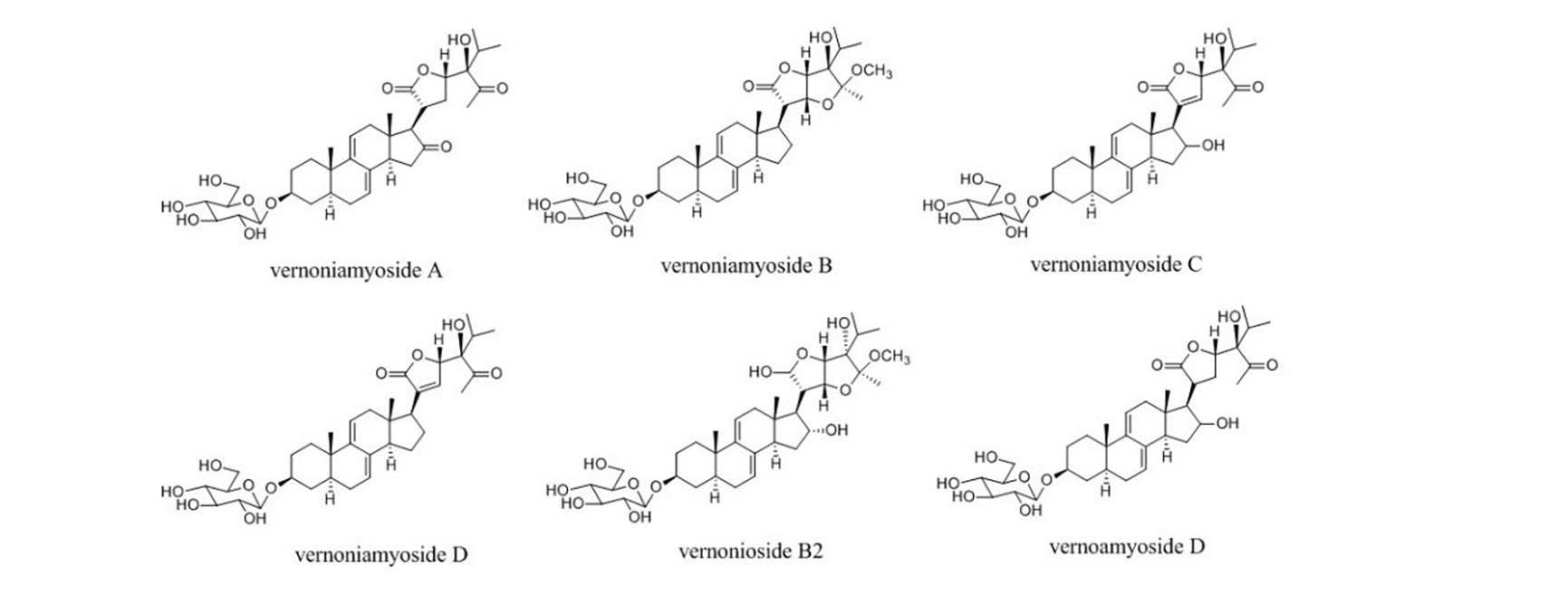
Figure 1.
Structures of compounds 1–6.
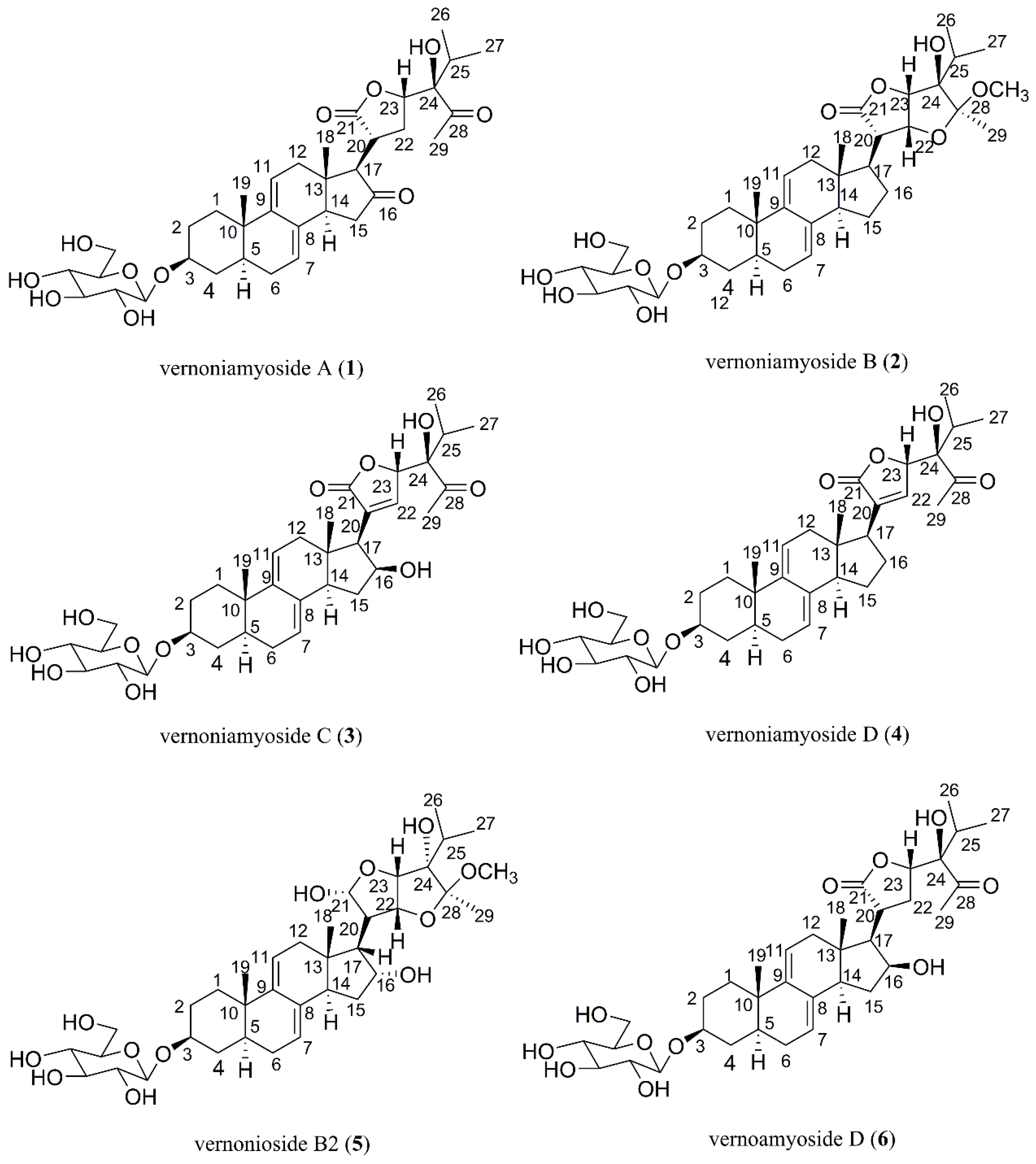
Figure 2.
Major 1H-1H correlation spectroscopy (1H-1H COSY) correlations of compound 1 (bold lines).
Figure 2.
Major 1H-1H correlation spectroscopy (1H-1H COSY) correlations of compound 1 (bold lines).
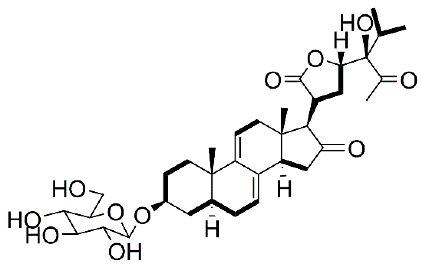
Figure 3.
Major HMBC and NOESY correlations of compound 1.
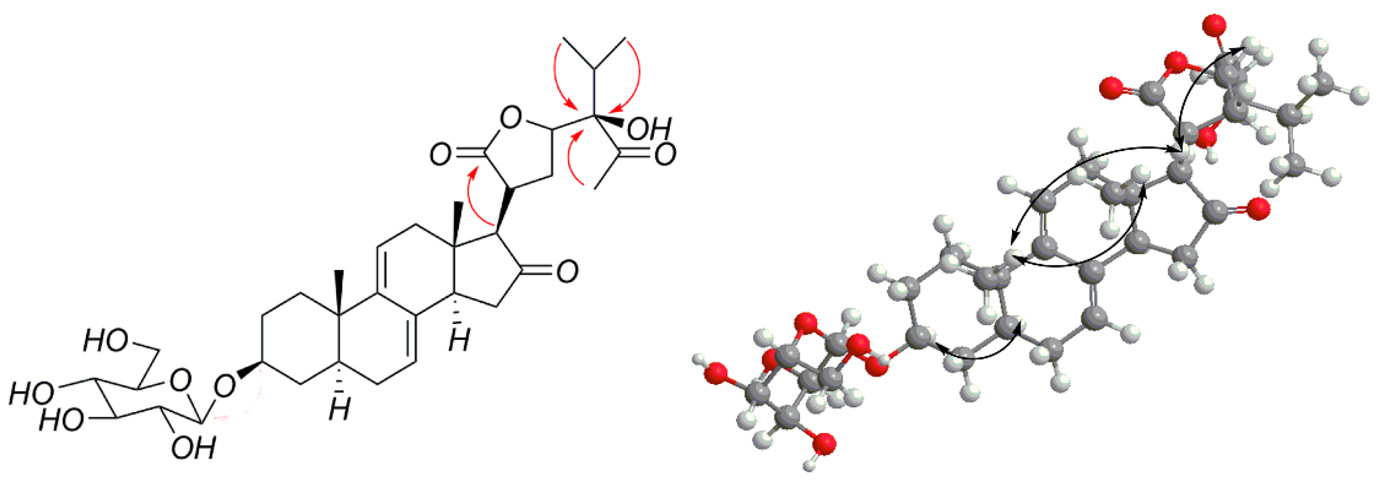
Figure 4.
Major HMBC and NOESY correlations of compound 2.
Figure 5.
Major HMBC and NOESY correlations of compound 3.
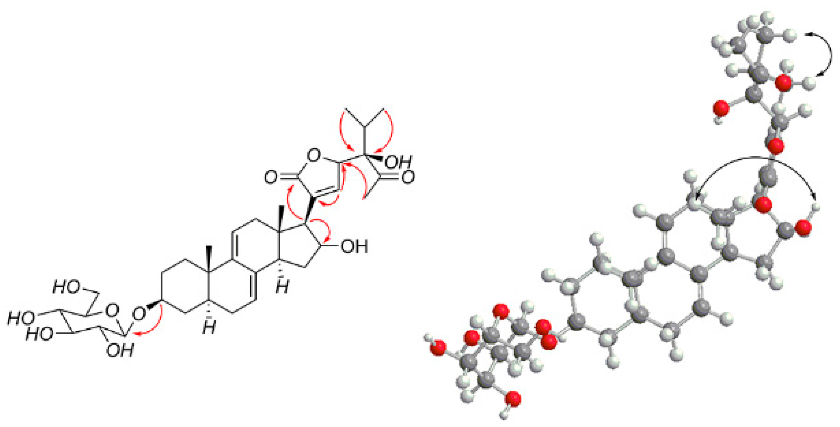
Figure 6.
Major HMBC and NOESY correlations of compound 4.

Table 1.
1H (600 MHz) and 13C (150 MHz) NMR data of 1–4 in CD3COCD3.
| No. | 1 a | 2 a | 3 a | 4 a | ||||
|---|---|---|---|---|---|---|---|---|
| δC | δH (J in Hz) | δC | δH (J in Hz) | δC | δH (J in Hz) | δC | δH (J in Hz) | |
| 1 | 34.1 | 1.27, 2.0, m | 34.3 | 1.23, 1.96, m | 34.3 | 1.24, 1.93, m | 34.3 | 1.24, 1.94, m |
| 2 | 29.2 | 1.48, 1.88, m | 29.3 | 1.45, 1.87, m | 29.2 | 1.44, 1.84,m | 29.2 | 1.46, 1.87, m |
| 3 | 76.3 | 3.57, m | 76.3 | 3.56, m | 76.2 | 3.56, m | 76.2 | 3.56, m |
| 4 | 33.7 | 1.22, d(11.6) | 33.7 | 1.19, d(11.6) | 33.7 | 1.18, d(11.7) | 33.7 | 1.18, d(11.7) |
| 1.83, m | 1.80, m | 1.79, m | 1.76, m | |||||
| 5 | 38.7 | 1.35 a | 38.7 | 1.29 a | 38.6 | 1.31 a | 38.6 | 1.31 a |
| 6 | 29.4 | 1.26, 1.88 a | 29.5 | 1.23, 1.87 a | 29.5 | 1.24, 1.84 a | 29.4 | 1.23, 1.87 a |
| 7 | 121.9 | 5.40, br s | 120.1 | 5.37, br s | 120.7 | 5.36, br s | 120.7 | 5.39, br s |
| 8 | 133.1 | 135.8 | 135.1 | 135.7 | ||||
| 9 | 143.9 | 143.3 | 143.6 | 143.7 | ||||
| 10 | 35.8 | 35.5 | 35.6 | 35.9 | ||||
| 11 | 117.6 | 5.62, d(5.8) | 119.0 | 5.53, d(6.2) | 117.9 | 5.47 a | 118.3 | 5.51 d(6.2) |
| 12 | 39.0 | 2.27, m | 40.8 | 2.01, d(7.4) | 39.6 | 1.83, d(6.6) | 39.4 | 1.94, d(6.6) |
| 2.84, m | 2.17, m | 2.16, m | ||||||
| 13 | 40.3 | 41.8 | 43.6 | 43.4 | ||||
| 14 | 45.0 | 2.65 a | 45.1 | 1.94 a | 48.7 | 2.54 a | 46.0 | 2.52 a |
| 15 | 37.0 | 2.02, 2.34 a | 28.0 | 1.47, 2.09 a | 34.5 | 1.66, 1.92 a | 26.5 | 1.91 a |
| 16 | 214.2 | 22.8 | 1.39, 1.76 a | 74.3 | 4.40 a | 23.1 | 1.49, 1.85 a | |
| 17 | 61.2 | 2.63, d(2.9) | 50.9 | 2.19 a | 56.6 | 2.41, d(7.5) | 50.8 | 2.27 a |
| 18 | 13.8 | 0.52, s, 3H | 11.8 | 0.54, s, 3H | 13.2 | 0.30, s, 3H | 12.1 | 0.32, s, 3H |
| 19 | 19.4 | 0.87, s, 3H | 19.4 | 0.85, s, 3H | 19.3 | 0.81, s, 3H | 19.3 | 0.82, s, 3H |
| 20 | 36.8 | 2.98, ddd, (12.2,8.7,3.2) | 47.6 | 2.76, dd, | 133.9 | 134.9 | ||
| (10.8, 6.6) | ||||||||
| 21 | 177.3 | 175.4 | 173.3 | 172.9 | ||||
| 22 | 26.0 | 1.88, 2.16 a | 79.1 | 4.58, t(6.0*2) | 146.8 | 7.49, br s | 146.9 | 7.44, br s |
| 23 | 80.6 | 4.63, d(5.5) | 78.7 | 4.62, d(5.1), | 82.8 | 5.24, m | 82.7 | 5.22, m |
| 24 | 83.0 | 82.6 | 83.3 | 83.3 | ||||
| 25 | 33.5 | 2.07, m | 30.8 | 1.91, m | 32.9 | 2.16, m | 32.8 | 2.16, m |
| 26 | 17.1 | 0.83, d(7.0) | 16.5 | 0.87, d(6.8) | 16.9 | 0.82, d(6.9) | 16.9 | 0.81, d(6.8) |
| 27 | 17.0 | 0.91, d(7.0) | 17.6 | 1.03, d(6.8) | 16.7 | 0.97, d(6.9) | 16.6 | 0.96, d(6.8) |
| 28 | 213.0 | 106.9 | 211.2 | 211.1 | ||||
| 29 | 29.2 | 2.15, s, 3H | 15.0 | 1.26, s, 3H | 28.1 | 2.09, s, 3H | 28.1 | 2.09, s, 3H |
| Glu | ||||||||
| 1′ | 100.9 | 4.24, d(8.1) | 100.9 | 4.23, d(8.1) | 100.9 | 4.22, d(8.0) | 100.9 | 4.22, d(8.1) |
| 2′ | 73.5 | 2.90 a | 73.5 | 2.90 a | 73.5 | 2.89 a | 73.5 | 2.89 a |
| 3′ | 76.7 | 3.10 a | 76.7 | 3.10 a | 76.8 | 3.10 a | 76.8 | 3.11 a |
| 4′ | 70.1 | 3.03 a | 70.1 | 3.02 a | 70.1 | 3.02 a | 70.1 | 3.02 a |
| 5′ | 76.8 | 3.10 a | 76.8 | 3.10 a | 76.8 | 3.10 a | 76.8 | 3.11 a |
| 6′ | 61.1 | 3.42, 3.65 a | 61.1 | 3.41, 3.65 a | 61.2 | 3.41, 3.65 a | 61.1 | 3.41, 3.65 a |
| OCH3 | 49.5 | 3.15, s, 3H | ||||||
a Resonance pattern unclear due to overlapping.
Table 2.
Results of the cytotoxicity assay.
| Sample | BT-549 | MDA-MB-231 | MCF-7 | Hela | ||||
|---|---|---|---|---|---|---|---|---|
| Average Absorbance | Inhibition (%) | Average Absorbance | Inhibition (%) | Average Absorbance | Inhibition (%) | Average Absorbance | Inhibition (%) | |
| Compound 1 | 0.2748 | 63.61 | 0.6102 | 28.97% | 0.4217 | 46.54 | 0.5212 | 42.05 |
| Compound 2 | 0.2836 | 62.17 | 0.6196 | 27.78% | 0.4893 | 37.07 | 0.6063 | 31.64 |
| Compound 3 | 0.4546 | 34.18 | 0.5803 | 32.74% | 0.4728 | 39.38 | 0.6464 | 26.73 |
| Compound 4 | 0.3946 | 44.00 | 0.5899 | 31.53% | 0.5301 | 31.36 | 0.5957 | 32.93 |
| Compound 5 | 0.4410 | 36.41 | 0.5734 | 33.61% | 0.3990 | 49.72 | 0.6893 | 21.48 |
| Compound 6 | 0.3510 | 51.14 | 0.5961 | 30.75% | 0.4750 | 39.08 | 0.5737 | 35.63 |
| Positive control | 0.1515 | 83.79 | 0.1789 | 83.39% | 0.0734 | 95.32 | 0.0996 | 92.70 |
| Negative control | 0.6634 | 0.8399 | 0.7540 | 0.8648 | ||||
| Blank | 0.0525 | 0.0470 | 0.0400 | 0.0477 | ||||
© 2018 by the authors. Licensee MDPI, Basel, Switzerland. This article is an open access article distributed under the terms and conditions of the Creative Commons Attribution (CC BY) license (http://creativecommons.org/licenses/by/4.0/).
Share and Cite
MDPI and ACS Style
Wang, J.; Song, H.; Wu, X.; Zhang, S.; Gao, X.; Li, F.; Zhu, X.; Chen, Q. Steroidal Saponins from Vernonia amygdalina Del. and Their Biological Activity. Molecules 2018, 23, 579. https://doi.org/10.3390/molecules23030579
AMA Style
Wang J, Song H, Wu X, Zhang S, Gao X, Li F, Zhu X, Chen Q. Steroidal Saponins from Vernonia amygdalina Del. and Their Biological Activity. Molecules. 2018; 23(3):579. https://doi.org/10.3390/molecules23030579
Chicago/Turabian StyleWang, Jing, Hua Song, Xiaoxue Wu, Shuyi Zhang, Xuemin Gao, Funan Li, Xuan Zhu, and Qing Chen. 2018. "Steroidal Saponins from Vernonia amygdalina Del. and Their Biological Activity" Molecules 23, no. 3: 579. https://doi.org/10.3390/molecules23030579