Review

Fungal Metabolites Antagonists towards Plant Pests and Human Pathogens: Structure-Activity Relationship Studies

Dipartimento di Scienze Chimiche, Università di Napoli Federico II, Complesso Universitario Monte Sant’Angelo, Via Cintia 4, 80126 Napoli, Italy
*
Author to whom correspondence should be addressed.
Molecules 2018, 23(4), 834; https://doi.org/10.3390/molecules23040834
Submission received: 5 March 2018 / Revised: 29 March 2018 / Accepted: 2 April 2018 / Published: 5 April 2018
(This article belongs to the Special Issue Structure-Activity Relationship of Natural Products 2018)

Abstract

:
Fungi are able to produce many bioactive secondary metabolites that belong to different classes of natural compounds. Some of these compounds have been selected for their antagonism against pests and human pathogens and structure–activity relationship (SAR) studies have been performed to better understand which structural features are essential for the biological activity. In some cases, these studies allowed for the obtaining of hemisynthetic derivatives with increased selectivity and stability in respect to the natural products as well as reduced toxicity in view of their potential practical applications. This review deals with the SAR studies performed on fungal metabolites with potential fungicidal, bactericidal, insecticidal, and herbicidal activities from 1990 to the present (beginning of 2018).

1. Introduction

Pests are one of the main concerns for plants, animals, and humans. Among them, invasive species such as weeds are a serious problem for agriculture while organisms that are human disease carriers such as mosquitoes cause significant diseases or even death, worldwide [1,2]. Strategies of pest management can vary but are still strongly dependent on the use of synthetic chemicals. The intensive use of these pesticides in the last fifty years has had negative environmental and ecological impacts [3,4,5]. The accumulation of the chemicals in the ecosystem and food and the rapidly evolving resistance of pests to commercial pesticides are two main problems to be solved [6]. Furthermore, many substances have been banned because of their hazardous effects and chemical companies have difficulties substituting them with safe and efficient synthetic compounds [7,8]. Thus, there is an increased requirement for novel and environmentally friendly alternatives and biological control could be an efficient tool for pest management [9,10]. Biocontrol agents are categorized into four groups: macroorganisms (for example, predators, parasitic insects, and nematodes), micro-organisms (for example, bacteria, fungi, and viruses), chemical mediators (for example, pheromones) and natural substances (originated from plant or animal). Biopesticide products can be either living organisms, and more specifically micro-organisms, or products derived from living organisms, including the metabolites produced during their growth and development [11]. Biopesticides are natural product based, therefore, they are expected to be more environment-friendly [12]. Furthermore, the half-life of biopesticides is usually shorter than that of chemicals but their activity spectrum should be carefully evaluated [13]. The available data show that natural products have had a substantial impact on pest control by providing compounds that are efficient as pest control agents. More importantly, identifying novel modes of action serves as inspiration/models for synthetic pesticides [6,14]. However, research, development, and regulation are necessary to improve the number of effective solutions on the bioherbicide world market [11]. Natural products have also been extensively studied to discover new drugs and to develop new formulations [15,16,17,18,19].
The antimicrobial resistance developed by human pathogens is one of the most urgent public health problems. For instance, members of the Enterobacteriaceae and Pseudomonas families—organisms that cause diarrhea, urinary infection, and sepsis—are now virtually resistant to all of the traditional antibiotics [20,21,22]. Bacteria can resist antibiotics by different mechanisms: chromosomal mutation; inductive expression of a latent chromosomal gene; by exchanging genetic material through transformation and/or transduction (bacteriophage); and through conjugation by plasmids. After a bacterium gains a gene resistance to protect itself from various antimicrobial agents, they can use several biochemical types of resistance mechanisms [23]. Several drugs that have recently been developed are still unable to overcome the resistance mechanisms of these pathogens [24,25].
New efforts are now dedicated to the discovery of new compounds with different mechanisms of action and in this battlefield, the biologically active natural products isolated from living organisms could play a fundamental role, being an efficient alternative to synthetic or semi-synthetic compounds [23,24,25,26,27,28].
Among the living organisms, fungi and bacteria are able to produce secondary metabolites with interesting biological activities [2,29,30]. These metabolites belong to all the different classes of natural compounds, such as terpenes, phenylpropanoids, polyketides, alkaloids, and so forth. [31,32]. Thus, these organisms represent a very good source of natural substances that could be used as an efficient tool to design natural biopesticides or antibiotics against human pathogens with new modes of action. Some selected metabolites have been used to carry out structure–activity relationship (SAR) studies to better understand which structural features are essential for their biological activity, to increase the selectivity and the stability of the natural products, as well as to reduce their toxicity in view of their potential practical applications.
This review reports the results of SAR studies performed from 1990 to the present, using fungal metabolites with potential practical applications as potential fungicides, bactericides, insecticides, and herbicides (Table S1, Supplementary Materials). The herbicides section is focused on the last results on SAR studies which were not covered by the review by Cimmino et al. 2015 [2].

2. Structure–Activity Relationship Studies Performed with Fungal Metabolites

2.1. Fungicides

Fungi are by far the most prevalent plant pathogens and several studies have been carried out to develop management strategies that minimize crop losses. Many natural products have shown potential fungicidal activity against phytopathogenic fungi and some of them have been selected to perform SAR studies. In particular, pathogenic fungi of forest trees are a good source of bioactive metabolites which have been extensively studied [33]. From the fungus Diplodia cupressi, involved in the cypress (Cupressus sempervirens) canker disease in the Mediterranean area, tri-, and tetra-cyclic un-rearranged pimarane diterpenes, namely sphaeropsidins A–F (16, Figure 1), were isolated together with the dimedone methyl ethers named sphaeropsidones (7 and 8, Figure 1) and their chlorinated analogs (9 and 10, Figure 1) [33].
Sphaeropsidin A is a well-known bioactive metabolite produced by different species of Diplodia [34,35,36,37] which showed strong anticancer activity against advanced melanomas [38,39]. Recently, its absolute configuration was confirmed by X-ray analysis of the toxin and its 6-O-p-bromobenzoyl derivative [40], while its anticancer activity was evaluated in combination with cytotoxic chemotherapeutics [41]. Sphaeropsidins A–C (13) showed a preliminary interesting activity when tested against several plant pathogenic fungi, including Seiridium cardinale and Seiridium cupressi, both agents of a different canker forms [42,43]. For this reason, their potential antimycotic activity was investigated [44]. In order to get information on the SAR and to identify which structural features are essential for the biological activities of sphaeropsidins, eight derivatives (1118, Scheme 1, Scheme 2 and Scheme 3) were prepared by chemical transformation of the functionalities present in 13 [44].
A suitable amount of sphaeropsidin B was obtained by stereoselective reduction of 1 as reported in Scheme 1 [44].
The antimycotic activity of 16 and 1118 was assayed at 100 μg/mL on eight plant pathogenic fungi, namely Botrytis cinerea, Fusarium oxysporum, Penicillium expansum, Verticillium dahliae, Phomopsis amygdali, S. cardinale, S. cupressi, and Seiridium unicorne. The results showed that the integrity of the tricyclic pimarane system, the preservation of the double bond from C-8 to C-14, the tertiary hydroxyl group at C-9, the vinyl group at C-13, the carboxylic group at C-10, and the integrity of the A-ring are structural features essential to impart activity against several plant pathogenic fungi [44].
These relations were also observed when the same compounds were tested (at a concentration of 100 μg/mL) to evaluate their phytotoxic activity against host (three cypress species) and non-host plants [44].
Sphaeropsidones (7 and 8, Figure 1) and their chlorinated analogs (9 and 10, Figure 1) were evaluated for their activities against five Phytophthora species (destructive pathogens of forest trees and shrubs) and for their phytotoxicity on Quercus ilex, Quercus rubra, Quercus suber, and tomato (Lycopersicon esculentum) leaves. In particular, eight derivatives (1926, Scheme 4 and Scheme 5) were prepared by chemical transformation of the functionalities present in 7 and 8 in order to carry out SAR studies [45].
The results obtained with either natural or synthetic analogs of sphaeropsidones (tested at 0.05, 0.1 and 0.2 mg/plug) showed that the specific structural features related to their toxicity are the C-5 hydroxy group, the epoxy ring, and the C-2 carbonyl group, along with the C-5 absolute configuration. In fact, compounds that were much less active and/or inactive in comparison to 7 were obtained with the opening of the epoxy ring and with the modifications of the C-5 hydroxy group and the reduction of the C-2 carbonyl group. However, the most important result was obtained with compound 21 which, among the synthesized derivatives, was found to be more effective than 7 in inhibiting the mycelial growth of Phytophthora species. In particular, its activity was very similar to that showed by the synthetic fungicide (mefenoxam) commonly employed for the control of diseases caused by oomycetes. Considering the development of a resistance to the phenylamides and the fact that the Phytophthora species are invasive pathogens on a global scale, compound 21 could be suitable for the development of an alternative strategy to manage these pathogens [45].
From Diplodia africana, the fungal pathogen responsible for branch dieback of Juniperus phoenicea in Italy, two phytotoxic dihydrofuropyran-2-ones, named afritoxinones A and B (27 and 28, Figure 2) were isolated together with the known oxysporone (29, Figure 2), sphaeropsidin A and epi-sphaeropsidone (1 and 8, Figure 1), R-(−)-mellein, (3R,4R)-4-hydroxymellein, and (3R,4S)-4-hydroxymellein (3032, Figure 2) [34]. The phytotoxic activity of afritoxinones A and B and the main compound oxysporone was evaluated on host (J. phoenicea) and non-host plants (Q. ilex, Q. suber, and tomato) by cutting and leaf puncture assays. Oxysporone proved to be the most phytotoxic compound [34]. Successively, eight derivatives (3340, Scheme 6) were hemisynthesized and assayed for their phytotoxic and antifungal activities in comparison to the parent compound oxysporone. In particular, oxysporone (29) and its derivatives (3340) were tested (at a concentration of 100 mg/mL) on four different plant pathogens including two fungal species (Athelia rolfsii and Diplodia corticola) and two oomycetes (Phytophthora cinnamomi and Phytophthora plurivora) which have a great impact in both agriculture and natural ecosystems. The same compounds were also tested (at a concentration of 1 mg/mL) against non-host plants, namely cork oak (Q. suber), holm oak (Q. ilex), and grapevine (Vitis vinifera) to evaluate their phytotoxic activity. The results of the latter assay showed that the dihydrofuropyranone carbon skeleton and both the double bond and the hydroxy group of the dihydropyran ring are structural features important in conferring phytotoxic activity [46].
Unfortunately, the antifungal activity data were not suitable to speculate on the SAR but the corresponding 4-O-benzoyl derivative of oxysporone 36 showed a good antifungal activity towards P. cinnamomi, P. plurivora, and A. rolfsii. However, its EC50 (concentration which inhibits mycelial growth by 50%) was one or two orders of magnitude lower than those of three specific commercial fungicides toclofos-methyl, pentachloronitrobenzene (PCNB), and metalaxyl-M [46].
From the culture filtrates of the fungus Ascochyta heteromorpha, the causal agent of a foliar disease of oleander (Nertum oleander), a new cytochalasin named ascochalasin (41, Figure 3), was isolated together with deoxaphomin (42, Figure 3) and cytochalasins A and B (43 and 44, Figure 3). Cytochalasins are a large group of fungal metabolites produced by several genera of fungi which showed different biological activities. These compounds have the ability to bind to actin filaments and block the polymerization and the elongation of actin [47,48,49,50].
These compounds were assayed against Geotrichum candidum together with two derivatives of cytochalasin B (namely 7,20-O,O′-diacetylcytochalasin B and 21,22-dihydrocytochalasin B (45 and 46, Figure 3)); 17-O-acetylcytochalasin A (47, Figure 3); and the natural cytochalasins C, D, E, H, and J (4852, Figure 3). However, only cytochalasin A (43) showed to be active (<5 μg/disk) against this fungus, suggesting the importance of the ketonic group at C-20 (probably involved in a Michael reaction) which plays a significant role in the induction of fungicidal activity [51].
Aspergillus fumigatus LN-4, an endophytic fungus isolated from the stem bark of Melia azedarach, was grown in vitro to evaluate its ability to produce secondary metabolites with antifungal activity. In particular, from its fermentation broth, 39 fungal metabolites were isolated, including the two new alkaloids 12β-hydroxy-13α-methoxyverruculogen TR-2 (53, Figure 4) and 3-hydroxyfumiquinazoline A (54, Figure 4). When tested against some phytopathogenic fungi (B. cinerea, Alternaria solani, Alternaria alternata, Colletotrichum gloeosporioides, Fusarium solani, Fusarium oxysporum f. sp. niveum, Fusarium oxysporum f. sp. vasinfectum, and Gibberella saubinettii), 16 compounds showed potent antifungal activity. For some compounds, these results were comparable to those obtained testing two commercial fungicides such as carbendazim and hymexazol, which were used as a positive control. Structure–activity relationships of the metabolites were also discussed for the tested indole diketopiperazine alkaloids (53, 5561, Figure 4) and the fumiquinazolines (54, 6265, Figure 4) [52]. In particular, the results obtained with the diketopiperazine alkaloids suggested that the introduction of a MeO group onto C-13 in these molecules give higher activity, regardless of the configuration of the OH group at C-12. Compound 53 (MIC (Minimum inhibitory concentration) = 6.25 μg/mL), having a 2-methylpropan-2-ol group at C-3, is more toxic than 57 (MIC = 12.5 μg/mL), which has an isobutenyl group at C-3, indicating that the 2-methylpropan-2-ol substituent at C-3 on ring C of the compounds 53, 58, and 59 appear to be necessary for activity. Furthermore, comparing the activity of 60 and 61 shows that the presence of the peroxide bridge is important to impart antifungal activity. Among the five fumiquinazolines (54, 6265), fumiquinazolines F (62), G (63), A (65), and 3-hydroxyfumiquinazoline A (54) have good antifungal activities (MICs = 12.5−25 μg/mL), whereas, fumiquinazoline D (64) weakly inhibited the growth of phytopathogenic fungi (MICs = 25−50 μg/mL), indicating that the presence of a C−N bridge between C-3 and N-22 in compound 64 could be detrimental to the activity. In addition, the brine shrimp (Artemia salina) toxicity was determined and compounds 60 and 61 both showed significant toxicities with median lethal concentration (LC50) values of 13.6 and 15.8 μg/mL, respectively. Furthermore, among nine metabolites that were found to possess antifeedant activity against armyworm larvae, compounds 60 and 61 gave the best activity with antifeedant indexes (AFI) of 50.0% and 55.0%, respectively. All these results allowed the authors to consider the compounds 12β-hydroxy-13α-methoxyverruculogen TR-2 (53) and fumitremorgin B (60) as promising lead compounds for developing new fungicides [52]. However, considering the toxicity shown by 60, its potential application as a natural fungicide seems very difficult. For the other compounds, further studies need to be carried out in order to better understand the mechanism of action associated with the antifungal and antifeedant effects and to show some selectivity.
From the rice cultures of the soil fungus Fusarium semitectum obtained from maize stalk rot in southern Italy, two 3-substituted 4-hydroxy-6-alkyl-2-pyrones were isolated and named fusapyrone (66, Figure 5) and deoxyfusapyrone (67, Figure 5), respectively. Fusapyrone showed a preliminary antifungal activity and low zootoxicity in an A. salina larvae mortality bioassay, while 67 had a higher zootoxicity [53]. Thus, a SAR study was performed by preparing seven different chemical derivatives of fusapyrone (68,7075, Figure 5) and one derivative of deoxyfusapyrone (69, Figure 5) and testing their antifungal and zootoxic activities in comparison with the natural compounds. In particular, the antifungal activity was tested against two yeasts (Pichia guilliermondii and Rhodotorula glutinis) and against three filamentous fungi B. cinerea, Aspergillus parasiticus and Penicillium brevi-compactum that are agents of pre- and post-harvest plant diseases. The derivatives were prepared by trying to modify the glycosyl residue, the 2-pyrone ring, and the aliphatic chain of 66 and 67. Among the derivatives of 6865, only compounds 72, 74, and 75 retained some activity against B. cinerea, while the others were inactive against the yeasts. However, the activity was shown only at the highest concentration (25–50 μg/mL). These results highlighted the importance of the hydrophilic sugar residue for the activity against B. cinerea. The same derivatives were also tested against A. salina (brine shrimp) to evaluate the zootoxic activity. In this case, the toxicity was related to the increased hydrophobicity of some derivatives [54].
Considering the biological activities of 66 and 67 and the results obtained, the potential use of these molecules in combination with biocontrol agents for plant disease control was proposed [54]. The importance of α-pyrone moiety to impart antifungal activity in natural compounds is also confirmed by the fungicidal activity of 6-n-pentyl-2H-pyran-2-one (76, Figure 6), which is a metabolite produced by different Trichoderma species. In fact, compound 76 has been demonstrated to inhibit the growth of several plant-pathogenic fungi: B. cinerea, Fusarium oxysporum f. sp. lycopersici, Fusarium verticillioides (moniliforme), Phytophthora megasperma, Rhizoctonia solani, and Armillaria mellea [55,56]. Compound 76 was also previously isolated by some of the authors together with the new 6-substituted-2H-pyran-2-one, named viridepyronone (77, Figure 6), from the cultural filtrates of a Trichoderma viride strain showing in vitro antagonistic activity toward Sclerotium rolfsii, which is the causal agent of crown and stem rot of artichoke. Viridepyronone inhibited the growth of S. rolfsii by 48% with a MIC of 196 μg/mL [57].
Dung-inhabiting fungi are also an underexplored reservoir of bioactive compounds [58]. Recently, the cultures of the two fimicolous fungi Cleistothelebolus nipigonensis and Neogymnomyces virgineus showed strong antifungal activity against Alternaria brassicicola, B. cinerea, and Fusarium graminearum. From the bioguided purification of their organic extracts, fusaproliferin (78, Scheme 7) and terpestacin (79, Scheme 8) were isolated and identified. A SAR study was conducted to understand the role of each functional group of these natural products to the imparted activity. In particular, four fusaproliferin derivatives (8083, Scheme 7) and four terpestacin derivatives (8487, Scheme 8) were prepared and tested (using a concentration of 10−3 M) against A. brassicicola, B. cinerea, and F. graminearum [59]. These results showed that the three fungi have different sensitivities towards fusaproliferin and terpestacin. Furthermore, the hydroxy enolic group at C-17 and the conformational freedom of the macrocyclic ring, due to the presence/absence of the three double bonds, are structural features important to impart activity [59].
However, fusaproliferin resulted in being toxic to A. salina, against the lepidopteran cell line SF-9, and also against the human non-neoplastic B-lymphocyte cell line IARC/LCL 171. Thus, it seems very hard to hypothesize on its application as a biopesticide, although the SAR study performed was useful in order to speculate on the mechanisms of action of these natural products. The functions in nature of these two mycotoxins have not been clearly established, but they are believed to play a role in eliminating other microorganisms competing in the same environment [59].

2.2. Bactericides

Several bacterial species are able to cause a large number of different plant diseases, some of which are devastating for agricultural crops [60]. Among the pathogens associated with rice diseases, Xanthomonas oryzae pv. oryzae is the causal agent of bacterial blight [61,62]. This disease is one of the most serious bacterial diseases in many of the rice-growing regions of the world. Every year, fungal and bacterial diseases cause damage to crops and lead to severe economic losses of rice production. Thus, different studies were carried out to investigate the potential antibacterial activity of fungal metabolites. In particular, sphaeropsidin A (1, Figure 1)—the main phytotoxin produced by D. cupressi—showed good activity against several bacterial rice pathogens (X. oryzae pv. oryzae, Pseudomonas fuscovaginae, and Burkholderia glumae). For this reason, fourteen derivatives were prepared by chemical transformation of the functionalities present in sphaeropsidins A, B, and C (1114, Scheme 1; 15 Scheme 2; 1618 Scheme 3; 8891 Scheme 9; 9294, Scheme 10) and tested (at a concentration range of 0.5−6.0 × 10⁻³ M) against three rice bacterial pathogens to identify which structural features are essential for this biological activity [63].
The results obtained showed a strong and specific activity of sphaeropsidin A against X. oryzae pv. oryzae, while no activity was observed against the other two pathogens. The results of the SAR study showed that the structural features important to impart this antibacterial activity are the presence of the ketone at C-7 (probably involved in a Michael reaction) and the hemiketal lactone functionality. The C-13 vinyl group, the double bond of ring C, and/or the tertiary hydroxyl group at C-9, as well as the pimarane arrangement of tricylic carbon skeleton, are also important for the antibacterial activity. These findings may be useful in designing novel compounds for practical applications in agriculture as bactericides. However, selectivity studies are necessary to understand if 1 is a suitable compound to develop a new biopesticide formulation. On the other hand, it is important to take into account its other biological activities with particular focus on the concentrations under which sphaeropsidin A turns out to be active [63].
Some bacteria species are also pathogenic for humans and the bacterial resistance to antimicrobial drugs is an increasing health and economic problem [23,64]. Natural products continue to provide key scaffolds for drug development and new bactericides could be discovered studying the fungal secondary metabolism [26]. Recently, the biosynthetic potential of the marine fungus Neosartorya pseudofischeri (isolated from the inner tissue of starfish Acanthaster planci) has been evaluated in different culture conditions to find new bactericides [65]. N. pseudofischeri was previously studied for the production of secondary bioactive metabolites possessing in vitro anticancer activity, and a new pyrroloindole sesquiterpenoid named fischerindoline (95, Figure 7) was isolated together with the promising compound eurochevalierine (96, Figure 7) [66,67].
Three new metabolites named neosartins A–C (9799, Figure 7) were isolated together with twelve other already-known compounds (100111, Figure 7) from in vitro cultures of N. pseudofischeri [65].
Compounds 98110 were evaluated for their antibacterial activity against three multidrug-resistant bacteria, that is, the Gram-positive Staphylococcus aureus and Methicillin-resistant Staphylococcus aureus and the Gram-negative Escherichia coli. Compounds 104 and 106 displayed significant inhibitory activities against these three bacteria with MIC values ranging from 1.52 to 97.56 μM. Compounds 104 and 106, especially, showed potent inhibition against Methicillin-resistant S. aureus with MIC values of 1.53 and 1.52 μM, respectively. Compounds 101 and 105 inhibited the growth of both S. aureus strains with MIC values of 283.11, 70.70 μM and 86.91, 21.73 μM, respectively. The remaining nine compounds were inactive in this assay (MIC > 256 μg/mL). Compounds 104 and 106 only inhibited the growth of S. aureus. Considering the results obtained, a preliminary analysis of the structure-activity relationships of these twelve diketopiperazines suggests that the disulfide bridge, the α-methylene ketone group, the hydroxyl group at C-6, and the thiol groups are important to impart the activity. In particular, the presence of the disulfide bridge or the reduced disulfide bond are essential for the inhibitory activity because when the thiol groups are substituted, the inhibitory effects disappeared. Furthermore, the intensity of the antibacterial activity is influenced by the substitution at the six-membered ring containing two conjugated double bonds and the analogs with a hydroxyl group at C-6 enhance the antibacterial activity compared to the analogs with an acetyl group at the same position [52].
From the culture filtrates of the fungus Pseudallescheria boydii residing in the gut of a coleopteran (Holotrichia parallela) larva, four new epipolythiodioxopiperazines named boydines A–D (112115, Figure 8) were isolated together with six other known and new metabolites [68]. All the isolated fungal metabolites were assayed for in vitro antibacterial activity against pathogenic anaerobes isolated from clinical specimens. Only boydine B (113) showed a strong inhibition against Peptostreptococcus sp., Bifidobacterium sp., Anaerostreptococcus sp., and Veillonella parvula with a MIC range of 0.2–0.8 μM. The SAR study on the cyclic dipeptides suggested that the antibacterial activity might be dependent on the combination of the acyl residue with a diketopiperazine nucleus condensed from two (2R,7S,7aS)-7-hydroxy-2-(methylthio)-2,3,7,7a-tetrahydro-1Hindole-2-carboxylic acids [68].
The above reported natural and hemisynthetic cytochalasins (4352) were also tested against two bacteria: Pseudomonas syringae (gram-negative) and Bacillius megaterium (gram-positive) [51]. Only cytochalasin A (43) and its 17-O-acetyl derivative (47) were active against B. megaterium, while no effects were observed on P. syringae. This may be attributed to the different cell walls of these two classes of bacteria which caused a different permeability of the compounds assayed. Furthermore, the activity of 47 could be explained by considering the fact that the acetylation of 43 increased the lypophilicity of the compound. However, in this case, the results obtained correlating the structures and the activity of compounds 4251 highlight the importance of the ketone group at C-20 in 44. The same compounds (4352) were also tested in the brine shrimp bioassay to evaluate their toxicity at concentrations of 20, 2, and 0.2 μg/mL. Cytochalasin E (50) was the most active mycotoxin, but generally, at low concentrations, the cytochalasans with the (11)-macrocyclic ring are more active than those with the (14)-macrocyclic ring [51]. Considering that cytochalasins are primarily known to act against eukaryotes as they strongly inhibit actin polymerization, the activity described in this study against bacteria may be an off target effect.
From the culture filtrates of the marine-derived fungus ZJ-2010006, isolated from a sea anemone and identified as a Nigrospora sp., two new hydroanthraquinone analogs, 4a-epi-9α-methoxydihydrodeoxybostrycin (126, Figure 9) and 10-deoxybostrycin (127, Figure 9), were obtained together with seven known anthraquinone derivatives (118124, Figure 9). The antibacterial activity of these compounds was evaluated in vitro together with those of the ten acetyl derivatives (125134, Figure 9) prepared from the natural analogs and their structure–activity relationships were examined [69].
In particular, the antibacterial activities were tested against nine bacterial strains including the Gram-positive Bacillus cereus, Bacillus subtilis, Staphylococcus albus, S. aureus, Micrococcus luteus, and Micrococcus tetragenus and the Gram negative E. coli, Vibrio anguillarum, and Vibrio parahemolyticus. Nigrosporin B (118) showed strong antibacterial activity against B. subtilis (MIC = 312 nM) and B. cereus (MIC = 312 nM) while the acetylated derivative 125 exhibited strong activity against B. cereus and V. anguillarum with MIC values of 48.8 and 97.5 nM, respectively. The SAR study suggested that the cycloaliphatic ring C and the aromatic ring B are very important for the antibacterial activity while the presence of the hydroxyl groups at C-4, C-9, and C-10 are not essential to impart the same activity. Furthermore, the presence of an acetyl group at C-3 in 125 has a positive impact on the antibacterial activity [69].
From the fermentation broth of a deep-sea-derived fungus (Spiromastix sp.), 15 new depsidone analogs were isolated by Niu et al. in 2014 and named spiromastixones A–O (135149, Figure 10) [70]. They were classified into two subtypes based on the orientation of the C ring relative to the A ring and most analogs were substituted by various numbers of chlorine atoms [70].
Antimicrobial activities were measured against four bacterial strains, namely E. coli, S. aureus, Bacillus thuringensis, and B. subtilis, against a spectrum of multiresistant Gram-positive and Gram-negative bacteria species, including reference strains and clinical isolates (methicillin-sensitive and -resistant staphylococci S. aureus and Staphylococcus epidermis, vancomycin-sensitive and -resistant enterococci Enterococcus faecalis and Enterococcus faecium) and a number of Gram-negative bacteria containing an extended spectrum β-lactamase (E. coli, K. pneumoniae). All compounds exhibited significant inhibition against Gram-positive bacteria (MIC values ranging from 0.125 to 8.0 μg/mL) while none of the tested spiromastixones had any significant growth inhibitory effect against the Gram-negative bacterium E. coli. Furthermore, compounds 135149 displayed potent inhibitory effects (IC50 values ranging from 1 to 32 μM) against methicillin-resistant bacterial strains of S. aureus and S. epidermidis. Compound 144 also inhibited the growth (IC50 values = 4 μM) of the vancomycin-resistant bacteria E. faecalis and E. faecium. These results allowed for the speculation on the SARs which revealed that the inhibitory effects of the spiromastixones depend on the number of chlorine atoms and the substitution at ring C. In fact, increasing the number of chlorine atoms in the spiromastixones enhanced the inhibitory effects while the compounds with a methoxy substituent at ring C (141, 144146) were more active against the Gram-positive bacteria compared to the analogs that had a hydroxy group at this position (140, 143, 148 and 149). The selective inhibition against Gram-positive bacteria suggested that the spiromastixones could be promising lead compounds and the specific activity of 144 suggested that further investigation into its potential as an agent to treat multidrug-resistant bacterial infections is required [70]. The results of this SAR study were also confirmed when spiromastixones A–O (135149) were tested to evaluate their lipid-lowering activity. In particular, compounds 135149 inhibited foam cell formation via the regulation of cholesterol efflux and uptake in the RAW264.7 macrophages. In this study, the mechanistic investigation revealed that compounds 140 and 148 promoted cholesterol efflux through the upregulation of the PPARγ-ABCA1/G1 pathway and inhibited cholesterol uptake via the downregulation of the scavenger receptors CD36 and SR-A1. Thus, spiromastixones 140 and 148 were considered promising leads for the development of a new type of anti-atherosclerotic agent. However, further studies need to be carried out to investigate their specificity and toxicity [71].

2.3. Insecticides

Insecticides of chemical and biological origins constitute a large number of chemical classes which exert toxicity towards insects through different mechanisms of action. They are used in agriculture, forestry, horticulture, gardens, and homes, but also to control vectors such as mosquitoes and ticks [72]. Several insecticidal and nematicidal metabolites were isolated from fungi but very few examples of SAR studies are reported in the literature for these compounds [73]. Most of the work has been done trying to find natural compounds that could have insecticidal activity against Aedes aegypti, the major vector of the Zika virus and the viruses responsible for dengue and yellow fevers [1]. These devastating human diseases are some of the major concerns for public health safety. During the last few years, different secondary metabolites produced by plants, fungi, bacteria, and lichens have been studied for their activity against Ae. aegypti [74,75,76,77].
In a preliminary screening, some fungal phytotoxins were evaluated for their biting deterrent and larvicidal activities against Ae. aegypti and SAR studies were conducted with the active compounds preparing some known and new suitable derivatives [78,79]. In particular, starting from cyclopaldic acid (150, Scheme 11), produced by the fungus S. cupressi involved in the canker disease of Italian cypress (C. sempervirens), nine derivatives (151159, Scheme 11) were prepared and their insecticidal activity was tested in comparison with the parent compound (150) [78,80]. Compounds 155159 were active in biting-deterrence bioassays, confirming the importance of the CHO group at C-4 to impart the deterring activity. These data are in agreement with the results of a previous SAR study in which the phytotoxic activity of 150 and some derivatives was tested on host and non-host plants [81]. However, the lack of activity of 159 indicates that the primary OH group at C-8 should be free and not involved in an ether bond as in the 3,4,5-trisubstituted dihydrofuran ring [78].
Additionally, seiridin (160, Scheme 12), a metabolite produced by S. cardinale—another fungus involved in canker disease of cypress [82]—showed a promising activity against Ae. aegypti. Three of its derivatives (161163, Scheme 12) were prepared by modifying the main functional groups present in the molecule. These compounds were tested for their biting deterrence and larvicidal activities against Ae. aegypti in comparison with 160 and its natural analogue isoseiridin 164 (Scheme 12). The results showed that the presence of an unaltered butenolide ring is very important for the activity while the presence and the position of the OH group of the hydroxyheptyl side chain at C-4 are not important to impart the deterring activity. In addition, the increased activity of 2′-O-acetylseiridin (161) was probably due to its high lipophilicity (which can facilitate the membrane crossing) and its hydrolysis at the physiological pH (according to the known lethal metabolism) [78].
Additionally, the previously described sphaeropsidin A (1, Figure 1) showed interesting activities against Ae. aegypti and a SAR study was performed [78]. In particular, some new derivatives (165 and 166, Scheme 13) were prepared starting from 1 and 2 and tested together with the known 11 and 14 (Scheme 1) and the natural analogue 2. The results obtained were in total disagreement with the results of the previous SAR studies [44,63] and indicated that the lactone ring, the hemiketal OH group at C-6, and the C-8=C-14 bond are not relevant for the activity. Furthermore, the increased activity of the sphaeropsidin B (2) suggested that the presence of a secondary OH group at C-7 is more significant compared to the C=O group in 1. Furthermore, this result was not in agreement with the previous SAR studies, indicating a different mode of action for these natural products in imparting biological activities [44,63,78].
Because papyracillic acid (167, Figure 11) showed a strong mosquito biting deterrent activity, five already known and six new derivatives were prepared to carry out a SAR study (168178, Figure 11) [78,79]. Papyracillic acid (167) was isolated for the first time in 1996 from the cultures of the ascomycete Lachnum papyraceum [83] and then as the main phytotoxin produced by a strain of Ascochyta agropyrina var. nana. This latter fungus was obtained from the noxious perennial weed Elytrigia repens (quack grass) and for this reason, 167 has been previously studied for its potential mycoherbicide activity [84]. In particular, when tested by leaf disk-puncture assay at a concentration of 1 mg/mL, papyracillic acid was shown to be phytotoxic both for the host plant and a number of nonhost plants of the fungus. Papyracillic acid was also active against bacteria (Xanthomonas campestris and B. subtilis) and the fungus Candida tropicalis at 6 μg/disk [84]. When tested against Ae. aegypti, all the compounds showed biting deterrence but the activity of compounds 174176 and 178 were similar to the positive control DEET (97%, N,N-diethyl-3-methylbenzamide). None of these compounds showed any larvicidal activity at the highest testing dose of 100 ppm. These results indicated that the structural feature responsible for the activity of compound 167 is probably the furanone ring. The presence of the alternative substituted cyclobutene, oxiran, or substituted 4H-1,2,3-oxadiazine rings in some derivatives may be responsible for an increase in activity [79]. However, 167 is an analogue of penicillic acid, a mycotoxin produced by various fungi including strains of the genera Penicillium and Aspergillus, and its reactivity was extensively studied. In particular, it was demonstrated that its bioactivities are probably due to its reactivity towards nucleophiles. In fact, when 167 reacted with cysteine and cysteine methyl ester, it exclusively added the thiol group to the exomethylene double bond [85]. Thus, considering the high reactivity of 167 and its low specificity, it is important to further investigate the mode of action and the toxicity of its derivatives before proposing them as an alternative to common insecticides.
Six meroterpenoids (179185, Figure 12) were isolated from the solid cultures of a Penicillium sp. obtained from M. azedarach roots. These compounds were tested (at a concentration of 500 ppm) for their larvicidal effects in the control of Ae. aegypti, together with the related meroterpenoid austin (185, Figure 12). Compounds 182 and 183 exhibited in vitro larvicidal activities (LC50 values of 2.9 and 7.3 ppm, respectively) of 100% and 70%, respectively, after 24 h of exposure while 185 displayed a very low larval mortality. The larvicidal activity displayed by 182 and 183 was probably related to the δ-spirolactone system while the additional AcO group in 183 seems to significantly reduce the larvicidal activity. Furthermore, the very low activity of 185 compared to 182 and 183 suggests that the additional bridging furan ring also significantly influences the activity. This could indicate a hydrophobic binding/reactivity site in this part of the molecule. Since 182 is much more active than natural insecticides, it could have a great potential for the control of Ae. aegypti larvae [86]. However, it will first be necessary to more deeply investigate the larvicidal mode-of action and possible effects on non-target organisms before it can be practically used as a natural mosquito-control agent.
From Penicillium simplicissimum AK-40 (ATCC 90288), more than a dozen different prenylated indole alkaloids, named okaramines, were isolated [87,88]. These compounds exhibited insecticidal activity against silkworm larvae of Bombyx mori [88]. Okaramine A (186, Figure 13) is a heptacyclic compound containing a hexahydropyrrolindole and dihydroazocinoindole while okaramine B (187, Figure 13) possesses an unusual octacyclic ring system, including a four-membered azetidine ring and an eight-membered azocine ring [89]. Compound 187 exhibited insecticidal activity against the third instar larvae of silkworm at 0.1 ppm. At a concentration of 0.3 ppm, 100% of the larvae were killed within 24 h. On the other hand, 186 showed much lower activity than 187, indicating that an azetidine ring and/or a methoxyl in 188 play(s) an important role in expressing the physiological activity. Other SAR studies conducted in order to compare the activities of 187 with those of other natural analogs have suggested that the two characteristic ring structures of 187 play important roles in the insecticidal activity of the compound. A molecular target of 187 was identified as B. mori GluCl (BmGluCl) [90]. In order to explore the biosynthetic potential of okaramines, gene knockout experiments of an okaramine-producer fungus were performed. Ten compounds (186195, Figure 13) were isolated and tested for BmGluCl-activating activity. Analysis of the structure–activity relationships of 186195 revealed that the 1,4-dihydroazocine and N-aliphatic groups attached to the indole are crucial for the GluCl-activating activity. This provided insights into the further derivatization of the complex structure of okaramines in order to facilitate the development of new insecticides [88].

2.4. Herbicides

Weeds (including parasitic, annual, and perennial) are able to compete with a large number of important crops causing severe yield losses worldwide [60]. In order to avoid the use of synthetic chemicals, the phytotoxins produced by weed pathogenic fungi could be an efficient tool to design natural and safe bioherbicides. A recent review has described, in detail, the chemical and biological characterization of fungal phytotoxins with potential herbicidal activity [2]. The same review has also reported the SAR studies conducted on some promising phytotoxins such as chenopodolin (196, Figure 14) produced by Phoma chenopodiicola for the control of Cirsium arvense [91,92]; nonenolides (197199, Figure 14) and cytochalasans (4244, Figure 3 and 200204, Figure 14) isolated from phytopathogenic Stagonospora, Phoma, and Ascochyta spp. for the control of C. arvense and Sonchus arvensis [93]; papyracillic acid (167, Figure 11) and agropyrenol (205, Figure 14), produced by A. agropyrina var. nana for the control of E. repens [84,94,95]; and phomentrioloxin (206, Figure 14), isolated from the liquid culture of Phomopsis sp., a fungal pathogen proposed for the biological control of Carthamus lanatus [96,97]. The last results which were not covered by the review of Cimmino et al. [2] are reported below.
Among weeds, parasitic plants are of great economic importance causing huge crop losses worldwide and the urgent development of innovative control strategies is due to the lack of effective control methods [98,99,100]. A SAR study was carried out to evaluate the efficacy to stimulate seed germination of the parasitic plant Orobanche ramosa. Between the diterpene fusicoccin A (207, Figure 15), its aglycone, several fusicoccin A derivatives, and natural analogs were assayed. In this study, the most active compounds appeared to be the dideacetylfusicoccin A and the isopropylidene derivative of fusicoccin aglycone (208 and 209, Figure 15) [2,101]. Successively, the effect of fusicoccin A derivatives were evaluated on the seed germination of nine different Orobanche species. The results showed that the stimulation of seed germination was species-dependent and also affected by the concentration of the stimulant. Among fusicoccin and its seven derivatives, the highest stimulatory effect was observed for the hexacetyl and pentacetyl isomers of 16-O-demethyl-de-tert-pentenylfusicoccin A (210 and 211, Figure 15) [102].
Recently a SAR study was performed on the above cited sphaeropsidone and epi-sphaeropsidone (7 and 8, Figure 1) which showed the ability to induce haustorium development in radicals of the parasitic weeds Striga hermonthica, Orobanche crenata, and Orobanche cumana [100].
The haustorium is a plant organ that the parasitic weeds use to invade the host and to withdraw the plant nutrients. Thus, the stimulation of a haustorium development in radicles of parasitic plants in the absence of the host could be a suitable strategy to manage these weeds. For this reason, seven already known (1925, Scheme 4 and Scheme 5) and four new hemisynthetic derivatives (212215, Scheme 14) were prepared starting from 7 and 8. SAR studies were carried out by testing (at different decreasing concentrations between 100 and 0.01 μM) their haustorium-inducing activity in comparison with that of the natural cyclohexene oxides. The results suggested that the haustorium-inducing activity is due to the possibility to convert the natural sphaeropsidone and natural and hemisynthetic derivatives in the corresponding 3-methoxyquinone and that the stereochemistry at C-5 also seems to affect this activity [100].
Buffelgrass (Pennisetum ciliare, syn. Cenchrus ciliare) is a perennial forage grass introduced from Africa that is widely planted for livestock forage in Texas and northern Mexico. However, buffelgrass has become a very serious invader in the Sonoran Desert of southern Arizona where it increases the frequency of fire and has the potential to destroy the iconic saguaro woodland ecosystem. Considering that buffelgrass is difficult to eradicate with chemical herbicides without major damage to native vegetation, two primary foliar pathogens in its introduced range, namely Cochliobolus australiensis and Pyricularia grisea, were studied to evaluate their ability to produce phytotoxic secondary metabolites which could be used as potential natural herbicides [103,104,105]. In particular, from the liquid culture of C. australiensis, a new phytotoxin named cochliotoxin (216, Figure 16) was isolated together with radicinin, radicinol, and their 3-epimers (217220, Figure 16). The fungus produced these compounds in two different liquid media together with two new tetrasubstituted 3-chromanonacrylic acids named chloromonilinic acids C and D, and the known chloromonilinic acid B and chloromonilicin (221224, Figure 16). However, the radicinin-related compounds were also produced when the fungus was grown in wheat seed solid culture, but chloromonilinic acids were not found in the solid culture organic extract. These results were not surprising because it is known that some fungi are able to produce metabolites belonging to disparate classes of natural compounds when grown in different culture conditions as recently demonstrated for the seed pathogen Pyrenophora semeniperda. In fact, this fungus showed the ability to produce cytochalasins and pyrenophoric acids when grown on cheatgrass and wheat seed cultures but spirostaphylotrichins when grown in liquid cultures (PDB) [48,49,106,107,108].
Cochliotoxin was characterized by spectroscopic methods as 3-hydroxy-2-methyl-7-(3-methyloxiranyl)-2,3-dihydropyrano[4,3-b]pyran-4,5-dione. Its relative stereochemistry was assigned by 1H NMR techniques, while the absolute configuration (2S,3S) was determined by applying the advanced Mosher’s method by esterification of its hydroxy group at C-3 [103,109].
When bioassayed in a buffelgrass coleoptile elongation test and by leaf puncture bioassay against the host weed and two nontarget kinds of grass, cochliotoxin, radicinin, and 3-epi-radicinin showed phytotoxicity while radicinol and 3-epi-radicinol were largely inactive. On the basis of these results, some preliminary structure–activity relationships between 216 and its analogs (217220) were considered. In particular, the presence at C-4 of the α,β-unsaturated ketone in 216218 seems to play a central role in the strong phytotoxic activities of these compounds. In fact, the absence of this moiety in 219 and 220 causes a noticeable activity reduction at the higher concentration used on buffelgrass and the complete inactivity in the leaf puncture assay at lower concentrations on the native grasses. Furthermore, the stereochemistry of the chiral C-3 in 216218 as well as the presence of the epoxy group in 216 seem also to be important features involved in modulating the activity of these compounds [103].

3. Conclusions

Natural products have been used since ancient times in different fields due to their abundant scaffold diversity and the several biological activities that they show. These compounds have also had a substantial impact on pest control and drug discovery, identifying novel modes of action and serving as inspiration/models for synthetic compounds. Among the living organisms, fungi represent a very good source of natural substances that could be used as an efficient tool to design natural biopesticides and drugs against human pathogens. Unfortunately, their application into practice as commercial products is still very limited due to the lack of funding to this research field and for the severe regulations adopted by different countries. However, to better understand the mechanisms and modes of action of selected active metabolites, SAR studies are necessary to identify the structural features essential for their biological activities. Furthermore, these studies could help find a derivative of the natural bioactive compound with an increased activity, selectivity, and the stability associated to reduce toxicity, all important properties for potential practical applications.

Supplementary Materials

A table (Table S1) with all the fungal metabolites cited in the text, their hemisynthetic derivatives and the relative biological activities is reported in the Supplementary Materials available online.

Acknowledgments

This research was carried out in the frame of Programme STAR 2017 financially supported by UniNA and Compagnia di San Paolo. Antonio Evidente is associated with “Istituto di Chimica Biomolecolare del CNR”, Pozzuoli, Italy.

Author Contributions

M.M. designed the whole structure of the review and wrote a draft; P.N. and P.R. helped in the bibliographic research and figures draft; A.C. wrote part of the review and finalized the draft; A.E. revised the manuscript.

Conflicts of Interest

The authors declare no conflict of interest.

Abbreviations

Ac2OAcetic anhydride
at.p.Atmospheric pressure
CH2N2Diazomethane
CH3CNAcetonitrile
DCCN,N′-Dicyclohexylcarbodiimide
DMAP4-Dimethylaminopyridine
EtOAcEthyl acetate
Et2ODiethyl ether
MeOHMethanol
Me2COAcetone
MICMinimum inhibitory concentration
PDBPotato dextrose broth
p-TSAp-Toluenesulfonic acid
rtRoom temperature
THFTetrahydrofuran

References

  1. Benelli, G.; Jeffries, C.L.; Walker, T. Biological control of mosquito vectors: Past, present, and future. Insects 2016, 7, 52. [Google Scholar] [CrossRef] [PubMed]
  2. Cimmino, A.; Masi, M.; Evidente, M.; Superchi, S.; Evidente, A. Fungal phytotoxins with potential herbicidal activity: Chemical and biological characterization. Nat. Prod. Rep. 2015, 32, 1629–1653. [Google Scholar] [CrossRef] [PubMed]
  3. Finizio, A.; Villa, S. Environmental risk assessment for pesticides. Environ. Impact Assess. Rev. 2002, 22, 235–248. [Google Scholar] [CrossRef]
  4. Aktar, W.; Sengupta, D.; Chowdhury, A. Impact of pesticides use in agriculture: Their benefits and hazards. Interdiscip. Toxicol. 2009, 2, 1–12. [Google Scholar] [CrossRef] [PubMed]
  5. Vivekanandhan, N.; Duraisamy, A. Ecological impact of pesticides principally organochlorine insecticide endosulfan: A review. Univ. J. Environ. Res. Technol. 2012, 2, 369–376. [Google Scholar]
  6. Gerwick, B.C.; Sparks, T.C. Natural products for pest control: An analysis of their role, value and future. Pest Manag. Sci. 2014, 70, 1169–1185. [Google Scholar] [CrossRef] [PubMed]
  7. Gilden, R.C.; Huffling, K.; Sattler, B. Pesticides and health risks. JOGNN J. Obstet. Gynecol. Neonatal. Nurs. 2010, 39, 103–110. [Google Scholar] [CrossRef] [PubMed]
  8. Eddleston, M.; Bateman, D.N. Pesticides. Medicine 2012, 40, 147–150. [Google Scholar] [CrossRef]
  9. Pal, K.K.; Mc Spadden Gardener, B. Biological control of plant pathogens. Plant Health Instr. 2006, 2, 1117–1142. [Google Scholar] [CrossRef]
  10. Bale, J.S.; van Lenteren, J.C.; Bigler, F. Biological control and sustainable food production. Philos. Trans. R. Soc. Lond. B Biol. Sci. 2008, 363, 761–776. [Google Scholar] [CrossRef] [PubMed]
  11. Balog, A.; Hartel, T.; Loxdale, H.D.; Wilson, K. Differences in the progress of the biopesticide revolution between the EU and other major crop-growing regions. Pest Manag. Sci. 2017, 73, 2203–2208. [Google Scholar] [CrossRef] [PubMed]
  12. Gupta, S.; Dikshit, A.K. Biopesticides: An ecofriendly approach for pest control. J. Biopestic. 2010, 3, 186–188. [Google Scholar] [CrossRef]
  13. Dayan, F.E.; Duke, S.O. Natural compounds as next generation herbicides. Plant Physiol. 2014, 166, 1090–1105. [Google Scholar] [CrossRef] [PubMed]
  14. Seiber, J.N.; Coats, J.; Duke, S.O.; Gross, A.D. Biopesticides: State of the art and future opportunities. J. Agric. Food Chem. 2014, 62, 11613–11619. [Google Scholar] [CrossRef] [PubMed]
  15. Mishra, B.B.; Tiwari, V.K. Natural products: An evolving role in future drug discovery. Eur. J. Med. Chem. 2011, 46, 4769–4807. [Google Scholar] [CrossRef] [PubMed]
  16. Harvey, A.L. Natural products in drug discovery. Drug Discov. Today 2008, 13, 894–901. [Google Scholar] [CrossRef] [PubMed]
  17. Newman, D.J.; Cragg, G.M. Natural products as sources of new drugs from 1981 to 2014. J. Nat. Prod. 2016, 79, 629–661. [Google Scholar] [CrossRef] [PubMed]
  18. Harvey, A.L.; Edrada-Ebel, R.; Quinn, R.J. The re-emergence of natural products for drug discovery in the genomics era. Nat. Rev. Drug Discov. 2015, 14, 111–129. [Google Scholar] [CrossRef] [PubMed] [Green Version]
  19. Lahlou, M. The success of natural products in drug discovery. Pharmacol. Pharm. 2013, 4, 17–31. [Google Scholar] [CrossRef]
  20. Rossolini, G.M.; Arena, F.; Pecile, P.; Pollini, S. Update on the antibiotic resistance crisis. Curr. Opin. Pharmacol. 2014, 18, 56–60. [Google Scholar] [CrossRef] [PubMed]
  21. Ventola, C.L. The antibiotic resistance crisis. Pharm. Ther. 2015, 40, 277–283. [Google Scholar]
  22. Neu, H.C. The crisis in antibiotic resistance. Science 1992, 257, 1064–1073. [Google Scholar] [CrossRef] [PubMed]
  23. Giedraitienė, A.; Vitkauskienė, A.; Naginienė, R.; Pavilonis, A. Antibiotic resistance mechanisms of clinically important bacteria. Medicina 2011, 47, 137–146. [Google Scholar] [PubMed]
  24. Taiwo, S.S. Antibiotic-resistant bugs in the 21st century: A public health challenge. World J. Clin. Infect. Dis. 2011, 1, 11–16. [Google Scholar] [CrossRef]
  25. Smith, K.F.; Guégan, J.F. Changing geographic distributions of human pathogens. Annu. Rev. Ecol. Evol. Syst. 2010, 41, 231–250. [Google Scholar] [CrossRef]
  26. Katz, L.; Baltz, R.H. Natural product discovery: Past, present, and future. J. Ind. Microbiol. Biotechnol. 2016, 43, 155–176. [Google Scholar] [CrossRef] [PubMed]
  27. Di Santo, R. Natural products as antifungal agents against clinically relevant pathogens. Nat. Prod. Rep. 2010, 27, 1084–1098. [Google Scholar] [CrossRef] [PubMed]
  28. Gyawali, R.; Ibrahim, S.A. Natural products as antimicrobial agents. Food Control 2014, 46, 412–429. [Google Scholar] [CrossRef]
  29. Narsing Rao, M.P.; Xiao, M.; Li, W.J. Fungal and bacterial pigments: Secondary metabolites with wide applications. Front. Microbiol. 2017, 8, 1113. [Google Scholar] [CrossRef] [PubMed]
  30. Kornienko, A.; Evidente, A.; Vurro, M.; Mathieu, V.; Cimmino, A.; Evidente, M.; van Otterlo, W.A.L.; Dasari, R.; Lefranc, F.; Kiss, R. Toward a cancer drug of fungal origin. Med. Res. Rev. 2015, 35, 937–967. [Google Scholar] [CrossRef] [PubMed]
  31. Dewick, P.M. Medicinal Natural Products: A Biosynthetic Approach, 3rd ed.; John Wiley & Sons, Ltd.: Hoboken, NJ, USA, 2009; ISBN 9780470741689. [Google Scholar]
  32. Cole, R.J.; Cox, R.H. Handbook of Toxic Fungal Metabolites; Elsevier Inc.: Amsterdam, The Netherlands, 1981; ISBN 9780121797607. [Google Scholar]
  33. Masi, M.; Maddau, L.; Linaldeddu, B.T.; Scanu, B.; Evidente, A.; Cimmino, A. Bioactive metabolites from pathogenic and endophytic fungi of forest trees. Curr. Med. Chem. 2018, 25, 208–252. [Google Scholar] [CrossRef] [PubMed]
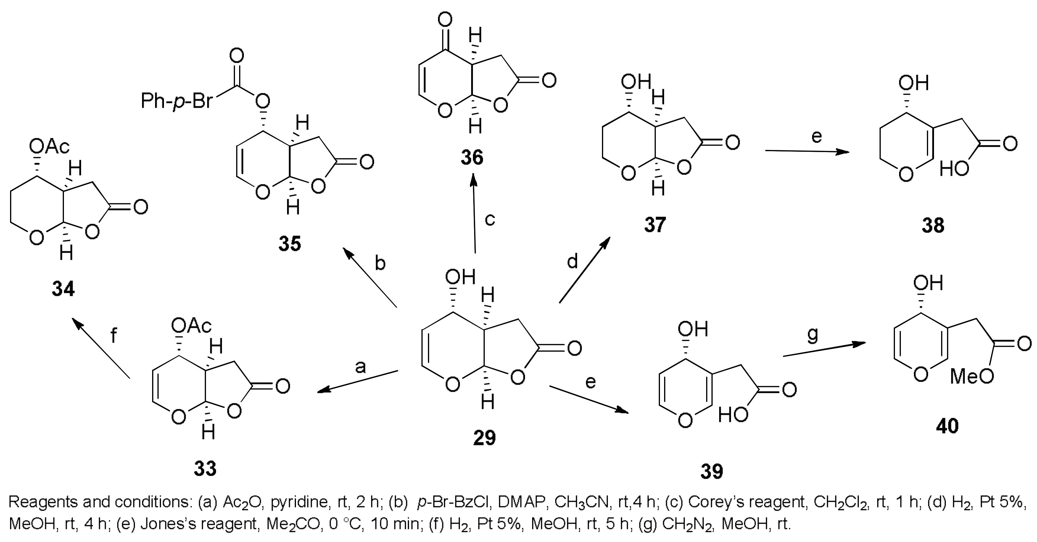
  34. Evidente, A.; Masi, M.; Linaldeddu, B.T.; Franceschini, A.; Scanu, B.; Cimmino, A.; Andolfi, A.; Motta, A.; Maddau, L. Afritoxinones A and B, dihydrofuropyran-2-ones produced by Diplodia africana the causal agent of branch dieback on Juniperus phoenicea. Phytochemistry 2012, 77, 245–250. [Google Scholar] [CrossRef] [PubMed]
  35. Andolfi, A.; Maddau, L.; Basso, S.; Linaldeddu, B.T.; Cimmino, A.; Scanu, B.; Deidda, A.; Tuzi, A.; Evidente, A. Diplopimarane, a 20-nor-ent-pimarane produced by the oak pathogen Diplodia quercivora. J. Nat. Prod. 2014, 77, 2352–2360. [Google Scholar] [CrossRef] [PubMed]
  36. Masi, M.; Maddau, L.; Linaldeddu, B.T.; Cimmino, A.; D’Amico, W.; Scanu, B.; Evidente, M.; Tuzi, A.; Evidente, A. Bioactive secondary metabolites produced by the oak pathogen Diplodia corticola. J. Agric. Food Chem. 2016, 64, 217–225. [Google Scholar] [CrossRef] [PubMed]
  37. Cimmino, A.; Maddau, L.; Masi, M.; Evidente, M.; Linaldeddu, B.T.; Evidente, A. Further secondary metabolites produced by Diplodia corticola, a fungal pathogen involved in cork oak decline. Tetrahedron 2016, 72, 6788–6793. [Google Scholar] [CrossRef]
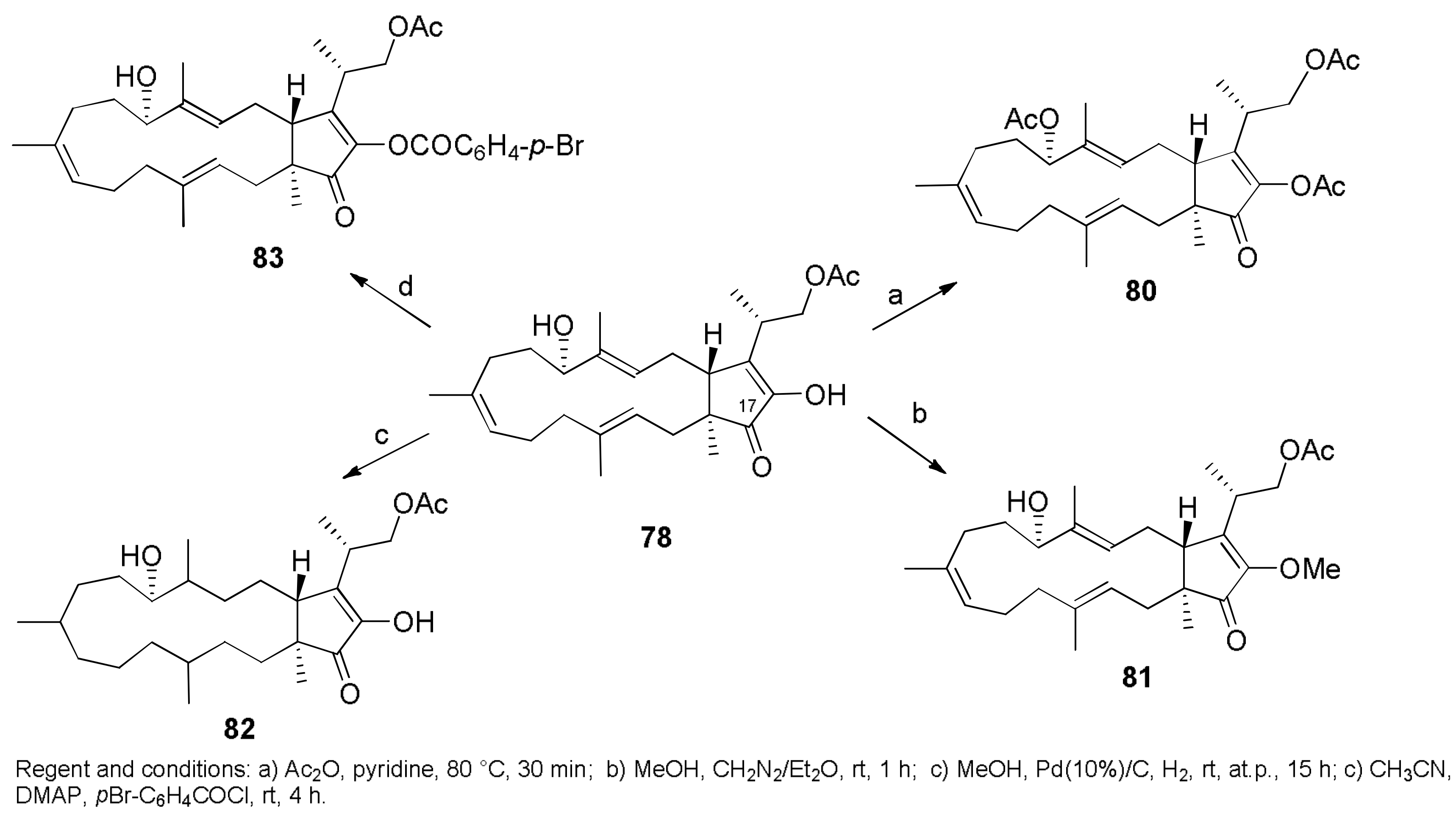
  38. Lallemand, B.; Masi, M.; Maddau, L.; De Lorenzi, M.; Dam, R.; Cimmino, A.; Moreno, Y.; Banuls, L.; Andolfi, A.; Kiss, R.; et al. Evaluation of in vitro anticancer activity of sphaeropsidins A–C, fungal rearranged pimarane diterpenes, and semisynthetic derivatives. Phytochem. Lett. 2012, 5, 770–775. [Google Scholar] [CrossRef]
  39. Mathieu, V.; Chantôme, A.; Lefranc, F.; Cimmino, A.; Miklos, W.; Paulitschke, V.; Mohr, T.; Maddau, L.; Kornienko, A.; Berger, W.; et al. Sphaeropsidin A shows promising activity against drug-resistant cancer cells by targeting regulatory volume increase. Cell. Mol. Life Sci. 2015, 72, 3731–3746. [Google Scholar] [CrossRef] [PubMed]
  40. Masi, M.; Cimmino, A.; Maddau, L.; Kornienko, A.; Tuzi, A.; Evidente, A. Crystal structure and absolute configuration of sphaeropsidin A and its 6-O-p-bromobenzoate. Tetrahedron Lett. 2016, 57, 4592–4594. [Google Scholar] [CrossRef]
  41. Ingels, A.; Dinhof, C.; Garg, A.D.; Maddau, L.; Masi, M.; Evidente, A.; Berger, W.; Dejaegher, B.; Mathieu, V. Computed determination of the in vitro optimal chemocombinations of sphaeropsidin A with chemotherapeutic agents to combat melanomas. Cancer Chemother. Pharmacol. 2017, 79, 971–983. [Google Scholar] [CrossRef] [PubMed]
  42. Evidente, A.; Sparapano, L.; Motta, A.; Giordano, F.; Fierro, O.; Frisullo, S. A phytotoxic pimarane diterpene of Sphaeropsis sapinea f. sp. cupressi, the pathogen of a canker disease of cypress. Phytochemistry 1996, 42, 1541–1546. [Google Scholar] [CrossRef]
  43. Evidente, A.; Sparapano, L.; Fierro, O.; Bruno, G.; Giordano, F.; Motta, A. Sphaeropsidins B and C, phytotoxic pimarane diterpenes from Sphaeropsis sapinea f. sp. cupressi and Diplodia mutila. Phytochemistry 1997, 45, 705–713. [Google Scholar] [CrossRef]
  44. Sparapano, L.; Bruno, G.; Fierro, O.; Evidente, A. Studies on structure-activity relationship of sphaeropsidins A–F, phytotoxins produced by Sphaeropsis sapinea f. sp. cupressi. Phytochemistry 2004, 65, 189–198. [Google Scholar] [CrossRef] [PubMed]
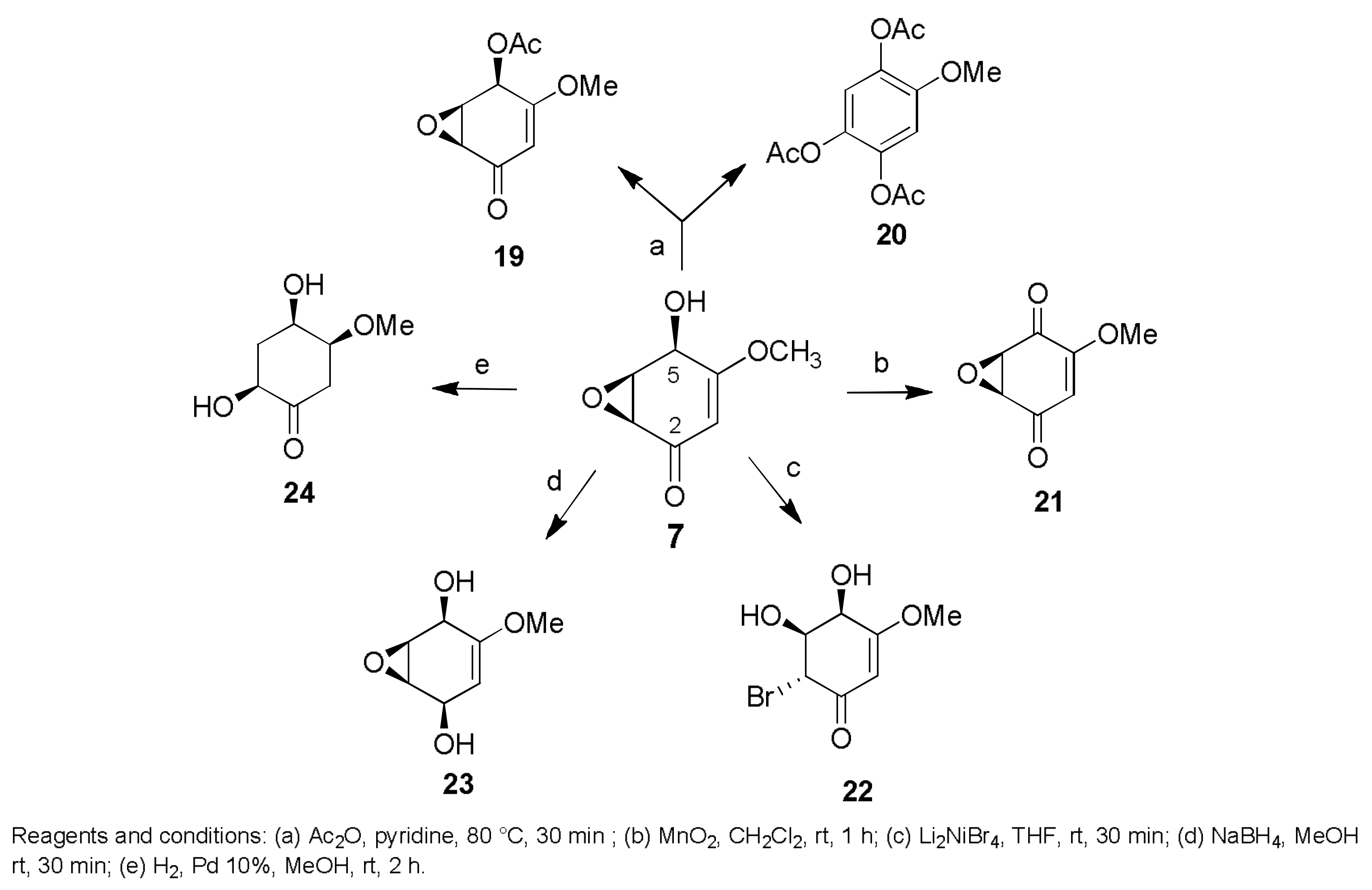
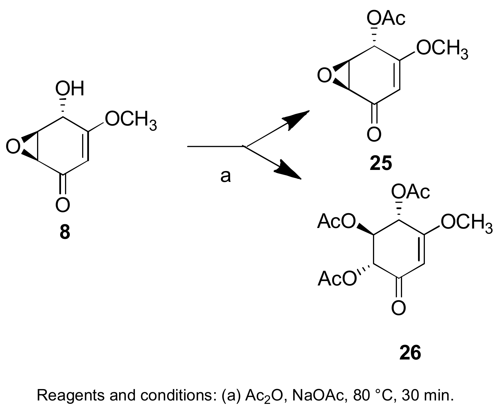
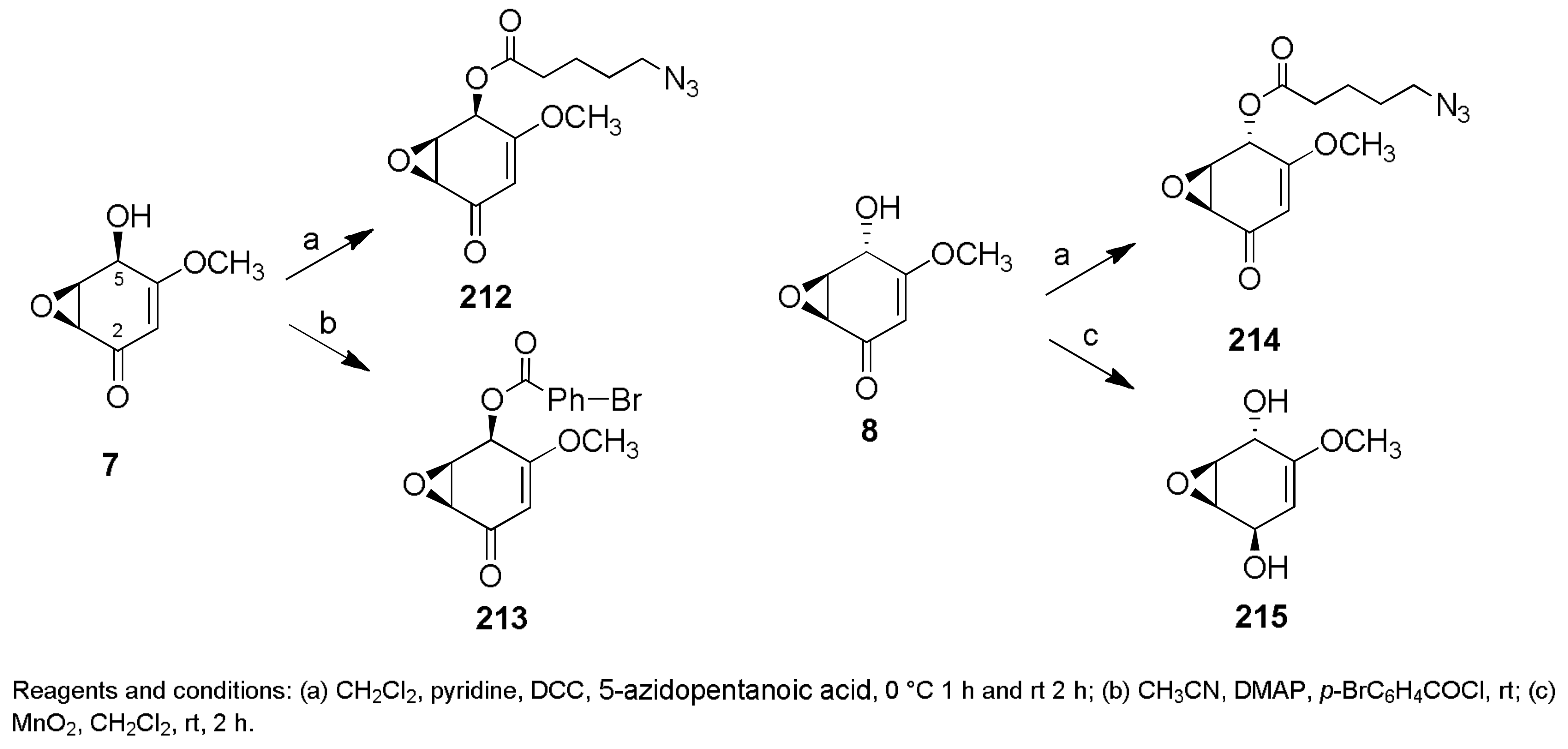
  45. Evidente, A.; Maddau, L.; Scanu, B.; Andolfi, A.; Masi, M.; Motta, A.; Tuzi, A. Sphaeropsidones, phytotoxic dimedone methyl ethers produced by Diplodia cupressi: A structure—Activity relationship study. J. Nat. Prod. 2011, 74, 757–763. [Google Scholar] [CrossRef] [PubMed]
  46. Andolfi, A.; Maddau, L.; Linaldeddu, B.T.; Scanu, B.; Cimmino, A.; Basso, S.; Evidente, A. Bioactivity studies of oxysporone and several derivatives. Phytochem. Lett. 2014, 10, 40–45. [Google Scholar] [CrossRef]
  47. Scherlach, K.; Boettger, D.; Remme, N.; Hertweck, C. The chemistry and biology of cytochalasans. Nat. Prod. Rep. 2010, 27, 869–886. [Google Scholar] [CrossRef] [PubMed]
  48. Masi, M.; Evidente, A.; Meyer, S.; Nicholson, J.; Munoz, A. Effect of strain and cultural conditions on the production of cytochalasin B by the potential mycoherbicide Pyrenophora semeniperda (Pleosporaceae, Pleosporales). Biocontrol. Sci. Technol. 2014, 24, 53–64. [Google Scholar] [CrossRef]
  49. Masi, M.; Meyer, S.; Cimmino, A.; Clement, S.; Black, B.; Evidente, A. Pyrenophoric acids B and C, two new phytotoxic sesquiterpenoids produced by Pyrenophora semeniperda. J. Agric. Food Chem. 2014, 62, 10304–10311. [Google Scholar] [CrossRef] [PubMed]
  50. Skellam, E. The biosynthesis of cytochalasans. Nat. Prod. Rep. 2017, 34, 1252–1263. [Google Scholar] [CrossRef] [PubMed]
  51. Bottalico, A.; Capasso, R.; Evidente, A.; Randazzo, G.; Vurro, M. Cytochalasins: Structure-activity relationships. Phytochemistry 1990, 29, 93–96. [Google Scholar] [CrossRef]
  52. Li, X.J.; Zhang, Q.; Zhang, A.L.; Gao, J.M. Metabolites from Aspergillus fumigatus, an endophytic fungus associated with Melia azedarach, and their antifungal, antifeedant, and toxic activities. J. Agric. Food Chem. 2012, 60, 3424–3431. [Google Scholar] [CrossRef] [PubMed]
  53. Evidente, A.; Conti, L.; Altomare, C.; Bottalico, A.; Sindona, G.; Segre, A.L.; Logrieco, A. Fusapyrone and deoxyfusapyrone, two antifungal α-pyrones from Fusarium semitectum. Nat. Toxins 1994, 2, 4–13. [Google Scholar] [CrossRef] [PubMed]
  54. Altomare, C.; Pengue, R.; Favilla, M.; Evidente, A.; Visconti, A. Structure-activity relationships of derivatives of fusapyrone, an antifungal metabolite of Fusarium semitectum. J. Agric. Food Chem. 2004, 52, 2997–3001. [Google Scholar] [CrossRef] [PubMed]
  55. Cooney, J.M.; Lauren, D.R. Trichoderma/pathogen interactions: Measurement of antagonistic chemicals produced at the antagonist/pathogen interface using a tubular bioassay. Lett. Appl. Microbiol. 1998, 27, 283–286. [Google Scholar] [CrossRef] [PubMed]
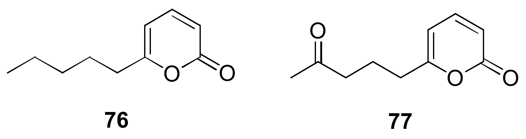
  56. Jeleń, H.; Błaszczyk, L.; Chełkowski, J.; Rogowicz, K.; Strakowska, J. Formation of 6-n-pentyl-2H-pyran-2-one (6-PAP) and other volatiles by different Trichoderma species. Mycol. Prog. 2014, 13, 589–600. [Google Scholar] [CrossRef]
  57. Evidente, A.; Cabras, A.; Maddau, L.; Serra, S.; Andolfi, A.; Motta, A. Viridepyronone, a New antifungal 6-substituted 2H-Pyran-2-one produced by Trichoderma viride. J. Agric. Food Chem. 2003, 51, 6957–6960. [Google Scholar] [CrossRef] [PubMed]
  58. Sarrocco, S. Dung-inhabiting fungi: A potential reservoir of novel secondary metabolites for the control of plant pathogens. Pest Manag. Sci. 2016, 72, 643–652. [Google Scholar] [CrossRef] [PubMed]
  59. Cimmino, A.; Sarrocco, S.; Masi, M.; Diquattro, S.; Evidente, M.; Vannacci, G.; Evidente, A. Fusaproliferin, terpestacin and their derivatives display variable allelopathic activity against some Ascomycetous fungi. Chem. Biodivers. 2016, 13, 1593–1600. [Google Scholar] [CrossRef] [PubMed]
  60. Oerke, E.C. Crop losses to pests. J. Agric. Sci. 2006, 144, 31–43. [Google Scholar] [CrossRef]
  61. Xu, Y.; Zhu, X.F.; Zhou, M.G.; Kuang, J.; Zhang, Y.; Shang, Y.; Wang, J.X. Status of streptomycin resistance development in Xanthomonas oryzae pv. oryzae and Xanthomonas oryzae pv. oryzicola in China and their resistance characters. J. Phytopathol. 2010, 158, 601–608. [Google Scholar] [CrossRef]
  62. Mansfield, J.; Genin, S.; Magori, S.; Citovsky, V.; Sriariyanum, M.; Ronald, P.; Dow, M.; Verdier, V.; Beer, S.V.; Machado, M.A.; et al. Top 10 plant pathogenic bacteria in molecular plant pathology. Mol. Plant Pathol. 2012, 13, 614–629. [Google Scholar] [CrossRef] [PubMed]
  63. Evidente, A.; Venturi, V.; Masi, M.; Degrassi, G.; Cimmino, A.; Maddau, L.; Andolfi, A. In vitro antibacterial activity of sphaeropsidins and chemical derivatives toward Xanthomonas oryzae pv. oryzae, the causal agent of rice bacterial blight. J. Nat. Prod. 2011, 74, 2520–2525. [Google Scholar] [CrossRef] [PubMed]
  64. Pan, X.; Yang, Y.; Zhang, J.-R. Molecular basis of host specificity in human pathogenic bacteria. Emerg. Microbes Infect. 2014, 3, e23. [Google Scholar] [CrossRef] [PubMed]
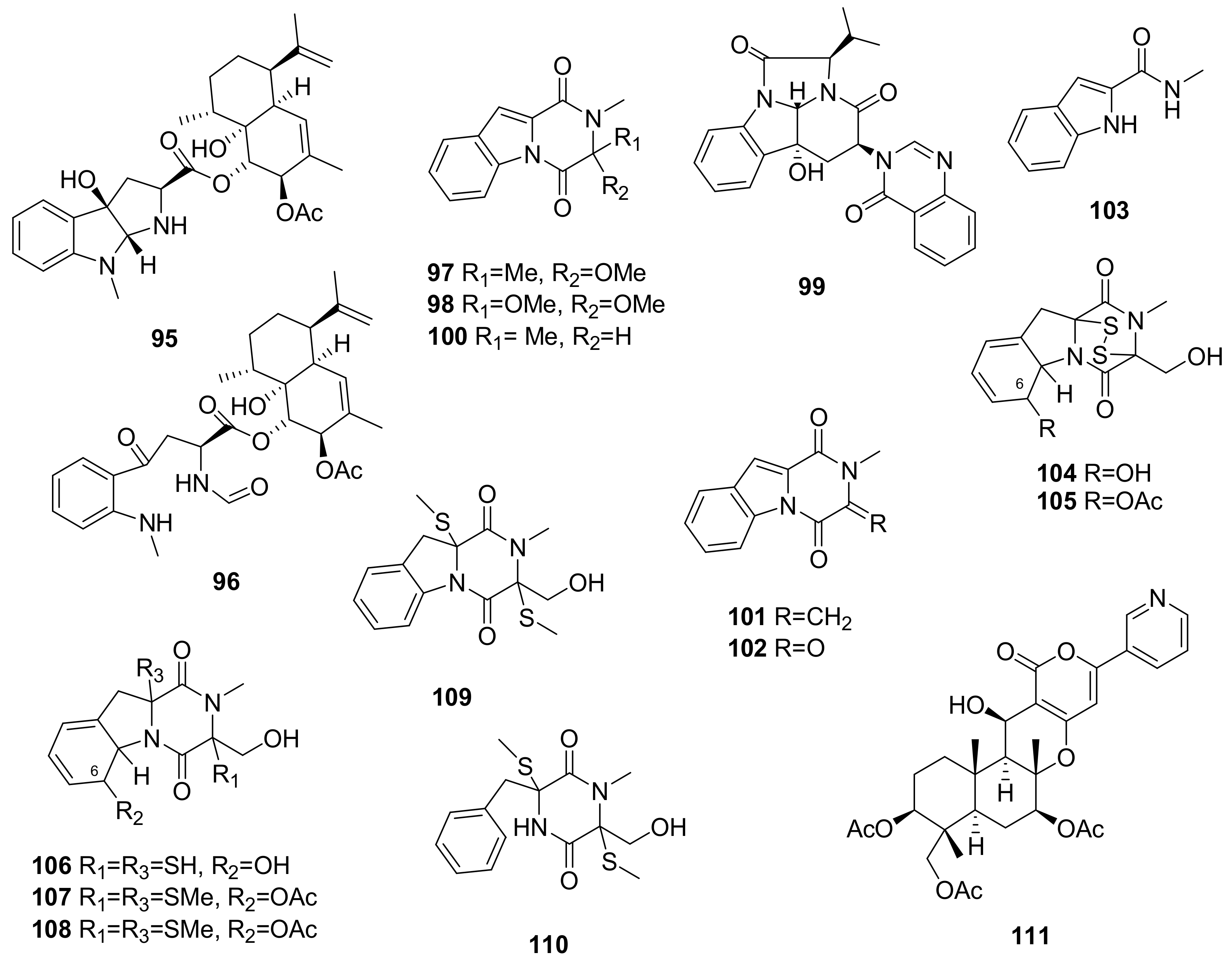
  65. Liang, W.L.; Le, X.; Li, H.J.; Yang, X.L.; Chen, J.X.; Xu, J.; Liu, H.L.; Wang, L.Y.; Wang, K.T.; Hu, K.C.; et al. Exploring the chemodiversity and biological activities of the secondary metabolites from the marine fungus Neosartorya pseudofischeri. Mar. Drugs 2014, 12, 5657–5676. [Google Scholar] [CrossRef] [PubMed]
  66. Masi, M.; Andolfi, A.; Mathieu, V.; Boari, A.; Cimmino, A.; Moreno, Y.; Banuls, L.; Vurro, M.; Kornienko, A.; Kiss, R.; et al. Fischerindoline, a pyrroloindole sesquiterpenoid isolated from Neosartorya pseudofischeri, with in vitro growth inhibitory activity in human cancer cell lines. Tetrahedron 2013, 69, 7466–7470. [Google Scholar] [CrossRef]
  67. Schnekenburger, M.; Mathieu, V.; Lefranc, F.; Jang, J.Y.; Masi, M.; Kijjoa, A.; Evidente, A.; Kim, H.-J.; Kiss, R.; Dicato, M.; et al. The fungal metabolite eurochevalierine, a sequiterpene alkaloid, displays anti-cancer properties through selective sirtuin 1/2 inhibition. Molecules 2018, 23, 333. [Google Scholar] [CrossRef] [PubMed]
  68. Wu, Q.; Jiang, N.; Han, B.; Mei, Y.; Ge, H.; Guo, Z.; Weng, N.; Tan, R. Antibacterial epipolythiodioxopiperazine and unprecedented sesquiterpene from Pseudallescheria boydii, a beetle (coleoptera)-associated fungus. Org. Biomol. Chem. 2014, 12, 9405–9412. [Google Scholar] [CrossRef] [PubMed]
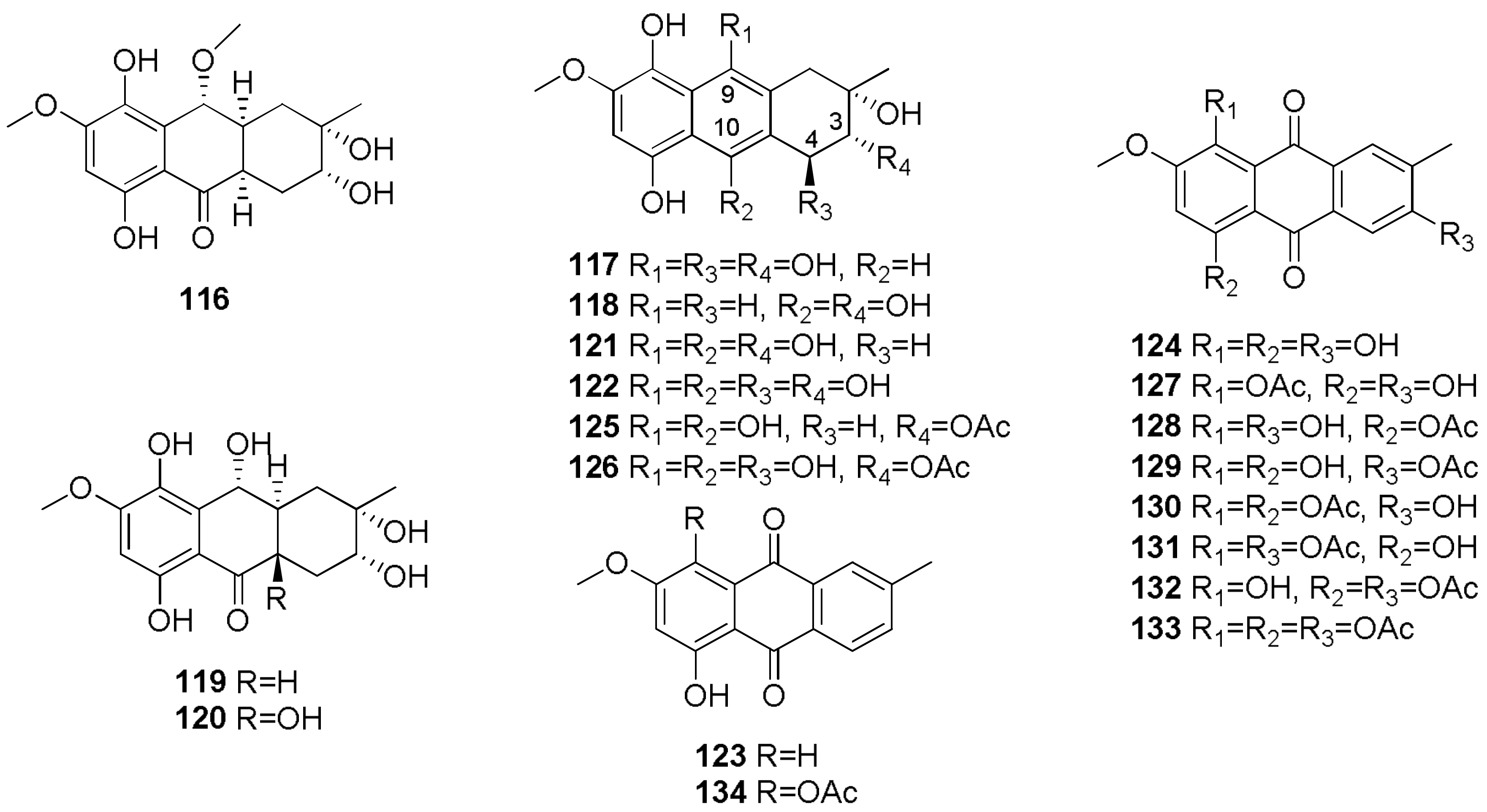
  69. Yang, K.L.; Wei, M.Y.; Shao, C.L.; Fu, X.M.; Guo, Z.Y.; Xu, R.F.; Zheng, C.J.; She, Z.G.; Lin, Y.C.; Wang, C.Y. Antibacterial anthraquinone derivatives from a sea anemone-derived fungus Nigrospora sp. J. Nat. Prod. 2012, 75, 935–941. [Google Scholar] [CrossRef] [PubMed]
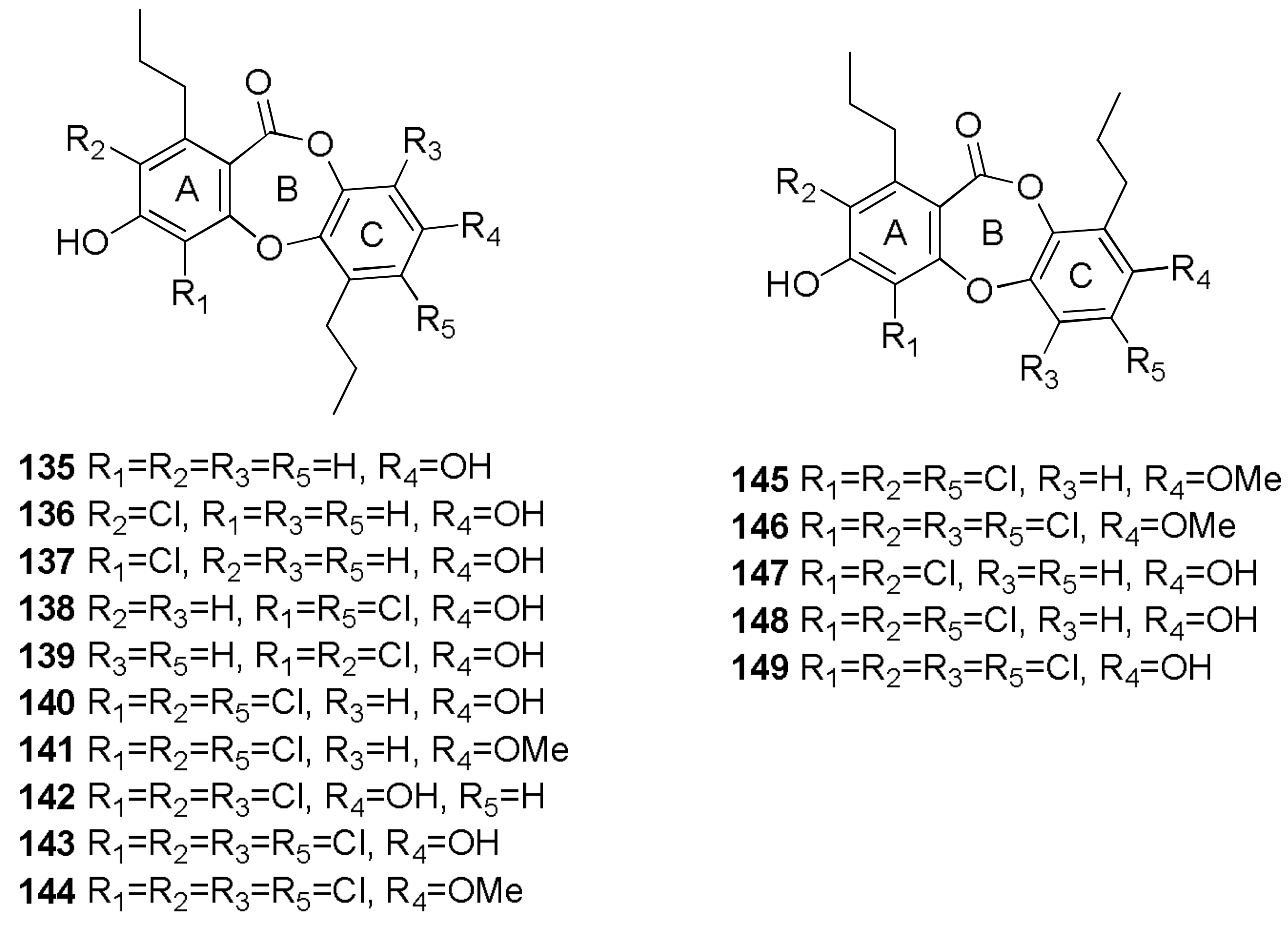
  70. Niu, S.; Liu, D.; Hu, X.; Proksch, P.; Shao, Z.; Lin, W. Spiromastixones A–O, antibacterial chlorodepsidones from a deep-sea-derived Spiromastix sp. fungus. J. Nat. Prod. 2014, 77, 1021–1030. [Google Scholar] [CrossRef] [PubMed]
  71. Wu, C.; Chen, R.; Liu, M.; Liu, D.; Li, X.; Wang, S.; Niu, S.; Guo, P.; Lin, W.; Jacobson, P.B. Spiromastixones inhibit foam cell formation via regulation of cholesterol efflux and uptake in RAW264.7 macrophages. Mar. Drugs 2015, 13, 6352–6365. [Google Scholar] [CrossRef] [PubMed]
  72. Gupta, R.C.; Milatovic, D. Insecticides. In Biomarkers in Toxicology; Academic Press: Cambridge, MA, USA, 2014; pp. 389–407. ISBN 9780124046306. [Google Scholar]
  73. Anke, H.; Sterner, O. Insecticidal and nematicidal metabolites from fungi. In The Mycota X. Industrial Applications; Osiewacz, H.D., Ed.; Springer: Berlin, Germany, 2002; pp. 109–127. [Google Scholar]
  74. Geris, R.; Ribeiro, P.R.; Da Silva Brandão, M.; Da Silva, H.H.G.; Da Silva, I.G. Chapter 10—Bioactive Natural Products as Potential Candidates to Control Aedes aegypti, the Vector of Dengue; Elsevier: New York, NY, USA, 2012; Volume 37. [Google Scholar]
  75. Masi, M.; Cala, A.; Tabanca, N.; Cimmino, A.; Green, I.R.; Bloomquist, J.R.; Van Otterlo, W.A.L.; Macias, F.A.; Evidente, A. Alkaloids with activity against the zika virus vector Aedes aegypti (L.)-crinsarnine and sarniensinol, two new crinine and mesembrine type alkaloids isolated from the South African plant Nerine sarniensis. Molecules 2016, 21, 1432. [Google Scholar] [CrossRef] [PubMed]
  76. Masi, M.; van der Westhuyzen, A.E.; Tabanca, N.; Evidente, M.; Cimmino, A.; Green, I.R.; Bernier, U.R.; Becnel, J.J.; Bloomquist, J.R.; van Otterlo, W.A.L.; et al. Sarniensine, a mesembrine-type alkaloid isolated from Nerine sarniensis, an indigenous South African Amaryllidaceae, with larvicidal and adulticidal activities against Aedes aegypti. Fitoterapia 2017, 116, 34–38. [Google Scholar] [CrossRef] [PubMed]
  77. Masi, M.; Cimmino, A.; Tabanca, N.; Becnel, J.J.; Bloomquist, J.R.; Evidente, A. A survey of bacterial, fungal and plant metabolites against Aedes aegypti (Diptera: Culicidae), the vector of yellow and dengue fevers and Zika virus. Open Chem. 2017, 15, 156–166. [Google Scholar] [CrossRef]
  78. Cimmino, A.; Andolfi, A.; Avolio, F.; Ali, A.; Tabanca, N.; Khan, I.A.; Evidente, A. Cyclopaldic acid, seiridin, and sphaeropsidin A as fungal phytotoxins, and larvicidal and biting deterrents against Aedes aegypti (Diptera: Culicidae): Structure-activity relationships. Chem. Biodivers. 2013, 10, 1239–1251. [Google Scholar] [CrossRef] [PubMed]
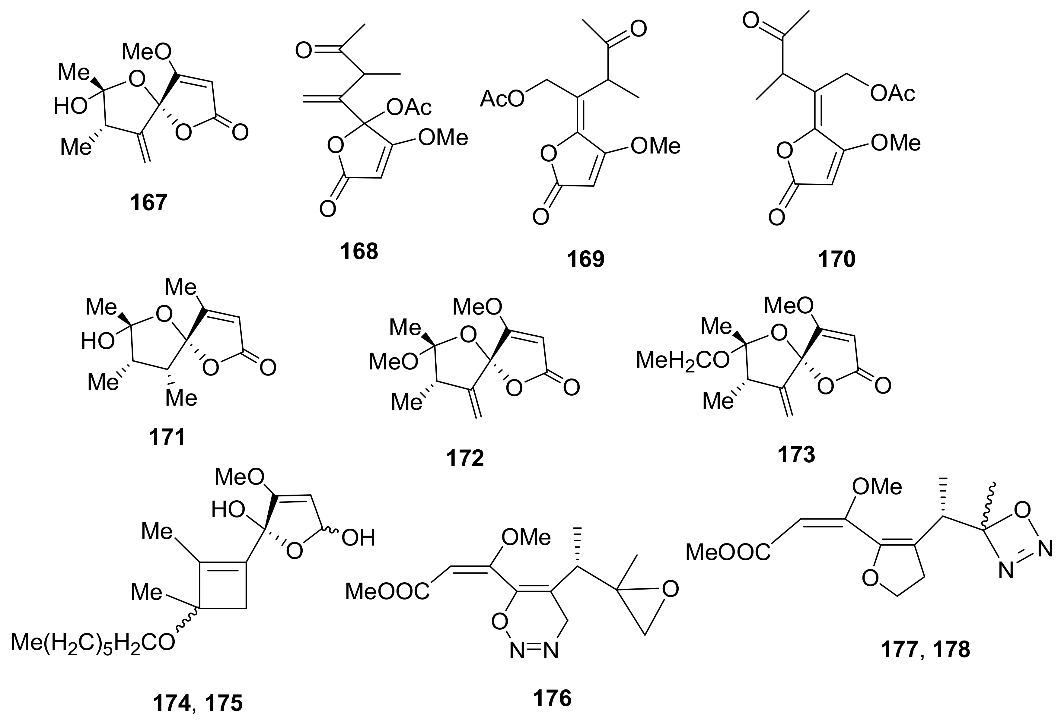
  79. Cimmino, A.; Evidente, M.; Masi, M.; Ali, A.; Tabanca, N.; Khan, I.A.; Evidente, A. Papyracillic acid and its derivatives as biting deterrents against Aedes aegypti (Diptera: Culicidae): Structure-activity relationships. Med. Chem. Res. 2015, 24, 3981–3989. [Google Scholar] [CrossRef]
  80. Graniti, A.; Sparapano, L.; Evidente, A. Cyclopaldic acid, a major phytotoxic metabolite of Seiridium cupressi, the pathogen of a canker disease of cypress. Plant Pathol. 2007, 41, 563–568. [Google Scholar] [CrossRef]
  81. Sparapano, L.; Evidente, A. Biological activity of cyclopaldic acid, a major toxin of Seiridium cupressi, its six derivatives, andiso-cyclopaldic acid. Nat. Toxins 1995, 3, 156–165. [Google Scholar] [CrossRef] [PubMed]
  82. Evidente, A.; Randazzo, G.; Ballio, A. Structure determination of seiridin and isoseiridin, phytotoxic butenolides from culture filtrate of Seiridium cardinale. J. Nat. Prod. 1986, 49, 593–601. [Google Scholar] [CrossRef]
  83. Shan, R.; Anke, H.; Stadler, M.; Sterner, O. Papyracillic acid, a new penicillic acid analogue from the ascomycete Lachnum papyraceum. Tetrahedron 1996, 52, 10249–10254. [Google Scholar] [CrossRef]
  84. Evidente, A.; Berestetskiy, A.; Cimmino, A.; Tuzi, A.; Superchi, S.; Melck, D.; Andolfi, A. Papyracillic acid, a phytotoxic 1,6-dioxaspiro[4,4]nonene produced by Ascochyta agropyrina var. nana, a potential mycoherbicide for Elytrigia repens biocontrol. J. Agric. Food Chem. 2009, 57, 11168–11173. [Google Scholar] [CrossRef] [PubMed]
  85. Shan, R.; Stadler, M.; Anke, H.; Sterner, O. The reactivity of the fungal toxin papyracillic acid. Tetrahedron 1997, 53, 6209–6214. [Google Scholar] [CrossRef]
  86. Geris dos Santos, R.M.; Rodrigues-Fo, E. Meroterpenes from Penicillium sp. found in association with Melia azedarach. Phytochemistry 2002, 61, 907–912. [Google Scholar] [CrossRef]
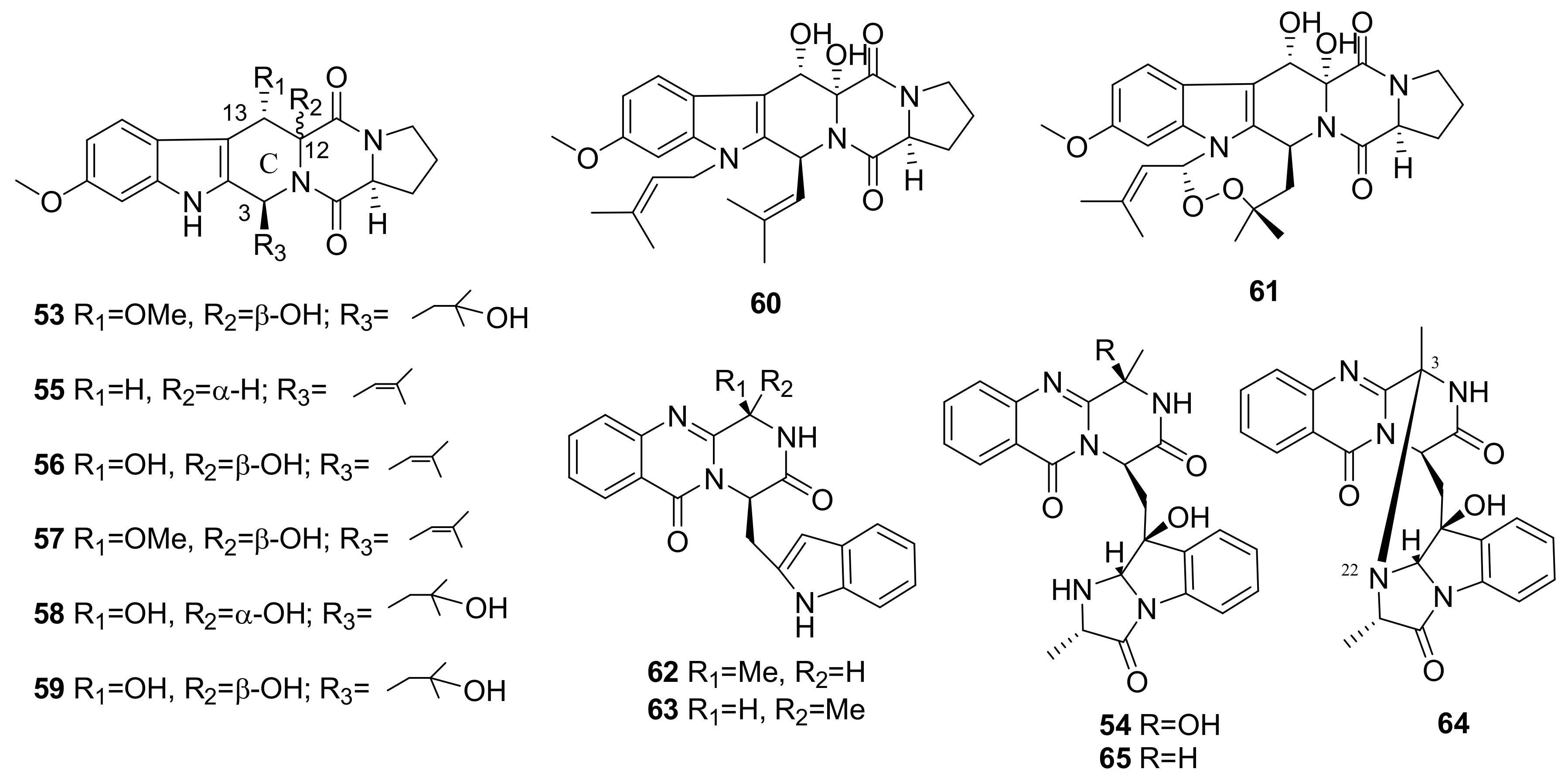
  87. Wang, X.; Li, Y.; Zhang, X.; Lai, D.; Zhou, L. Structural diversity and biological activities of the cyclodipeptides from fungi. Molecules 2017, 22, 2026. [Google Scholar] [CrossRef] [PubMed]
  88. Kato, N.; Furutani, S.; Otaka, J.; Noguchi, A.; Kinugasa, K.; Kai, K.; Hayashi, H.; Ihara, M.; Takahashi, S.; Matsuda, K.; et al. Biosynthesis and structure–activity relationship studies of okaramines that target insect glutamate-gated chloride channels. ACS Chem. Biol. 2018, 13, 561–566. [Google Scholar] [CrossRef] [PubMed]
  89. Hayashi, H.; Takiuchi, K.; Murao, S.; Arai, M. Structure and insecticidal activity of new indole alkaloids, okaramines A and B, from Penicillium simplicissimum ak-40. Agric. Biol. Chem. 1989, 53, 461–469. [Google Scholar] [CrossRef]
  90. Harvey, R.J.; Vreugdenhill, E.; Zaman, S.H.; Bhandal, N.S.; Usherwood, P.N.R.; Barnard, E.A.; Darlison, M.G.; Harvey, R.J.; ffrench-Constant, R.H.; Rocheleau, T.A.; et al. GluCl a target of indole alkaloid okaramines: A 25 year enigma solved. Br. J. Pharmacol. 1996, 119, 62–67. [Google Scholar] [CrossRef]
  91. Cimmino, A.; Andolfi, A.; Zonno, M.C.; Avolio, F.; Santini, A.; Tuzi, A.; Berestetskyi, A.; Vurro, M.; Evidente, A. Chenopodolin: A phytotoxic unrearranged ent-pimaradiene diterpene produced by phoma chenopodicola, a fungal pathogen for Chenopodium album biocontrol. J. Nat. Prod. 2013, 76, 1291–1297. [Google Scholar] [CrossRef] [PubMed]
  92. Evidente, M.; Cimmino, A.; Zonno, M.C.; Masi, M.; Berestetskyi, A.; Santoro, E.; Superchi, S.; Vurro, M.; Evidente, A. Phytotoxins produced by Phoma chenopodiicola, a fungal pathogen of Chenopodium album. Phytochemistry 2015, 117, 482–488. [Google Scholar] [CrossRef] [PubMed]
  93. Berestetskiy, A.; Dmitriev, A.; Mitina, G.; Lisker, I.; Andolfi, A.; Evidente, A. Nonenolides and cytochalasins with phytotoxic activity against Cirsium arvense and Sonchus arvensis: A structure-activity relationships study. Phytochemistry 2008, 69, 953–960. [Google Scholar] [CrossRef] [PubMed]
  94. Andolfi, A.; Cimmino, A.; Vurro, M.; Berestetskiy, A.; Troise, C.; Zonno, M.C.; Motta, A.; Evidente, A. Agropyrenol and agropyrenal, phytotoxins from Ascochyta agropyrina var. nana, a fungal pathogen of Elitrigia repens. Phytochemistry 2012, 79, 102–108. [Google Scholar] [CrossRef] [PubMed]
  95. Cimmino, A.; Zonno, M.C.; Andolfi, A.; Troise, C.; Motta, A.; Vurro, M.; Evidente, A. Agropyrenol, a phytotoxic fungal metabolite, and its derivatives: A structure-activity relationship study. J. Agric. Food Chem. 2013, 61, 1779–1783. [Google Scholar] [CrossRef] [PubMed]
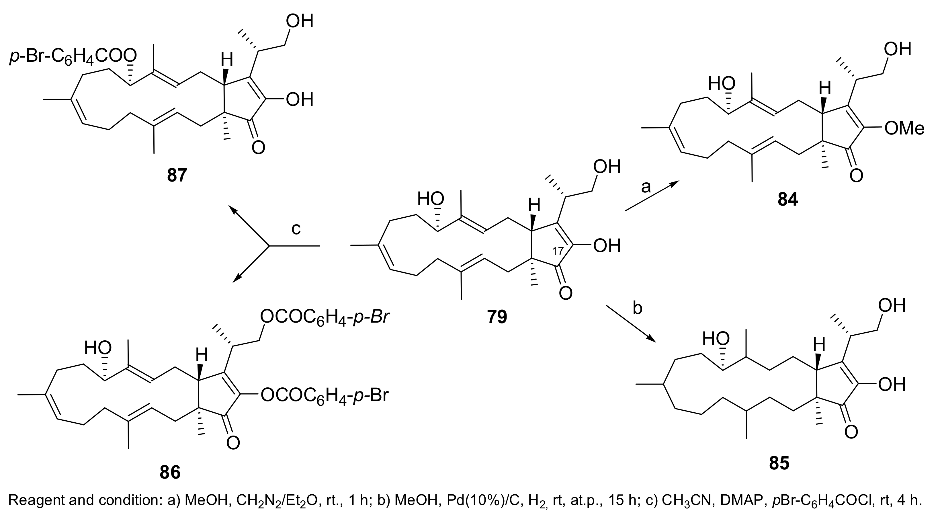
  96. Cimmino, A.; Andolfi, A.; Zonno, M.C.; Troise, C.; Santini, A.; Tuzi, A.; Vurro, M.; Ash, G.; Evidente, A. Phomentrioloxin: A phytotoxic pentasubstituted geranylcyclohexentriol produced by Phomopsis sp., a potential mycoherbicide for Carthamus lanatus biocontrol. J. Nat. Prod. 2012, 75, 1130–1137. [Google Scholar] [CrossRef] [PubMed]
  97. Cimmino, A.; Andolfi, A.; Zonno, M.C.; Boari, A.; Troise, C.; Motta, A.; Vurro, M.; Ash, G.; Evidente, A. Phomentrioloxin, a fungal phytotoxin with potential herbicidal activity, and its derivatives: A structure-activity relationship study. J. Agric. Food Chem. 2013, 61, 9645–9649. [Google Scholar] [CrossRef] [PubMed]
  98. Evidente, A.; Andolfi, A.; Fiore, M.; Boari, A.; Vurro, M. Stimulation of Orobanche ramosa seed germination by fusicoccin derivatives: A structure-activity relationship study. Phytochemistry 2006, 67, 19–26. [Google Scholar] [CrossRef] [PubMed]
  99. Fernández-Aparicio, M.; Andolfi, A.; Cimmino, A.; Rubiales, D.; Evidente, A. Stimulation of seed germination of Orobanche species by ophiobolin a and fusicoccin derivatives. J. Agric. Food Chem. 2008, 56, 8343–8347. [Google Scholar] [CrossRef] [PubMed]
  100. Fernandez-Aparicio, M.; Masi, M.; Maddau, L.; Cimmino, A.; Evidente, M.; Rubiales, D.; Evidente, A. Induction of haustorium development by sphaeropsidones in radicles of the parasitic weeds Striga and Orobanche. A structure-activity relationship study. J. Agric. Food Chem. 2016, 64, 5188–5196. [Google Scholar] [CrossRef] [PubMed]
  101. Nickrent, D.L. Parasitic plants. Ecology 1997, 78, 1612–1613. [Google Scholar] [CrossRef]
  102. Press, M.C.; Phoenix, G.K. Impacts of parasitic plants on natural communities. New Phytol. 2005, 166, 737–751. [Google Scholar] [CrossRef] [PubMed]
  103. Masi, M.; Meyer, S.; Clement, S.; Cimmino, A.; Cristofaro, M.; Evidente, A. Cochliotoxin, a dihydropyranopyran-4,5-dione, and its analogues produced by Cochliobolus australiensis display phytotoxic activity against buffelgrass (Cenchrus ciliaris). J. Nat. Prod. 2017, 80, 1241–1247. [Google Scholar] [CrossRef] [PubMed]
  104. Masi, M.; Meyer, S.; Clement, S.; Pescitelli, G.; Cimmino, A.; Cristofaro, M.; Evidente, A. Chloromonilinic acids C and D, phytotoxic tetrasubstituted 3-chromanonacrylic acids isolated from Cochliobolus australiensis with potential herbicidal activity against buffelgrass (Cenchrus ciliaris). J. Nat. Prod. 2017, 80, 2771–2777. [Google Scholar] [CrossRef] [PubMed]
  105. Masi, M.; Meyer, S.; Górecki, M.; Mandoli, A.; Di Bari, L.; Pescitelli, G.; Cimmino, A.; Cristofaro, M.; Clement, S.; Evidente, A. Pyriculins A and B, two monosubstituted hex-4-ene-2,3-diols and other phytotoxic metabolites produced by Pyricularia grisea isolated from buffelgrass (Cenchrus ciliaris). Chirality 2017, 29, 726–736. [Google Scholar] [CrossRef] [PubMed]
  106. Masi, M.; Meyer, S.; Clement, S.; Andolfi, A.; Cimmino, A.; Evidente, A. Spirostaphylotrichin W, a spirocyclic γ-lactam isolated from liquid culture of Pyrenophora semeniperda, a potential mycoherbicide for cheatgrass (Bromus tectorum) biocontrol. Tetrahedron 2014, 70, 1497–1501. [Google Scholar] [CrossRef]
  107. Masi, M.; Meyer, S.; Cimmino, A.; Andolfi, A.; Evidente, A. Pyrenophoric acid, a phytotoxic sesquiterpenoid penta-2,4-dienoic acid produced by a potential mycoherbicide Pyrenophora semeniperda. J. Nat. Prod. 2014, 77, 925–930. [Google Scholar] [CrossRef] [PubMed]
  108. Meyer, S.E.; Masi, M.; Clement, S.; Davis, T.L.; Beckstead, J. Mycelial growth rate and toxin production in the seed pathogen Pyrenophora semeniperda: Resource trade-offs and temporally varying selection. Plant Pathol. 2015, 64, 1450–1460. [Google Scholar] [CrossRef]
  109. Cimmino, A.; Masi, M.; Evidente, M.; Superchi, S.; Evidente, A. Application of Mosher’s method for absolute configuration assignment to bioactive plants and fungi metabolites. J. Pharm. Biomed. Anal. 2017, 144, 59–89. [Google Scholar] [CrossRef] [PubMed]
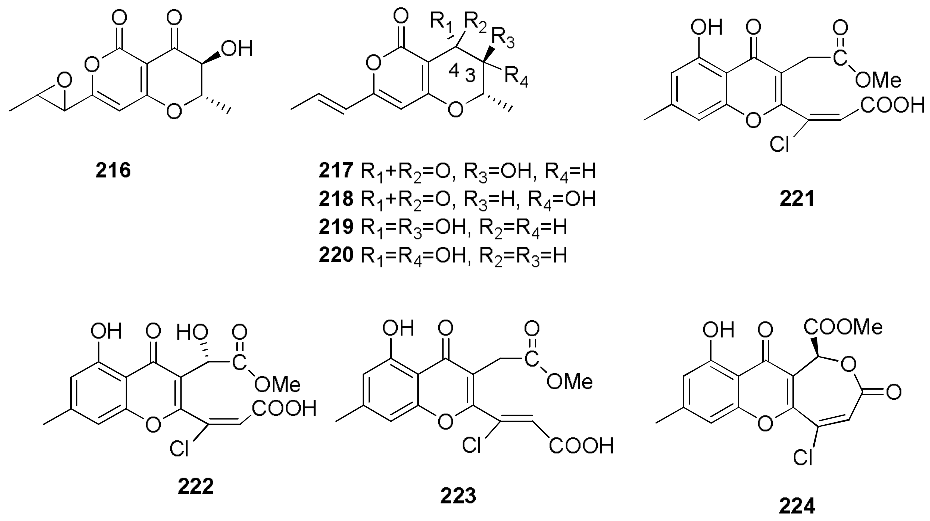
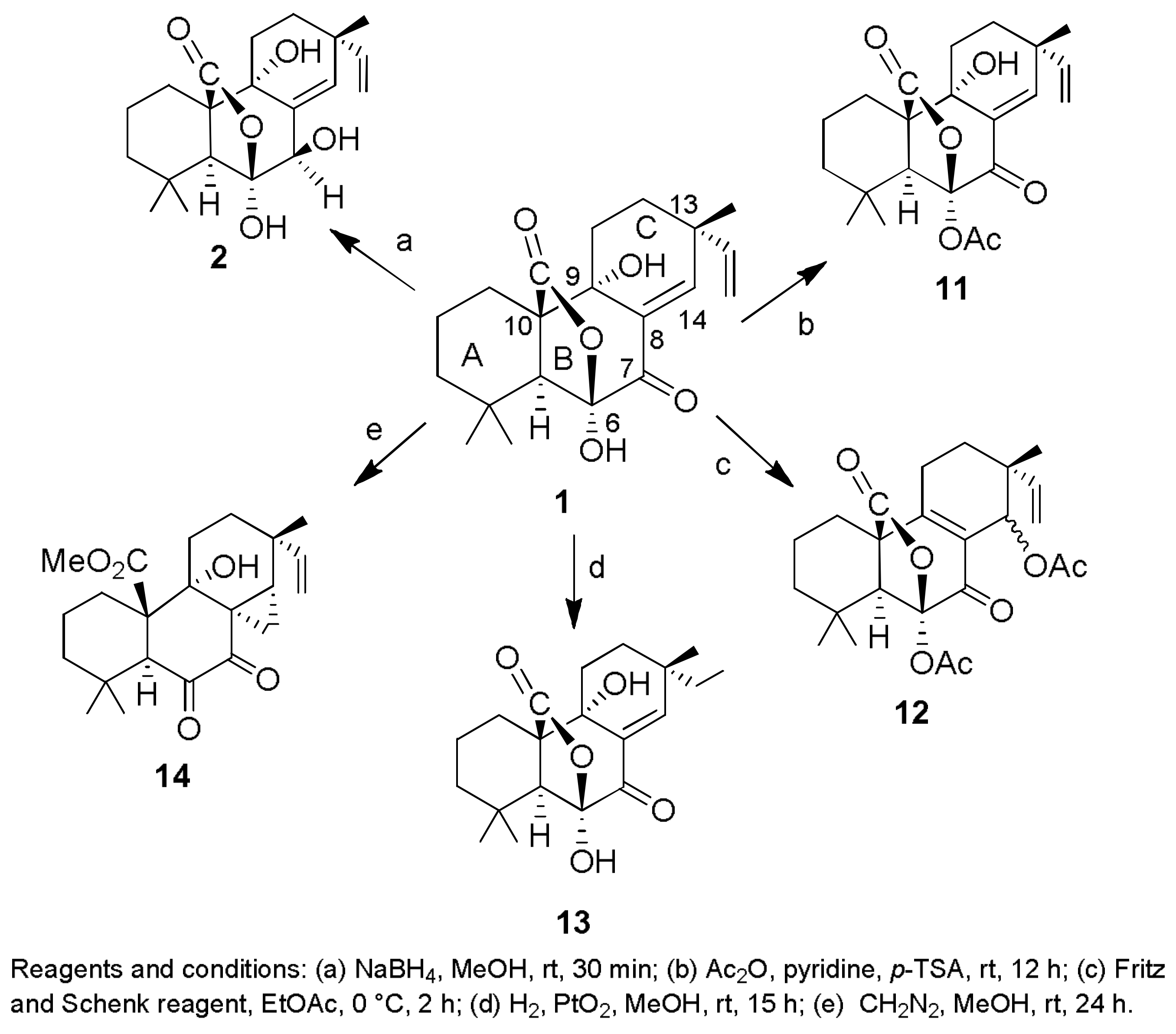
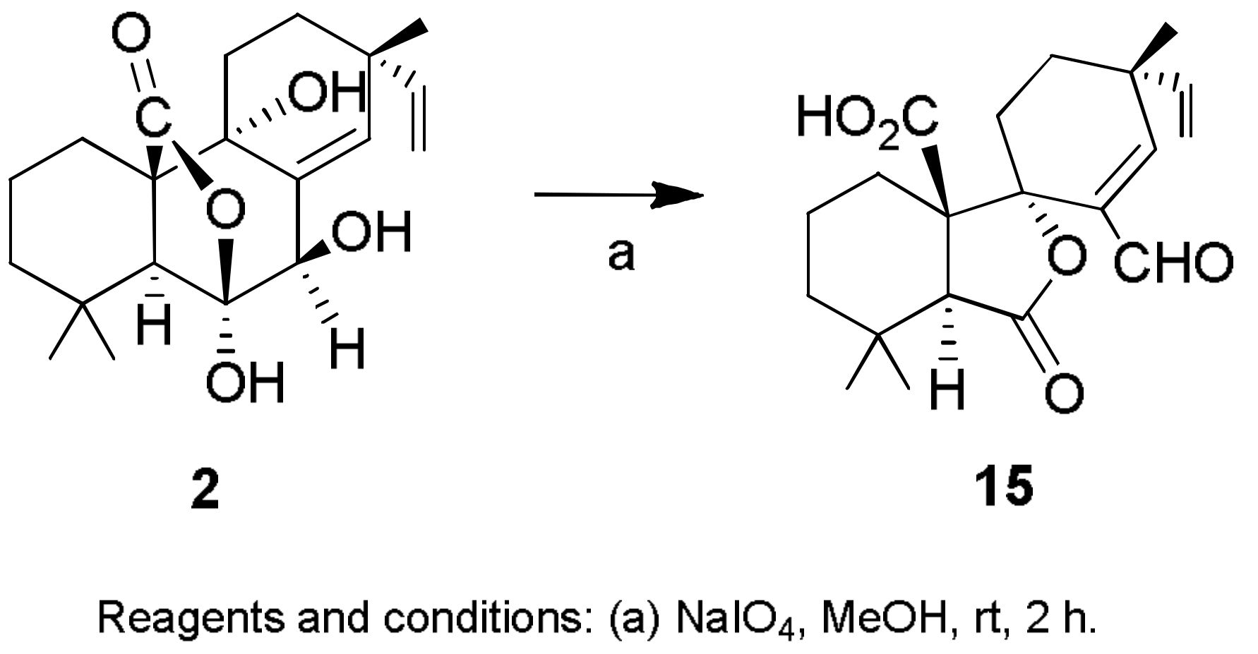
Sample Availability: Samples of the compounds 1, 2, 7, 8, 2932, 43, 44, 150, 160, 167, 207, 216224 are available from the authors.
Figure 1. The structures of sphaeropsidins A–F (16) and sphaeropsidones (7 and 8) and their chlorinated analogs (9 and 10).
Molecules 23 00834 g001
Scheme 1. The structures of sphaeropsidin A (1) and its derivatives (2, 1114).
Molecules 23 00834 sch001
Scheme 2. The structures of sphaeropsidin B (2) and its derivative (15).
Molecules 23 00834 sch002
Scheme 3. The structures of sphaeropsidin C (3) and its derivatives (1618).
Molecules 23 00834 sch003
Scheme 4. The structures of sphaeropsidone (7) and its derivatives (1924).
Molecules 23 00834 sch004
Scheme 5. The structures of epi-sphaeropsidone (8) and its derivatives (25 and 26).
Molecules 23 00834 sch005
Figure 2. The structures of afritoxinones A and B (27 and 28), oxysporone (29), R-(−)-mellein, (3R,4R)-4-hydroxymellein, and (3R,4S)-4-hydroxymellein (3032).
Molecules 23 00834 g002
Scheme 6. The structures of oxysporone (29) and its derivatives (3340).
Molecules 23 00834 sch006
Figure 3. The structures of ascochalasin (41); deoxaphomin (42); cytochalasin A (43); cytochalasin B (44); 7,20-O,O′-diacetylcytochalasin B (45); 21,22-dihydrocytochalasin B (46); 17-O-acetylcytochalasin A (47); and cytochalasins C, D, E, H, and J (4852, respectively).
Molecules 23 00834 g003
Figure 4. The structures of 12β-hydroxy-13α-methoxyverruculogen TR-2 (53); 3-hydroxyfumiquinazoline A (54); fumitremorgin C (55); cyclotryprostatins A (56) and B (57); verruculogen TR-2 (58); 12β-hydroxyverruculogen TR-2 (59); fumitremorgin B (60); verruculogen (61); and fumiquinazolines F, G, D, and A (6265).
Molecules 23 00834 g004
Figure 5. The structures of fusapyrone (66), its derivatives (68,7075), deoxyfusapyrone (67), and its derivative (69).
Molecules 23 00834 g005
Figure 6. The structures of 6-n-pentyl-2H-pyran-2-one and viridepyronone (76 and 77).
Molecules 23 00834 g006
Scheme 7. The structures of fusaproliferin (78) and its derivatives (8083).
Molecules 23 00834 sch007
Scheme 8. The structures of terpestacin (79) and its derivatives (8487).
Molecules 23 00834 sch008
Scheme 9. The structures of sphaeropsidin A (1) and its derivatives (8891).
Molecules 23 00834 sch009
Scheme 10. The structures of sphaeropsidin B (2) and its derivatives (9294).
Molecules 23 00834 sch010
Figure 7. The structures of fischerindoline (95), eurochevalierine (96), neosartins A–C (9799), 1,2,3,4-tetrahydro-2,3-dimethyl-1,4-dioxopyrazino[1–a]indole (100), 1,2,3,4-tetrahydro-2-methyl- 3-methylene-1,4-dioxopyrazino[1–a]indole (101), 1,2,3,4-tetrahydro-2-methyl-1,3,4-trioxopyrazino [1–a]indole (102), N-methyl-1H-indole-2-carboxamide (103), gliotoxin (104), acetylgliotoxin (105), reduced gliotoxin (106), 6-acetylbis(methylthio)gliotoxin (107), bisdethiobis(methylthio)gliotoxin (108), didehydrobisdethiobis(methylthio)gliotoxin (109), bis-N-norgliovictin (110), and pyripyropene A (111).
Molecules 23 00834 g007
Figure 8. The structures of boydines A–D (112115).
Molecules 23 00834 g008
Figure 9. The structures of 4a-epi-9α-methoxydihydrodeoxybostrycin (116), 10-deoxybostrycin (117), nigrosporin B (118), 9α-hydroxydihydrodesoxybostrycin (119), 9α-hydroxyhalorosellinia A (120), 4-deoxybostrycin (121), bostrycin (122), austrocortirubin (123), 3,5,8-trihydroxy-7-methoxy-2-methylanthracene-9,10-dione (124), 3-acetoxy-4-deoxybostrycin (125), 3-acetoxybostrycin (126), 8-acetoxy-3,5-dihydroxy-7-methoxy-2-methylanthracene-9,10-dione (127), 5-acetoxy-3,8-dihydroxy-7-methoxy-2-methylanthracene-9,10-dione (128), 3-acetoxy-5,8-dihydroxy-7-methoxy-2-methylanthracene-9,10-dione (129), 5,8-diacetoxy-3-hydroxy-7-methoxy-2-methylanthracene-9,10-dione (130), 3,8-diacetoxy-5-hydroxy-7-methoxy-2-methylanthracene-9,10-dione (131), 3,5-diacetoxy-8-hydroxy-7-methoxy-2-methylanthracene-9,10-dione (132), 3,5,8-triacetoxy-7-methoxy-2-methylanthracene-9,10-dione (133), and 8-acetoxyaustrocortirubin (134).
Molecules 23 00834 g009
Figure 10. The structures of spiromastixones A–O (135149).
Molecules 23 00834 g010
Scheme 11. The structures of cyclopaldic acid (150) and its derivatives (151159).
Molecules 23 00834 sch011
Scheme 12. The structures of seiridin (160), its derivatives (161163), and isoseiridin (164).
Molecules 23 00834 sch012
Scheme 13. The structures of sphaeropsidins A and B (1 and 2) and their derivatives (165 and 166).
Molecules 23 00834 sch013
Figure 11. The structures of papyracillic acid (167) and some of its derivatives (168178).
Molecules 23 00834 g011
Figure 12. The structures of preaustinoid A (179), preaustinoid B (180), preaustinoid A2 (181), dehydroaustin (182), acetoxydehydroaustin (183), neoaustin (184), and austin (185).
Molecules 23 00834 g012
Figure 13. The structures of okaramines A, B, C, G, H, I, N, and Q (186193); 2-Dehydroxy-3-demethoxy okaramine B (194); and cyclo(N8-(α,α-dimethylallyl)-L-Trp-6a′-(α,α-dimethylallyl)-L-Trp) (195).
Molecules 23 00834 g013
Figure 14. The structures of chenopodolin (196); stagonolide (197); putaminoxin (198); pinolidoxin (199); cytochalasins F, T, Z1, Z2, and Z3 (200204); agropyrenol (205); and phomentrioloxin (206).
Molecules 23 00834 g014
Figure 15. The structures of fusicoccin A (207), dideacetylfusicoccin A (208), the isopropylidene derivative of fusicoccin aglycone (209), and the hexacetyl and pentacetyl isomers of 16-O-demethyl-de-tert-pentenylfusicoccin A (210 and 211).
Molecules 23 00834 g015
Scheme 14. The structures of sphaeropsidone (7), its derivatives (212 and 213), epi-sphaeropsidone (8), and its derivatives (214 and 215).
Molecules 23 00834 sch014
Figure 16. The structures of cochliotoxin (216), radicinin (217), 3-epi-radicinin (218), radicinol (219), 3-epi-radicinol (220), chloromonilinic acids B–D (221223), and chloromonilicin (224).
Molecules 23 00834 g016

