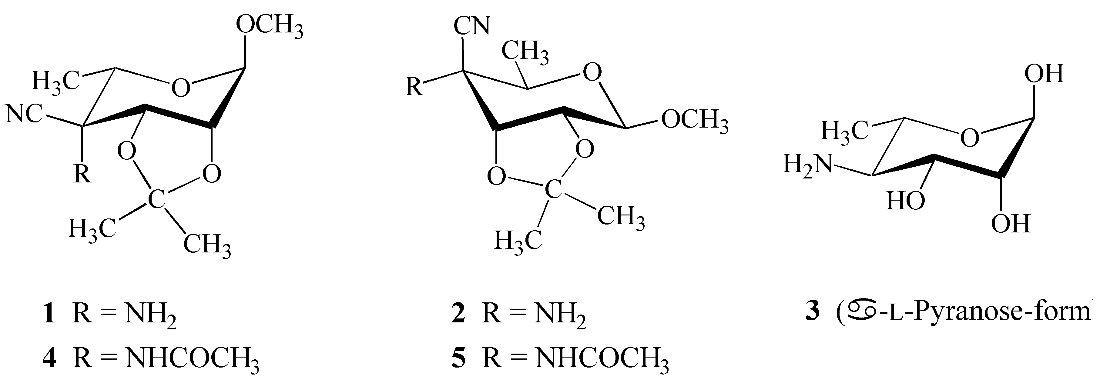
Crystal Structure of Methyl 4-amino-4-cyano-4,6-dideoxy-2,3-O-isopropylidene-α-L-talopyranoside

Abstract
:Introduction
Results and Discussion
Structure Elucidation
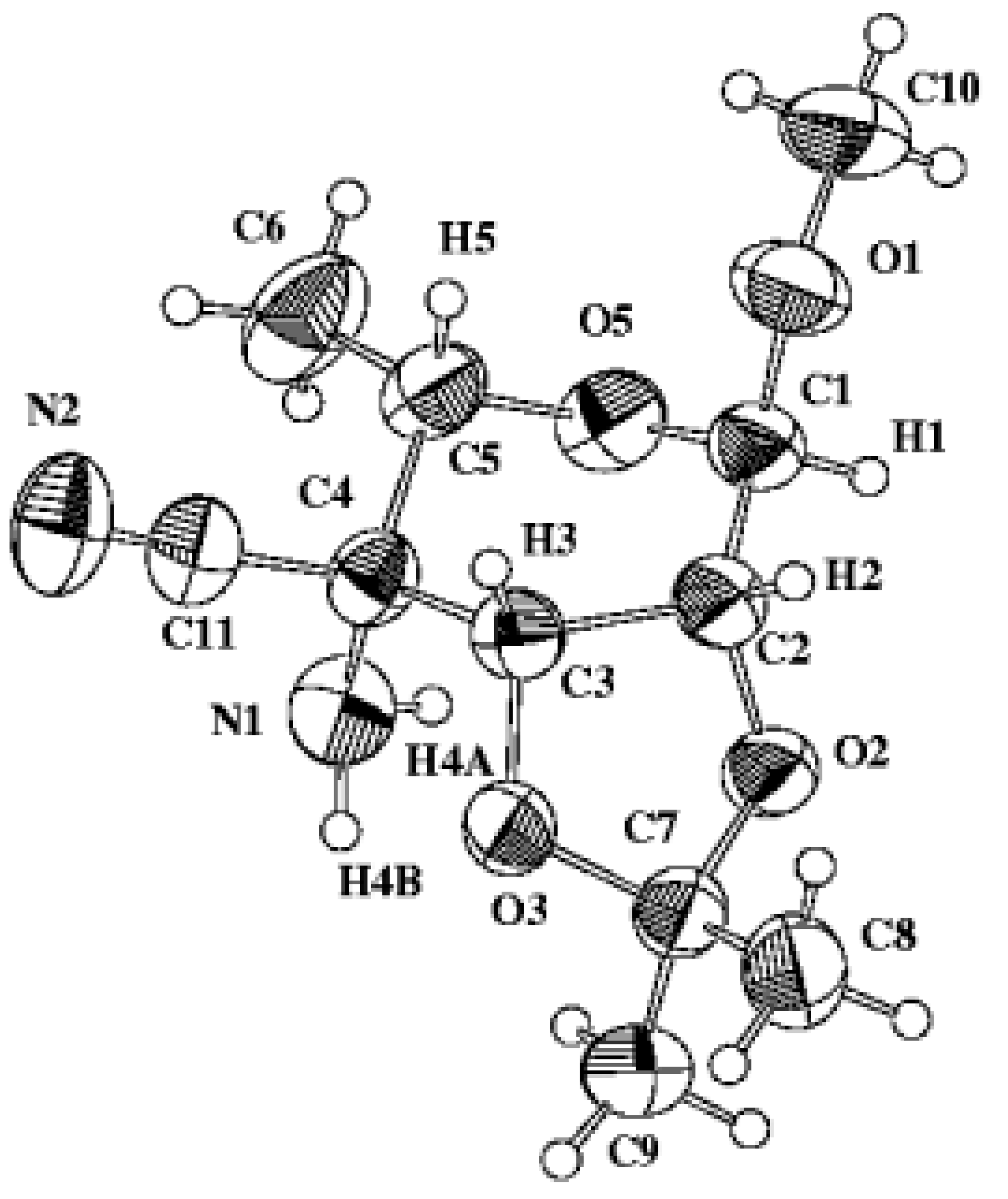
X-ray Analysis
Experimental
General
X-ray Analysis
Acknowledgements
References and Notes
- Steiner, B.; Koóš, M.; Langer, V.; Gyepesová, D.; Smrčok, Ľ. 4-Amino-4-cyano-4,6-dideoxy Sugar Derivatives from Methyl 6-deoxy-2,3-O-isopropylidene-α-l-lyxo-hexopyranosid-4-ulose via Strecker-type Reaction. Carbohydr. Res. 1998, 311, 1–9. [Google Scholar] [CrossRef]
- Koóš, M.; Steiner, B.; Gajdoš, J.; Langer, V.; Gyepesová, D.; Smrčok, Ľ; Ďurík, M. Crystal Structure of Methyl 4-acetamido-4-cyano-4,6-dideoxy-2,3-O-isopropylidene-β-d-allopyranoside. Molecules 2000, 5, 219–226. [Google Scholar] [CrossRef]
- Cremer, D.; Pople, J. A. A General Definition of Ring Puckering Coordinates. J. Am. Chem. Soc. 1975, 97, 1354–1358. [Google Scholar] [CrossRef]
- Köll, P.; Saak, W.; Pohl, S.; Steiner, B.; Koóš, M. Preparation and crystal and molecular structure of 6-O-[(2S)-2,3-epoxypropyl]-1,2:3,4-di-O-isopropylidene-α-d-galactopyranose. Pyranoid ring conformation in 1,2:3,4-di-O-isopropylidene-galactopyranose and related systems. Carbohydr. Res. 1994, 265, 237–248. [Google Scholar] [CrossRef]
- Boeyens, J.C.A. The conformation of six-membered rings. J. Cryst. Mol. Struct. 1978, 8, 317–320. [Google Scholar] [CrossRef]
- Hesse, M.; Meier, H.; Zeeh, B. Spectroscopic Methods in Organic Chemistry; Enders, D., Noyori, R., Trost, B.M., Eds.; Georg Thieme Verlag Stuttgart: New York, 1997; p. 108. [Google Scholar]
- Bruker AXS Inc. SHELXTL Version 5.10, Madison, Wisconsin: USA, 1997.
- Zsolnai, L.; Huttner, G. Program ZORTEP; University of Heidelberg: Germany, 1994. [Google Scholar]
- Sample Availability: The title compound is available from the corresponding author.


| Empirical formula | C11H18N2O4 |
| Formula weight | 242.27 |
| Temperature, T (K) | 296(2) |
| Wavelength, λ (Å) | 0.71073 |
| Crystal system | Trigonal |
| Space group | P32 |
| Unit cell dimensions (Å) | a = 10.29620(10) α = β = 90° |
| b = 10.29620(10) | |
| c = 10.8118(2) γ = 120° | |
| Volume, V (Å3) | 996.62(2) |
| Formula units per unit cell, Z | 3 |
| Calculated density, Dx (g cm−3) | 1.216 |
| Absorption coefficient, μ (mm−1) | 0.093 |
| F(000) | 390 |
| Crystal size (mm) | 1.00 (max) 0.21 (min) |
| Diffractometer) | Siemens SMART CCD |
| Theta range for data collection (°) | 2.28—27.17 |
| Index ranges | –13 ≤ h ≤ 13, –13 ≤ k ≤ 13, –13 ≤ l ≤ 13 |
| Reflections collected | 11057 |
| Independent reflections [ I > 2σ(I)] | 2902 (Rint = 0.0246) |
| Refinement method | Full-matrix least-squares |
| Minimization of | Σ w (|Fo| − |Fc|)2 |
| Data / parameters | 2902 / 182 |
| Goodness of fit (all) | 1.028 |
| Final R indices [I > 2σ(I)] | R1 = 0.0375 wR2 = 0.0897 |
| R indices (all data) | R1 = 0.0475 wR2 = 0.0963 |
| Largest diff. peak and hole | 0.086 and –0.149 (e Å−3) |
| O5–C1 | 1.406(2) | C2–C3–C4 | 114.65(14) |
| O5–C5 | 1.437(2) | O1–C1–O5 | 112.29(15) |
| O2–C2 | 1.419(2) | O1–C1–C2 | 106.30(15) |
| O2–C7 | 1.423(2) | O5–C1–C2 | 113.09(14) |
| O3–C3 | 1.420(2) | O2–C7–O3 | 104.82(12) |
| O3–C7 | 1.454(2) | O2–C7–C9 | 108.34(17) |
| O1–C1 | 1.404(2) | O3–C7–C9 | 109.91(16) |
| O1–C10 | 1.433(3) | O2–C7–C8 | 111.78(16) |
| C3–C2 | 1.530(2) | O3–C7–C8 | 109.10(17) |
| C3–C4 | 1.563(2) | C9–C7–C8 | 112.60(18) |
| N1–C4 | 1.450(2) | N2–C11–C4 | 176.6(2) |
| C1–C2 | 1.519(2) | O5–C5–C6 | 107.37(17) |
| C7–C9 | 1.505(3) | O5–C5–C4 | 106.70(13) |
| C7–C8 | 1.510(3) | C6–C5–C4 | 113.9(2) |
| C11–N2 | 1.136(3) | N1–C4–C11 | 107.76(16) |
| C11–C4 | 1.485(2) | N1–C4–C5 | 109.84(16) |
| C5–C6 | 1.510(3) | C11–C4–C5 | 108.38(15) |
| C5–C4 | 1.545(3) | N1–C4–C3 | 116.12(15) |
| C1–O5–C5 | 113.91(14) | C11–C4–C3 | 106.49(14) |
| C2–O2–C7 | 106.33(13) | C5–C4–C3 | 107.98(15) |
| C3–O3–C7 | 109.19(12) | O2–C2–C1 | 108.93(14) |
| C1–O1–C10 | 113.01(19) | O2–C2–C3 | 102.59(12) |
| O3–C3–C2 | 103.59(13) | C1–C2–C3 | 116.68(15) |
| O3–C3–C4 | 109.92(13) |
| C1–C2 – C3–C4 | 28.0(2) |
| H1–C1 – C2–H2 | 83.2 |
| H2–C2 – C3–H3 | 29.4 |
| C3–C4 – C5–O5 | −37.35(16) |
| C3–C4 – C5–C6 | −179.59(19) |
| O1–C1 – C2–O2 | −152.71(14) |
| O2–C2 – C3–O3 | 28.82(16) |
| C10–O1 – C1–O5 | −61.5(2) |
| C10–O1 – C1–C2 | 174.32(19) |
| O3–C3 – C4–C11 | 85.07(18) |
| Atom | x | y | z | U(eq) |
|---|---|---|---|---|
| O5 | 6111.7(13) | 247.3(16) | 4509.9(11) | 57.5(3) |
| O2 | 9554.1(13) | 2224.3(14) | 4448.5(11) | 53.4(3) |
| O3 | 9820.0(12) | 1926.3(14) | 6505.3(11) | 51.6(3) |
| O1 | 6342.0(17) | 2618.0(19) | 4597.9(14) | 70.8(4) |
| C3 | 8375.7(18) | 1759.9(18) | 6340.1(15) | 45.4(4) |
| H3 | 8203 | 2334 | 6977 | 50(5) |
| N1 | 7550(2) | –998.2(18) | 5960(2) | 59.9(4) |
| H4A | 7490(30) | –1010(30) | 5170(30) | 70(7) |
| H4B | 8550(40) | –690(40) | 6170(30) | 107(10) |
| C1 | 7050.2(19) | 1793(2) | 4307.8(17) | 52.2(4) |
| H1 | 7328 | 1948 | 3431 | 49(5) |
| C7 | 10646.0(18) | 2418(2) | 5348.3(17) | 54.8(4) |
| C11 | 6810(2) | –251(2) | 7778.6(18) | 57.1(4) |
| N2 | 6545(3) | –434(2) | 8805.6(18) | 80.0(5) |
| C5 | 5720(2) | –146(2) | 5785.1(18) | 57.7(5) |
| H5 | 5411 | 527 | 6153 | 64(6) |
| C4 | 7147.1(19) | 67.8(18) | 6441.0(15) | 47.7(4) |
| C2 | 8479.5(19) | 2465(2) | 5071.8(16) | 48.3(4) |
| H2 | 8874 | 3545 | 5170 | 54(5) |
| C6 | 4424(3) | –1738(3) | 5817(3) | 88.0(8) |
| H6A | 3566 | –1780 | 5427 | 119(11) |
| H6B | 4186 | –2064 | 6660 | 93(8) |
| H6C | 4696 | –2381 | 5383 | 89(9) |
| C9 | 11261(3) | 1410(3) | 5015(2) | 73.3(6) |
| H9A | 10447 | 406 | 4896 | 79(8) |
| H9B | 11895 | 1421 | 5670 | 97(9) |
| H9C | 11833 | 1761 | 4265 | 91(8) |
| C8 | 11857(2) | 4041(3) | 5468(2) | 75.1(7) |
| H8A | 12362 | 4392 | 4689 | 120(12) |
| H8B | 12565 | 4130 | 6087 | 86(8) |
| H8C | 11414 | 4632 | 5704 | 69(7) |
| C10 | 5039(3) | 2210(4) | 3857(3) | 94.5(9) |
| H10A | 5288 | 2233 | 2999 | 117(11) |
| H10B | 4692 | 2906 | 4001 | 110(11) |
| H10C | 4264 | 1217 | 4075 | 107(10) |
© 2000 by MDPI (http://www.mdpi.org). Reproduction is permitted for noncommercial purposes.
Share and Cite
Koóš, M.; Steiner, B.; Mičová, J.; Langer, V.; Ďurík, M.; Gyepesová, D.; Smrčok, Ľ. Crystal Structure of Methyl 4-amino-4-cyano-4,6-dideoxy-2,3-O-isopropylidene-α-L-talopyranoside. Molecules 2000, 5, 1113-1120. https://doi.org/10.3390/51001113
Koóš M, Steiner B, Mičová J, Langer V, Ďurík M, Gyepesová D, Smrčok Ľ. Crystal Structure of Methyl 4-amino-4-cyano-4,6-dideoxy-2,3-O-isopropylidene-α-L-talopyranoside. Molecules. 2000; 5(10):1113-1120. https://doi.org/10.3390/51001113
Chicago/Turabian StyleKoóš, Miroslav, Bohumil Steiner, Júlia Mičová, Vratislav Langer, Marián Ďurík, Dalma Gyepesová, and Ľubomír Smrčok. 2000. "Crystal Structure of Methyl 4-amino-4-cyano-4,6-dideoxy-2,3-O-isopropylidene-α-L-talopyranoside" Molecules 5, no. 10: 1113-1120. https://doi.org/10.3390/51001113