Cyclohexenonic Long-Chain Fatty Alcohols as Neuronal Growth Stimulators

1
Laboratoire de Chimie Organique des Substances Naturelles, UMR CNRS 7509, Centre de Neurochimie, Université Louis Pasteur, 5, rue Blaise Pascal, 67084 Strasbourg cedex, France
2
Laboratoire de Neurophysiologie Cellulaire et Integrée, UMR CNRS 7519, Université Louis Pasteur, Strasbourg, France
*
Author to whom correspondence should be addressed.
Molecules 2000, 5(12), 1439-1460; https://doi.org/10.3390/51201439
Submission received: 13 November 2000
/
Revised: 7 December 2000
/
Accepted: 7 December 2000
/
Published: 22 December 2000
Abstract
:Neurotrophic factors play an important role in the development and maintenance of neurons, thus providing a suitable therapeutic approach for the treatment of neurodegenerative diseases. However, their clinical use has revealed problematic because of a number of technical and biological disadvantages. Among the different strategies proposed to overcome such difficulties, the search for non-peptide substances with neurotrophic potential is giving promising results. Here we will expose major findings in this field, drawing special attention to cyclohexenonic long-chain fatty alcohols, a novel family of compounds that promote neuronal survival and neurite outgrowth.
- 1. Introduction
- 2. Neurotrophic compounds as therapeutic agents for neurodegenerative diseases
- 2.1. Natural neurotrophic factors
- 2.2. Non-peptide neurotrophic factor analogues
- 3. Long-chain fatty alcohols
- 4. Cyclohexenonic long-chain fatty alcohols
- 4.1. Synthesis of cyclohexenonic long-chain fatty alcohols
- 4.2. Biological activity of cyclohexenonic long-chain fatty alcohols
- 5. Conclusions
- 6. Experimental section: synthesis of t-CFAs
- 6.1. Materials and Methods
- 6.2. Synthesis of 1-(12-hydroxydodecyl-1-phenylsulfonyl)-2,6,6-trimethyl-1-cyclohexene
- 6.3. Synthesis of 1-(12-hydroxydodecyl)-2,6,6-trimethyl-1-cyclohexene
- 6.4. Synthesis of 1-(12-acetoxydodecyl)-2,6,6-trimethyl-1-cyclohexene
- 6.5. Synthesis of 3-(12-acetoxydodecyl)-2,4,4-trimethyl-2-cyclohexen-1-one
- 6.6. Synthesis of 3-(12-hydroxydodecyl)-2,4,4-trimethyl-2-cyclohexen-1-one
1. Introduction
Since the discovery of the nerve growth factor (NGF) in the 50’s [1], numerous neurotrophic factors (NFs) have been proven to play a crucial role in the survival and differentiation of discrete neuronal populations. The origin of these naturally occurring factors is diverse, including the neurotrophin, fibroblast growth factor, epidermal growth factor, insulin-like growth factor and transforming growth factor β families, cytokines, proteases, and protease inhibitors [2]. The fact that such substances are able to restrain neuronal loss has generated over the past several years a great interest in their therapeutic potential for the treatment of neurodegenerative diseases. However, the clinical use is at present restricted because of a number of technical and biological disadvantages. In this respect, the production of synthetic compounds that mimic the biological actions of natural NFs represents an attractive approach to bypass these difficulties. In this review, we will outline the limitations of NF therapies, and the effects of various non-peptide NF analogues. Specifically, we will focus on the chemistry and biological activity of cyclohexenonic long-chain fatty alcohols, a new family of compounds generated in our laboratory.
2. Neurotrophic compounds as therapeutic agents for neurodegenerative diseases
2.1. Natural neurotrophic factors
As mentioned above, the ability of naturally occurring NFs to promote the survival and development of neurons provides strong evidence for their therapeutic utilisation. Thus, an increasing number of preclinical and clinical studies have concerned the application of different NFs to the treatment of neurodegenerative disorders. In some cases, clinical trials have been conducted with variable success (Table 1). The main obstacle for using natural NFs as therapeutic agents resides in their chemical structure. They are hydrophilic, high-molecular weight proteins that can be rapidly degraded by endogenous peptidases, thus limiting to a large extent oral, subcutaneous or intravenous administration. In addition, they cannot cross the blood-brain barrier, which impedes severely the treatment of central nervous system (CNS) disorders. It should be also noted that systemic administration may provoke adverse side effects (i.e. injection site reactions, cough, asthenia, nausea, anorexia, weight loss) that worsen the situation of the patients, as has been illustrated by injecting recombinant human ciliary neurotrophic factor in patients affected of amyotrophic lateral sclerosis [3, 4].
To overcome these problems, diverse strategies have been considered. Intracranial injection of NFs allows them to reach directly specific brain areas. However, studies that used this aggressive, uncommon method to treat Alzheimer’s disease patients have reported uncertain results [5, 6].
In contrast, the local implantation of polymer-encapsulated cells genetically modified to produce and secrete NFs minimises the incidence of negative immune reactions, and makes possible a continuous delivery of the therapeutic agent [7, 8]. Likewise, injection into the affected area of recombinant adenoviruses that encodes for NFs also provides a local and constant delivery system, although strong inflammatory reactions can be expected [9, 10]. At present, these gene therapies, already tested in experimental models of neurodegenerative disorders, need further improvements to gain in applicability and reduced costs. An additional strategy to transport NFs into the CNS consists of fusing the therapeutic agent with a transferrin receptor antibody, thus forming a conjugate capable of crossing the blood-brain barrier [11]. Finally, small peptide sequences that contain the active site of NFs should be able to mimic their biological actions, and solve many of the difficulties resulting from the use of protein macromolecules [12, 13]. However, these techniques are still at a basic research level.
2.2. Non-peptide neurotrophic factor analogues
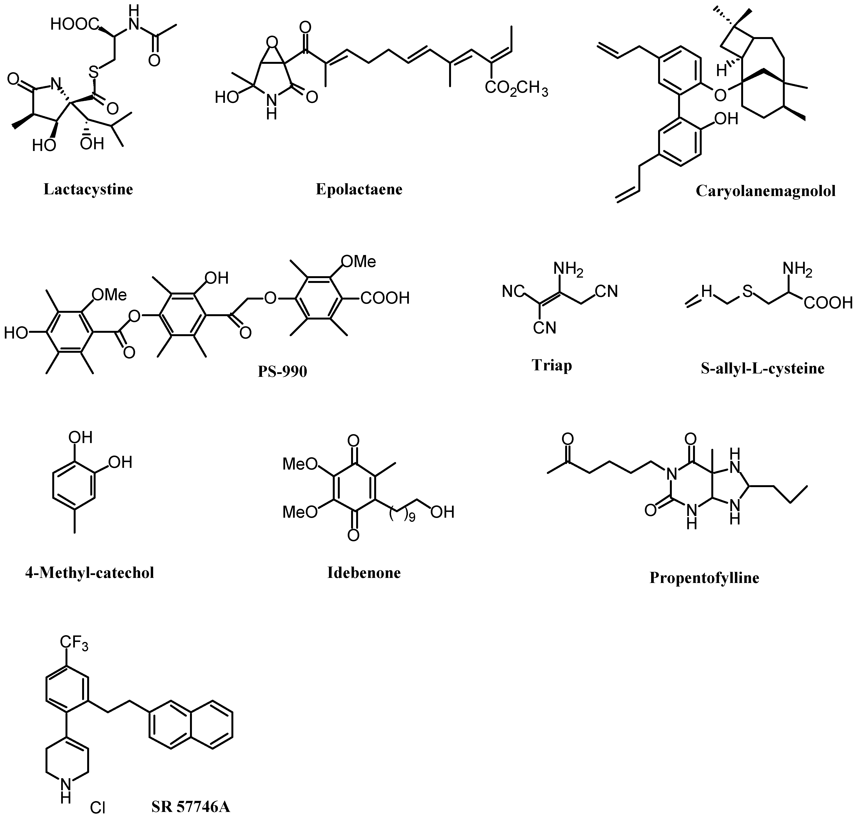
The synthesis or isolation from biological specimens of non-peptide organic compounds is emerging as a novel and promising approach in the treatment of neurodegenerative disorders. These substances, that either stimulate NF production or act directly on neurons to promote survival and/or neurite outgrowth, present a number of advantageous properties. In principle, they are low-molecular weight compounds that can be chemically manipulated to cross the blood-brain barrier, and to ameliorate their tolerance and pharmacokinetic parameters. In addition, large-scale production technologies may be applied. Over the last decade, diverse studies have reported the neurotrophic activity of numerous non-peptide molecules in vitro (Fig. 1). In some cases, they have also been tested in animal models of neurodegeneration, and in clinical trials. We will expose now the major results in this field.
A first group of substances possessing a direct neuritogenic action were originally isolated from microbial extracts. Thus, lactacystin, a sulphur-containing γ-lactam from Streptomyces sp. OM6519 [14], and PS-990, a metabolite of Acremonium sp. KY12702 [15], induce neurite sprouting in mouse neuroblastoma Neuro 2A cells. Moreover, epolactaene, an epoxide-containing γ-lactam from Penicillium sp. BM1689P, stimulates neuritogenesis in human neuroblastoma SH-SY5Y cells [16]. Interestingly, recent studies have suggested that the differentiating neuritogenic effect elicited by these compounds could result from their ability to prevent cell cycle progression, through as yet unclear mechanisms that include inhibition of either the multicatalytic protease complex proteasome [17] or DNA replication enzymes [18]. Plants commonly used in traditional medicine constitute another source of molecules with NF-like activity. Examples of these substances inducing neurite extension in primary cultures of foetal rat neurons are caryolanemagnolol, a sesquiterpene neolignan from the bark Magnolia obovata [19], and S-allyl-L-cysteine, an organosulphur compound with a thioallyl group from the garlic Allium sativum [20]. Finally, Triap (1,1,3-tricyano-2-amino-1-propene), a small molecule largely studied in the past, stimulates survival of sympathetic ganglion neurons of chick embryos, and neurite outgrowth in rat pheochromocytoma PC12 cells [21]. In vivo, Triap promotes the differentiation of cholinergic neurons after systemic administration [22]. At present, little is known about the mechanisms of action of all these compounds, and in particular further efforts must be done to evaluate their specific actions and toxic side effects under pathological conditions.
Astroglial cells participate actively in the homeostasis and neuronal plasticity of the CNS, by maintaining an appropriate extracellular medium, and producing a series of metabolic precursors, immunological mediators and NFs. Thus, substances that induce the synthesis and release of endogenous NFs by astroglial cells may present a valuable therapeutic potential. In this respect, the non-peptide compound SR57746A [1-[2-beta-naphthylethyl]-4-(3-trifluoromethylphenyl)-1,2,5,6-tetrahydropyridine hydrochloride] has been shown to exhibit both NF-like and NF-releasing properties. On the one hand, SR57746A stimulates, by itself or through potentiation of NGF activity, neuritogenesis and/or survival in several cell culture systems [23, 24, 25].
On the other hand, it increases NGF mRNA and protein contents in astrocytoma cells [26], and cultured astrocytes from neonatal rat cortex [27]. In vivo, SR57746A, either orally administered or injected, has demonstrated protective effects in a variety of experimental models of neurodegenerative disorders, such as transient global ischaemia, vincristine-induced septohippocampal lesion, sciatic nerve crushing, acrylamide-induced neuropathy [28], β-amyloid pepdide-induced memory deficit [29], progressive motor neuropathy [30], and motor neurone axotomy [31]. 4-Methylcatechol, a non-amine catechol analogue, has also been shown to induce the synthesis and secretion of NGF in mouse astroglial cells [32], and to protect neurons against experimental injury [33, 34, 35, 36, 37].
Alzheimer’s disease, the main cause of senile dementia, is a neurological disorder characterised by the presence of β-amyloid pepdide-containing plaques in specific brain areas, and the degeneration of cholinergic neurons, which are thought to be responsible for cognitive impairments [38]. Because early studies suggested that NGF may ameliorate this cholinergic dysfunction [39, 40], the use of NGF stimulators as potential therapeutic agents constitute a very valuable strategy in the treatment of Alzheimer’s disease. In this respect, two organic compounds, the coenzyme Q analogue idebenone [2,3-dimethoxy-5-methyl-6-(10-hydroxydecyl)-1,4-benzoquinone], and the alkylated xanthine derivative propentofylline [1-(5’-oxohexyl)-3-methyl-7-propylxanthine] have been the focus of basic and clinical research. These substances induce the synthesis and secretion of NGF in cultured mouse astroglial cells [41, 42], and numerous studies have reported their protective effects in experimentally-induced neurodegenerative disorders [43, 44, 45]. Specifically, both idebenone [46] and propentofylline [47] ameliorate learning and memory deficits provoked by β-amyloid pepdides. In addition, their ability to improve cognitive impairments, and delay the progression of Alzheimer’s disease has been evaluated in a number of clinical trials with promising results [48, 49, 50]. At present, however, growing evidence supports additional neuroprotective mechanisms distinct from stimulation of NGF synthesis and release. These mechanisms include inhibitioin of adenosine uptake by propentofylline [51], and a remarkable antioxidant activity of both molecules [52, 53].
3. Long-chain fatty alcohols
The curative capacity of plants is known for centuries, and people over the world uses commonly many of them for the treatment of diverse illnesses. Nowadays, research laboratories and pharmaceutical industries make continuous efforts to characterise novel vegetable substances of pharmacological application. Our early interest in this field led us to the search for non-peptide NF analogues in plants reported to heal wounds. Interestingly, the process of cutaneous wound healing requires the participation of a variety of growth factors that contribute actively to the repair and remodeling of the injured tissue [61]. In particular, NGF has been shown to be locally released in response to skin injury, and its topical application accelerates the rate of wound healing in both normal and diabetic mice [62]. Thus, one can expect that the ability of certain plants to cure wounds may reside in the existence of compounds with NF-like effects.
In 1987, we reported the in vitro neuritogenic activity of long-chain fatty alcohols isolated from the tropical plant Hygrophila erecta Hochr. (Acanthaceae), which is used in Vietnam and India to treat wounds. Among these alcohols, n-hexacosanol (Fig. 2; 1), containing 26 carbon atoms, was the most potent in inducing neuronal maturation. In contrast, n-tetracosanol and n-triacontanol were not effective, while n-octacosanol presented a slight effect. Similarly, saturated fatty acids were ten times less potent than the corresponding alcohols, thus supporting the specificity of the molecular structure in determining the biological activity [63]. In vivo, n-hexacosanol has been shown to promote the survival of cholinergic neurons after fimbria-fornix transection [64], and to reduce kainic acid-induced hippocampal damage [65].
Although these findings predict a potential use of n-hexacosanol as a therapeutic agent, an important limitation must be taken into account. Due to its highly lipophilic nature, n-hexacosanol is hardly soluble in both aqueous and organic solvents, which brings about considerable technical and biological difficulties.
In an attempt to improve the physicochemical properties of n-hexacosanol, we undertook the synthesis of new molecules that, resembling the structure of long-chain fatty alcohols, could manifest a neurotrophic activity. Because vitamin A (or retinol; Fig. 2; 2), which is involved in visual function, cell morphogenesis, differentiation and proliferation [66], bears a long-chain fatty alcohol in its structure, we used this molecule as starting-point for developing a series of compounds with longer, saturated hydrocarbon chains. These elongated analogues of perhydroretinol (Fig. 2; 3-7) have been proved to exhibit NF-like activity in different experimental paradigms. In vitro, 3, 4 and 5 promote the survival of cultured CNS neurons, and, in addition, 4 and 5 stimulate the proliferation of newborn rat astrocytes. In vivo, intraperitoneal administration of 3 and 4 reduce memory deficits provoked by lesion of the basal magnocellular nucleus. From a structural point of view, configuration studies revealed that the four diastereomers 7a, 7b, 7c and 7d present a similar neurite outgrowth-stimulating effect. Moreover, removal of the asymmetric methyl groups in the hydrocarbon chain showed that non-methylated perhydroretinols are as active as their methylated counterparts, which strongly suggests that the presence of methyl groups is not essential for the biological activity [67, 68, 69].
4. Cyclohexenonic long-chain fatty alcohols
4.1. Synthesis of cyclohexenonic long-chain fatty alcohols
From our findings on the neurotrophic action of perhydroretinol analogues, we considered the possibility of modifying the cyclohexene ring in order to evaluate structure/activity relationships, and further improve the efficiency of the molecules. Thus, a series of novel, neurotrophic compounds, known as cyclohexenonic long-chain fatty alcohols (CFAs; Fig. 3), were synthesised.
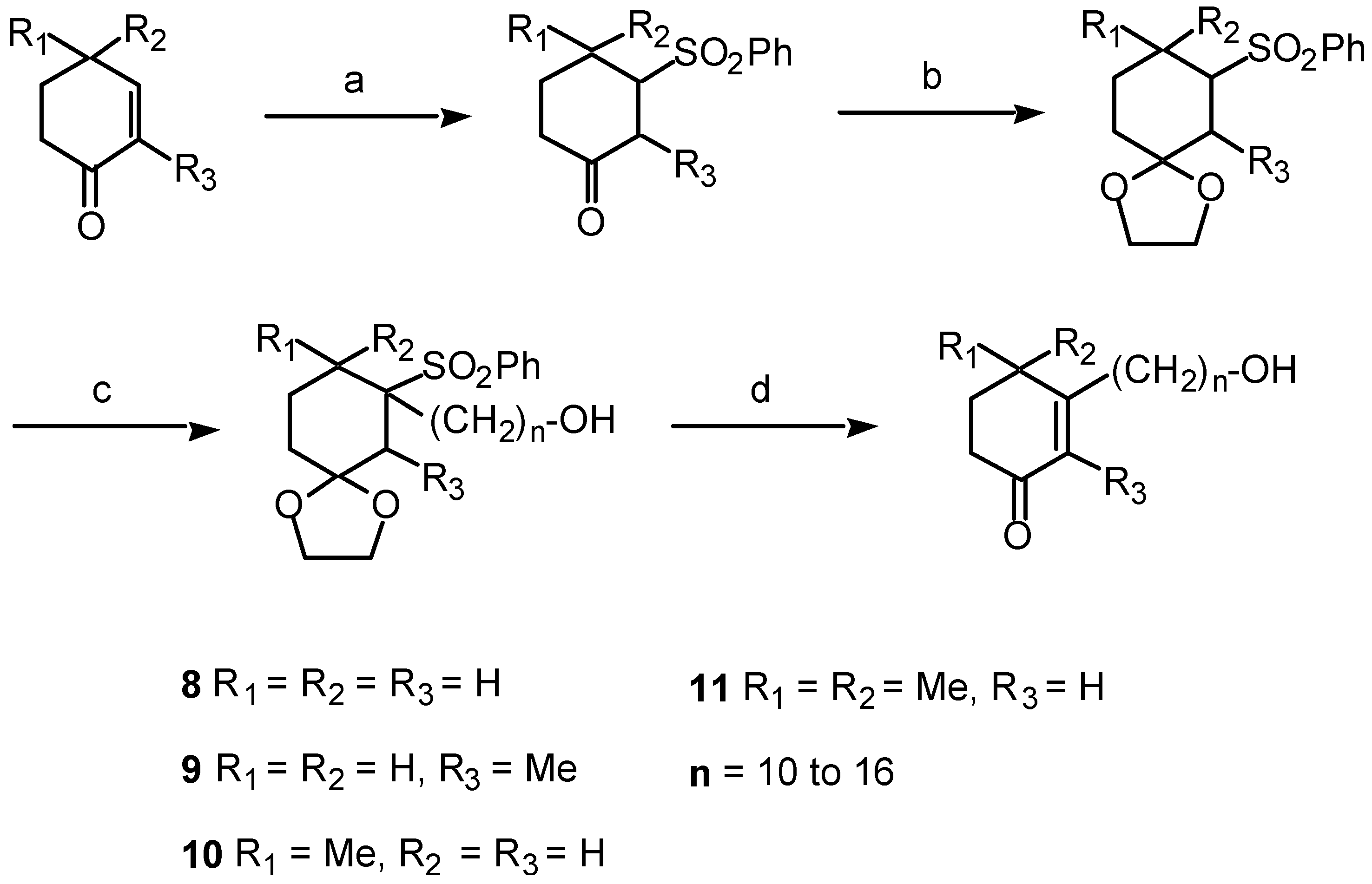
These CFAs contain basically a cyclohexenonic ring, and an ω-alkanol side chain ranging from 10 to 18 carbon atoms (Fig. 3; 8). In addition, the cyclic moiety may present one (2m- or 4m-CFA; Fig. 3; 9 and 10, respectively), two (dm-CFA; Fig. 3; 11), or three (t-CFA; Fig. 3; 12) methyl groups. The synthesis of the CFA series 8, 9, 10 and 11 was accomplished using the Umpolung strategy, which allows the inversion of the polarity in the cyclohexenone ring to introduce the hydrocarbon side chain by nucleophilic displacement (Fig. 4) [70, 71].
The synthesis of t-CFAs [3-(ω-hydroxy-alkyl)-2,4,4-trimethyl-2-cyclohexen-1-one; 12], according to the retrosynthetic analysis shown in Fig. 5, was carried out in seven steps from commercially available geranylbromide 13 (see Section 5 for details) [72]. Briefly, 13 reacted with benzenesulfinic acid sodium salt in methanol medium to give the corresponding geranylphenylsulfone 14, which was cyclised as described by Krischna et al. [73] and Torii et al. [74] to afford a mixture of 15 and 16 (85/15) in 90% yield (Fig. 6). These two isomers were separated by successive recrystallizations in hexane, and the desired 15 was obtained in 74% yield with a purity over 99%. Using a one-pot procedure, and n-butyllithium in the presence of hexamethylphosphoramide (HMPA), 15 was coupled with the unprotected ω-bromoalkanol to give 17 (79-88%). After reductive desulfonation by means of 6% Na/Hg amalgam [75] (88-93%), the resulting alcohol 18 was protected by an acetate to give 19 (97-98%), which was oxidized to the corresponding α,β-unsaturated ketone 20 using RuCl3 as catalyst (0,7% mol), and 70% tBuOOH [76] (49-53%). Deprotection of the alcohol by K2CO3 in methanol gave 12 in 81-96% yield [77] (Fig. 7).
4.2. Biological activity of cyclohexenonic long-chain fatty alcohols
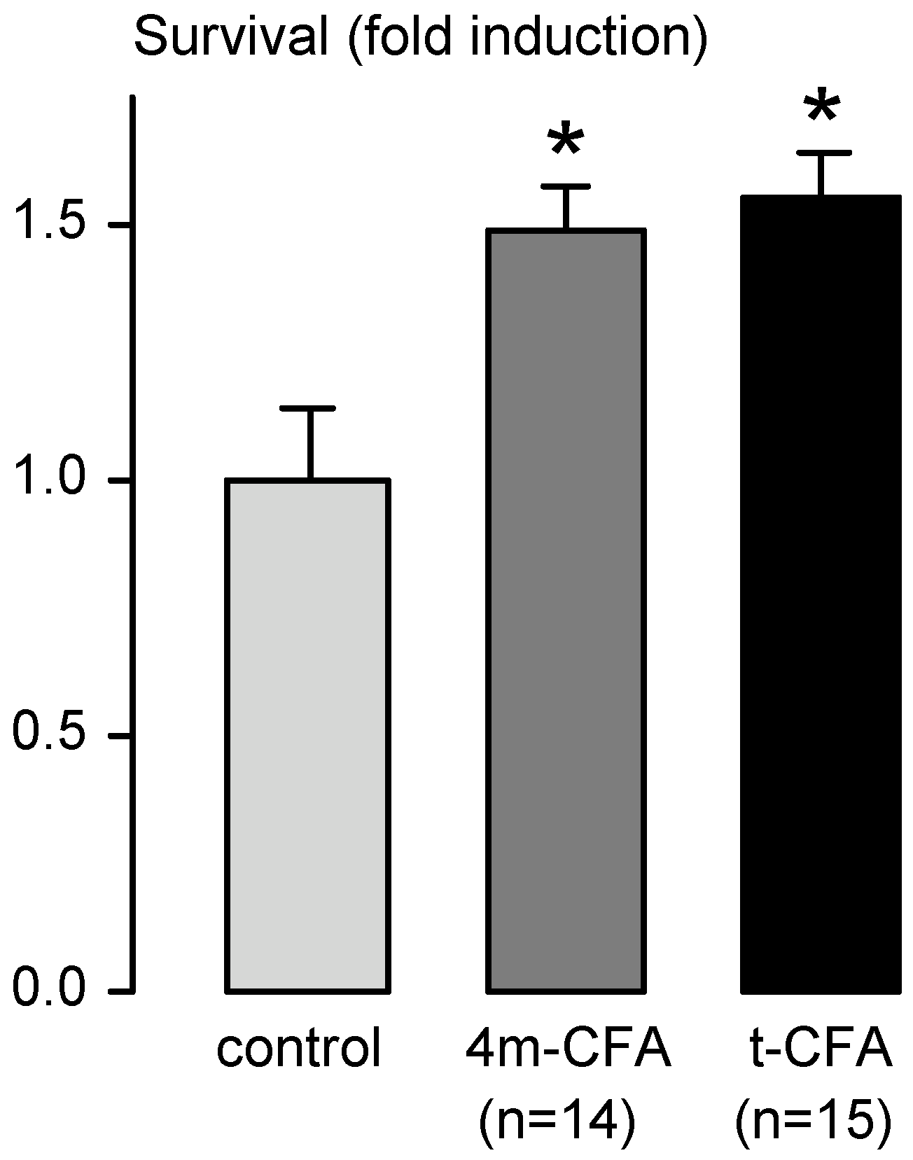
The neurotrophic action of the synthesised CFAs is first assessed by in vitro studies that serve as a means to evaluate their potential, and to establish the most appropriate structure/activity relationships. To this aim, and based on previous findings [78], we developed a low-density primary culture that uses neurons from foetal rat cerebral hemispheres, and allows analysis of both neuronal survival and neurite outgrowth. Fig. 8 illustrates an example of the protective action of two CFAs, 4m-CFA (n=14) and t- CFA (n=15), on the survival of cultured CNS neurons.
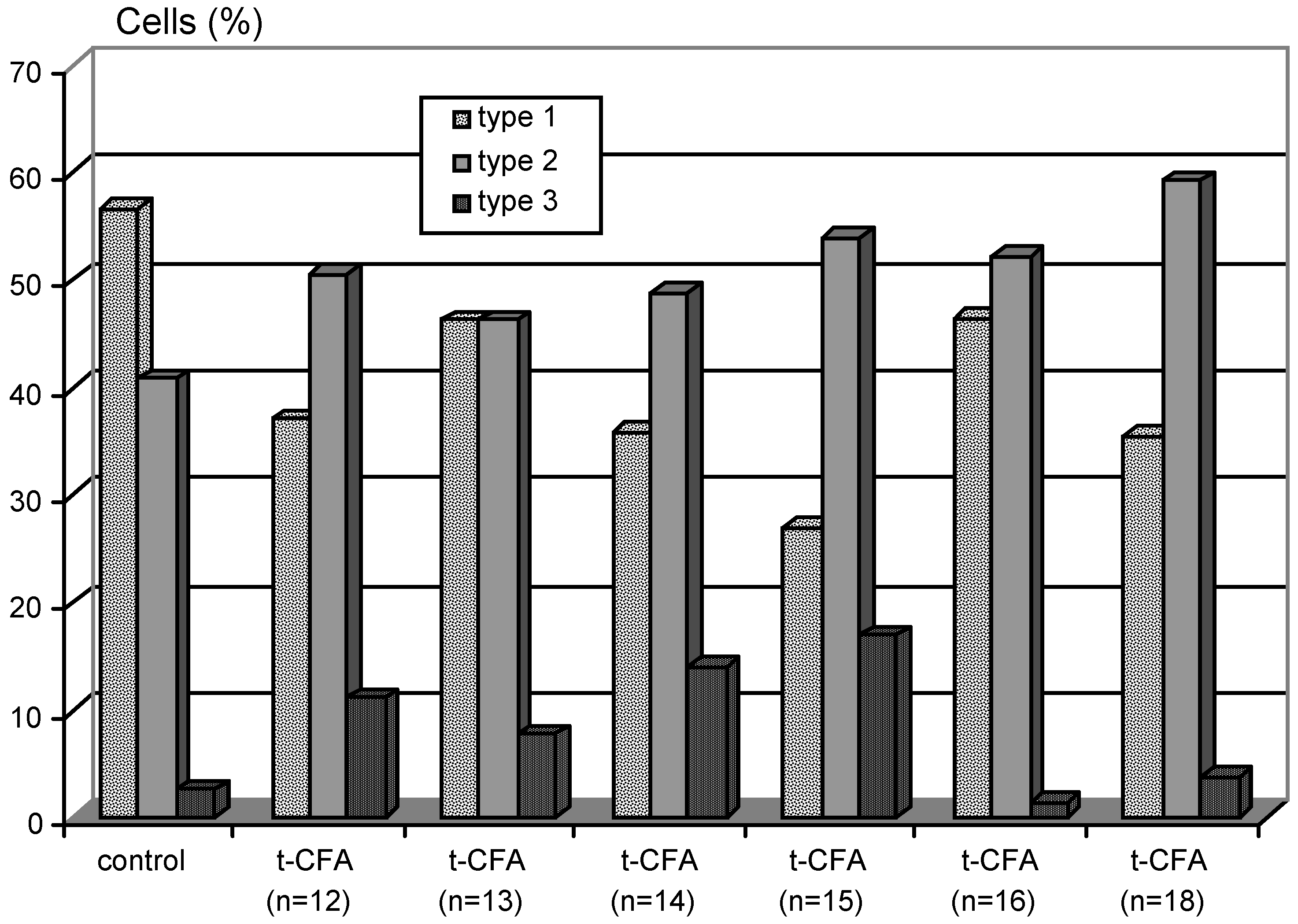
Concerning neuritogenesis, important considerations must be taken into account that support the existence of specificity in the stimulation of neurite sprouting by different CFAs. Thus, comparative studies on the neuritogenic effect elicited by the series of compounds 8, 9, 10, and 11 (Table 2) revealed that monomethylated CFAs with 13 or 14 carbon atoms in the aliphatic side chain were the most potent molecules, while non-methylated CFAs provoked toxicity. Similarly, in the series of t-CFAs (12), the hydrocarbon chains n=14 and n=15 seem to be the most active (Fig. 9). It is noteworthy that other studies dealing with the neurotrophic potential of non-peptide compounds have postulated that aliphatic side chains may potentiate both NF-like [79] and NF-releasing [41, 54, 80] activities. At present, the intracellular mechanisms underlying these structure requirements are still unknown, and further investigation is needed. Besides, the in vitro actions of CFAs provides evidence for their utilisation as neurotrophic factors in vivo, and suggest possible applications in the treatment of neurological disorders. Our preliminary findings on in vivo administration of CFAs in a transgenic mouse model of amyotrophic lateral sclerosis [81, 82] have shown that treated animals respond more rapidly to behavioural tasks (Fig. 10). New experiments are currently in progress.
5. Conclusions
In summary, the findings presented herein provide strong evidence that cyclohexenonic long-chain fatty alcohols exhibit a very promosing NF-like activity, thus establishing the basis for future investigations. Further research by testing this neurotrophic potential in experimental and transgenic models of neurodegenerative diseases is currently in progress, in order to evaluate possible therapeutic effects.
6. Experimental section: synthesis of t-CFAs
6.1. Materials and Methods
Tetrahydrofuran (THF) was distilled from sodium/benzophenone under argon atmosphere prior to use. Methanol, pyridine, benzene and HMPA were distilled from calcium hydride. All reactions involving moisture-sensitive reactants were executed under argon atmosphere using oven- and/or flame-dried glassware. 1H NMR spectra were recorded on a Bruker SY 200 spectrometer at 200 MHz as solutions in deuterochloroform (CDCl3). Chemical shifts are expressed in parts per million (ppm, ☐) downfield from tetramethylsilane (TMS), and are referenced to CHCl3 (7.26 ppm) as internal standard. Splitting patterns are designed as s (singlet), d (doublet), t (triplet), q (quartet), qn (quintuplet), m (multiplet), or br (broad). Coupling constants are given in hertz (Hz). 13C NMR spectra were recorded on the Bruker SY 200 spectrometer at 50 MHz as solutions in CDCl3. Chemical shifts are reported in ppm downfield from TMS, and are referenced to the centre line of CDCl3 (77.0 ppm) as internal standard. The attribution of the different carbons (C, CH, CH2, and CH3) was determined by 13C to 1H polarisation transfer (DEPT). IR spectra were recorded on sodium chloride using a Perkin-Elmer 881 FT-IR spectrometer, and are reported in wave numbers (cm-1). UV spectra were obtained in acetonitrile solution using a Kontron-Uvikon 810 UV-Vis spectrometer. Mass spectra (MS) were measured on a Trio 2000 apparatus by direct introduction [ionisation potential 70 eV; M/z relative intensities (in %) are noted in brackets]. GC chromatograms were obtained by dissolving the sample in ethyl acetate using a Carlo-Erba apparatus. Microanalyses were performed by the Service Central de Microanalyse du CNRS (Strasbourg). Routine chromatography monitoring of reactions was performed using 60F254 silica gel TLC plates, which were dipped in a solution of vanillin (1 g) in EtOH/H2SO4 (95/5), and heated on a hot plate.
6.2. Synthesis of 1-(12-hydroxydodecyl-1-phenylsulfonyl)-2,6,6-trimethyl-1-cyclohexene
To a solution containing sulfone 15 (1 g, 3.5 mmol, 2 eq.), and 4 mg triphenylmethane in dry THF (8 mL) was added n-butyllithium (4 mL, 1.4 M in hexane, 3 eq.). After stirring for 10 min at -78°C under argon atmosphere, the mixture was further stirred at room temperature. Then, HMPA (1.5 mL) was added, and after 90 min at room temperature, the mixture was recooled at -78°C. 11-Bromo-undecanol (439 mg, 1.75 mmol, 1 eq.) was added slowly, and the mixture was stirred for 180 min at - 20°C. After pouring into saturated NH4Cl (40 mL), the solution was extracted with ether. The organic layer was then washed with brine, dried with MgSO4, and concentrated in vacuo. The residue was purified by silica-gel chromatography, and eluted with hexane-AcOEt (8-2 to 6-4) to give 17 (n=12) as a white solid (622 mg, 79%). TLC: (hexane-AcOEt: 5-5) Rf=0.43; 1H-NMR (200 MHz), δ: 0.87 (s, 3H, H-19); 0.97 (s, 3H, H-20); 1.16 (s br, 14H, H-10 to H-16); 1.2-1.57 (m, 8H, H-4, 5, 9, 17); 1.94 (s, 3H, H-21); 1.98-2.25 (m, 4H, H-8, 3); 3.61 (t, J=6.8 Hz, 1H, H-18); 3.71 (t, J=6.8 Hz, 1H, H-7); 7.48-7.65 (m, 3H, H ar-3', 4'); 7.86-7.92 (m, 2H, H ar-2'); 13C-NMR (50 MHz), δ: 19 (C-4); 23 (C-21); 25.7 (C-16); 28.5 (C-8); 28.9 (C-19, 20); 29.4 (C-9 to C-15); 31.2 (C-17); 32.7 (C-3); 35.9 (C-6); 39.8 (C-5); 63 (C-18); 67.9 (C-7); 128.4 (C ar-2'); 130.5 (C-2); 133 5 (C ar-4'); 137.8 (C-1); 141.8 (C ar-1'); m.p.: 77-78°C; microanalysis (%): calcd for C27H44O3S (448.7028) C: 72.27, H: 9.88, O: 10.7; found C: 72.19, H: 9.92, O: 11.09. Coupling with the other hydrocarbon side chains from n=13 to 18 is shown in Table 3.
6.3. Synthesis of 1-(12-hydroxydodecyl)-2,6,6-trimethyl-1-cyclohexene
To a solution of 17 (n=12) (579 mg, 1.29 mmol, 1 eq.) in dry methanol (25 mL) was added Na2HPO4 (366 mg, 2 eq.), and mercury-sodium amalgam (6% Na, 4 g) at 0°C under argon atmosphere. The heterogeneous mixture was stirred for 240 min at room temperature, and then quenched with 1N HCl. After extraction with ether (3 times), the solution was washed with saturated NaHCO3, dried with MgSO4, and concentrated in vacuo. The residue was purified by silica-gel chromatography, and eluted with hexane-AcOEt (8-2-) to give 18 (n=12) as a colorless oil (350 mg, 88%). TLC: (hexane-AcOEt: 5-5) Rf=0.59; GC: 40-280°C (20°C/min) 10.50 min; 1H-NMR (200 MHz), δ: 0.96 (s, 6H, H-19, 20); 1.27 (s br, 16H, H-9 to H-16); 1.35-1.54 (m, 8H, H-4, 5, 8, 17); 1.57 (s, 3H, H-21); 1.83-2.03 (m, 4H, H-3, 7); 3.62 (t, J=6.5 Hz, 2H, H-18); 13C-NMR (50 MHZ), δ: 19.6 (C-4); 19.8 (C-21); 25.7 (C-16); 28.6 (C-19,20); 28.9 (C-8); 29.6 (C-9 to C-15); 30.5 (C-17); 32.8 (C-3*); 32.81 (C-7*); 34.8 (C-6); 39.8 (C-5); 63 (C-18); 126.3 (C-2); 137.8 (C-1); IR ν: 3330 (broad, O-H); 2925, 2852 (w, C-H); 1466 (s, C-H); 1112 (s, C-O).
6.4. Synthesis of 1-(12-acetoxydodecyl)-2,6,6-trimethyl-1-cyclohexene
To a solution of 18 (n=12) (316 mg, 1.026 mmol) was added acetic anhydride (7 mL), and pyridine (7 mL). The mixture was stirred for 60 min at room temperature, and quenched with 5% HCl. The mixture was then extracted with ether, washed with water, dried with MgSO4, and concentrated in vacuo to obtain 19 (n=12) as a colorless oil (353 mg, 98%). TLC: (hexane-AcOEt: 5-5) Rf=0.75; GC: 40-280°C (20°/min) 11.02; 1H-NMR (200 MHz), δ: 0.96 (s, 6H, H-19, 20); 1.27 (s br, 16H, H-9 to H-16); 1.35-1.54 (m, 8H, H-4, 5, 8, 17); 1.57 (s, 3H, H-21); 1.83-2.02 (m, 4H, H-3, 7); 2.04 (s, 3H, CH3-COO); 4.04 (t, J=6.6 Hz, 2H, H-18); 13C-NMR (50 MHz), δ: 19.5 (C-4); 19.8 (C-21); 20.9 (CH3-COO); 25.9 (C-16); 28.6 (C-19, 20); 28.9 (C-8); 29.6 (C-9 to C-15); 30.5 (C-17); 32.7 (C-7*); 32.75 (C-3*); 34.8 (C-6); 39.9 (C-5); 64.6 (C-18); 126.3 (C-2); 137.8 (C-1); 171.2 (CH3-COO); IR ν: 2925, 2854 (w, C-H); 1744 (w, C=O); 1466 (s, C-H); 1238 (w, C-O).
6.5. Synthesis of 3-(12-acetoxydodecyl)-2,4,4-trimethyl-2-cyclohexen-1-one
To a solution containing 19 (n=12) (321 mg, 0.92 mmol, 1 eq.) in cyclohexane (6 mL) was added water (0.8 mL), ruthenium trichloride hydrate (1.3 mg, 0.7% mol), and 70% t-BuOOH (1.26 mL, 10 eq.). The solution was stirred for 360 min at room temperature, filtered through a pad of Celite, and poured into 10% Na2SO3. The solution was then extracted with ether, washed with brine, dried with MgSO4, and concentrated in vacuo. The residue was purified by silica-gel chromatography, and eluted with hexane-AcOEt (95-15 to 90-10) to give 20 (n=12) as a colorless oil (227 mg, 53%). TLC: (hexane-AcOEt: 7-3) Rf=0.50; GC: 40-280°C (20°C/min) 12.2 min, 99%; 1H-NMR (200 MHz), δ: 1.13 (s, 6H, H-19, 20); 1.26 (s br, 16H, H-9 to H-16); 1.35-1.69 (m, 4H, H-8, 17); 1.73 (s, 3H, H-21); 1.78 (t, J=7.5 Hz, 2H, H-5); 2.02 (s, 3H, CH3-COO); 2.11-2.19 (m, 2H, H-7); 2.43 (t, J=6.8 Hz, 2H, H-6); 4.03 (t, J=6.8 Hz, 2H, H-18); 13C-NMR (50 MHz), δ: 11.5 (C-21); 20.9 (CH3-COO); 25.8 (C-16); 26.8 (C-19, 20); 28.8 (C-8); 29.1 (C-17); 29.5 (C-9 to C-15); 30.45 (C-7); 34.2 (C-5); 36.2 (C-4); 37.3 (C-6); 64.5 (C-18); 130.5 (C-2); 165 (C-3); 171 (CH3-COO); 199 (C-1); IR ν: 2925, 2854 (w, C-H); 1741 (w, C=O); 1667 (w, C=O); 1607 (s, C=C); 1468 (s, C-H); 1239 (w, C-O).
6.6. Synthesis of 3-(12-hydroxydodecyl)-2,4,4-trimethyl-2-cyclohexen-1-one
To a solution of 20 (n=12) (132 mg, 0.36 mmol, 1 eq.) in dry methanol (8 mL) was added 3 drops of water, and K2CO3 (74 mg, 0.54 mmol, 1.5 eq.). After stirring for 150 min at room temperature, the solution was neutralized at pH=7 with 5% HCl. Then, the solution was extracted with ether, dried with MgSO4, and concentrated in vacuo. The residue was purified by silica-gel chromatography, and eluted with hexane-AcOEt (8-2 to 7-3) to give 12 (n=12) as a colorless oil (94 mg, 81%). TLC: (hexane- AcOEt: 7-3) Rf=0.2; GC: 40-280°C (20°/min) 13.94 min, 99%; 1H-NMR (200 MHz), δ: 1.13 (s, 6H, H-19, 20); 1.26 (s br, 16H, H-9 to H-16); 1.35-1.59 (m, 4H, H-8, 17); 1.73 (s, 3H, H-21); 1.77 (t, J=7.5 Hz, 2H, H-5); 2.11-2.19 (m, 2H, H-7); 2.43 (t, J=6.8 Hz, 2H, H-6); 3.61 (t, J = 6.5 Hz, 2H, H-18); 13C-NMR (50 Mhz), δ: 11.4 (C-21); 25.7 (C-16); 26.8 (C-19, 20); 28.8 (C-8); 29.5 (C-9 to C-15); 30.45 (C-7); 32.7 (C-17); 34.2 (C-5); 36.2 (C-4); 37.3 (C-6); 62.9 (C-18); 130.4 (C-2); 165.4 (C-3); 199 (C-1); IR ν: 3440 (broad OH); 2925,2853 (w, C-H); 1666 (w, C=O); 1605 (s, C=C); 1467 (s, C-H). UV λmax: 244.5 nm (ε 10420); microanalysis (%): calcd for C21H38O2 (322.530) C: 78.02, H: 11.87; found C: 78.34, H: 12.03.
Acknowledgements:
This study was supported by Meiji Milk Products Co., LTD. J.L.G.A. is the recipient of a grant Aide aux Etudes from the Association Française contre les Myopathies.
References
- Levi-Montalcini, R. Science 1987, 237, 1154–1162. [PubMed]
- Lindsay, R.M.; Wiegand, S.J.; Altar, C.A.; DiStefano, P.S. Trends Neurosci. 1994, 17, 182–190. [PubMed]
- Miller, R.G.; Bryan, W.W.; Dietz, M.A.; Munsat, T.L.; Petajan, J.H.; Smith, S.A.; Goodpasture, J.C. Neurology 1996, 47, 1329–1331. [PubMed]
- Miller, R.G.; Petajan, J.H.; Bryan, W.W.; Armon, C.; Barohn, R.J.; Goodpasture, J.C.; Hoagland, R.J.; Parry, G.J.; Ross, M.A.; Stromatt, S.C. Annu. Neurol. 1996, 39, 256–260.
- Seiger, A.; Nordberg, A.; von Holst, H.; Backman, L.; Ebendal, T.; Alafuzoff, I.; Amberla, K.; Hartvig, P.; Herlitz, A.; Lilja, A. Behav. Brain Res. 1993, 57, 255–261.
- Eriksdotter, J.M.; Nordberg, A.; Amberla, K.; Backman, L.; Ebendal, T.; Meyerson, B.; Olson, L.; Seiger, A.; Shigeta, M.; Theodorsson, E.; Viitanen, M.; Winblad, B.; Wahlund, L.O. Dement. Geriatr. Cogn. Disord. 1998, 9, 246–257.
- Maysinger, D.; Piccardo, P.; Liberini, P.; Jalsenjak, I.; Cuello, C. Neurochem. Int. 1994, 24, 495–503.
- Aebischer, P.; Schluep, M.; Deglon, N.; Joseph, J.M.; Hirt, L.; Heyd, B.; Goddard, M.; Hammang, J.P.; Zurn, A.D.; Kato, A.C.; Regli, F.; Baetge, E.E. Nat. Med. 1996, 2, 696–699. [CrossRef] [PubMed]
- Bilang-bleuel, A.; Revah, F.; Colin, P.; Locquet, I.; Robert, J.J.; Mallet, J.; Horellou, P. Proc. Natl. Acad. Sci. USA 1997, 94, 8818–8823. [CrossRef]
- Haase, G.; Kennel, P.; Pettmann, B.; Vigne, E.; Akli, S.; Revah, F.; Schmalbruch, H.; Kahn, A. Nat. Med. 1997, 3, 429–436.
- Friden, P.M.; Walus, L.R.; Watson, P.; Doctrow, S.R.; Kozarich, J.W.; Backman, C.; Bergman, H.; Hoffer, B.; Bloom, F.; Granholm, A.C. Science 1993, 259, 373–377. [PubMed]
- LeSauteur, L.; Wei, L.; Gibbs, B.F.; Saragovi, H.U. J. Biol. Chem. 1995, 270, 6564–6569.
- Xie, Y.; Tisi, M.A.; Yeo, T.T.; Longo, F.M. J. Biol. Chem. 2000, 275, 29868–29874. [CrossRef] [PubMed]
- Omura, S.; Fujimoto, T.; Otoguro, K.; Matsuzaki, K.; Moriguchi, R.; Tanaka, H.; Sasaki, Y. J. Antibiot. 1991, 44, 113–116. [CrossRef]
- Toki, S.; Ando, K.; Yoshida, M.; Matsuda, Y. J. Antibiot. 1994, 47, 1175–1181. [CrossRef]
- Kakeya, H.; Takahashi, I.; Okada, G.; Isono, K.; Osada, H. J. Antibiot. 1995, 48, 733–735. [CrossRef] [PubMed]
- Fenteany, G.; Schreiber, S.L. J. Biol. Chem. 1998, 273, 8545–8548. [CrossRef]
- Mizushina, Y.; Kobayashi, S.; Kuramochi, K.; Nagata, S.; Sugawara, F.; Sakaguchi, K. Biochem. Biophys. Res. Commun. 2000, 273, 784–788. [CrossRef] [PubMed]
- Fukuyama, Y.; Otoshi, Y.; Miyoshi, K.; Nakamura, K.; Kodama, M.; Nagasawa, M.; Hasegawa, T.; Okazaki, H.; Sugawara, M. Tetrahedron 1992, 48, 377–392. [CrossRef]
- Moriguchi, T.; Matsuura, H.; Kodera, Y.; Itakura, Y.; Katsuki, H.; Saito, H.; Nishiyama, N. Neurochem. Res. 1997, 22, 1449–1452.
- Paul, J.W.; Quach, T.T.; Duchemin, A.M.; Schrier, B.K.; DaVanzo, J.P. Dev. Brain Res. 1990, 55, 21–27.
- Paul, J.W.; DaVanzo, J.P. Brain Res. Dev. Brain Res. 1992, 67, 113–120.
- Pradines, A.; Magazin, M.; Schiltz, P.; Le Fur, G.; Caput, D.; Ferrara, P. J. Neurochem. 1995, 64, 1954–1964. [CrossRef]
- Magazin, M.; Schiltz, P.; Zachayus, J.L.; Cavrois, E.; Caput, D.; Ferrara, P. Brain Res. Mol. Brain Res. 1998, 53, 301–306. [CrossRef]
- Duong, F.H.; Warter, J.M.; Poindron, P.; Passilly, P. Br. J. Pharmacol. 1999, 128, 1385–1392.
- Gauthier, T.; Bornia, J.; Reith, Y.; Keane, P.E.; Le Fur, G.; Soubrie, P. Fund. Clin. Parmacol. 1993, 7, 359.
- Labie, C.; Lafon, C.; Marmouget, C.; Saubusse, P.; Fournier, J.; Keane, P.E.; Le Fur, G.; Soubrie, P. Br. J. Pharmacol. 1999, 127, 139–144.
- Fournier, J.; Steinberg, R.; Gauthier, T.; Keane, P.E.; Guzzi, U.; Coude, F.X.; Bougault, I.; Maffrand, J.P.; Soubrie, P.; Le Fur, G. Neuroscience 1993, 55, 629–641. [PubMed]
- Terranova, J.P.; Kan, J.P.; Storme, J.J.; Perreaut, P.; Le Fur, G.; Soubrie, P. Neurosci. Lett. 1996, 213, 79–82.
- Duong, F.H.; Fournier, J.; Keane, P.E.; Guenet, J.L.; Soubrie, P.; Warter, J.M.; Borg, J.; Poindron, P. Br. J. Pharmacol. 1998, 124, 811–817.
- Iwasaki, I.; Shiojima, T.; Kinoshita, M.; Ikeda, K. J. Neurol. Sci. 1998, 160, 92–96. [CrossRef]
- Furukawa, Y.; Tomioka, N.; Sato, W.; Satoyoshi, E.; Hayashi, K.; Furukawa, S. FEBS Lett. 1989, 247, 463–467. [PubMed]
- Hanaoka, Y.; Ohi, T.; Furukawa, S.; Furukawa, Y.; Hayashi, K.; Matsukura, S. J. Neurol. Sci. 1994, 122, 28–32. [CrossRef]
- Kaechi, K.; Ikegami, R.; Nakamura, N.; Nakajima, M.; Furukawa, Y.; Furukawa, S. J. Pharmacol. Exp. Ther. 1995, 272, 1300–1304. [PubMed]
- Saita, K.; Ohi, T.; Hanaoka, Y.; Furukawa, S.; Furukawa, Y.; Hayashi, K.; Matsukura, S. Neurotoxicology 1995, 16, 403–412. [PubMed]
- Saita, K.; Ohi, T.; Hanaoka, Y.; Furukawa, S.; Furukawa, Y.; Hayashi, K.; Matsukura, S. J. Pharmacol. Exp. Ther. 1996, 276, 231–237.
- Kimura, N.; Nishizaki, K.; Orita, Y.; Masuda, Y. Acta Otolaryngol. Suppl. 1999, 540, 12–15.
- Coyle, J.T.; Price, D.L.; Delong, M.R. Science 1983, 219, 1184–1190. [PubMed]
- Hefti, F. J. Neurosci. 1986, 6, 2155–2162.
- Olson, L.; Nordberg, A.; von Holst, H.; Backman, L.; Ebendal, T.; Alafuzoff, I.; Amberla, K.; Hartvig, P.; Herlitz, A.; Lilja, A.; Lundqvist, H.; Langstrom, B.; Meyerson, B.; Persson, A.; Viitanen, M.; Winblad, B.; Seiger, A. J. Neural Transm. Park. Dis. Dement. Sec. 1992, 4, 79–95. [CrossRef]
- Takeuchi, R.; Murase, K.; Furukawa, Y.; Furukawa, S.; Hayashi, K. FEBS Lett. 1990, 261, 63–66. [PubMed]
- Shinoda, I.; Furukawa, Y.; Furukawa, S. Biochem. Pharmacol. 1990, 39, 1813–1816.
- Rego, A.C.; Santos, M.S.; Oliveira, C.R. Free Radic. Biol. Med. 1999, 26, 1405–1417.
- Fuji, K.; Hiramatsu, M.; Kameyama, T.; Nabeshima, T. Eur. J. Pharmacol. 1993, 236, 411–417.
- Haag, P.; Schneider, T.; Schabitz, W.R.; Hacke, W. J. Neurol. Sci. 2000, 175, 52–56. [CrossRef]
- Yamada, K.; Tanaka, T.; Han, D.; Senzaki, K.; Kameyama, T.; Nabeshima, T. Eur. J. Neurosci. 1999, 11, 83–90.
- Yamada, K.; Tanaka, T.; Senzaki, K.; Kameyama, T.; Nabeshima, T. Eur. J. Pharmacol. 1998, 349, 15–22.
- Gutzmann, H.; Hadler, D. J. Neural Transm. Suppl. 1998, 54, 301–310.
- Rother, M.; Erkinjuntti, T.; Roessner, M.; Kittner, B.; Marcusson, J.; Karlsson, I. Dement. Geriatr. Cogn. Disord. 1998, 9, 36–43. [CrossRef]
- Mielke, R.; Moller, H.J.; Erkinjuntti, T.; Rosenkranz, B.; Rother, M.; Kittner, B. Alzheimer Dis. Assoc. Disord. 1998, 12, 29–35.
- Parkinson, F.E.; Paterson, A.R.P.; Young, J.D.; Cass, C.E. Biochem. Pharmacol. 1993, 46, 891–896.
- Mordente, A.; Martorana, G.E.; Minotti, G.; Giardina, B. Chem. Res. Toxicol. 1998, 11, 54–63. [CrossRef] [PubMed]
- Banati, R.B.; Schubert, P.; Rothe, G.; Gehrmann, J.; Rudulphi, K.; Valet, G.; Kreutzberg, G.W. J. Cereb. Blood Flow Metab. 1994, 14, 145–149. [CrossRef] [PubMed]
- Kawagishi, H.; Ando, M.; Sakamoto, H.; Yoshida, S.; Ojima, F.; Ishiguro, Y.; Ukai, N.; Furukawa, S. Tetrahedron Lett. 1991, 32, 4561–45-64.
- Yamaguchi, K.; Tsuji, T.; Wakuri, S.; Yazawa, K.; Kondo, K.; Higemori, H.; Kobayashi, J. Biosci. Biotech. Biochem. 1993, 57, 195–199. [CrossRef] [PubMed]
- Doi, Y.; Shigemori, H.; Ishibashi, M.; Mizobe, F.; Kawashima, A.; Nakaike, S.; Kobayashi, J. Chem. Pharm. Bull. 1993, 41, 2190–2191. [CrossRef]
- Yamaguchi, K.; Sasano, A.; Urakami, T.; Tsuji, T.; Kondo, K. Biosci. Biotech. Biochem. 1993, 57, 1231–1233. [CrossRef] [PubMed]
- Kawagishi, H.; Shimada, A.; Hosokawa, S.; Mori, H.; Sakamoto, H.; Ishiguro, Y.; Sakemi, S.; Bordner, J.; Kojima, N.; Furukawa, S. Tetrahedron Lett. 1996, 37, 7399–7402.
- Kawagishi, H.; Ishiyama, D.; Mori, H.; Sakamoto, H.; Ishiguro, Y.; Furukawa, S.; Li, J. Phytochemistry 1997, 45, 1203–1205. [PubMed]
- Kita, T.; Takaya, Y.; Oshima, Y. Tetrahedron 1998, 54, 11877–11886.
- Martin, P. Science 1997, 276, 75–81. [PubMed]
- Matsuda, H.; Koyama, H.; Sato, H.; Sawada, J.; Itakura, A.; Tanaka, A.; Matsumoto, M.; Konno, K.; Ushio, H.; Matsuda, K. J. Exp. Med. 1998, 187, 297–306. [CrossRef]
- Borg, J.; Toazara, J.; Hietter, H.; Henry, M.; Schmitt, G.; Luu, B. FEBS Lett. 1987, 213, 406–410. [PubMed]
- Borg, J.; Kesslak, P.J.; Cotman, C.W. Brain Res 1990, 518, 295–298. [PubMed]
- Borg, J. J. Neurosci. Res. 1991, 29, 62–67. [CrossRef] [PubMed]
- Dawson, M.I. Curr. Pharm. Des. 2000, 6, 311–325.
- Borg, J. International Patent WO91/05754, 1991. PCT/FR90/00742 (15/10/90).
- Luu, B.; Borg, J.; Beck, A.; Laabich, A.; Crestani, F.; Neveux, O. International Patent WO94/11343, 1994. PCT/FR93/01059 (28/10/93).
- Keyling-Bilger, F.; Schmitt, G.; Beck, A.; Luu, B. Tetrahedron 1996, 52, 14891–14904.
- Girlanda-Junges, C.; Keyling-Bilger, F.; Schmitt, G.; Luu, B. Tetrahedron 1998, 54, 7735–7748.
- Luu, B.; Schmitt, G.; Keyling, F.; Girlanda, C.; Yamada, M.; Suma, Y. International Patent WO99/08987, 1999. PCT/JP98/03560 (11/08/98).
- Luu, B.; Schmitt, G.; Keyling, F.; Girlanda-Junges, C.; Chabert, P.; Loeffler, J.P.; Lutz-Bucher, B.; Gonzalez de Aguilar, J.L.; Yamada, M.; Suma, Y. International Patent WO00/47199, 2000. PCT/JP00/00742 (10/02/00).
- Krishna, H.J.V.; Joshi, B.N. J. Org. Chem. 1957, 22, 224–225. [CrossRef]
- Torii, S.; Uneyama, K.; Isihara, M. Chem Lett. 1975, 479–782.
- Trost, B.M.; Arndt, H.C.; Strege, P.E.; Verhoeven, T.H. Tetrahedron Lett. 1976, 39, 3477–3478. [CrossRef]
- Miller, R.A.; Li, W.; Humphrey, G.R. Tetrahedron Lett. 1996, 37, 3429–3432.
- Barua, N.C.; Schmidt, R.R. Synthesis 1986, 891–893.
- Borg, J.; Spitz, B.; Hamel, G.; Mark, J. Dev. Brain Res. 1985, 18, 37–49. [CrossRef]
- Kakeya, H.; Onozawa, C.; Sato, M.; Arai, K.; Osada, H. J. Med. Chem. 1997, 40, 391–394. [CrossRef] [PubMed]
- Furukawa, Y.; Furukawa, S.; Ikeda, F.; Satoyoshi, E.; Hayashi, K. FEBS Lett. 1986, 208, 258–262. [PubMed]
- Ripps, M.E.; Huntley, G.W.; Hof, P.R.; Morrison, J.H.; Gordon, J.W. Proc. Natl. Acad. Sci. USA 1995, 92, 689–693. [CrossRef]
- Gonzalez de Aguilar, J.L.; Gordon, J.W.; Rene, F.; De Tapia, M.; Lutz-Bucher, B.; Gaiddon, C.; Loeffler, J.P. Neurobiol. Dis. 2000, 7, 406–415.
- Samples Availability: Available from the authors.
Figure 1.
Structure of non-peptide molecules with NF-like and/or NF-releasing activity.

Figure 2.
Structure of n-hexacosanol (1), retinol (2), and its elongated analogues (3-7).
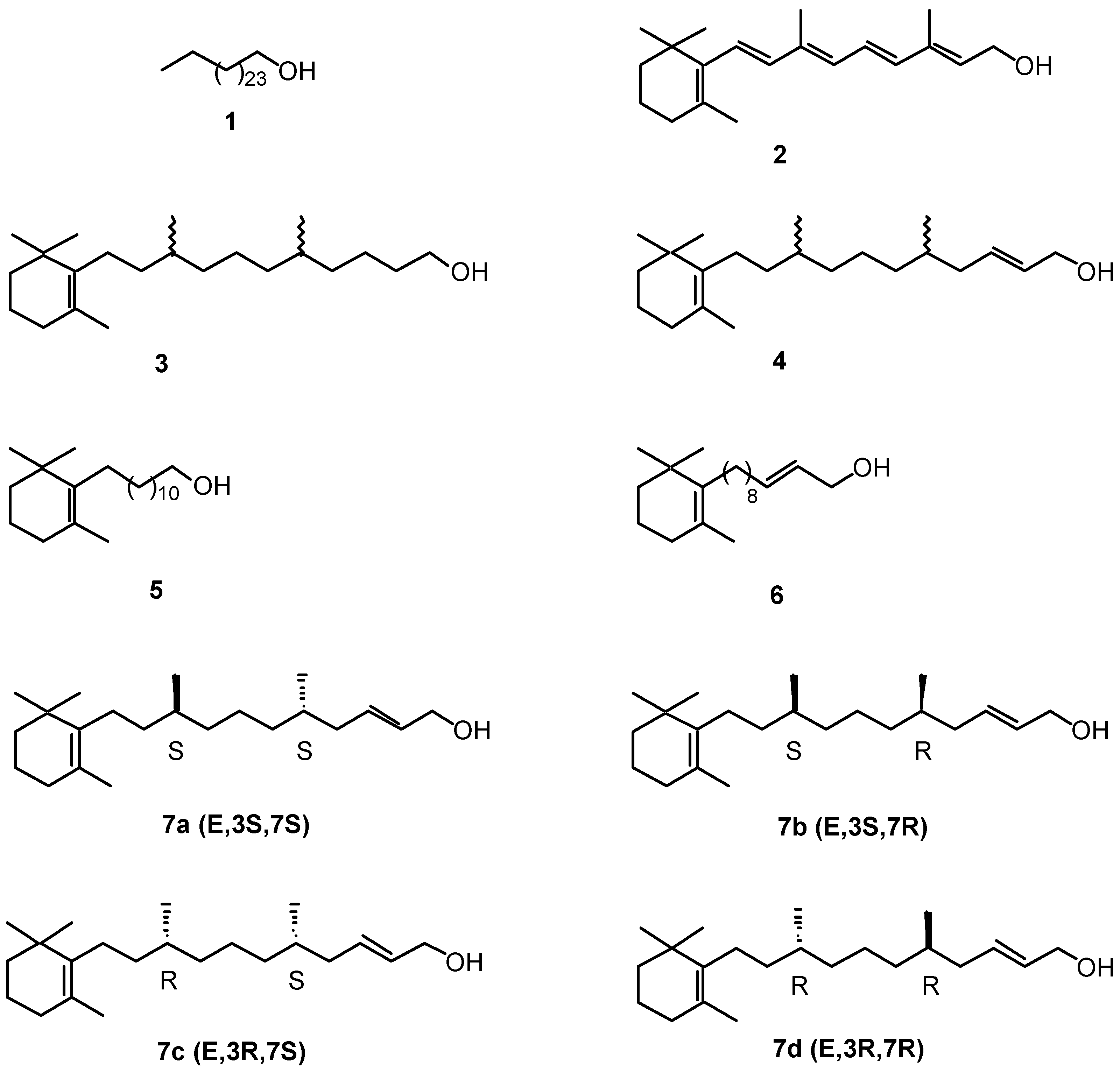
Figure 3.
Structure of diverse cyclohexenonic long-chain fatty alcohols.
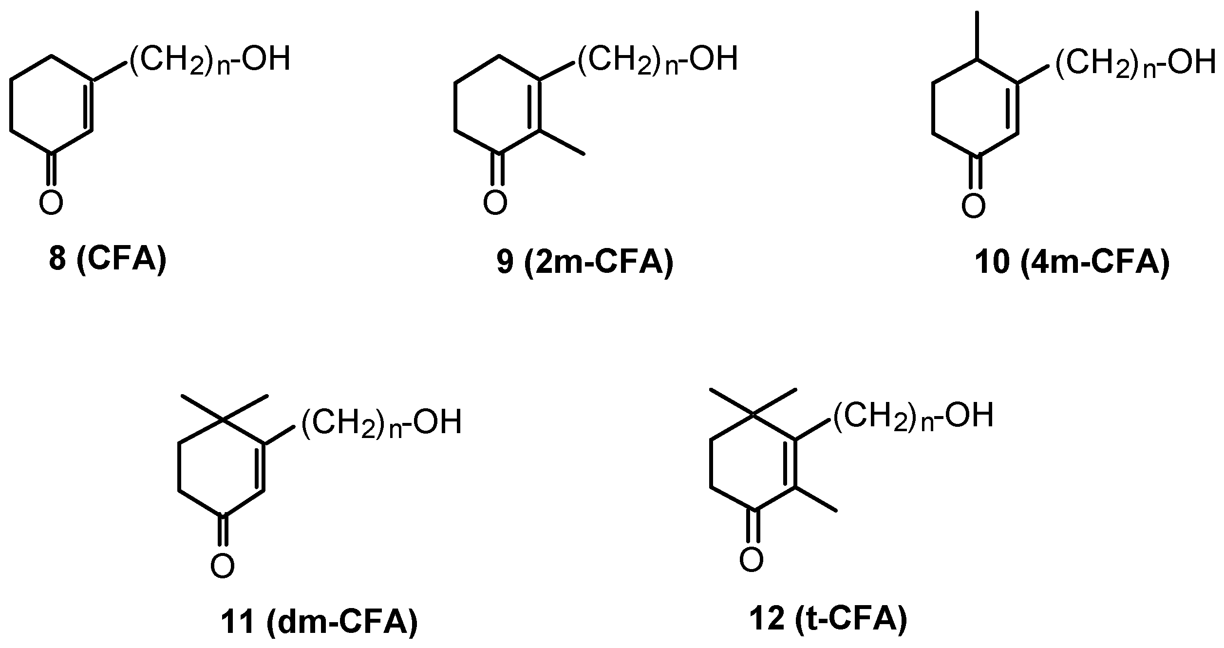
Figure 4.
Synthesis of CFA (8), 2m-CFA (9), 4m-CFA (10), and dm-CFA (11). Reagents: (a) PhSO2Na, HCl, H2O, room temperature (r.t.), 24 hours. (b) HO-(CH2)n-OH, pTsOH, benzene, reflux, 4 hours. (c) n-BuLi, THF, HMPA, CHPh3, -78°C to r.t., 1 hour; Br(CH2)nOH; THF, -78°C to -20°C, 2 hours. (d) CHCl3-acetone (5-1), pTsOH, 50°C, 24 hours.

Figure 5.
Retrosynthetic analysis of t-CFAs (12).
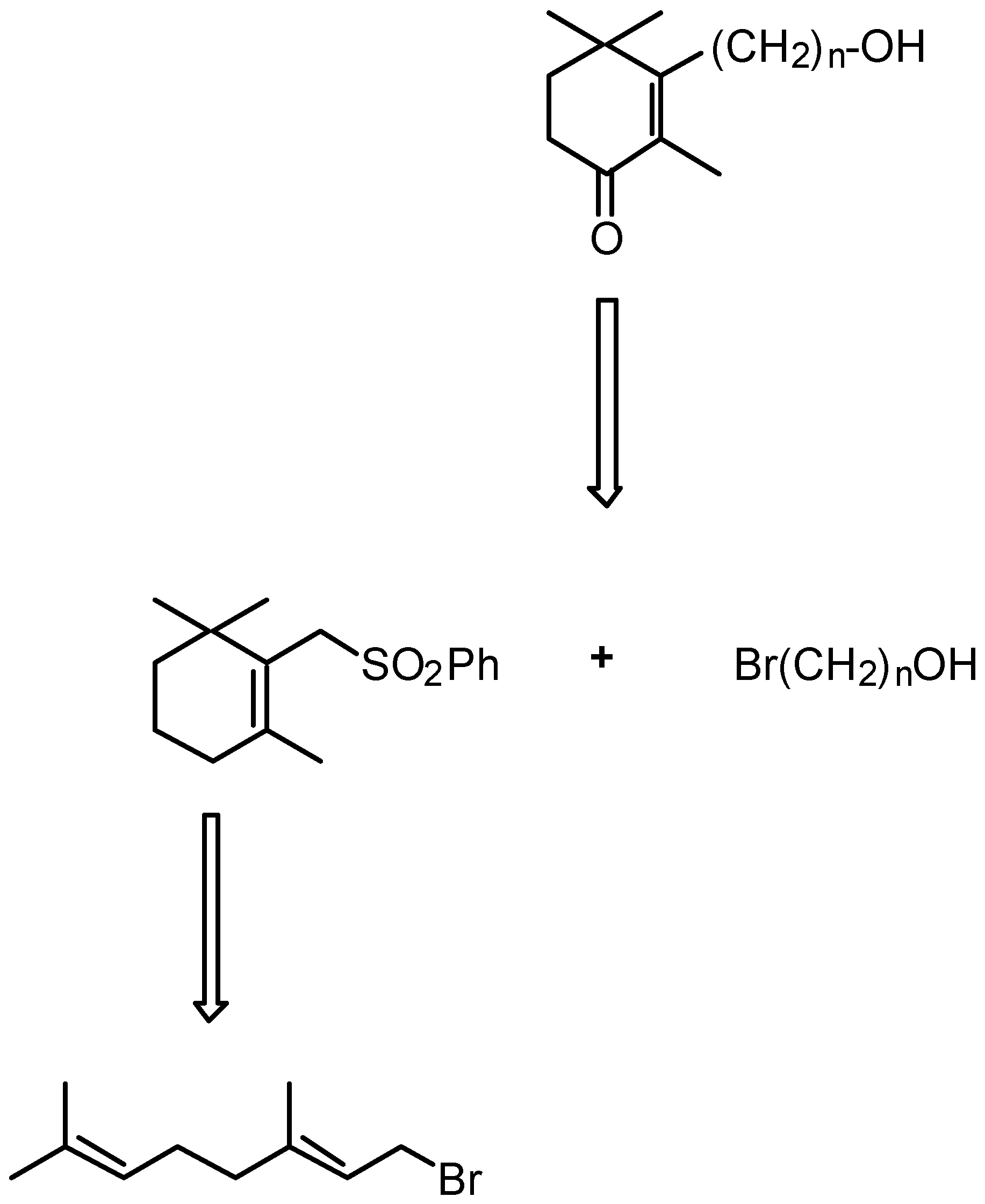
Figure 6.
Synthesis of sulfones 15 and 16 from geranylbromide 13. Reagents: (a) PhSO2Na, MeOH, 0°C, 1 hour (80%). (b) H2SO4 32 eq., AcOH, 12°C, 30 min (90%).
Figure 6.
Synthesis of sulfones 15 and 16 from geranylbromide 13. Reagents: (a) PhSO2Na, MeOH, 0°C, 1 hour (80%). (b) H2SO4 32 eq., AcOH, 12°C, 30 min (90%).
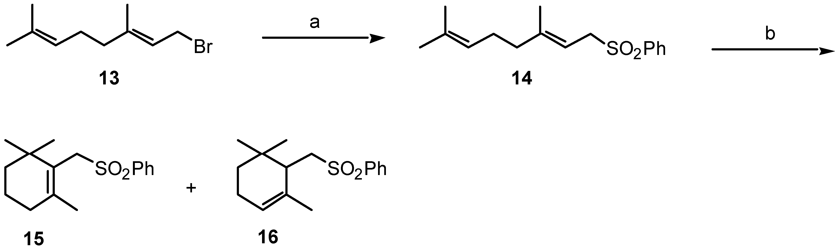
Figure 7.
Synthesis of t-CFAs (12) from sulfone 15. Reagents: (a) n-BuLi, THF, HMPA, CHPh3, - 78°C to r.t., 1 hour; Br(CH2)nOH, THF, -78°C to –20°C, 2 hours, (79-88%). (b) Na/Hg 6%, MeOH, Na2HPO4, 0°C to r.t., 3 hours (88-93%). (c) Ac2O, pyridine, r.t., 1 hour (97-98%). (d) RuCl3 0.7% mol, tBuOOH 70%, cyclohexane, H2O, r.t., 6 hours (49-53%). (e) K2CO3, MeOH, H2O, r.t., 2 hours (81-96%).

Figure 8.
Effect of 4m-CFA (n=14), and t-CFA (n=15) on the survival of cultured CNS neurons. Substances were tested at 100 nM. The data represent means ± SE of three independent experiments. *p<0.05 vs Control; one-way ANOVA, followed by Student-Newman-Keuls test.
Figure 8.
Effect of 4m-CFA (n=14), and t-CFA (n=15) on the survival of cultured CNS neurons. Substances were tested at 100 nM. The data represent means ± SE of three independent experiments. *p<0.05 vs Control; one-way ANOVA, followed by Student-Newman-Keuls test.

Figure 9.
Effect of t-CFAs on neurite outgrowth. Six t-CFAs bearing an aliphatic side chain that ranged from 12 to 18 carbon atoms were tested at 100 nM. After morphometric analysis, neurons were divided into three categories according to their total neurite length (Type 1 ≤ l00 μm; Type 2 101-200 μm; Type 3 201-300 μm). Note that t-CFA (n=14) and (n=15) are the most potent molecules in stimulating neurite sprouting. The data correspond to two pooled independent experiments (50-70 cells / experiment).

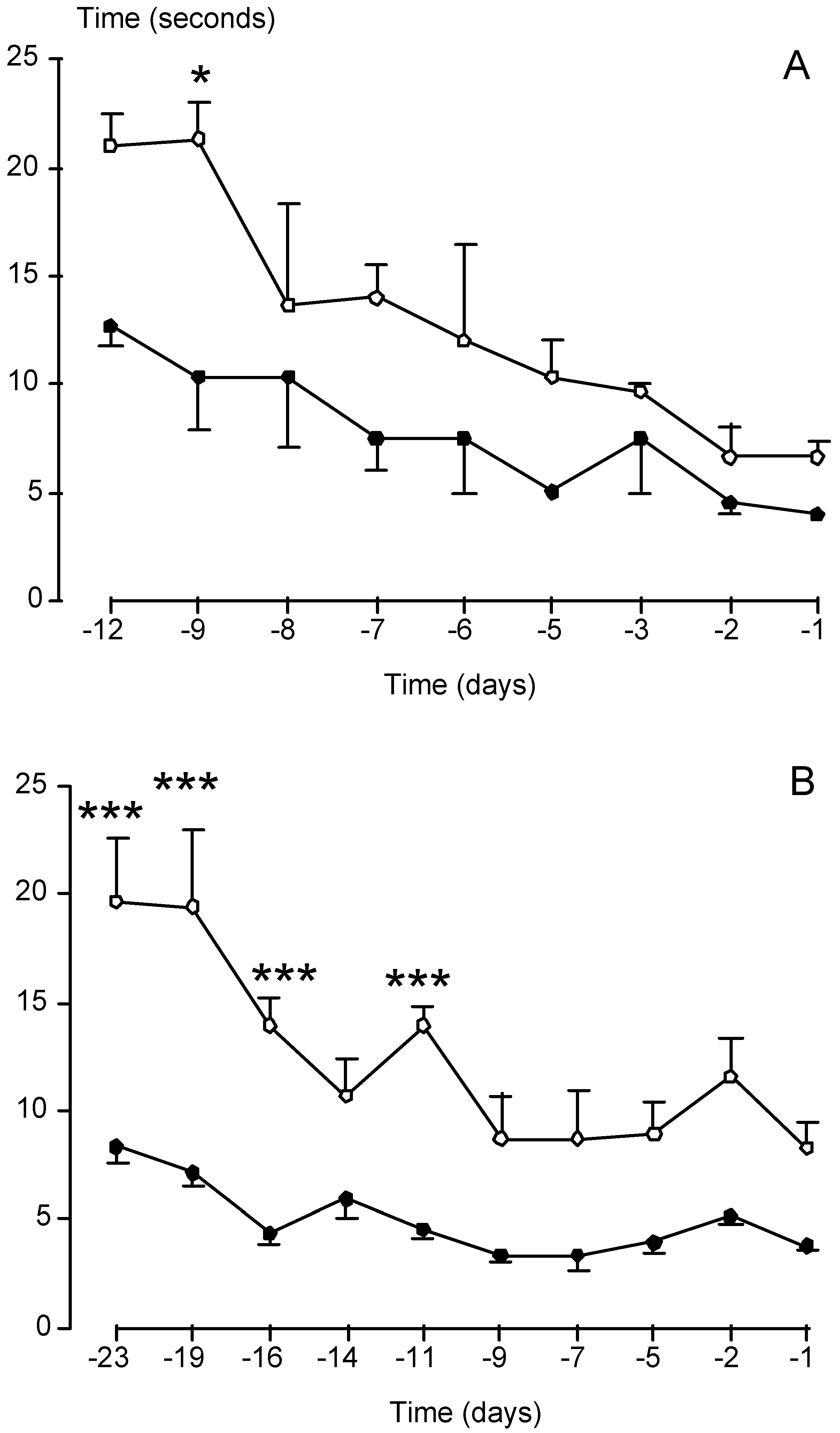
Figure 10.
Effect of 4m-CFA (n=14) (A), and t-CFA (n=15) (B) on the ability of mice to perform an exercise. The mice used in these experiments overexpress a mutant Cu/Zn superoxide dismutase gene associated with the neurodegenerative disease amyotrophic lateral sclerosis. The task consisted in determining the time (in seconds) necessary to cross a metallic bar of 45 cm long. Substances were injected intraperitoneally at 8 mg/kg 4m-CFA (n=14) or 32 mg/kg t-CFA (n=15) three times per week during 50-60 days, and meaurements were taken at days indicated in the abscissas before disease onset and death of the animals through motor paralysis. Note that treated mice (filled circles in both graphs) are more active than control animals. *p<0.05, ***p<0.001 vs corresponding control; one-way ANOVA, followed by Student-Newman-Keuls test.

| Diseases and affected neurons | Tested Neurotrophic Factors |
|---|---|
| Alzheimer’s disease: Cholinergic neurons | NGF (Neurotrophic Growth Factor) BDNF (Brain-Derived Neurotrophic Factor) NT-4/5 (Neurotrophin-4/5) bFGF (basic Fibroblast Neurotrophic Factor) |
| Parkinson’s disease: Dopaminergic neurons | GDNF (Glial-Derived Neurotrophic Factor) BDNF NT-4/5 |
| Ischemia: Striatum, hippocampus and cortex | TGF-β1 (Transforming Growth Factor) IGF-1 (Insulin-like Growth Factor) bFGF NT-4/5 |
| Amyotrophic lateral sclerosis: Motor neurons | IGF-1 BDNF CNTF (Ciliary Neurotrophic Factor) |
Table 2.
Structure/activity relationships of the CFA series 8, 9, 10, and 11 according to their neuritogenic effect in cultured CNS neurons.
 |
Stimulation of neurite sprouting by 10 ng/mL bFGF was used as positive control. Compounds were tested at 10 nM. (0) No effect; (+) Stimulatory effect; (-) Toxic effect. The table summarises data from at least two independent experiments.
Table 3.
Analysis of compounds 17 bearing an aliphatic side chain that ranged from 13 to 18 carbon atoms (TLC: hexane-AcOEt: 5-5).
| Product (n) | Yield (%) | R.f* | m.p (°C) | Analysis |
|---|---|---|---|---|
| (13) | 7 | .46 | 56-57 | Microanalysis (%): C28H46O3S (462.7296) Calcd: C: 72.68; H: 10.02; O: 10.37 Found: C: 72.49; H: 10.06; O: 10.62 |
| (14) | 4 | .47 | 75-76 | Microanalysis (%): C28H46O3S (462.7296) Calcd: C: 73.06; H: 10.15; O: 10.07 Found: C: 72.83; H: 10.18; O: 9.85 MS (EI): 334.9 (M-SO2Ph, 100); 136.5 (C10H16); 122.5 (C9H15); 108.5;94.5; 80.5 |
| (15) | 6 | .46 | 58-59 | Microanalysis (%): C30H50O3S (490.7832) Calcd: C: 73.42; H: 10.27; O: 9.78 Found: C: 73.34; H: 10.34; O: 9.92 |
| (16) | 6 | .48 | 85-87 | Microanalysis (%): C31H52O3S (504.810) Calcd: C: 73.76; H: 10.38; O: 9.51 Found: C: 73.56; H: 10.37; O: 9.63 |
| (18) | 8 | .56 | 93-95 | Microanalysis (%): C33H56O3S (532.8636) Calcd: C: 74.38; H: 10.59; O: 9.01 Found: C: 73.90; H: 10.74; O: 9.15 |
© 2000 by MDPI (http://www.mdpi.org).
Share and Cite
MDPI and ACS Style
Luu, B.; De Aguilar, J.-L.G.; Girlanda-Junges, C. Cyclohexenonic Long-Chain Fatty Alcohols as Neuronal Growth Stimulators. Molecules 2000, 5, 1439-1460. https://doi.org/10.3390/51201439
AMA Style
Luu B, De Aguilar J-LG, Girlanda-Junges C. Cyclohexenonic Long-Chain Fatty Alcohols as Neuronal Growth Stimulators. Molecules. 2000; 5(12):1439-1460. https://doi.org/10.3390/51201439
Chicago/Turabian StyleLuu, Bang, José-Luis González De Aguilar, and Céline Girlanda-Junges. 2000. "Cyclohexenonic Long-Chain Fatty Alcohols as Neuronal Growth Stimulators" Molecules 5, no. 12: 1439-1460. https://doi.org/10.3390/51201439