Photomediated Transformation of Salannin, a Tetranortriterpenoid from Azadirachta indica A. Juss

Centre for Natural Products, SPIC Science Foundation, Guindy, Chennai - 600 032, India
*
Author to whom correspondence should be addressed.
Molecules 2001, 6(6), 551-556; https://doi.org/10.3390/60600551
Submission received: 6 December 2000
/
Revised: 11 May 2001
/
Accepted: 14 May 2001
/
Published: 31 May 2001
Abstract
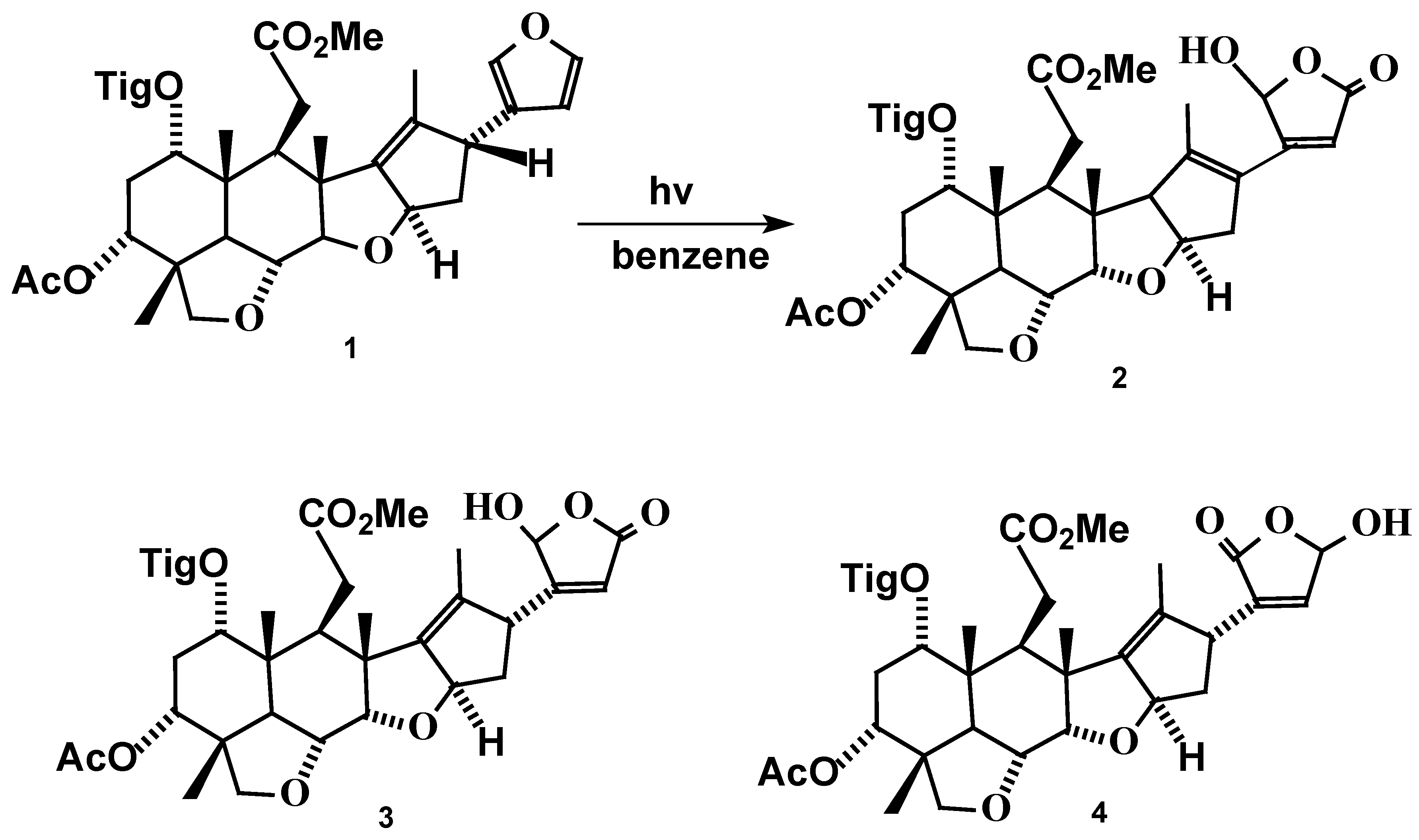
:Photolysis of Salannin (1), an important bioactive compound from Azadirachta indica A.Juss (Neem), affords Δ17-isosalanninolide (2), a hitherto unknown compound, along with isosalanninolide (3) and salanninolide (4). A probable mechanism has been suggested. While the mechanism of formation of 2 involves both a [4+2] cycloaddition and a [1,3] sigmatropic shift in the furan and D-rings, respectively, formation of 3 and 4 involves the decomposition of an ozonoide-like peroxide intermediate.
Introduction
Neem, Azadirachta indica A. Juss (Meliaceae) seeds contain a number of complex triterpenoids which are of great interest as they exhibit a variety of biological properties [1,2,3,4]. Several of these contain UV chromophores such as α,β - unsaturated ketone, furan, α,β -unsaturated ester and tigloyl groups. The bio-efficacy of .neem formulations is altered due to degradation of the triterpenoids under light exposure [5,6]. Hence, it is relevant to study the formation, isolation and bio-activity of the products formed when pure samples of these limonoids are exposed to sunlight. Herein the photolysis of salannin, one of the major triterpenoids present in the neem seed is reported.
Irradiation of salannin (1) using a sunlamp with a wider spectrum of irradiation equivalent to exposure to sunlight yielded three compounds (Figure 1). The present paper describes the sequence of events leading to the formation of Δ17-isosalanninolide (2) and its complete characterization. It may be noted that the compound 2 exhibited antifeedancy against Spodoptera litura comparable to Azadirachtin A, the most potent antifeedant in neem extract. Compounds isosalanninolide (3) and salanninolide (4) [7] have been previously reported to be formed in the photolysis of salannin (1) using UV irradiation in presence of oxygen.
Figure 1.
Results and Discussion
Salannin (1) isolated from seed kernel of A. indica [8,9] was irradiated using a sunlamp in benzene for eight hours. Purification of the crude product by HPLC yielded compounds 2, 3 and 4. The mechanism for the formation of 2 is envisaged as given below (Figure 2) while the mechanism for 3 and 4 is assumed to be similar to that of the the reported photooxygenation of cedrelone[10].
Figure 2.
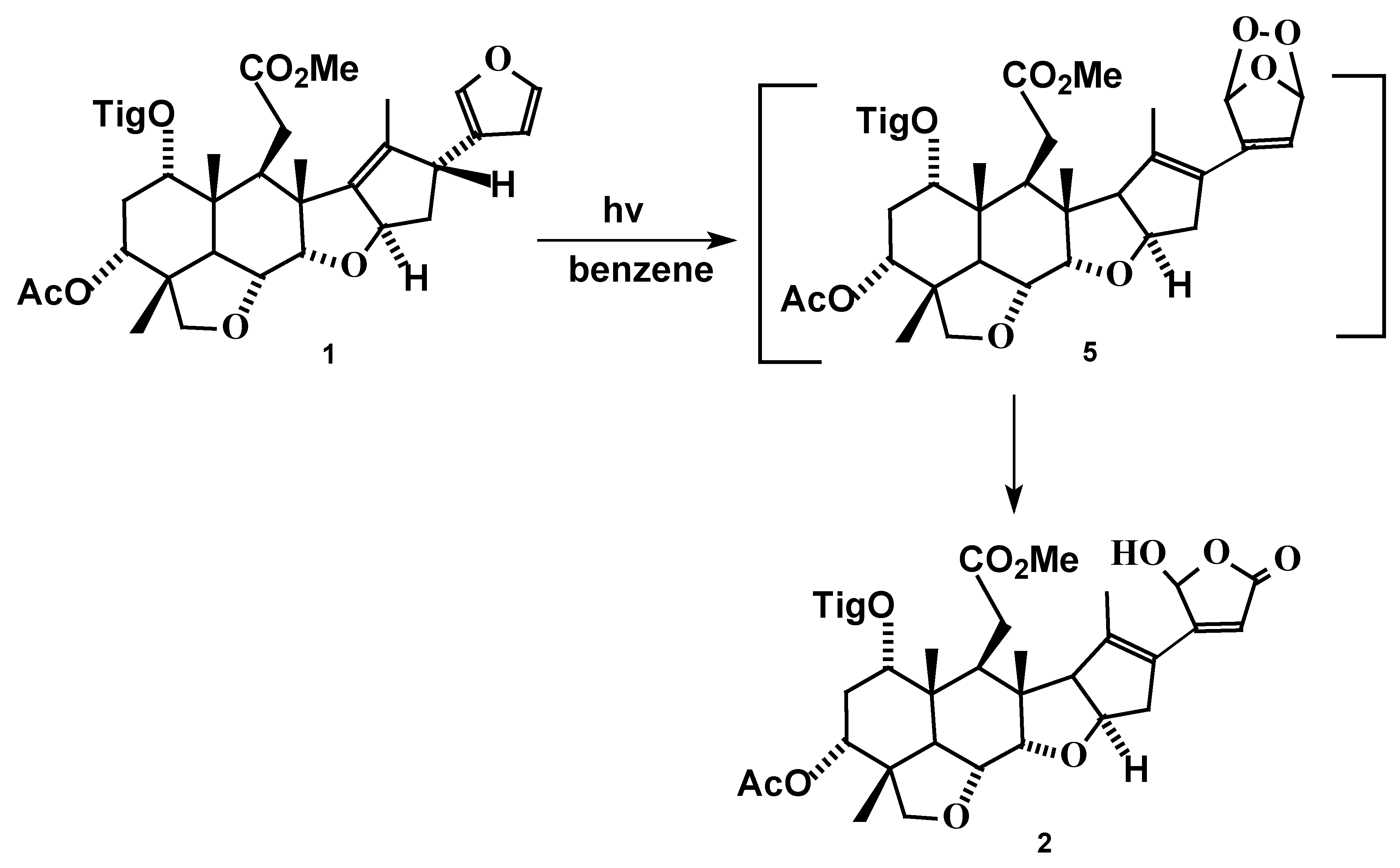
The formation of the intermediate 5 can be explained by: a) the addition of 1O2 to furan generating an unstable endoperoxide similar to that of Diels-Alder reaction.[11]; b) [1,3] suprafacial sigmatropic hydrogen shift resulting in the 1,2-shift of the double bond in the D-ring. Further the unstable endoperoxide functionality in compound 5 rearranges to yield hydroxy butenolide in ring E of compound 2 [12].
The structure of the compound 2 was established on the basis of the following spectral evidence. Compound 2 exhibited a λ max at 298, 227 nm (log ε 3.782, 4.395) indicating an extra conjugation, compared to salannin (1) (λ max at 225 nm, log ε 4.356). The infrared spectrum of 2 showed additional absorptions at 1708 cm-1 (α,β- unsaturated ketone) and 3376 cm-1 (hydroxyl group). The 1H- and 13C-NMR spectrum of 2 was similar to that of 1 except for changes observed in the D and furan rings. The 13C-NMR exhibited signals due to a lactone (169.2 ppm), a deoxygenated methine at 88.1 ppm (proton of which resonated at 5.52 ppm as a triplet), an allylic methylene at 31.1 ppm (attached to protons at 2.31 and 2.37 ppm as multiplets) and 4 olefinic carbons (154.0, 119.5, 130.9, 98.1) among which only the signal at 98.1 showed connectivity with a hydrogen at 5.92 ppm (as a singlet). The changes observed in the olefinic region indicated that the double bond between C-13 and C-14 has now been reorganized to C-13 and C-17 and is in conjugation with C-20, C-22 which lie in the same plane. The D2O exchanged 1H-NMR spectrum revealed the presence of a -OH at 5.93 ppm, The structure assigned was further supported by the mass spectra of 2 which showed a M+ at m/z 628.
It must be noted that though salannin 1, the major constituent in neem formulations, did not exhibit significant antifeedant activity against Spodoptera litura, its photoproduct 2 exhibits remarkable activity comparable to that of Azadirachtin A, the results of which are published elsewhere[13].
Acknowledgments
The authors thank the Department of Biotechnology, Government of India, for financial assistance and Dr.G.Suresh for useful suggestions.
Experimental
General
UV spectra were recorded on a Shimadzu 160A instrument. NMR spectra were recorded on a BRUKER 200 MHz instrument using TMS as the internal standard and CDCl3 as solvent. Chemical shifts are given in terms of parts per million (δ scale). LRMS were recorded on a Shimadzu QP 5000 mass spectrometer. Precoated silica gel plates (E-Merck, Germany, Art. 5554 Kieselgel 60 F254, 0.2 mm thickness) were used for thin layer chromatography. Ammonium molybdate – ceric sulphate in H2SO4 was used as the visualization agent.
Analytical high performance liquid chromatography was carried out using a C18 RP column (E-Merck, 10μm, 4.6 mm x 25 cm) eluting with CH3CN: H2O (50: 50) at a flow rate of 1 mL/min and UV detection (215 nm). For semi-preparative HPLC runs, a C18 RP column (E-Merck, 10μm, 9.6 mm x 25 cm) was used with CH3CN: H2O (50: 50) as eluant at a flow rate of 5 mL/min and the peaks were again detected at 215 nm.
Irradiation of Salannin (1).
Salannin (1) (50mg) dissolved in benzene (80mL) was irradiated with a sunlamp equipped with a cooling device (Phillips, 300W) under aerated conditions for eight hours, at the end of which complete decomposition of salannin was noted by analytical HPLC. The solvent was removed under reduced pressure and the products were purified by semi-preparative HPLC. Three major peaks eluted with retention times of 5.6, 7.4 and 15.7 min., respectively. The fractions with Rt 5.6 and 15.1min. were identified as compounds 3 (8mg) and 4 (14mg) by comparison with reported data [7]. The fraction with Rt 7.4 min yielded 2 as a white solid (12 mg) upon concentration.
Analytical and spectral data of compound 2.
Mp 114 - 116oC; UV: λ max (MeOH) / nm 298 (log ε 3.782), 227 (log ε 4.395); IR: υmax/ cm-1 3376, 2976, 2880, 1760, 1708, 1696, 1651, 1596, 1488, 1465, 1379, 1315, 1273, 1068, 1001, 969, 880, 838, 646, 611; 1H-NMR (200MHz: CDCl3; Me4Si): δH 6.93 (1H, m, 3’-H), 5.93 (1H, br s, -OH), 5.92 (1H, s, 22-H), 5.52 (1H, t, 21-H), 4.95 (1H, t, 1-H), 4.71 (1H, t, 3-H), 4.24 (1H, d, J 3.0, 7-H), 3.98 (1H, dd, J 9.4 and 3.1, 6-H), 3.79 (1H, d, J 8.9, 15-H), 3.66 (1H, d, J 7.5, 28b-H), 3.58 (1H, d, J 7.5, 28a-H), 3.52 (3H, s, COOMe), 2.68 (1H, d, J 9.1, 5-H), 2.66 (2H, m, 9-H and 14-H), 2.37 (2H, m, 11b-H and 16b-H), 2.31 (2H, m, 11a-H and 16a-H), 2.21 (2H, m, 2-H), 1.93 (3H, s, OCOMe), 1.87 (3H, s, Me), 1.81(3H, s, Me), 1.73 (3H, s, Me), 1.28 (3H, s, Me), 1.23 (3H, s, Me), 1.03 (3H, s, Me); 13C-NMR (50MHz: CDCl3; Me4Si): δC 174.19 (C-12), 170.33 (OCOMe), 169.20 (C-23), 166.54 (C-1’), 154.00 (C-13), 137.99 (C-3’), 130.85 (C-20), 128.74 (C-2’), 119.53 (C-17), 98.12 (C-22), 88.09 (C-21), 85.82 (C-7), 77.63 (C-28), 72.39 (C-6), 71.48 (C-3),71.21(C-1), 52.47 (C-15 and COOMe), 49.56 (C-8), 42.67 (C-4), 40.57 (C-10), 40.05 (C-5), 39.51 (C-9 and C-14), 31.13 (C-11and C-16), 27.28 (C-2), 20.82 (OCOMe), 19.65 (C-29), 16.79 (C-30), 14.87 (C-19), 14.43 (C-4’), 13.03 (C-18), 11.86 (C-5’). Mass spectum: m/z 628 (M+).
References
- Kraus, W. The Neem Tree, Azadirachta indica A.Juss and other Meliaceous Plants; Schmutterer, H., Ed.; Verlag Chemie: Weinheim, 1995; pp. 35–38. [Google Scholar]
- Blaney, W.M.; Simmonds, M.S.J.; Ley, S.V.; Anderson, J.C.; Toogood, P.L. Antifeedant effects of Azadirachtin and structurally related compounds on lepidoptera larvae. Entomol. Exp. Appl. 1990, 55, 149–160. [Google Scholar] [CrossRef]
- Butterworth, J.H.; Morgan, E.D. Isolation of a substance that suppresses feeding in locust. J. Chem. Soc. Chem. Commun. 1968, 23–24. [Google Scholar] [CrossRef]
- Govindachari, T.R.; Narasimhan, N.S.; Suresh, G.; Partho, P.D.; Geetha Gopalakrishnan. Insect antifeedant and growth regulating activities of salannin and other C-seco limonoids from neem oil in relation to azadirachtin. J. Chem. Ecol. 1996, 22, 1453–1461. [Google Scholar] [CrossRef] [PubMed]
- Barnaby, M.A.; Yamasaki, R.B.; Klocke, J.A. Biological activity of azadirachtin, three derivatives, and their ultraviolet radiation degradation products against tobacco bud worm (Lepidoptera: Noctuidae) larvae. J. Econ. Entomol. 1989, 82, 58–63. [Google Scholar] [CrossRef]
- Stokes, J.B.; Redfern, R.E. Effect of sunlight on azadirachtin: antifeeding potency. J. Environ. Sci. Health 1982, A17, 57–65. [Google Scholar] [CrossRef]
- Jarvis, A. P.; Johnson, S.; Morgan, E.D.; Simmonds, M.S.J.; Blaney, W.M. Photooxidation of nimbin and salannin-tetranortriterpenoid from neem tree. J. Chem. Ecol. 1997, 23, 2841–2860. [Google Scholar] [CrossRef]
- Henderson, R.; McCrindle, R.; Melera, A.; Overton, K.W. Tetranortriterpenoids-IX The constitution and stereochemistry of salannin. Tetrahedron 1968, 24, 1525–1528. [Google Scholar] [CrossRef]
- Govindachari, T.R.; Suresh, G.; Geetha Gopalakrishnan. A direct preparative high performance liquid chromatography procedure for the isolation of major triterpenoids and their quantitative determination in neem oil. J. Liq. Chromatogra. 1995, 18, 3465–3471. [Google Scholar] [CrossRef]
- Geetha Goplakrishnan.; Pradeep Singh, N.D.; Kasinath, V.; Malathi, R.; Rajan, S.S. Photooxidation of cedrelone,a tetranortriterpenoid from Toona ciliata. Photochem. Photobio 2000, 72, 464–466. [Google Scholar]
- Foote, C.S.; Wuesthoff, M.T.; Wexler, S.; Burstain, I.G.; Denny, R.; Schenck, G.O.; Schulte-Elte, K-M. Photosensitized oxigenation of alkyl substituted furans. Tetrahedron 1967, 23, 2583–2599. [Google Scholar] [CrossRef]
- Gilbert, A. Photochemistry in Organic Synthesis; Coyle, J.D., Ed.; Royal Society of Chemistry: Cambridge, U.K., 1986; pp. 141–163. [Google Scholar]
- Pradeep Singh, N.D. Ph.D. Thesis, University of Madras, November 2000.
- Sample Availability: Both the starting compound (50 mg.) and the photoproducts (5 mg. each) are available from the author.
© 2001 by MDPI (http://www.mdpi.org). Reproduction is permitted for noncommercial purposes
Share and Cite
MDPI and ACS Style
Gopalakrishnan, G.; Pradeep Singh, N.D.; Kasinath, V. Photomediated Transformation of Salannin, a Tetranortriterpenoid from Azadirachta indica A. Juss. Molecules 2001, 6, 551-556. https://doi.org/10.3390/60600551
AMA Style
Gopalakrishnan G, Pradeep Singh ND, Kasinath V. Photomediated Transformation of Salannin, a Tetranortriterpenoid from Azadirachta indica A. Juss. Molecules. 2001; 6(6):551-556. https://doi.org/10.3390/60600551
Chicago/Turabian StyleGopalakrishnan, Geetha, N. D. Pradeep Singh, and V. Kasinath. 2001. "Photomediated Transformation of Salannin, a Tetranortriterpenoid from Azadirachta indica A. Juss" Molecules 6, no. 6: 551-556. https://doi.org/10.3390/60600551