An Attempt to Assign the NMR Spectra in 7-Methyl-and 7-Benzyl-substituted 7H-Purine 1-Oxides Using the ACD/Labs Software Package

Institute of Chemistry, University of Podlasie, ul. 3-Maja 54, PL-08-110 Siedlce, Poland; Fax: (+48) 25-644-2045 and Institute of Organic Chemistry, Polish Academy of Sciences, ul. Kasprzaka 44/52, PL-01-224 Warszawa, Poland
Molecules 2003, 8(9), 649-654; https://doi.org/10.3390/80900649
Submission received: 11 March 2003
/
Revised: 17 July 2003
/
Accepted: 20 July 2003
/
Published: 15 August 2003
Abstract
:Commercially available 4-nitroimidazole can be transformed in a few simple steps into 4-aminoimidazoyl-5-carboximes, from which the respective purine mono-1-oxides may be synthesized. The N→O functionality in these products significantly changes the electron density in the purine skeleton and consequently, the determination of the 1H- and 13C-NMR spectra of these simple compounds is somewhat troublesome. The use of the ACD/Labs software package for this purpose is discussed.
Introduction
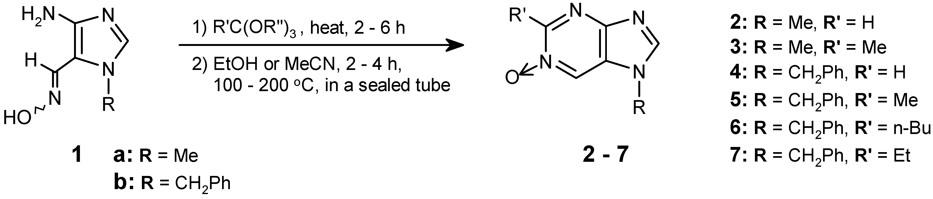
Aromatic or heteroaromatic ortho-aminocarboximes of type 1 are very valuable intermediates for the preparation of fused pyrimidine mono-N-oxides by cyclisation with the use of orthoesters [1]. This selectively introduces the N→O function into the desired position in heteroaromatic compounds having more than one nitrogen atom. Intermediates of type 1 are now easily obtainable [2,3,4,5] by Vicarious Nucleophilic Substitution methodology [6], thus allowing development of this efficient cyclisation. This has been exemplified by synthesis of a series of heterocyclic systems [4,5].
This synthetic approach was discussed in our previous publication [5], concerning application to the synthesis of purine mono-N-oxides (2-7; as outlined on Scheme 1). The structures of these N-oxides were confirmed by 1H-NMR and 13C-NMR spectra. We determined the spectra of these newly prepared compounds by simple comparisons within a series of the respective derivatives. It is believed that this is the first attempt (in the case of 13C-NMR) to assign chemical shifts to the carbon atoms of these structures. Herein, we would like to present the application of the ACD/Labs software package [7] for the quick determination of the corresponding signals, exploring both its utility and limitations.
Scheme 1.
Results and Discussion
Spectra of Purine mono-N-Oxides: 1H-NMR spectra
The assignment of the proton spectra in the purine skeleton 1-oxides was relatively simple. The proton chemical shifts of H-6 and H-8 were easily differentiated because the H-8 signal should always be shifted upfield, according to the literature [8]. Indeed we observed its shift to a higher field (at δ = 8.12 - 8.32 ppm) in all the compounds investigated. In 2-substituted products (R’ = Me, Et, n-Bu), the remaining signals (corresponding to H-6) were found at δ = 8.43 - 8.65 ppm for the 7-CH2Ph substituted derivatives 5-7; however, for the 7-Me substituted compound 3, the corresponding signal was shifted downfield to δ = 8.85 ppm. This observation additionally allows us to differentiate between H-6 and H-2 in 2-unsubstituted derivatives. The H-2 signal in 4 (δ = 8.91 ppm) displays the greatest downfield shift (for comparison, in 2, at δ = 8.96 ppm, the value was identical to δ(H-6)). The remaining signals (CH3, NCH3, and NCH2Ph) are of minor importance, and they were easily identified.
The ACD/Labs program is helpful for many compounds, but in this case we observed poor agreement between the calculated data and the experimentally determined results (Table 1; calculated data - in parentheses). In some cases of H-2/H-6/H-8, the differences were very significant (up to 0.9 ppm). Unexpectedly, the largest deviation was observed for H-8 in compounds substituted with the CH2Ph group, e.g. 8.22 ppm (found) versus 9.17 ppm (calculated) for compound 6. Also, the observed order of the chemical shifts δ(H-2) > δ(H-6) > δ(H-8) was inverted. Hence, we conclude that the usefulness of the ACD/Labs program for the determination of 1H-NMR chemical shifts of purine-1-oxides is very limited.
Figure 1.

| Compound | H-2 | H-6 | H-8 | Other protons | |
|---|---|---|---|---|---|
| 2 | 8.96 (9.09) | 8.96 (8.85) | 8.32 (8.32) | 7-CH3− 3.93 | |
| 3 | – | 8.85 (8.64) | 8.12 (8.31) | 2-CH3− 2.85 7-CH3− 3.95 | |
| 4 | 8.91 (J = 1.8) (9.11) | 8.43 (J = 1.8) (8.87) | 8.28 (9.14) | 7.1-CH2− 5.34 | |
| 5 | – | 8.43 (8.66) | 8.22 (9.12) | 2-CH3 − 2.80 | |
| 6 | – | 8.47 (8.72) | 8.22 (9.17) | 2.4-CH3− 0.96; t, J = 7.2 | |
| 7 | – | 8.65 (8.61) | 8.24 (9.12) | 2.2-CH3− 1.40; t, J = 7.6 | |
* compound 2 in CDCl3/DMSO-d6 (5:1).
13C-NMR spectra
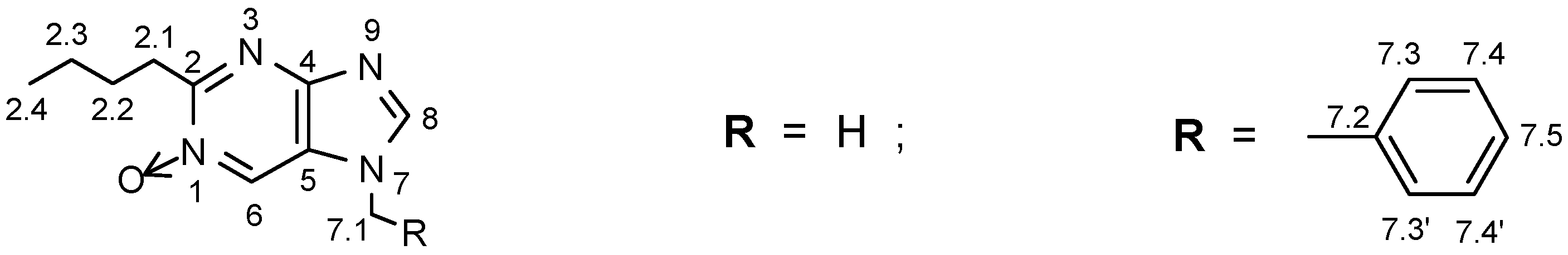
The previously published spectra of these particular types of purine mono-N-oxides [5] represent the first attempts at assigning chemical shifts to carbon atoms of the structures. The correct assignment of C-2.1, C-2.2, C-2.3, C-2.4, C-7.1, and C-8 was a trivial problem. However, it was difficult to correctly assign the remaining values on the basis of 13C decoupled spectra because of considerable C-6 upfield shifts (in compounds 4,5,6), and the proximity of these signals to the aromatic peaks of benzyl groups. Also, a pair of weak C-4 and C-5 quaternary carbon atom signals, despite significant differences in their chemical shifts (over 25 ppm), was difficult to assign correctly (in the case of 2 and 3 some signals were not even detected). We finally assigned the values of chemical shifts for the structures in this series of compounds on the basis of the spectra of N-oxides measured by the GHMBC technique [5]. The most valuable diagnostic chemical shift assignments for carbon atoms in the phenyl ring of the CH2Ph substituent were the correlations between the more easily identified protons of the CH2 group and the respective aromatic carbon atoms. Details of this assignment protocol were discussed in [5].
Definitive assignment of all benzyl group signals allows us to identify the rest of the peaks - which belong to carbon atoms of the purine skeleton (C-2, C-4, C-5, and C-6). In this case, the ACD/Labs program, despite only moderate agreement of calculated and experimental data, was very helpful in the differentiation of the signals originating from quaternary C-4 and C-5 atoms. Hence δ(C-4) = 151.3 ppm (calculated - 157.3 ppm) and δ(C-5) = 123.4 ppm (calculated - 130.0 ppm) were definitively assigned. Also the C-2 signal (when C-2 is unsubstituted; i.e., in compounds 2 and 4) can be efficiently predicted on the basis of the calculated data with the use of the above program. All of the 13C NMR parameters are listed in Table 2.
 |  |  |  |  | ||
|---|---|---|---|---|---|---|
| Carbon Atom | 2 | 3 | 4 | 5 | 6 | |
| C-2 | 151.8 (147.8) | 153.4 (150.8) | 146.0 (147.6) | 156.1 (150.7) | 159.0 (155.8) | |
| C-4 | ** (156.1) | ** (156.7) | 151.9 (156.7) | 151.3 (157.3) | 151.4 (157.3) | |
| C-5 | 125.0 (129.8) | ** (130.2) | 124.3 (129.6) | 123.4 (130.0) | 123.0 (129.5) | |
| C-6 | 139.1 (131.7) | 139.5 (130.4) | 129.8 (132.3) | 129.6 (131.0) | 129.8 (130.9) | |
| C-8 | 148.3 (149. 8) | 148.4 (149.8) | 148.9 (146.3) | 148.3 (146.3) | 148.3 (146.3) | |
| C-2.1 | ― | 26.0 (24.5) | ― | 20.1 (24.5) | 31.9 (36.8) | |
| C-2.2 | ― | ― | ― | ― | 27.5 (26.0) | |
| C-2.3 | ― | ― | ― | ― | 22.4 (24.9) | |
| C-2.4 | ― | ― | ― | ― | 13.9 (14.2) | |
| C-7.1 | 31.1 (31.6) | 32.0 (31.6) | 50.7 (49.4) | 50.5 (49.4) | 50.5 (49.4) | |
| C-7.2 | ― | ― | 132.6 (138.1) | 132.9 (138.1) | 133.0 (138.1) | |
| C-7.3; C-7.3' | ― | ― | 127.6 (130.3) | 127.5 (130.3) | 127.5 (130.3) | |
| C-7.4; C-7.4' | ― | ― | 129.7 (128.8) | 129.5 (128.8) | 129.6 (128.8) | |
| C-7.5 | ― | ― | 129.6 (128.0) | 129.3 (128.0) | 129.4 (128.0) | |
* compound 2 in CDCl3/DMSO-d6 (5:1); ** not detected.
Generally, the chemical shift calculations obtained for all of the investigated purine N-oxides when using the ACD/Labs program (Table 2, in parentheses) are unsatisfactory as compared to the data determined experimentally. The best agreement was observed for C-2, C-8, and C-7.1, and for some of the benzyl group signals; the largest deviations were observed for C-4, C-6 (in 2 and 3), and for C-7.2 (in the NCH2Ph substituent). The presence of the N-oxide function in the purine ring system is the likely factor causing the observed poor correlation – its dipolar character (N+→O−) probably significantly changes the electronic situation therein, especially in the neighbouring fragment to the oxidized nitrogen atom.
Also the N-7 CH2Ph substituent changes the magnetic environment at the C-6 position (due to its bulkiness, or local ”ring-current effect” generated by the aromatic phenyl substituent). This long-distance influence of the N-7 group on the chemical shifts of C-6 was reasonably substantial (ca 139 ppm → 129 ppm).
Conclusions
The N-oxide function caused significant electronic disturbances in the purine skeleton of all investigated structures. This was the likely reason that the ACD/Labs software package could not be applied efficiently to predict chemical shifts of some protons and carbon atoms of these derivatives, hence its limitations for this purpose.
On the other hand, this program was very helpful for bulk signal assignment in the title compounds; thus, it may be applied simultaneously with other spectroscopic techniques. In our case, some direct proofs came finally from GHMBC spectra.
It is worth mentioning that when utilizing some other similar softwares, e.g. ChemOffice, we cannot predict the discussed chemical shifts at all, because this program does not simulate the values at the positions neighbouring to N-oxide function [9]. Calculations with the use of the CAChe program gave also very poor agreement [10]. We believe that our observations described herein will receive more attention and will be very helpful for determination of the 1H- and 13C-NMR spectra of purine N-oxides.
Experimental
General
NMR spectra were recorded with a Varian GEMINI-200 spectrometer operating at 200 MHz for 1H and 50 MHz for 13C. Coupling constants J are expressed in hertz [Hz]. Two-dimensional GHMBC spectra were recorded with a Varian INOVA-500 spectrometer (500 MHz / 125 MHz).
Preparation of Purine mono-N-Oxides 2-7.
References and Notes
- Katritzky, A.R.; Lagowski, J.M. Chemistry of the Heterocyclic N-Oxides; Blomquist, A.T., Ed.; Academic Press: London – New York, 1971; pp. 19–141. [Google Scholar]
- Ostrowski, S. Polish J. Chem. 1994, 68, 2237–2247.
- Ostrowski, S. Synlett 1995, 253–254.
- Ostrowski, S. Heterocycles 1996, 43, 389–396.
- Ostrowski, S. Molecules 1999, 4, 287–309.
- Mąkosza, M. Synthesis 1991, 103–111. Mąkosza, M.; Wojciechowski, K. Liebigs Ann. / Recueil 1997, 1805–1816.
- Calculations courtesy of deCODE genetics, Inc., Woodridge, IL, USA; ACD/Labs 6.00, ACD / CNMR and HNMR Predictors, 16 May 2002, Lic. ID: 20587.
- Lister, J.H. The Chemistry of Heterocyclic Compounds, Fused Pyrimidines; Part II: ”Purines”; Brown, D.J., Ed.; vol. 24/2, Brown, D.J., Ed.; Wiley – Interscience: New York – London – Sydney – Toronto, 1971. [Google Scholar] Lister, J.H. ibid.; Supplement 1, Wiley – Interscience: New York – Chichester – Brisbane – Toronto – Singapore, 1996; pp. 61–82. [Google Scholar] Lythgoe, D.J.; Ramsden, Ch.A. Adv. Heterocycl. Chem. 1994, 61, 1–58.
- ChemOffice calculations; for example for compound 2 (calculated data in brackets): C-2 – 151.8 (no data); C-4 – undetected (122.6); C-5 – 125.0 (125.9); C-6 – 139.1 (no data); C-8 – 148.3 (139.8); C-7.1 – 31.1 (37.5).
- CAChe program calculations, LORG B88-LYP method; for compound 2 (calculated data in brackets): C-2 – 151.8 (155.4); C-4 – undetected (151.4); C-5 – 125.0 (128.6); C-6 – 139.1 (132.0); C-8 – 148.3 (140.9); C-7.1 – 31.1 (37.2).
- Sample Availability: Available from the author.
© 2003 by MDPI ( http://www.mdpi.org). Reproduction is permitted for noncommercial purposes.
Share and Cite
MDPI and ACS Style
Ostrowski, S. An Attempt to Assign the NMR Spectra in 7-Methyl-and 7-Benzyl-substituted 7H-Purine 1-Oxides Using the ACD/Labs Software Package. Molecules 2003, 8, 649-654. https://doi.org/10.3390/80900649
AMA Style
Ostrowski S. An Attempt to Assign the NMR Spectra in 7-Methyl-and 7-Benzyl-substituted 7H-Purine 1-Oxides Using the ACD/Labs Software Package. Molecules. 2003; 8(9):649-654. https://doi.org/10.3390/80900649
Chicago/Turabian StyleOstrowski, Stanisław. 2003. "An Attempt to Assign the NMR Spectra in 7-Methyl-and 7-Benzyl-substituted 7H-Purine 1-Oxides Using the ACD/Labs Software Package" Molecules 8, no. 9: 649-654. https://doi.org/10.3390/80900649