Article

Eu-Doped BaTiO3 Powder and Film from Sol-Gel Process with Polyvinylpyrrolidone Additive

by
Margarita García-Hernández
1,
Antonieta García-Murillo
1,2,
Felipe de J. Carrillo-Romo
1,2,*,
David Jaramillo-Vigueras
2,
Geneviève Chadeyron
3,
Elder De la Rosa
4 and
Damien Boyer
3
1
Instituto Politécnico Nacional, CICATA Unidad Altamira, Km. 14.5, Carretera Tampico-Puerto Industrial Altamira, C.P. 89600 Altamira, Tamps, Mexico
2
Instituto Politécnico Nacional, CIITEC, Cerrada CECATI S/N Col. Sta. Catarina, Del. Azcapotzalco, México D.F. 02250, Mexico
3
Université Blaise Pascal, Laboratoire des Matériaux Inorganiques, CNRS-UMR 6002, 63177 Aubière, France
4
Centro de Investigaciones en Óptica, A.P. 1-948, 37150 León, Gto., Mexico
*
Author to whom correspondence should be addressed.
Int. J. Mol. Sci. 2009, 10(9), 4088-4101; https://doi.org/10.3390/ijms10094088
Submission received: 16 July 2009 / Revised: 13 August 2009 / Accepted: 3 September 2009 / Published: 17 September 2009
(This article belongs to the Special Issue Sol-Gel Technique)

Abstract

:
Transparent BaTiO3:Eu3+ films were prepared via a sol-gel method and dip-coating technique, using barium acetate, titanium butoxide, and polyvinylpyrrolidone (PVP) as modifier viscosity. BaTiO3:Eu3+ films ~500 nm thick, crystallized after thermal treatment at 700 ºC. The powders revealed spherical and rod shape morphology. The optical quality of films showed a predominant band at 615 nm under 250 nm excitation. A preliminary luminescent test provided the properties of the Eu3+ doped BaTiO3.

1. Introduction

In recent years, lanthanide-doped ultrafine and nanocrystalline oxide materials have been widely investigated due to their optical properties, which make them promising candidates for applications in optoelectronic devices and flat panel displays [13]. Additionally, perovskite-structure materials are attractive as host matrices for rare earth doping because they present promising properties in integrated light-emission devices, field emission displays (FEDs), all-solid compact laser devices operating in the blue-green region, and positive coefficient (PCT) resistors [47]. Research has been particularly active for binary oxides systems such as cubic Y2O3 [8] and Lu2O3 [9], mainly doped with the technologically important Eu3+ ions. These investigations have been extended to mixed oxides and, in particular, the optical spectroscopy of BaTiO3 powders doped with Eu3+ has been extensively studied [1014]. The formation of Eu3+ doped BaTiO3 films is attractive due to its revealed luminescent properties [15]. BaTiO3 thin films have previously been prepared by different methods such as MOCVD [16], sputtering [17], electrophoretic deposition [18] and sol-gel [1921]. Among these methods, the sol-gel route has been intensively studied because, in general, this process is flexible enough to produce ceramic powders, fibers, and monoliths, as well as advantageously elaborate films of complex oxides [2225]. This method implies the formation of a colloidal (‘sol’) solution which is traditionally based on dissolved organometallic molecular precursors. In the hydrolysis and polycondensation reactions, the macromolecular oxides species M-O-M occurs via oxolation [26]. The formation of M-O-M products degrades the stability of the precursors and fails to yield the final ceramic products: precipitate, gel or stable colloidal solution. The hydrolysis problem has been investigated by the addition of glacial acetic acid and acetylacetone to Ti(OR)4 [27,28].
The present study describes a modified sol-gel process for preparing europium doped BaTiO3 films using monosubstituting agents like acetic acid [29] and acetylacetone [11] to change the precursors formed in the conventional sol-gel process [30], and using polyvinylpyrrolidone (PVP), which has proved to have significant advantages in the elaboration of uniform non-doped BaTiO3 thick films [3133]. Juan Li et al. reported the elaboration of BaTiO3:Eu3+ crack-free films by sol-gel process [15]; nevertheless, there have been no reports related on BaTiO3:Eu3+ sol-gel films in presence of PVP. The aim of this study is to provide the ability to disperse europium ions in the BaTiO3 film structure and investigate the effect of PVP on structure, morphology, thickness and luminescence properties.

2. Results and Discussion

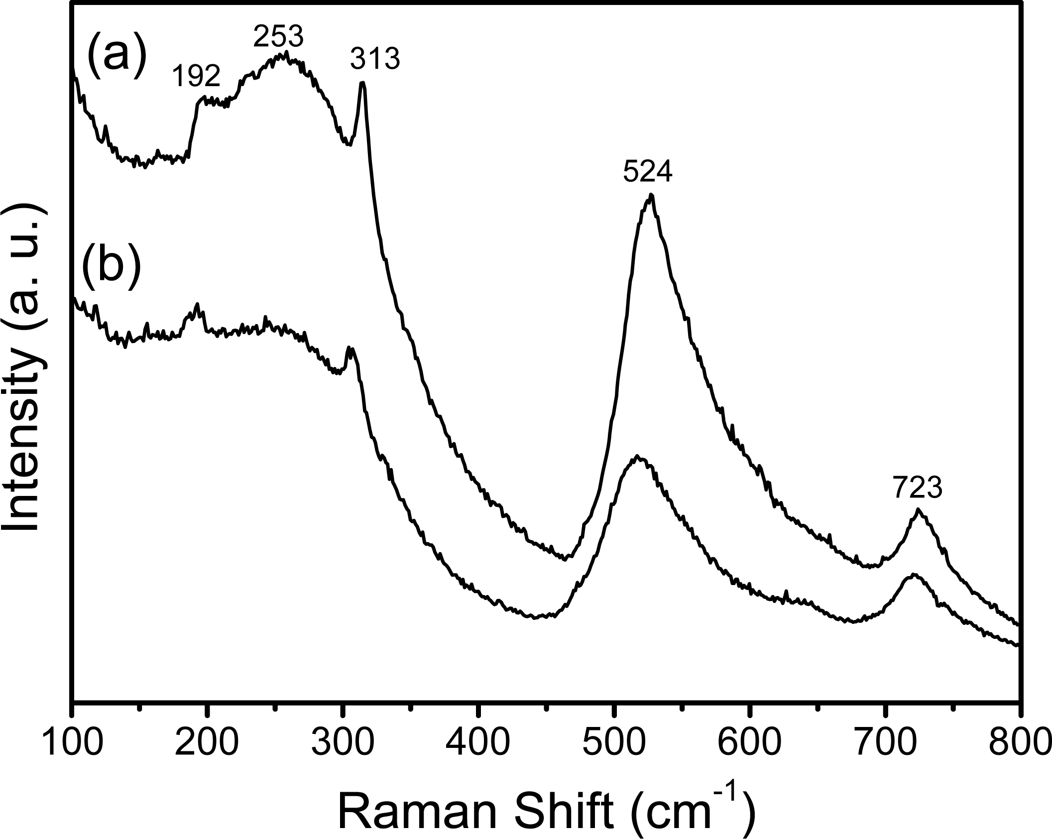
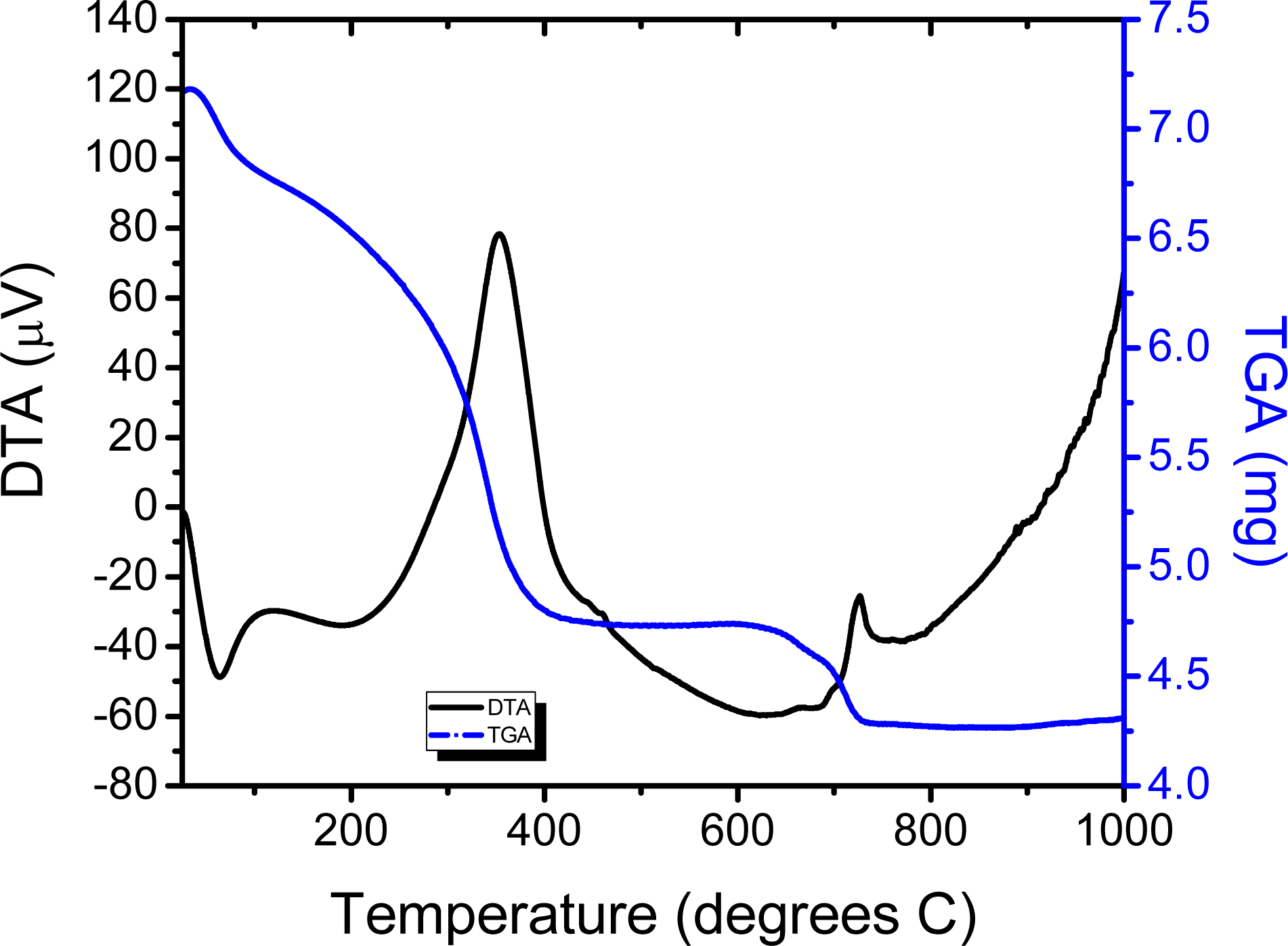
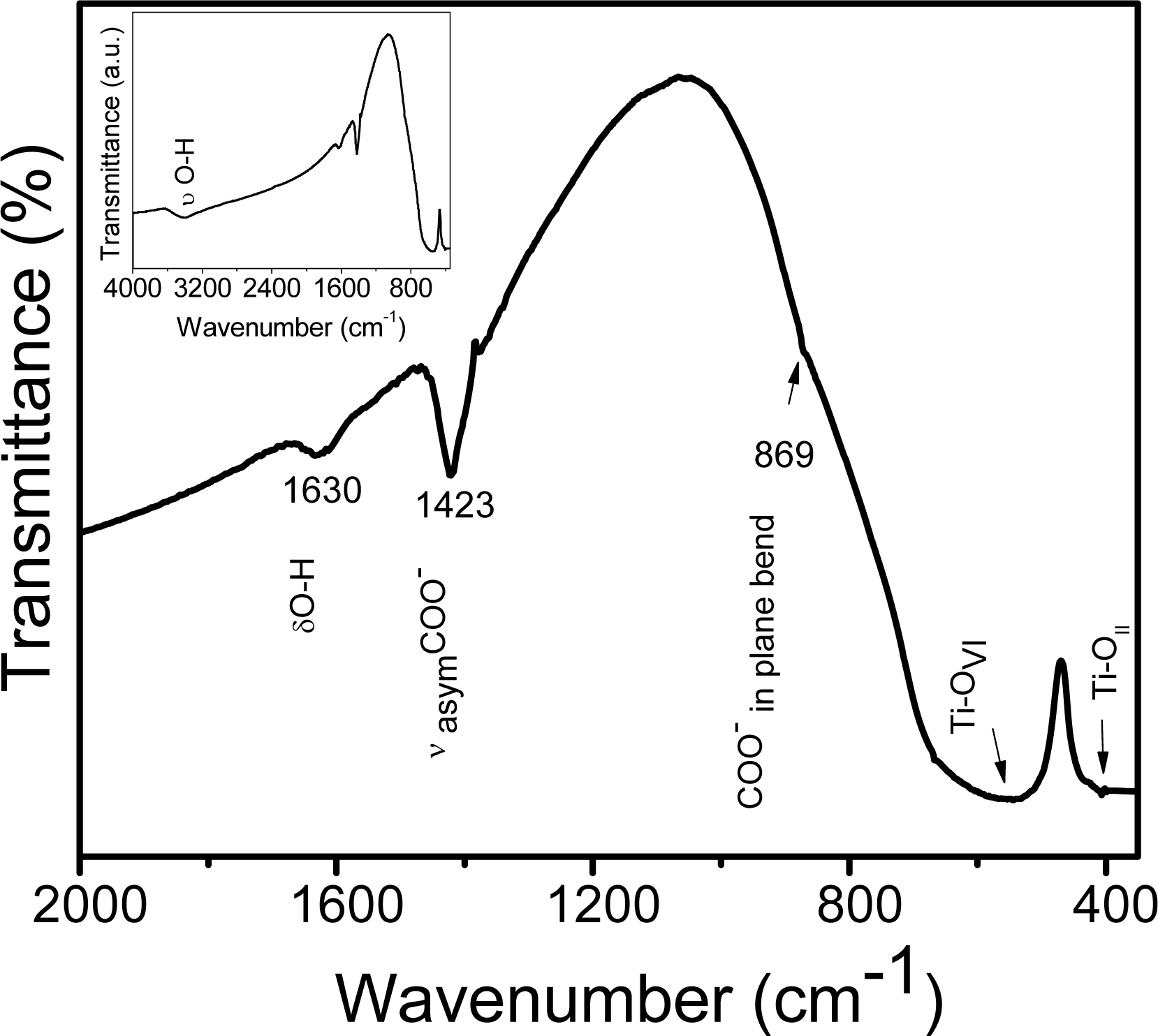
2.1. Chemical Studies

TG and DTA curves of the europium-doped BaTiO3 powders dried at 100 °C for 24 h are presented in Figure 1. In the first temperature region up to 200 °C, the endothermic peak situated at 115 °C results from the evaporation of alcohol and excess water. One endothermic peak situated at ~350 °C represents the decomposition of organic compounds. The third weight loss at 700–750 °C, associated with an exothermic peak, points out the transformation of amorphous decomposition products in BaTiO3. During decomposition, there is not significant loss of titanium according to Madarász [34] for the TiO(acac)1.0 hydroxo complexes for samples with acetylacetone content higher than 0.49. The acetylacetone anion deficiency is compensated with hydroxide ions considered products of hydrolysis (acac-Ti, + H2O-Ti.. + acacH) according to the following reaction [35]:
M   ( OR ) 4 + ( AcAc ) H M ( OR ) 3 ( AcAc ) + R OH
where M represent the metal atom M and ROH the leaving group. According to the XRD results, crystallization of barium titanate starts at 700 °C Moreover, the weight loss is minimal and the weight remans unchanged afterwards. It was stated that Ba2+ ions in the A site are mainly replaced by rare earth elements [36]. Eu3+ ions (0.098 nm) are most probably replaced Ba2+ (0.156 nm) cations rather than Ti4+ (0.065 nm). When Eu3+ was introduced to BaTiO3, three Ba2+ sites were substituted by two Eu3+ neighbors to maintain electrical neutrality, and so one vacancy was created, then the composition expected is Ba(1-0.05)Eu0.05TiO3, as observed by Rath [14].
Figure 2 shows the FTIR spectrum of BaTiO3:Eu powders, calcined at 700 °C for 2 h. The IR spectrum consisted mainly of three regions: the first region (Figure 2 inset) shows bands at 3,428 and 1,630 cm−1, which are due to the OH stretching vibration (υ) and OH deformation vibration (δ), respectively, arising from the water and isopropanol present in the porous structure of the barium titanante xerogel. The second region corresponds to the absorption bands at 1,423 and 869 cm−1, characteristic for the symmetrical vibrations and bending vibrations (in plane) of COO- groups arising from two types of ligands (the acetylacetone and acetic acid).The third region, 600-380 cm−1, represents the characteristic infrared absorptions of the Ti-O vibrations. The band situated around 565 cm−1 is due to TiO6 stretching vibration connected to the barium [37]. Finally, the peak at 414 cm−1 can be attributed to normal TiOII bending vibrations [38].
In order to complete the investigations of the local structure of the sol-gel BaTiO3:Eu3+ derived powders (Figure 3a) and films (Figure 3b), Raman spectra were used to measure the samples annealed at 700 °C. Both recorded spectra contain characteristic bands: (a) one weak band [A1(TO), E(LO)] at 192 cm−1, and (b) two intense broad bands A1(TO1) at ~253 cm−1 and A1(TO4) at 524 cm−1, with sharp peaks at ~313 cm−1 (TO3-LO3) and for the LO4 band at ~723 cm−1. Referring to Amami et al. [39], the sharp peaks situated at ~185 and ~235 cm−1 are associated with the cubic phase. Nevertheless, it is accepted by many researchers that the Raman peak at around 260 cm−1, which is somewhat variable in relation to particles size, shape, and aggregation, is due to the characteristics of tetragonal BaTiO3 [4043]. Finally, the origin of the above described bands in the cubic phase has been disputed due to the presence of Raman modes in this phase, indicating that it does not have perfect cubic symmetry but has small distortions [44]. By using the Raman-active modes discussed above, it was observed that the tetragonal phase is present in both BaTiO3 powder and film. However, the Raman band positions in the powder spectrum do not exactly match the peak positions in the films, which could be attributed to the internal stress from the surface tension in the nanocrystals [45].

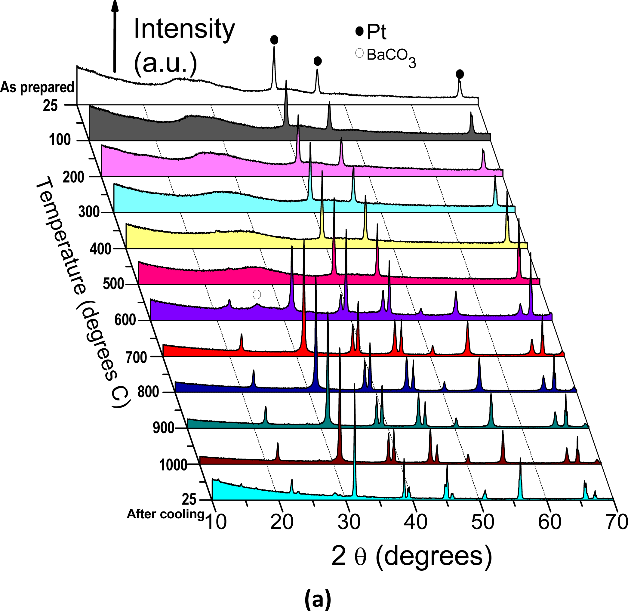
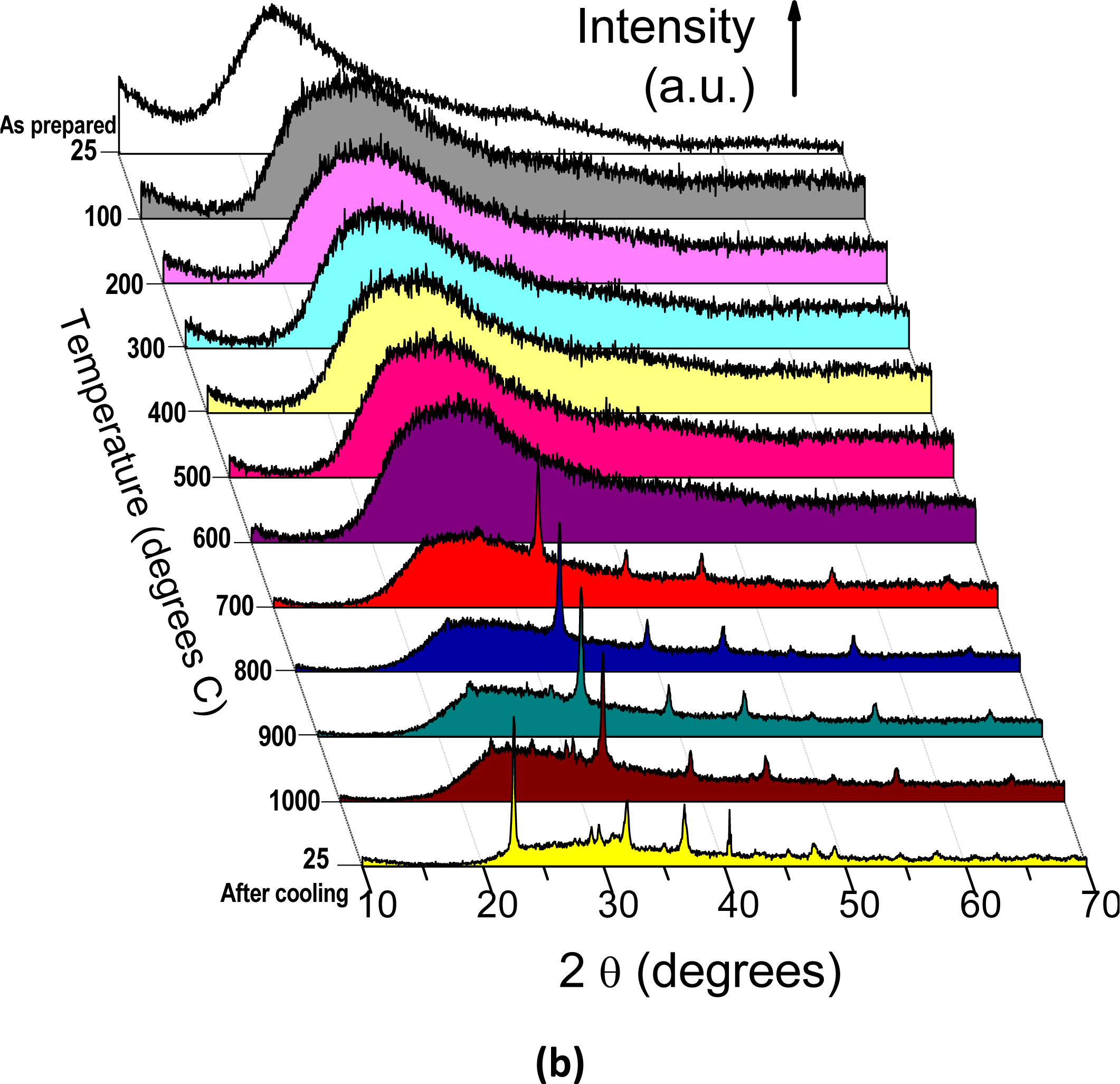
2.2. HT-XRD and XRD Studies

HT-XRD is used to follow in situ the formation of the BaTiO3:Eu3+ oxide from the BaTiO3 precursor gel as powder and film (see Figures 4a and b, respectively). The first scan is the pattern for the as-synthesized xerogel at room temperature, along with the intense peaks at 2θ values of about 40, 46 and 67 (results from the Pt sample holder). In the scan corresponding to 600 °C (Figure 4a), it is shown that some barium carbonate is formed during the decomposition of the precursor into the BaTiO3.
This carbonate decomposes between 650 and 700 °C. This led to our conclusion that pure BaTiO3 is fully crystallized after 2 h at 700 °C. Figure 5c shows the full XRD 2θ range of the crystallized powders, showing that two distinct peaks characterize BaTiO3 tetragonal structure [46]. Additionally, in situ HT-XRD experiments were performed to understand the phase stability of nanocrystalline BaTiO3:Eu3+ sol-gel films. Figure 4b shows the multiple plots of the barium titanate gel films scanned in air at various temperatures; from room temperature to 1000 °C, and again at room temperature after cooling. The gel film patterns indicate presence of an amorphous structure character up to 600 °C (Figure 5a) and the crystallization of BaTiO3:Eu3+ after the films were heated to 700 °C (Figure 4b and Figure 5b), in agreement with DTA analyses. The patterns of films calcined from 700 °C to 1000 °C are characterized by nanocrystalline BaTiO3 samples. Due to the very broad diffraction peaks attributed to the presence of the support or to the film thickness, it is not a sensitive enough technique to easily distinguish between ferroelectric tetragonal and paraelectric pseudocubic structure (Figure 5b). Nevertheless, it was found that after cooling from 1000 °C to room temperature, the BaTiO3:Eu3+ films transform from (1 1 0) orientation dominated to (1 0 0) dominated orientation, probably due to the textured densified films. The rates of cooling are mainly responsible for the high preferential orientation achieved in the BaTiO3 films. The transformation of orientation has been also observed in the case of PBxT layers films deposited on MgO (1 0 0) substrates [47].

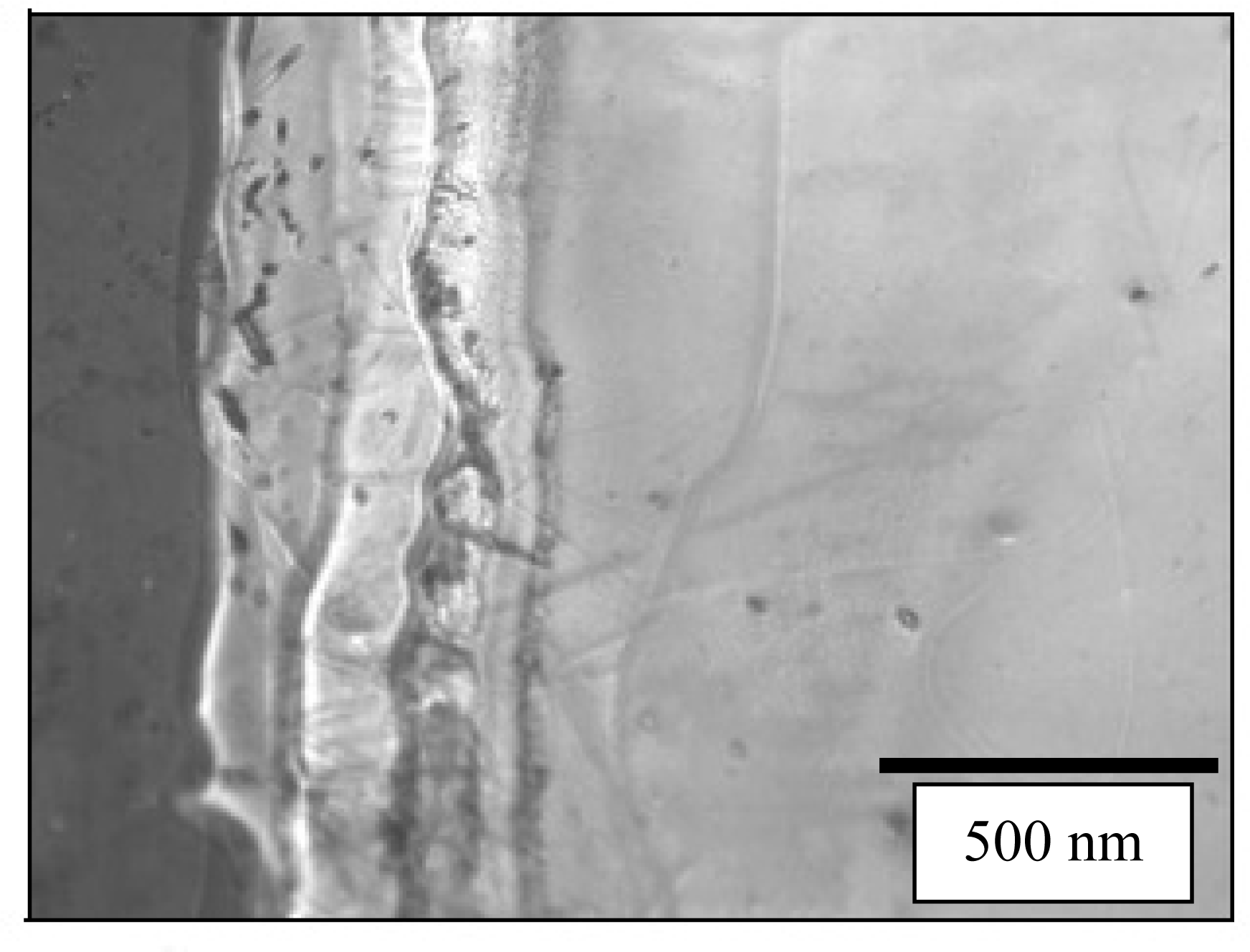
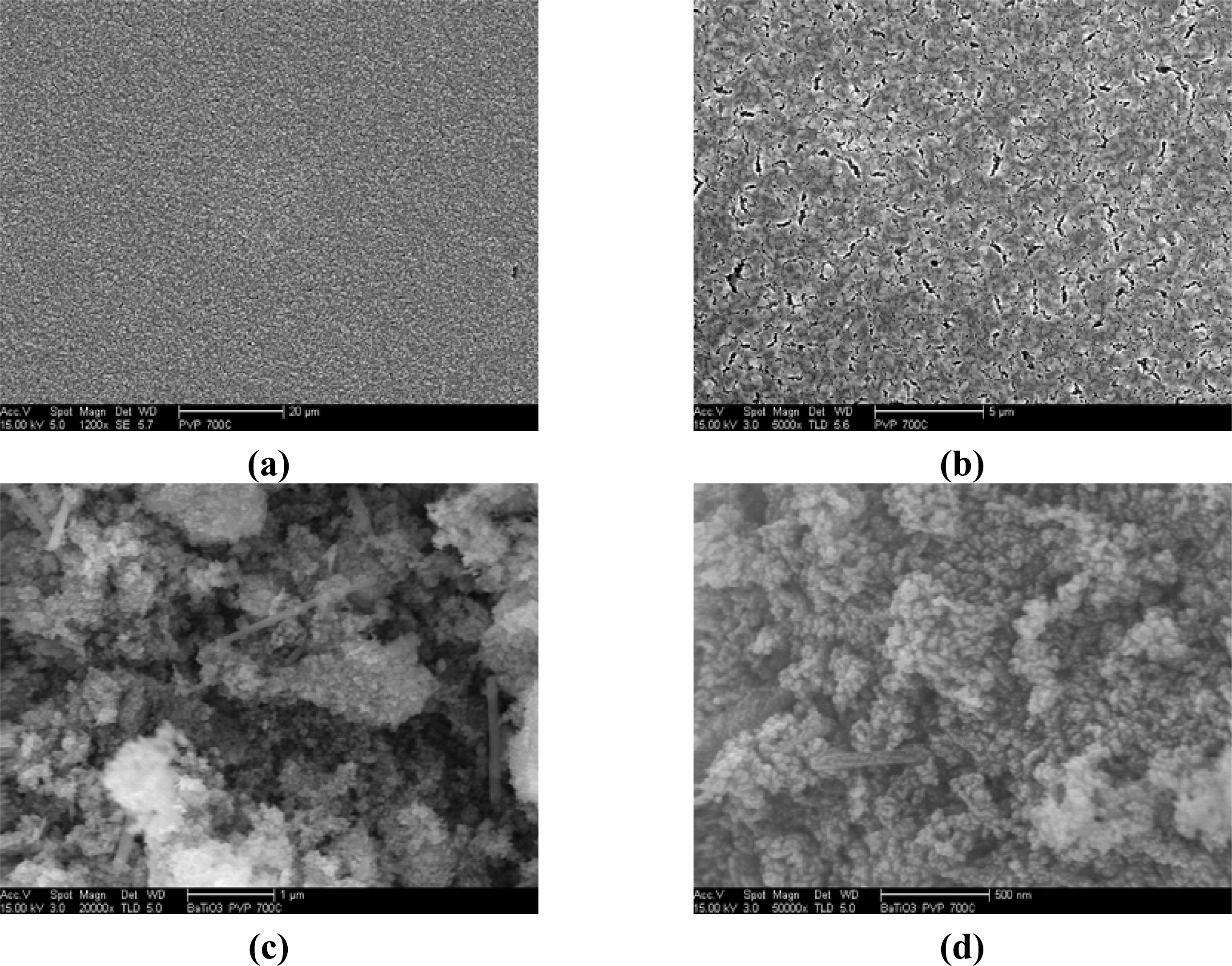
2.3. Microscopy Observations

The morphology of BaTiO3:Eu3+ powder and film calcined at 700 °C was investigated by SEM and is shown in Figure 6. The SEM images of BaTiO3:Eu3+ powders shown in Figures 6a and 6b reveal closely-packed fine equiaxed particles, about 100 nm in size. Aditionally, non-equiaxed powders (nanorod shaped structures) reaching ~800 nm in length were observed. On the other hand, equiaxed particles consisted of the approximately spherical type. For example, the shape of BaTiO3:Eu3+ powders at 700 °C results in a combination of non-equiaxed and equiaxed morphology due to the coexistence of the tetragonal and cubic phase. From the surface images of BaTiO3:Eu3+ films shown in Figures 6c and 6d, the microstructures were homogeneous, continuous and crack-free. At high magnification, as shown in Figure 6d, there was no evidence of cracks; however, the presence of pores can be associated with the presence of PVP. Kozuka et al. [48] have reported that when the film is heated directly at 700 °C, the decomposition of PVP and the crystallization of the film may occur concurrently. This can lead to crystallization with much less densification, which provides higher porosity and smaller tensile stress. Profilometry is a very common method of post-process measurement of films thickness [49]. The thickness of BaTiO3:Eu3+ calcined at 700 °C thin film was about 500 nm is in agreement with the determined by cross section SEM, as can be observed in Figure 7.

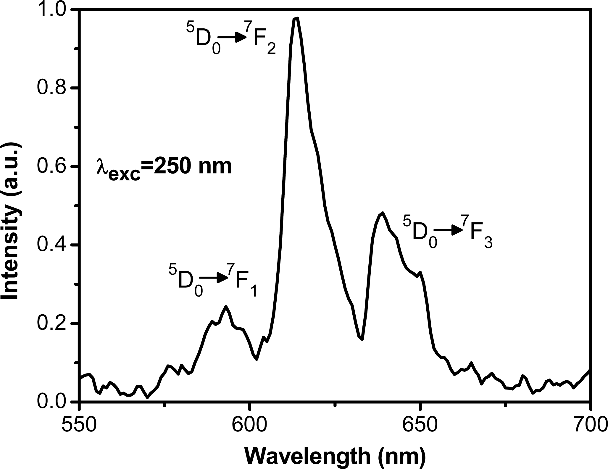
2.4. Luminescence Properties

The room temperature photoluminescence emission spectrum of BaTiO3:Eu3+ (5 mol %) film in the range of 550–700 nm and excited at 250 nm wavelength is shown in Figure 8. The red emission from the film is easily seen to the naked eye when excited with 254 nm from UV lamp (see the inset of Figure 8). Peaks centered at 595, 615 and 645 nm are assigned to 5D07F1, 5D07F2 and 5D07F3, respectively, arising from the lowest excited 5D0 level into the split by the crystal field 7FJ (where J = 0, 1, 2, 3, 4, 5, 6) as observed by other authors and is in agreement with results reported for bulk Eu3+ doped cubic yttria [5051].
In most cases, transitions to the higher laying levels (7F5, 7F6) are difficult to detect due to their low intensity [52]. The 5D07F1 band originates from magnetic-dipole transition and, in this case, the change of the crystal field strength has very little influence on it. The dominant peak is observed around 615 nm (5D07F2) and is attributed to the forced electric-dipole transition allowed only at low symmetries with no inversion center. Its intensity is sensitive to the local structure surrounding the Eu3+ ions. Thus the ratio R = (5D07F2)/(5D07F1) > 1 suggest that Eu3+ occupy sites with low symmetry. The origin of these transitions (electric dipole or magnetic dipole) from emitting to terminating levels depends upon the site where Eu3+ is located in the host lattice, and the type of these transitions is determined by the selection rules. It must be mentioned that the signal emitted of BaTiO3:Eu3+ films are not very strong, probably due to the final thickness, influencing directly the suitable effective value of providing high emission [11].

3. Experimental Section

3.1. Experimental Procedure

Europium doped BaTiO3 films were prepared using the sol-gel process and the dip-coating technique. The starting materials were: barium acetate, Ba(CH3COO)2 [Aldrich], titanium butoxide, Ti(C4H10O)4 [Aldrich], europium III chloride, EuCl3 [Alfa Aesar] acetylacetone, C5H8O2 [Aldrich], acetic acid, C2H4O2, [99.8%, Fermont], distilled water, H2O, isopropyl alcohol, C3H7OH [99.9%, Fermont] and polyvinylpyrrolidone, PVP; FW: 630,000 g mol−1 [Alfa Aesar]. Barium acetate and europium chloride were dissolved in water in the following molar ratio: Ba:Eu:H2O [1:0.05:40]. Titanium butoxide was mixed with acetylacetone in a molar ratio [1:1], and the reaction was continued under reflux at 60 °C for 6 h. PVP was dissolved in isopropyl alcohol in a molar ratio [1:18] for 2 hours at room temperature. The molar ratios were calculated with respect to Ba. The titanium and barium solutions were mixed and stirred for 2 h. Thereafter, the PVP solution was added to the previous solution drop by drop under vigorous magnetic stirring for 2 h at room temperature. In this step, a transparent and stable BaTiO3:Eu3+ sol was formed. Uniform coatings of BaTiO3:Eu3+ were dip coated on high polished and carefully cleaned silica substrates (Herasil from Heraeus®) with a constant withdrawal speed of 5 cm min−1 for five cycles. BaTiO3:Eu3+ films were dried at 100 °C between each coating under O2 flow for 10 min. In this step, one film was taken for HT-XRD studies. The BaTiO3:Eu3+ films were finally calcined at 700 °C for 10 min. The remaining solution was dried in order to obtain a xerogel at 100 °C for 24 h (xerogel was analyzed in HT-XRD). After drying, the xerogel was thermally treated at 700 °C for 2 h in order to densify and crystallize the sol-gel powders.

3.2. Apparatus

The xerogel was investigated by Thermogravimetric (TGA)-Differential Thermal (DTA) and High-Temperature X-Ray Diffraction (HT-XRD) analyses. The thermograms were recordered from 23 °C to 1000 °C using a Mettler Toledo TGA/SDTA 851e apparatus at a scan rate of 2 °C min −1 in a flux of nitrogen. The HT-XRD studies were carried out on a Philips Xpert Pro diffractometer operating with the Cu Kα radiation and equipped with a high temperature chamber, over a temperature range from 25 to 1000 °C in air atmosphere. The structural phases of the crystallized powder and film were recorded on a Siemens D5000 powder diffractometer using the Bragg–Brentano configuration and the Cu Kα radiation. The IR transmittance spectra were recorded on the crystallized powders calcined at 700 °C using a FTIR 2000 Perkin-Elmer in the range 4,000-400 cm−1, with the aim of completing DRX and DTA-TGA experiments. In order to determine the powder microstructure and quality of the derived BaTiO3:Eu3+ crystallized sol-gel films, SEM images were obtained using a JEOL 3200 scanning electron microscope (SEM) with a field emission gun operating at 15 and 8 kV. The thickness of BaTiO3 films was determined via an Alpha-step IQ profilometer (Tencor Instruments). The fluorescence emission spectra for BaTiO3:Eu3+ film as obtained with a modular Spectra Pro (Acton Research) fluorometer with a PM tube R955 (Hamamatsu).

4. Conclusions

The sol-gel method and dip-coating techniques have been successfully employed to prepare BaTiO3:Eu3+ (5 mol%) powder and film incorporating a viscosity modifier (PVP) in the sol. Both systems exhibited crystalline BaTiO3 phase at 700 °C within 2 h as revealed the HT-XRD studies. The powders were mainly spherical with some rod shapes; however, the films presented good surface morphology as detected by SEM. The obtained films exhibit the room temperature photoluminescence of the europium ions, with the predominant band at 615 nm (5D07F2 transition). The obtained thickness (~500 nm) of BaTiO3:Eu3+ films must be optimized to be promising for luminescent applications.

Acknowledgments

The authors gratefully acknowledge the financial support of this work by the SEP-CONACYT project 59408 and SIP-IPN projects 20090546 and 20090528. M. García Hernández thanks Conacyt for her Ph.D scholarship grant. The authors wish to acknowledge Daniel Ramírez González from IPICYT for the SEM analysis support, Joël Cellier (University Clermont II) for many hours of helpful discussions concerning HT-XRD measurements, Thierry ENCINAS (ENSCCF) for the DTA analyses. The authors would like to thank David Nentwick for his editing work on this paper. The authors would also like to thank M. García Murillo for her assistance.

References and notes

  1. Battisha, IK; Beyally, AEl; El Mongy, SA; Nahrawi, AM. Development of the FTIR properties of nano-structure silica gel doped with different rare earth elements, prepared by sol-gel route. J. Sol-Gel Sci. Technol 2007, 41, 129–137. [Google Scholar]
  2. Battisha, K; El Beyally, A; Soliman, SL; El Nahrawi, AS. Indian, structural and optical studies of activated thin film and monolith nano-structure silica gel with different rare earth elements prepared by sol-gel techniques. Indian J. Pure Appl. Phys 2007, 45, 441–453. [Google Scholar]
  3. Tissue, BM. Synthesis and luminescence of lanthanide ions in nanoscale insulating hosts. Chem. Mater 1998, 10, 2837–2845. [Google Scholar]
  4. Zhou, J; Li, L; Gui, Z; Buddhudu, S; Zhou, Y. Photoluminescence of CdSe nanocrystallites embedded in BaTiO3 matrix. Appl. Phys. Lett 2000, 76, 1540–1542. [Google Scholar]
  5. Huang, CH; McCaughan, L. Polarization-dependent enhancement of population inversion and of green upconversion in Er:LiNbO3 by Yb codoping. IEEE Photonics Technol. Lett 1997, 9, 599–601. [Google Scholar]
  6. Itoh, S; Toki, H; Tamura, K; Kataoka, F. A new red-emitting phosphor, SrTiO3:Pr3+, for low-voltage electron excitation. J. Appl. Phys 1999, 38, 6387–6391. [Google Scholar]
  7. Allak, HM; Brinkman, AW; Russell, GJ; Woods, J. The effect of Mn on the positive temperature coefficient of resistance characteristics of donor doped BaTiO3 ceramics. J. Appl. Phys 1988, 63, 4530–4535. [Google Scholar]
  8. Morales Ramírez, AdeJ; García Murillo, A; Carrillo Romo, FdJ; Ramírez Salgado, J; Le Luyer, C; Chadeyron, G; Boyer, D; Moreno Palmerin, J. Preparation and studies of Eu3+ and Tb3+ co-doped Gd2O3 and Y2O3 sol–gel scintillating films. Thin Solid Films 2009. [Google Scholar]
  9. García Murillo, A; de J. Carrillo Romo, F; Le Luyer, C; de J. Morales Ramirez, A; García Hernández, M; Moreno Palmerin, J. Sol–gel elaboration and structural investigations of Lu2O3:Eu3+ planar waveguides. J. Sol-Gel Sci. Technol 2009, 50, 359–367. [Google Scholar]
  10. Hreniak, D; Strek, W; Amami, J; Guyot, Y; Boulon, G; Goutaudier, C; Pazik, R. The size-effect on luminescence properties of BaTiO3:Eu3+ nanocrystallites prepared by the sol–gel method. J. Alloys Comp 2004, 380, 348–351. [Google Scholar]
  11. Badr, Y; Salah, A; Battisha, I. Effect of europium ion concentrations on the photoluminescence emission of nano-crystalline BaTiO3 prepared by sol–gel technique. J. Sol-Gel Sci. Technol 2005, 34, 219–226. [Google Scholar]
  12. Hreniak, D; Strek, W; Chmielowiec, J; Pasciak, G; Pazik, R; Gierlotka, S; Lojkowski, W. Preparation and conductivity measurement of Eu doped BaTiO3 nanoceramic. J Alloys Comp 2006, 408, 412, 637–640. [Google Scholar]
  13. Pazik, R; Hreniak, D; Strek, W; Kessler, VG; Seisenbaeva, GA. Photoluminescence investigations of Eu3+ doped BaTiO3 nanopowders fabricated using heterometallic tetranuclear alkoxide complexes. J. Alloys Comp 2008, 451, 557–562. [Google Scholar]
  14. Rath, MK; Pradhan, GK; Pandey, B; Verma, HC; Roul, BK; Anand, S. Synthesis, characterization and dielectric properties of europium-doped barium titanate nanopowders. Mat. Lett 2008, 62, 2136–2139. [Google Scholar]
  15. Li, J; Kuwabara, M. Preparation and luminescent properties of Eu-doped BaTiO3 thin films by sol–gel process. Sci. Technol. Adv. Mat 2003, 4, 143–148. [Google Scholar]
  16. Hoerman, H; Ford, GM; Kaufmann, LD; Wessels, BW. Dynamic response of the dielectric and electro-optic properties of epitaxial ferroelectric thin films. J. Appl. Phys. Lett 1998, 73, 2248–2250. [Google Scholar]
  17. Yoon, SG; Safari, A. (Ba0,5, Sr0.5)TiO3 thin film preparation by r.f. magnetron sputtering and its electric properties. Thin Solid Films 1995, 254, 211–215. [Google Scholar]
  18. Li, J; Wu, YJ; Yamamoto, T; Kuwabara, M. Electrophoretic deposition and photoluminescent properties of Eu-doped BaTiO3 thin film from a suspension of monodispersed nanocrystallites. Sci. Technol. Adv. Mat 2004, 5, 393–398. [Google Scholar]
  19. Battisha, IK; Speghini, A; Polizzi, S; Agnoli, F; Bettinelli, M. Molten chloride synthesis, structural characterisation and luminescence spectroscopy of nanocrystalline Eu3+ doped BaTiO3 and SrTiO3. Mat. Lett 2002, 57, 183–187. [Google Scholar]
  20. Xu, J; Zhai, J; Yao, X; Xue, J; Huang, Z. Dielectric and optical properties of BaTiO3 thin films prepared by low-temperature process. J. Sol-Gel Sci. Technol 2007, 42, 209–212. [Google Scholar]
  21. Matsuda, H; Kobayashi, N; Kobayashi, T; Miyazaya, K; Kuwabara, M. Room-temperature synthesis of crystalline barium titanate thin films by high–concentration sol–gel method. J. Non-Cryst. Sol 2000, 271, 162–166. [Google Scholar]
  22. Manso-Silván, M; Fuentes-Cobas, L; Martín-Palma, RJ; Hernández-Vélez, M; Martínez-Duart, JM. BaTiO3 thin films obtained by sol–gel spin coating. Surf Coat Technol 2002, 151, 152, 118–121. [Google Scholar]
  23. Qifang, L; Dairong, C; Xiuling, J. Preparation and characterization of BaTiO3 long fibers by sol-gel process using catechol-complexed alkoxide. J. Sol-Gel Sci. Technol 2002, 25, 243–248. [Google Scholar]
  24. Shimooka, H; Ken-ichi, Y; Seiji, T. Preparation of transparent, partially-crystallized BaTiO3 monolithic xerogels by sol-gel processing. J. Sol-Gel Sci. Technol 1998, 13, 873–876. [Google Scholar]
  25. Sharma, HB; Mansingh, A. Sol-gel processed barium titanate ceramics and thin films. J. Mater. Sci 1998, 33, 4455–4459. [Google Scholar]
  26. Henry, M; Jolivet, JP; Livage, J. Structure and Bonding; Springer-Verlag Berlin Heidelberg: New York, NY, USA, 1992. [Google Scholar]
  27. Henry, M; Jolivet, JP; Livage, J. Ultrastructure Processing of Advance Materials; John Wiley & Sons, Inc: Hoboken, NJ, USA, 1992. [Google Scholar]
  28. Doeuff, S; Henry, M; Sanchez, C; Livage, J. Hydrolysis of titanium alkoxides: Modification of the molecular precursor by acetic acid. J. Non-Cryst. Solids 1987, 89, 206–216. [Google Scholar]
  29. Mosset, A; Luneau, I; Galy, J. Sol-gel processed BaTiO3: Structural evolution from the gel to the crystallisation powder. J Non-Cryst Solids 1988, 100, 339–344. [Google Scholar]
  30. Bernier, JC; Rehspringer, JL; Vilminot, S; Poix, P. Synthesis and sintering comparison of cordierite powders. Mat. Res. Soc. Symp. Proc 1986, 73, 129–134. [Google Scholar]
  31. Kozuka, H; Takenaka, S; Tokita, H; Okubayashi, M. PVP-assisted sol-gel deposition of single layer ferroelectric thin films over submicron or micron in thickness. J. Eur. Ceram. Soc 2004, 24, 1585–1588. [Google Scholar]
  32. Kozuka, H; Kajimura, M; Hirano, T; Katayama, K. Crack-free thick ceramic coating films via non-repetitive dip-coating using polyvinylpyrrolidone as stress-relaxing agent. J. Sol-Gel Sci. Technol 2000, 19, 205–209. [Google Scholar]
  33. Kozuka, H; Higuchi, A. Stabilization of poly(vinylpyrrolidone)-containing alkoxide solutions for thick sol-gel barium titanate films. J. Am. Ceram. Soc 2003, 86, 33–38. [Google Scholar]
  34. Madarasz, J; Kaneko, S; Okuya, M; Pokol, G. Comparative evolved gas analyses of crystalline and amorphous titanium(IV)oxo-hydroxo-acetylacetonates by TG-FTIR and TG/DTA-MS. Thermochim. Acta 2009, 489, 37–44. [Google Scholar]
  35. Legrand-Buscema, C; Malibert, C; Bach, S. Elaboration and characterization of thin films of TiO2 prepared by sol-gel process. Thin Sol. Films 2002, 418, 79–84. [Google Scholar]
  36. Watanabe, K; Ohsato, H; Kishi, H; Okino, Y; Kohzu, N; Iguchi, Y; Okuda, T. Solubility of La–Mg and La–Al in BaTiO3. Solid State Ionics 1998, 108, 129–135. [Google Scholar]
  37. Harizanov, OA. Sol-gel BaTiO3 from a peptized solution. Mat. Lett 1998, 34, 232–236. [Google Scholar]
  38. Ghosh, S; Dasgupta, S; Sen, A; Maiti, HS. Synthesis of barium titanate nanopowder by a soft chemical process. Mat. Lett 2007, 61, 538–541. [Google Scholar]
  39. Amami, J; Hreniak, D; Guyot, Y; Pazik, R; Goutaudier, C; Boulon, G; Ayadi, M; Strek, W. Second harmonic generation and Yb3+ cooperative emission used as structural probes in size-driven cubic–tetragonal phase transition in BaTiO3 sol–gel nanocrystals. J Lumin 2006, 119, 120, 383–387. [Google Scholar]
  40. Zhu, X; Wang, J; Zhang, Z; Zhu, J; Zhou, S; Liu, Z; Ming, N. Atomic-scale characterization of barium titanate powders formed by the hydrothermal process. J. Am. Ceram. Soc 2008, 91, 1002–1008. [Google Scholar]
  41. An, C; Liu, C; Wang, S; Liu, Y. Generalized large-scale synthesis of MTiO3 (M = Ba, Sr, Pb) nanocrystals. Mat. Res. Bull 2008, 43, 932–938. [Google Scholar]
  42. Song-Wei, L; Burtrand, IL; Zhong-Lin, W; William, DS. Hydrothermal synthesis and structural characterization of BaTiO3 nanocrystals. J. Crystal Growth 2000, 219, 269–276. [Google Scholar]
  43. Asiaie, R; Zhu, W; Akbar, SA; Dutta, PK. Characterization of submicron particles of tetragonal BaTiO3. Chem. Mater 1996, 8, 226–234. [Google Scholar]
  44. Amami, J; Hreniak, D; Guyot, Y; Pazik, R; Strek, W; Goutaudier, C; Boulon, G. New optical tools used for characterization of phase transitions in nonlinear nano-crystals. Example of Yb3+-doped BaTiO3. J. Phys. Condens. Matter 2007, 19, 1–14. [Google Scholar]
  45. Ma, WH; Zhang, MS; Yin, Z. Phonon characteristics of thin film and nanophase lead titanate. J. Korean Phys. Soc 1998, 32, 1137–1139. [Google Scholar]
  46. García-Murillo, A; Carrillo-Romo, FJ; García-Hernández, M; Barbosa-García, O; Meneses-Nava, A; Palomares-Sánchez, S; Flores-Vela, A. Structural and optical characteristics of BaTiO3:Yb3+ powders. Mat Trans 2009, 50, 1850–1854. [Google Scholar]
  47. Wein-Duo, Y; Haile, SM. Highly preferred oriented lead barium titanate thin films using acetylacetone as chelating agent in a sol-gel process. Rev. Adv. Mater. Sci 2005, 10, 143–148. [Google Scholar]
  48. Kozuka, H; Takenaka, S. Single-step deposition of gel-derived lead zirconate titanate films: Critical thickness and gel film to ceramic film conversion. J. Am. Ceram. Soc 2002, 85, 2696–2702. [Google Scholar]
  49. Gary, SM; Costas, JS. Fundamentals of Semiconductor Manufacturing and Process Control; Wiley-Interscience, A John wiley & Sons, Inc: Hoboken, NJ, USA, 2006; p. 63. [Google Scholar]
  50. Hreniak, D; Strek, W; Amami, J; Guyot, Y; Boulon, G; Goutaudier, C; Pazik, R. The size-effect on luminescence properties of BaTiO3:Eu3+ nanocrystallites prepared by the sol–gel method. J. Alloys Comp 2004, 380, 348–351. [Google Scholar]
  51. Pązik, R; Hreniak, D; Stręk, W; Kessler, VG; Seisenbaeva, GA. Photoluminescence investigations of Eu3+ doped BaTiO3 nanopowders fabricated using heterometallic tetranuclear alkoxide complexes. J. Alloys Comp 2008, 451, 557–562. [Google Scholar]
  52. Blasse, G; Grabmaier, BC. Luminescent Materials; Springer-Verlag: New York, NY, USA, 1994. [Google Scholar]
Figure 1. DTA and TGA curves for BaTiO3:Eu xerogel powder.
Ijms 10 04088f1
Figure 2. FTIR spectrum of BaTiO3:Eu3+ sol-gel powder calcined at 700 °C.
Ijms 10 04088f2
Figure 3. Raman spectra of BaTiO3:Eu3+ powder (a) and film (b).
Ijms 10 04088f3
Figure 4. HT-XRD patterns of BaTiO3:Eu3+ powders (a) and films (b). Diffraction peaks related to the platinum ribbon are noted with a cross.
Ijms 10 04088f4aIjms 10 04088f4b
Figure 5. XRD patterns of BaTiO3:Eu3+ film calcined at 500 °C (a), 700 °C (b) and powder calcined at 700 °C (c).
Ijms 10 04088f5
Figure 6. SEM micrographs of BaTiO3:Eu3+ of films (a), (b) and powders (c), (d) calcined at 700 °C.
Ijms 10 04088f6
Figure 7. Cross section SEM micrograph of BaTiO3:Eu3+ film calcined at 700 °C.
Ijms 10 04088f7
Figure 8. PL emission spectrum of BaTiO3:Eu3+ film annealed at 700 °C under UV excitation. The inset shows a picture of the strong emission.
Ijms 10 04088f8

