Article

Pharmacological Proprieties of the Ethanol Extract of Muehlenbeckia platyclada (F. Muell.) Meisn. Leaves

by
Leopoldina Leonor Fagundes
1,
Glauciemar Del-Vechio Vieira
1,
José de Jesus R. G. De Pinho
1,
Célia Hitomi Yamamoto
1,
Maria Silvana Alves
2,
Paulo César Stringheta
3 and
Orlando Vieira De Sousa
1,*
1
Departamento Farmacêutico, Faculdade de Farmácia e Bioquímica, Universidade Federal de Juiz de Fora, Campus Universitário, Martelos, 36036-330, Juiz de Fora, MG, Brazil
2
Departamento de Análises Clínicas, Faculdade de Farmácia e Bioquímica, Universidade Federal de Juiz de Fora, Campus Universitário, Martelos, 36036-330, Juiz de Fora, MG, Brazil
3
Departamento de Tecnologia de Alimentos, Centro de Ciências Exatas e Tecnológicas, Universidade Federal de Viçosa, Campus Universitário, 36571-000 - Viçosa, MG, Brazil
*
Author to whom correspondence should be addressed.
Int. J. Mol. Sci. 2010, 11(10), 3942-3953; https://doi.org/10.3390/ijms11103942
Submission received: 17 September 2010 / Revised: 29 September 2010 / Accepted: 12 October 2010 / Published: 15 October 2010
(This article belongs to the Section Biochemistry)

Abstract

:
Antinociceptive and anti-inflammatory activities of the Muehlenbeckia platyclada leaves’ ethanol extract were investigated in animal models. The extract (p.o.) reduced the number of abdominal contortions induced by acetic acid by 21.57% (400 mg/kg). After intraplantar injection of formalin, a dose of 400 mg/kg (p.o.) inhibited the time spent paw licking in the first phase (26.43%), while the second phase was inhibited by 10.90 and 36.65% at the doses of 200 and 400 mg/kg, respectively. The extract (p.o.) increased the reaction time on a hot plate at a dose of 400 mg/kg (32.68 and 40.30%) after 60 and 90 minutes of treatment, respectively. The paw edema was reduced by extract (p.o.) at doses of 100 (15.46 and 16.67%), 200 (22.68 and 25.64%) and 400 mg/kg (29.50 and 37.33%) after 3 to 4 h of carrageenan application, respectively. Doses of 100, 200 and 400 mg/kg (p.o.), administered 4 h after the carrageenan injection, reduced the exudate volume (11.28, 21.54 and 45.13%), while leukocyte migration was reduced by 21.21 and 29.70% at the doses of 200 and 400 mg/kg, respectively. These results indicate that the ethanol extract from M. platyclada may constitute a potential target for the discovery of new molecules with antinociceptive and anti-inflammatory activities that can be explored for their therapeutic use.

1. Introduction

Medicinal plants are one of the most important sources of active substances with therapeutic potential and these are often used to cure a variety of diseases in humans [1,2]. Moreover, the evaluation of pharmacological effects can be used as a strategy for discovering new drugs of plant origin [2,3]. In particular, the use of medicinal plants as anti-inflammatory and analgesic agents is a common practice and have been the target of recent studies [4,5].
The genus Muehlenbeckia is constituted by four species belonging to the family Polygonaceae. Plants of this genus, such as Muehlenbeckia platyclada (F. Muell.) Meisn., commonly known as carqueja de jardim or fita de moça, are found in South America, and have been traditionally used as diuretic, hypotensive, antihemorragic, sedative, antirheumatic, abortive, cicatrizant, antiulcerogenic, anti-inflammatory and anthelmintic agents [6,7].
Among the active compounds found in M. platyclada, the flavonoids (morin-3-O-α-rhamnopyranoside, kaempferol-3-O-α-rhamnopyranoside, kaempferol-3-O-β-glucopyranoside, quercetin 3-O-α-rhamnopyranoside and (+)-catechin), were reported [8]. However, in species of Muehlenbeckia, such as M. tamnifolia, free anthraquinones (chrysophanic acid, emodin, and rhein) and glycoside anthraquinones have been identified [9]. Epicatechin, emodin-8-glycoside and rutin were identified from the aerial part and the root of M. hastulata [10]. Pheophorbide A, hypericin and protohypericin, isolated from this specie, demonstrated potent activity against influenza virus [11].
Considering pharmacological studies, M. hastulata possesses significant oxytoxic and analgesic activities [10]. The chloroform, butanol and aqueous extracts as well as flavonoids isolated from M. platyclada inhibited the generation of superoxide anion and elastase released by human neutrophils, indicating anti-inflammatory activity [8]. Based on the anthelmintic property, the methanolic extract of M. platyclada did not exhibit activity against Bursaphelenchus xylophilus [12].
In order to evaluate the pharmacological basis for a better understanding of the use of M. platyclada in folk medicine, the present study was designed to investigate the antinociceptive and anti-inflammatory effects of ethanol extract obtained from leaves using animal models.

2. Results and Discussion

2.1. Phytochemistry Screening

The phytochemical screening results of the ethanol extract showed the presence of different types of active constituents like flavonoids, terpenoids, sterols, coumarins, tannins, saponins and volatile oils.

2.2. Acute Toxicity

At the doses administered per oral route (p.o.), the ethanol extract from M. platyclada leaves was toxic to animals with LD50 of 2.67 g/kg (95% confidence intervals 1.65–4.31 g/kg). This result was applied as a parameter to establish the dosage definition in the experiments of pharmacological activities.

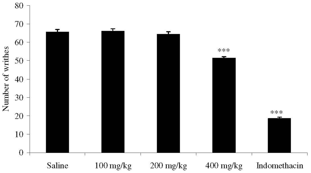
2.3. Writhing Response Induced by Acetic Acid in Mice

Dose (p.o.) of 400 mg/kg of M. platyclada extract significantly reduced (p < 0.001) the abdominal contortions induced by acetic acid to 51.37 ± 0.84 compared to the respective control (65.50 ± 1.50) (Figure 1).

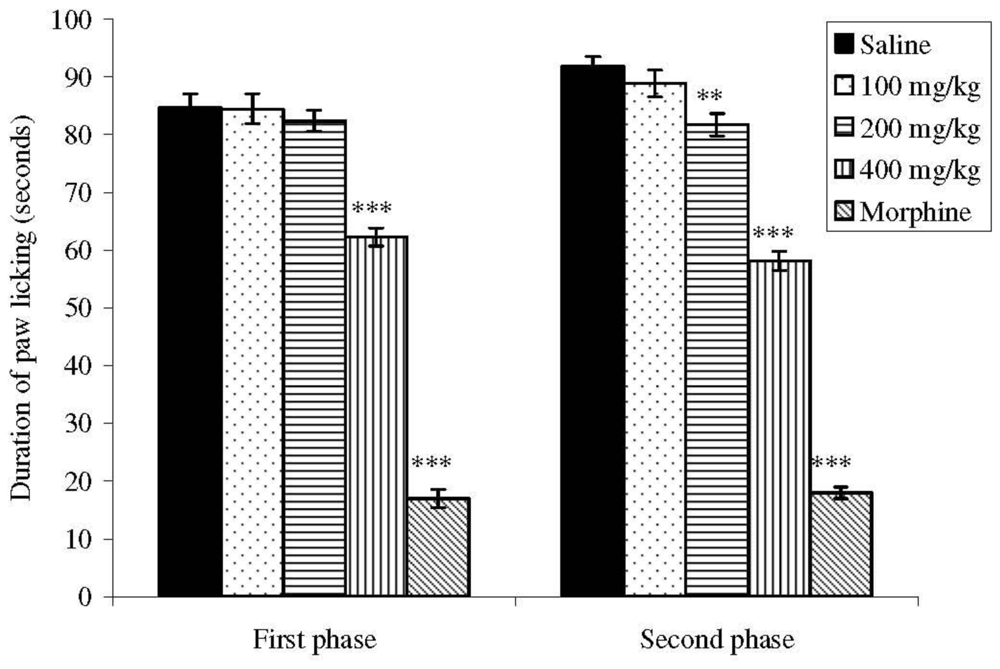
2.4. Effects on Formalin-Induced Nociception in Mice

The intraplantar injection of formalin promoted a biphasic characteristic response (Figure 2). The time spent licking in the first phase (0–5 min) was 84.62 ± 2.45 s and in the second phase (15–30 min) was 91.75 ± 1.79 s for the control group. After 60 min of treatment, a dose (p.o.) of 400 mg/kg of extract significantly inhibited (p < 0.001) the first phase at 26.43% and the doses of 200 and 400 mg/kg reduced the second phase at 10.90 and 36.65%, respectively, when compared to the control.

2.5. Effects on Hot-Plate Latency Assay in Mice

The M. platyclada ethanol extract increased the latency time of mice exposed to the hot plate (Table 1). After 60 and 90 min of treatment, dose (p.o.) of 400 mg/kg (32.68 and 40.31%) increased significantly (p < 0.05 and p < 0.01, respectively) the latency time in the respective control group. Morphine proved to be a potent analgesic, increasing the latency time within the evaluation periods. Naloxone, an opioid antagonist, blocked the morphine action but did not alter the antinociceptive effect of the tested extracts.

2.6. Effects on Carrageenan-Induced Edema in Rats

The M. platyclada ethanol extract anti-inflammatory effect evaluated by the paw edema method induced by carrageenan is shown in Table 2. Edema inhibition was observed 3 h after carrageenan application of doses (p.o.) of 100 (0.82 ± 0.03; 1.46 %; p < 0.05), 200 (0.75 ± 0.04; 22.70 %; p < 0.01) and 400 mg/kg (0.63 ± 0.04; 35.05 %; p < 0.001). 4 h after carrageenan injections, the doses of 100 (0.65 ± 0.02; p < 0.05), 200 (0.58 ± 0.03; p < 0.01) and 400 mg/kg (0.52 ± 0.02; p < 0.001) reduced the respective paw edema (16.67, 25.64 and 33.33%). In this time, indomethacin also reduced the paw edema (39.74%).

2.7. Effects on Carrageenan-Induced Pleurisy in Rats

The pleurisy effects demonstrated that doses (p.o.) of 100 (p < 0.05), 200 (p < 0.01) and 400 mg/kg (p < 0.001) of the extracts significantly reduced the exudate volume (Table 3). The number of total leukocytes was inhibited at the doses of 200 (p < 0.001) and 400 mg/kg (p < 0.001) (Table 3). The exudate volume was decreased by 11.28, 21.54 and 45.13% at doses (p.o.) of 100, 200 and 400 mg/kg compared to the respective control. Leukocyte migration inhibition also occurred from doses (p.o.) of 200 (13.00 ± 0.21 × 10³ cells/mm3; p < 0.001) and 400 mg/kg (11.60 ± 0.32 × 10³ cells/mm3; p < 0.001). Indomethacin reduced the exudate volume and the leukocyte migration.
The acute toxicity test showed that the M. platyclada leaves’ ethanol extract doses tested were toxic to mice. However, the largest dose administered (400 mg/kg) is less than the lowest dose applied for determination of the LD50 (0.5 g/kg or 500 mg/kg). This is the first time that the toxic effect on plants of the genus Muehlenbeckia is described. Probably, the toxic effect of the ethanol extract is due to the presence of compounds, such as saponins [13,14]. In the present study, the LD50 was used to define the doses that were administered to the animals.
Considering the antinociceptive activity, the intraperitoneal administration of acetic acid induces the synthesis of the prostaglandins and sympathomimetic system mediators like PGE2 and PGF2α [15]. Thus, the antinociceptive effect of the ethanol extract could be due to the inhibition of these mediators. This effect was also demonstrated by the biphasic response time of paw licking induced by formalin [16]. The first phase (0 to 5 min) corresponds to the neurogenic stage as an intensely painful process for the activation of nociceptive pathways, while inflammation mediators are produced after 15 minutes of formalin application (second phase) [16,17]. Substance P and bradykinin act as mediators in the first phase, while histamine, serotonin, prostaglandin and bradykinin are involved in the nociceptive response of the second stage [17].
The central action was confirmed in the hot plate test (400 mg/kg), showing that the maximum effect was reached after 90 minutes. This test is considered to be sensitive to drugs acting at the supraspinal modulation level of the pain response [18], suggesting a possible modulatory effect of the extract. Furthermore, antinociceptive action was not dependent on the opioid system, because naloxone treatment did not reverse the produced effect [19,20].
The algesia test induced by formalin also indicated a possible anti-inflammatory activity (the second phase was reduced from 200 mg/kg). This activity was confirmed by the paw edema induced by carrageenan in rats, an animal model widely used to investigate anti-inflammatory substances. Carrageenan induces paw edema resulting in the release of mediators such as histamine, serotonin, bradykinin, substance P, platelet activating factor and prostaglandins [2127]. Oral treatment with the M. platyclada ethanol extract significantly inhibited the paw edema from 100 mg/kg. This result suggests that the anti-inflammatory actions of the extract are related to inhibition of one or more signaling intracellular pathways involved with the release of mediators mentioned above.
The formation of exudate in the pleural cavity [28,29] and leukocyte migration [29,30] were produced by intrapleural injection of carrageenan and this method was used to confirm the obtained paw edema results. Non-steroidal anti-inflammatory drugs, such as indomethacin, inhibit the accumulation of exudates and mobilization of leukocytes between 3 and 6 h after application of carrageenan [29,31]. The ethanol extract from M. platyclada reduced the volume of exudate and the leukocyte migration, also corroborating the anti-inflammatory activity (Tables 2 and 3).
Our phytochemical screening demonstrated the presence of flavonoids, saponins, tannins, terpenoids, sterols, coumarins, volatile oils, triterpenes and steroids. Flavonoids, like morin-3-O-α-rhamnopyranoside, kaempferol-3-O-α-rhamnopyranoside, kaempferol-3-O-β-glucopyranoside, quercetin 3-O-α-rhamnopyranoside and (+)-catechin, have been isolated from M. platyclada [8]. These flavonoids inhibited the generation of superoxide anion and elastase released by human neutrophils, indicating anti-inflammatory activity [8]. Compounds like flavonoids [32] and triterpenes [33] have demonstrated anti-inflammatory activity, which may explain the antinociceptive activity of M. platyclada.
Several mechanisms of action could explain the pharmacological activities found in the present study. Flavonoids, for example, are potent inhibitors of nitric oxide synthase type 2 and protein tyrosine kinases are important enzymes involved in the NO/cGMP pathway [34]. Flavonoids also can stimulate NOS-2 via indirect inhibition of the COX and/or lipoxygenase pathways [35] and of the protein kinase C and/or L-arginine/NO pathways [36]. These pathways have been implicated in a series of molecular events related to the nociceptive [37] and inflammatory [32] processes. In addition, flavonoids have shown ability to block phospholipase A2 and phospholipase C, which are key enzymes in inflammation [38]. The ability of flavonoids and triterpenes to inhibit the nuclear factor-kappaB (NF-κB) could clarify the anti-inflammatory activity of the extract [39]. According to Suh et al. [40], the antinociceptive activity of saponin has been associated with the modulation of the GABAA, NMDA and non-NMDA receptors at the supraspinal level. These authors have also reported that the nonopioid-mediated saponin antinociception is modulated via activation of the descending serotonin and α2-adrenergic pathways. However, additional studies are necessary to establish the possible correlation between activities and chemical composition of M. platyclada to ensure the appropriate medicinal use of this plant.

3. Experimental Section

3.1. Plant Material and Extraction

The plant material used in this study was collected in Juiz de Fora, in the state of Minas Gerais, Brazil, in June 2008. The species was identified by Dr Fátima Regina Gonçalves Salimena and a voucher specimen (CESJ number 53055) was deposited in the Herbarium of the Universidade Federal de Juiz de Fora, Brazil. Dried and powdered leaves (726 g) were exhaustively extracted in 95% ethanol (2.5 L) by static maceration for three weeks at room temperature with renewal of solvent every two days. The ethanol extract was filtered and evaporated under a rotary evaporator at controlled temperature (50–60 °C). This material was placed in a desiccator with silica to yield 42.72 g. The dried extract was dissolved using 1% DMSO in normal saline for pharmacological studies.

3.2. Phytochemical Screening of the Ethanol Extract

The screening of chemical constituents was carried out with the ethanol extracts using chemical methods and thin-layer chromatography (TLC), according to the methodology suggested by Matos [41], including flavonoids, tannins, coumarins, alkaloids, saponins, terpenoids, steroids and volatile oils.

3.3. Chemicals

Drugs and reagents used in this study (and their sources) were as follows: acetic acid (Vetec Química Farm Ltda, Rio de Janeiro, RJ, Brazil), formaldehyde (Reagen Quimibrás Ind. Química S.A., Rio de Janeiro, RJ, Brazil), morphine hydrochloride (Merck Inc., Whitehouse Station, NJ, USA), naloxone and indomethacin (Sigma Chemical Co, St Louis, MI, U.S.).

3.4. Animals

Male Wistar rats (90–110 days) weighing 200–240 g and male Swiss albino mice (50–70 days) weighing 25–30 g were used in the experiments. The animals were provided by the Central Biotery of the Universidade Federal de Juiz de Fora. The animals were divided into groups and kept in plastic cages (47 × 34 × 18 cm) under a 12 h light/12 h dark cycle at room temperature (22 ± 2 ºC), with free access to Purina rations and water. Animal care and the experimental protocol followed the principles and guidelines suggested by the Brazilian College of Animal Experimentation (COBEA) and were approved by the local ethical committee.

3.5. Acute Toxicity

Groups of ten mice received oral doses of 0.5, 1, 1.5, 2 and 3 g/kg of ethanol extract from M. platyclada, while the control group received the vehicle (saline). The groups were observed for 48 h and mortality at end of this period was recorded for each group [42]. The LD50 (50% lethal dose) was determined by probit test using a log plot of percentage death versus dose [43]. The determination of LD50 served to define the doses used in experiments of pharmacological activities.

3.6. Acetic Acid-Induced Writhing Response in Mice

Antinociceptive activity was evaluated using the test of abdominal writhing induced by acetic acid in mice [44]. Animals were divided into groups of eight mice. Control mice received an i.p. injection of acetic acid 0.6% (0.25 mL) and 10 min later the writhes were counted over a period of 20 min. One group of mice received indomethacin (10 mg/kg) by the per oral route (p.o.) as a reference compound, and the other three groups received the extract at doses (p.o.) of 100, 200 and 400 mg/kg, 1 h before the acetic acid injection.

3.7. Formalin-Induced Nociception in Mice

Mice received subplantar injections of 20 μL 2.5% formalin (in 0.9% saline) and the time of paw licking (in seconds) was determined over 0–5 min (first phase -neurogenic) and 15–30 min (second phase -inflammatory) after formalin injection [16]. Animals (n = 8) were pretreated p.o. with extract (100, 200 or 400 mg/kg; 0.1 mL per 10 g body weight) or the reference compound, subcutaneous morphine (1 mg/kg), 1 h before administration of formalin. Control animals were treated with sterile saline (10 mL/kg).

3.8. Hot-Plate Latency Assay in Mice

Animals were placed on a hot-plate (Model LE 7406, Letica Scientific Instruments, Barcelona, Spain) heated at 55 ± 1 °C [45]. Three groups of mice (n = 8) were treated p.o. with ethanol extract (100, 200 or 400 mg/kg; 0.1 mL per 10 g body weight); the control group received sterile saline (10 mL/kg). Measurements were performed at time 0, 30, 60 and 90 min after drug administration, with a cut-off time of 40 s to avoid lesions to the animals’ paws. The effect of pretreatment with naloxone (1 mg/kg, subcutaneously) on the analgesia produced by the ethanol extract (400 mg/kg) was determined in a separate group of animals. Morphine (1 mg/kg, subcutaneously), in the absence and presence of naloxone treatment, was used as a reference.

3.9. Carrageenan-Induced Edema in Rats

Anti-inflammatory activity was assessed on the basis of inhibition of paw edema induced by the injection of 0.1 mL of 2% carrageenan (an edematogenic agent) into the subplantar region of the right hind paw of the rat [46]. Male Wistar rats were divided into groups of six animals which received p.o. doses of extract (100, 200 and 400 mg/kg; 0.1 mL per 10 g body weight), saline or indomethacin (10 mg/kg) 1 h before the injection of carrageenan. In the left paw, used as a control, 0.1 mL of sterile saline was injected. 1, 2, 3 and 4 h after injection of carrageenan, the measure of edema was made by the difference between the volume displaced by the right paw and the left paw using a plethysmometer (model LE 7500, Letica Scientific Instruments, Barcelona, Spain).

3.10. Carrageenan-Induced Pleurisy in Rats

Pleurisy was induced in male Wistar rats by intrapleural administration of 0.5 mL 2% carrageenan suspension in saline solution between the third and fifth ribs on the right side of the mediastinum [37]. Extract (100, 200 and 400 mg/kg), saline or indomethacin (10 mg/kg) p.o. were given 60 min before injection of the irritant. Animals were killed 4 h after carrageenan injection, and the skin and pectoral muscles were retracted. A longitudinal incision was made between the third and fifth ribs on each side of the mediastinum. The exudate was collected and transferred to a 15 mL conical centrifuge tube and the total volume determined. A 20 μL aliquot of the exudate was used to determine the total leucocyte count in Neubauer chambers.

3.11. Calculus and Statistical Analysis

Data are expressed as mean ± s.e.m. Statistical significance was determined by one-way analysis of variance followed by the Student–Newman–Keuls test. P values below 0.05 were considered significant. The percentage of inhibition was calculated by using
100 - T × 100 / C ( % ) or T × 100 / C - 100 ( % )
where C and T indicate non-treated (vehicle) and drug-treated, respectively.

4. Conclusions

The present study demonstrated that the ethanol extract from M. platyclada leaves possesses antinociceptive and anti-inflammatory activities that could be related to the synergistic activity of the bioactive compounds, particularly flavonoids, triterpenes, saponins, and steroids. The results support the folklore use of this plant, but phytochemical studies together with pharmacological and toxicological investigations are essential for complete understanding of the medicinal application.

Acknowledgements

This study was supported by Conselho Nacional de Desenvolvimento Científico e Tecnológico (CNPq), Fundação de Amparo à Pesquisa do Estado de Minas Gerais (FAPEMIG), and Universidade Federal de Juiz de Fora (UFJF).

References

  1. Kamboj, VP. Herbal medicine. Curr. Sci 2000, 78, 35–39. [Google Scholar]
  2. Samy, RP; Gopalakrishnakone, P. Therapeutic potential of plants as anti-microbials for drug discovery. Evid. Based Complement. Alternat. Med 2008, 5, 1–12. [Google Scholar]
  3. Patwardhan, B; Vaidya, ADB; Chorghade, M. Ayurved and natural products drug discovery. Curr. Sci 2004, 86, 789–799. [Google Scholar]
  4. Sousa, OV; Del-Vechio-Vieira, G; Pinho, JJRG; Yamamoto, CH; Alves, MS. Antinociceptive and anti-Inflammatory activities of the ethanol extract of Annona muricata L. leaves in animal models. Int. J. Mol. Sci 2010, 11, 2067–2078. [Google Scholar]
  5. Zakaria, ZA; Patahuddin, H; Mohamad, AS; Israf, DA; Sulaiman, MR. In vivo anti-nociceptive and anti-inflammatory activities of the aqueous extract of the leaves of Piper sarmentosum. J. Ethnopharmacol 2010, 128, 42–48. [Google Scholar]
  6. Houghton, PJ; Manby, J. Medicinal plants of the Mapuche. J. Ethnopharmacol 1985, 13, 89–103. [Google Scholar]
  7. Villegas, LF; Fernfindez, ID; Maldonado, H; Torres, R; Zavaleta, A; Vaisberg, AJ; Hammond, G. Evaluation of the wound-healing activity of selected traditional medicinal plants from Perú. J. Ethnopharmacol 1997, 55, 193–200. [Google Scholar]
  8. Yen, CT; Hsieh, PW; Hwang, TL; Lan, YH; Chang, FR; Wu, YC. Flavonol glycosides from Muehlenbeckia platyclada and their anti-inflammatory activity. Chem. Pharm. Bull 2009, 57, 280–282. [Google Scholar]
  9. Martinod, P; Garcia, L; Hidalgo, J; Guevara, C. Anthraquinone pigments in Muehlenbeckia tamnifolia and Muehlenbeckia vuleanica. Politécnica 1973, 3, 111–122. [Google Scholar]
  10. Erazo, S; Muñoz, O; García, R; Lemus, I; Backhouse, N; Negrete, R; San Feliciano, A; Delporte, C. Constituents and biological activities from Muehlenbeckia hastulata. Z. Naturforsch 2002, 57c, 801–804. [Google Scholar]
  11. Yasuda, T; Yamaki, M; Iimura, A; Shimotai, Y; Shimizu, K; Noshita, T; Funayama, S. Antiinfluenza virus principles from Muehlenbeckia hastulata. J. Nat. Med 2010, 64, 206–211. [Google Scholar]
  12. Mackeen, MM; Ali, AM; Abdullah, MA; Nasir, RM; Mat, NB; Razak, AR; Kawazu, K. Antinematodal activity of some malaysian plant extracts against the pine wood nematode, Bursaphelenchus xylophilus. Pestic. Sci 1997, 51, 165–170. [Google Scholar]
  13. Francis, G; Kerem, Z; Makkar, HPS; Becker, K. The biological action of saponins in animal systems: A review. Brit. J. Nutr 2002, 88, 587–605. [Google Scholar]
  14. Melzig, MF; Hebestreit, P; Gaidi, G; Lacaille-Dubois, MA. Structure-activity-relationship of saponins to enhance toxic effects of agrostin. Planta Med 2005, 71, 1088–1090. [Google Scholar]
  15. Deraedt, R; Jouquey, S; Delevallée, F; Flahaut, M. Release of prostaglandins E and F in an algogenic reaction and its inhibition. Eur. J. Pharmacol 1980, 51, 17–24. [Google Scholar]
  16. Hunskaar, S; Hole, K. The formalin test in mice: Dissociation between inflammatory and noninflammatory pain. Pain 1987, 30, 103–114. [Google Scholar]
  17. Shibata, M; Ohkubo, T; Takahashi, H; Inoki, R. Modified formalin test; characteristic biphasic pain response. Pain 1989, 38, 347–352. [Google Scholar]
  18. Yaksh, TL; Rudy, TA. Studies on direct spinal action of narcotics in production of analgesia in rat. J. Pharmacol. Exp. Ther 1977, 202, 411–428. [Google Scholar]
  19. Sousa, OV; Del-Vechio-Vieira, G; Amaral, MPH; Pinho, JJRG; Yamamoto, CH; Alves, MS. Efeitos antinociceptivo e antiinflamatório do extrato etanólico das folhas de Duguetia lanceolata St. Hil. (Annonaceae). Lat. Am. J. Pharm 2008, 27, 398–402. [Google Scholar]
  20. Silvério, MS; Sousa, OV; Del-Vechio-Vieira, G; Miranda, MA; Matheus, FC; Kaplan, MAC. Propriedades farmacológicas do extrato etanólico de Eremanthus erythropappus (DC.) McLeisch (Asteraceae). Rev. Bras. Farmacog 2008, 18, 430–435. [Google Scholar]
  21. Di Rosa, M; Giroud, JP; Willoughby, DA. Studies on the mediators of the acute inflammatory response induced in rats in different sites by carrageenan and turpentine. J. Pathol 1971, 104, 15–29. [Google Scholar]
  22. Seibert, K; Zhang, Y; Leahy, K; Hauser, S; Masferrer, J; Perkins, W; Lee, L; Isakson, P. Pharmacological and biochemical demonstration of the role of cyclooxygenase 2 in inflammation and pain. Proc. Natl. Acad. Sci. USA 1994, 91, 12013–12017. [Google Scholar]
  23. Nantel, F; Denis, D; Gordon, R; Northey, A; Cirino, M; Metters, KM; Chan, CC. Distribution and regulation of cyclooxygenase-2 in carrageenan-induced inflammation. Br. J. Pharmacol 1999, 128, 853–859. [Google Scholar]
  24. Stochla, K; Maślinśki, S. Carrageenan-induced oedema in the rat paw-histamine participation. Agent. Act 1982, 12, 201–202. [Google Scholar]
  25. Hwang, SB; Lam, MH; Li, CL; Shen, TY. Release of platelet activation factor and its involvement in the first phase of carrageenin-induced rat foot edema. Eur. J. Pharmacol 1986, 120, 33–41. [Google Scholar]
  26. De Campos, RO; Alves, RV; Kyle, DJ; Chakravarty, S; Mavunkel, BJ; Calixto, JB. Antioedematogenic and antinociceptive actions of NPC 18521, a novel bradykinin B2 receptor antagonist. Eur. J. Pharmacol 1996, 316, 277–286. [Google Scholar]
  27. Gilligan, JP; Lovato, SJ; Erion, MD; Jeng, AY. Modulation of carrageenan-induced hind paw edema by substance P. Inflammation 1994, 18, 285–292. [Google Scholar]
  28. Ammendola, G; Di Rosa, M; Sorrentino, L. Leucocyte migration and lysosomal enzymes release in rat carrageenin pleurisy. Agents Actions 1975, 5, 250–255. [Google Scholar]
  29. Almeida, AP; Bayer, BM; Horakova, Z; Beaven, MA. Influence of indomethacin and other anti-inflammatory drugs on mobilization and production of neutrophils: Studies with carrageenan induced inflammation in rats. J. Pharmacol. Exp. Ther 1980, 214, 74–79. [Google Scholar]
  30. Capasso, F; Dunn, CJ; Yamamoto, S; Willoughby, DA; Giroud, JP. Further studies on carrageenan-induced pleurisy in rats. J. Pathol 1975, 116, 117–124. [Google Scholar]
  31. Vinegar, R; Truax, JF; Selph, JL. Some quantitative temporal characteristics of carrageenin induced pleurisy in the rat. Proc. Soc. Exp. Biol. Med 1973, 143, 711–714. [Google Scholar]
  32. Kim, HP; Son, KH; Chang, HW; Kang, SS. Antiinflammatory plant flavonoids and cellular action mechanisms. J. Pharmacol. Sci 2004, 96, 229–245. [Google Scholar]
  33. Beirith, A; Santos, ARS; Calixto, JB; Hess, SC; Messana, I; Ferrari, F; Yunes, RA. Study of the antinociceptive action of the ethanolic extract and the triterpene 24-hydroxytormentic acid isolated from the stem bark of Ocotea suaveolens. Planta Med 1999, 65, 50–55. [Google Scholar]
  34. Olszanecki, R; Gêbska, A; Kozlovski, VI; Gryglewski, RJ. Flavonoids and nitric oxide synthase. J. Physiol. Pharmacol 2002, 53, 571–584. [Google Scholar]
  35. Robak, J; Shridi, F; Wolbis, M; Krolikowska, M. Screening of the influence of flavonoids on lipoxygenase and cyclooxygenase activity, as well as on nonenzymic lipid oxidation. Pol. J. Pharmacol. Pharm 1998, 40, 451–458. [Google Scholar]
  36. Meotti, FC; Luiz, AP; Pizzolatti, MG; Kassuya, CAL; Calixto, JB; Santos, ARS. Analysis of the antinociceptive effect of the flavonoid myricitrin. Evidence for a role of the l-argininenitric oxide and protein kinase C pathways. J. Pharmacol. Exp. Ther 2005, 316, 789–796. [Google Scholar]
  37. Machelska, H; Labuz, D; Przewlocki, R; Przewlocka, B. Inhibition of nitric oxide synthase enhances antinociception mediated by mu, delta and kappa opioid receptors in acute and prolonged pain in the rat spinal cord. J. Pharmacol. Exp. Ther 1997, 282, 977–984. [Google Scholar]
  38. Middleton, E, Jr; Kandaswami, C; Theoharides, TC. The effects of plant flavonoids on mammalian cells: Implications for inflammation, heart disease, and cancer. Pharmacol. Rev 2000, 52, 673–751. [Google Scholar]
  39. Nam, NH. Naturally occurring NF-kappaB inhibitors. Mini Rev. Med. Chem 2006, 6, 945–951. [Google Scholar]
  40. Suh, HW; Song, DK; Son, KH; Wie, MB; Lee, KH; Jung, KY; Do, JC; Kim, YH. Antinociceptive mechanisms of dipsacus saponin C administered intracerebroventricularly in the mouse. Gen. Pharmacol 1996, 27, 1167–1172. [Google Scholar]
  41. Matos, FJA. Introdução à Fitoquímica Experimental, 2nd ed; Edições UFC: Fortaleza, Brazil, 1997; pp. 41–75. [Google Scholar]
  42. Dietrich, L. A new approach to practical acute toxicity testing. Arch. Toxicol 1983, 54, 275–287. [Google Scholar]
  43. Litchfield, JT; Wilcoxon, F. A simplified method of evaluating dose-effect experiments. J. Pharmacol. Exp. Ther 1949, 96, 99–113. [Google Scholar]
  44. Collier, HDJ; Dinnin, LC; Johnson, CA; Schneider, C. The abdominal response and its suppression by analgesic drugs in the mouse. Br. J. Pharmacol. Chemother 1968, 32, 295–310. [Google Scholar]
  45. Eddy, NB; Leimbach, D. Synthetic analgesics. II. Dithienylbutenyl and dithienylbutilamines. J. Pharmacol. Exp. Ther 1953, 107, 385–393. [Google Scholar]
  46. Winter, CA; Risley, EA; Nuss, GW. Carrageenin-induced edema in hind paw of the rat as an assay for anti-inflammatory drugs. Proc. Soc. Exp. Biol. Med 1962, 111, 544–547. [Google Scholar]
Figure 1. Effects of the ethanol extract from M. platyclada leaves on acetic acid-induced writhing in mice. Data are mean ± s.e.m. of eight mice.***P < 0.001 vs. control group.
Ijms 11 03942f1
Figure 2. Effects of the ethanol extract from M. platyclada leaves on formalin-induced nociception in mice. First phase = 0–5 min after formalin injection; second phase = 15–30 min. Data are mean ± s.e.m. of eight mice. **P < 0.01; ***P < 0.001 vs. control group.
Ijms 11 03942f2
Table 1. Effects of the ethanol extract from M. platyclada leaves on the reaction time(s) of mice exposed to the hot-plate test.
GroupDose (mg/kg)Time after drug administration (seconds)
0 min30 min60 min90 min
ControlSaline5.25 ± 0.655.75 ± 0.596.12 ± 0.646.50 ± 0.57
1005.12 ± 0.486.00 ± 0.736.37 ± 0.466.62 ± 0.62
Ethanol Extract2005.37 ± 0.736.12 ± 0.746.50 ± 0.636.75 ± 0.59
4005.12 ± 0.836.37 ± 0.738.12 ± 0.40*9.12 ± 0.51**
Morphine15.50 ± 0.579.50 ± 0.82**14.37 ± 1.10***17.37 ± 0.86***
Naloxone + Morphine1 + 15.37 ± 0.707.50 ± 0.687.50 ± 0.386.87 ± 0.48
Naloxone + Extract1 + 4005.25 ± 0.886.50 ± 0.737.87 ± 0,40*8.50 ± 0.53*
Data are mean ± s.e.m. of eight mice.
*P < 0.05
**P < 0.01
***P < 0.001 vs. control group.
Table 2. Effects of the ethanol extract from M. platyclada leaves on carrageenan-induced paw edema in rats.
GroupDose (mg/kg)Volume of hind paw (mL)
1 h2 h3 h4 h
ControlSaline0.55 ± 0.040.70 ± 0.050.97 ± 0.050.78 ± 0.05
1000.55 ± 0.080.68 ± 0.070.82 ± 0.03*0.65 ± 0.02*
Ethanol Extract2000.53 ± 0.080.67 ± 0.080.75 ± 0.04**0.58 ± 0.03**
4000.52 ± 0.070.63 ± 0.070.63 ± 0.04***0.52 ± 0.02***
Indomethacin100.50 ± 0.090.60 ± 0.080.62 ± 0.05***0.47 ± 0.02***
Data are mean ± s.e.m. of six rats.
*P < 0.05
**P < 0.01
***P < 0.001 vs. control group.
Table 3. Effects of the ethanol extract from M. platyclada leaves on pleural exudation and number of leucocytes in carrageenan-induced pleurisy in rats.
GroupDose (mg/kg)Exudate volume (mL)Inhibition (%)Nº Leucocytes (× 103 cells/mm3)Inhibition (%)
ControlSaline1.95 ± 0.08-16.50 ± 0.50-
1001.73 ± 0.03*11.2815.70 ± 0.434.85
Ethanol Extract2001.53 ± 0.07**21.5413.00 ± 0.21***21.21
4001.07 ± 0.07***45.1311.60 ± 0.32***29.70
Indomethacin100.92 ± 0.08***52.8210.10 ± 0.35***38.80
Data are mean ± s.e.m. of six rats.
*P < 0.05
**P < 0.01
***P < 0.001 vs. control group.

