Article

Microbial Contamination of Orthodontic Buccal Tubes from Manufacturers

1
Department of General Dental Practice and Oral and Maxillofacial Imaging, Faculty of Dentistry, University of Malaya, 50603 Kuala Lumpur, Malaysia
2
Division of Genetics and Molecular Biology, Institute of Biological Sciences, Faculty of Science, University of Malaya, 50603 Kuala Lumpur, Malaysia
*
Author to whom correspondence should be addressed.
Int. J. Mol. Sci. 2010, 11(9), 3349-3356; https://doi.org/10.3390/ijms11093349
Submission received: 25 July 2010 / Revised: 19 August 2010 / Accepted: 10 September 2010 / Published: 16 September 2010
(This article belongs to the Section Biochemistry)

Abstract

:
This study aimed to test the sterility of new unused orthodontic buccal tubes received from manufacturers. Four different types of buccal tubes were used straight from the manufactures package without any additional sterilizing step. Of these buccal tubes tested, three genera of bacteria, implicated as opportunistic pathogens, namely Micrococcus luteus, Staphylococcus haemolyticus and Acinetobacter calcoaceticus were recovered from these buccal tubes. Our data showing microbial contamination on buccal tubes highlights the need of sterilization before clinical use. We also suggest that manufacturers should list the sterility state of orthodontic buccal tubes on their packaging or instructions stating the need for sterilization.

Graphical Abstract

1. Introduction

Human oral flora is a unique habitat which consists of at least 400 to 700 different bacterial species [1]. Oral bacteria represent a complex and dynamic community and are responsible for, inter alia, two human oral infectious diseases: caries and periodontal disease [2]. Orthodontic buccal tubes are used in orthodontic treatment to facilitate the movement of teeth along the archwire. They are attached to the molars but come in contact with intact mucous membranes. Sometimes they lacerate the mucosa and cause injury. The design of the buccal tubes and their various welded attachments present potential areas for bacterial adherence. Therefore, these buccal tubes can be considered as semi-critical medical instruments that require a high level of disinfection [3]. Lucas et al. [4] have demonstrated that orthodontic treatment procedures can cause bacteraemia by aerobic and anaerobic bacteria.
There is a clear guideline on the sterilization requirements for any instrument that comes into contact with human tissues, including those of dental use [5]. Sterilization can be achieved primarily by high heat treatment such as autoclave or irradiation, with the aim of destroying all forms of microorganisms to reduce the introduction of or spread of infectious diseases. This is a crucial step, including the sterilization of dental instrument such as orthodontic buccal tubes.
Although the dental literature presents an abundance of studies on microbial adherence onto orthodontic appliance and utilities [69], none of the studies have looked into buccal tubes that are new from the manufacturer. The manufacturers do not state on the packaging that the buccal tubes should be sterilized before clinical application. A review of the literature found no investigation to determine the presence of microbial contamination among a wide variety of new buccal tubes received from the manufacturer. Therefore, this study was designed to ascertain the sterility of new buccal tubes and to identify the presence of any pathological microorganisms, as some organisms are known to survive for more than a month in dry conditions and resist heat up to 60 °C [6].

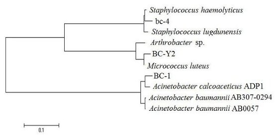
2. Results and Discussion

Three strains (labeled as BC-Y2, bc-4 and BC-1) were isolated from the buccal tubes. Six buccal tubes were found to be contaminated by bacteria. No bacteria were isolated from saline without the buccal tubes (data not shown). Based on 16S rRNA genes phylogenetic analyses, the identities of these bacterial strains (BC-Y2, bc-4 and BC-1) are: Micrococcus luteus, Staphylococcus haemolyticus and Acinetobacter calcoaceticus, respectively (Figure 1). The16S rRNA genes sequences for BC-Y2, bc-4 and BC-1 have been deposited in GenBank with the following accession numbers: GU370962, GU300765 and GU370964, respectively.
The evolutionary history was inferred using the Neighbor-Joining method [10]. The bootstrap consensus tree inferred from 1000 replicates [11] is taken to represent the evolutionary history of the taxa analyzed [11]. Branches corresponding to partitions reproduced in less than 50% of the bootstrap replicates are collapsed. The tree is drawn to scale, with branch lengths in the same units as those of the evolutionary distances used to infer the phylogenetic tree. The evolutionary distances were computed using the Kimura 2-parameter method [12] and are in the units of the number of base substitutions per site. All positions containing gaps and missing data were eliminated from the dataset (complete deletion option). There were a total of 538 positions in the final dataset. Phylogenetic analyses were conducted in MEGA4 [13].
Our study determined that buccal tubes received from manufacturers were biologically contaminated. However, the present work is not intended as a comprehensive survey for three reasons, namely: (1) Only four sets of buccal tubes from three manufacturers were tested; (2) only LB-agar was used for isolation of bacteria whereby most uncultivable bacteria will be missed; (3) the present study employed only one set of culture conditions where no attempt were made to isolate fungi, viruses and anaerobic bacteria in the present study. It must be pointed out that the present work is not aimed to determine the spectrum of microorganisms (such as viruses and fungi) and the microbial load on the buccal tubes. Neither do we attempt to identify from which manufacturer the contaminants are from because that was not our purpose. But rather, this work aimed specifically to study aerobic bacterial contamination by isolation and characterization of aerobic bacteria present on the surface of the available buccal tubes. This report established that some buccal tubes received from the manufacturers were contaminated with viable microorganisms. Contaminated buccal tubes may serve as a source for the subsequent cross-contamination of dental appliances; it will also result in the direct inoculation of micro-organisms into the mouths of patients.
Three manufacturers of the buccal tube types used in our study did not have any labeling with respect to sterility of the product. The packaging labels also did not have any suggestion whether these buccal tubes should be sterilized before clinical use. All these buccal tubes were packaged in a sealed plastic container; however, the majority of buccal tubes were not protected from the external environment by their packaging. Therefore, it is possible that contamination occurred during the manufacturing process, packaging and during transport from the manufacturers to the dental clinics or from repackaging by the local distributors. Metal orthodontic brackets bonded to the premolars were analyzed by scanning electron microscopy, indicating the presence of bacteria, predominantly streptococci, around the base of brackets [14], but no bacteria characterization was performed in that report. Another study reported that Streptococcus mutans were present in higher levels in patients with conventional and self-ligating brackets [15].
In the present work, the first microorganism isolated, M. luteus, is an obligate aerobe, that has been reported as the most common commensal species of Micrococcus found on human skin [16]. A previous study also reported that M. luteus can enter a profound dormancy period for 34,000 to 170,000 years in an oligotrophic environment such as amber [17]. M. luteus have numerous adaptations for survival in extreme, nutrient-poor environments, and M. luteus can be resuscitated from a dormancy state by a secreted protein called resuscitation-promoting factor [18]. Although human skin is now considered to be a primary habitat of the bacterium, and it has also been detected in water and soil, the mucous membranes, including the buccal cavity [19], septic arthritis [20], septic shock [21], meningitis [22] and pneumonia [23]. Considering all the reported work on M. luteus, this leads us to speculate that the reasons for the presence of M. luteus on the buccal tube are due to human contamination and its profound ability to survive even in nutrient-depleted condition.
The second microorganism found was Acinetobacter calcoaceticus. Although mostly found in the soil, Acinetobacters are part of the human skin flora also [24]. A. calcoaceticus is reported to be an opportunistic pathogen causing serious nosocomial diseases [25]. It has also been reported that Acinetobacter sp. has been recovered from used dental pumice [26].
The third microorganism identified, S. haemolyticus, is among the normal skin flora, and commonly isolated from the axillae, perineum and inguinal areas of humans [27]. Since this bacterium can be found among the normal skin flora, this suggests its origin in the buccal tubes can originate from contacts with the human skin or fingers. However, S. haemolyticus represents the second most commonly isolated bacteria recognized as important nosocomial pathogens [28,29]. Recently, multiple drug resistance has been documented in S. haemolyticus [30].
As all of the bacterial species cultured from the buccal tubes in this study are commonly associated with human being, we postulate that the presence of these bacteria are due to unhygienic practices of staff at the production line during manufacturing and packaging. We also speculate that the non-sterile packaging and improper handling of packages can also be a source of the bacterial contamination especially during logistics and storage. Lastly, the time of exposure of the buccal tubes upon being received in the clinic may explain the presence of these bacteria because these bacteria are common causes of nosocomial infections. These bacteria could have been acquired via exposure to the clinic environment. Our work highlights the need to consider sterilization of these buccal tubes as some of the bacteria isolated, such as A. calcoaceticus, is well recognized as a nosocomial species. Its long survival on a dry surface may be an additional factor aiding its transmission in hospitals [31] and suggests that more attention be paid to environmental surfaces including the buccal tubes as a source of significant nosocomial pathogens.
To our knowledge, no buccal tube sources have been identified in an oral health-care facility from which all three of these bacteria have been isolated. The potential spread of these organisms implicated as opportunistic in the dental operatory may pose a potential risk, and should pose a serious concern to dentists. This may increase the incidence of nosocomial bacteria and may cause serious implications on oral health.

3. Materials and Methods

3.1. Buccal Tube Samples

Four different types of buccal tubes from three manufacturers were selected for testing (Table 1). Upon inspection, there was no packaging information regarding sterility from any of the manufacturers. The original packages of the buccal tubes were opened using sterile latex gloves and the buccal tubes picked up with sterile tweezers and transferred to a sterile container. Six buccal tubes from each of the four types were tested (N = 24).

3.2. Isolation of Microorganisms

Each buccal tube was submerged in sterile saline (10 mL) in a sterile plastic tube and vortexed vigorously for 5 min. The resulting saline (100 μL) was serially diluted (ten-fold) with Luria Bertani (LB) broth and spread onto LB agar and incubated at 37 °C for 24–48 h. Pure colony was obtained by repeated streaking on LB agar and incubated at 37 °C for 24 h.

3.3. DNA Extraction, PCR and Sequencing

Extraction of bacterial genomic DNA was performed according to previously reported work [11]. The DNA extracted was subjected to PCR using the primers 27f (5′-AGAGTTTGATCMTGGCTCAG-3′) and 1525r (5′-AAGGAGGTGATCCAGCC-3′) as forward and reverse primers, respectively [32], at an annealing temperature of 55 °C, to selectively amplify the 16S rRNA gene region that is approximately 1500 bp in length. Purification and sequencing of PCR products were carried out according to published criteria [11]. All sequences obtained have been deposited in GenBank.

3.4. Analysis of Bacterial 16S rRNA Sequences

Alignment, editing and phylogenetic analysis of nucleotides sequences were performed according to the reported method [33]. Construction of phylogenetic tree was done using MEGA 4 [13].

4. Conclusions

Some buccal tubes received from the manufacturers were contaminated with viable aerobic bacteria, potentially nosocomial species. It is strongly suggested that buccal tubes should be sterilized by autoclave before clinical usage.

Acknowledgement

This work was supported by Research University Grants (TA048-2008A, RG003/09BIO, TB013-2009C) to KGC and (RG033/09HTM) to KP from the University of Malaya, which are gratefully acknowledged.

References

  1. Kuramitsu, HK; He, X; Lux, R; Anderson, MH; Shi, W. Interspecies interactions within oral microbial communities. Microbiol. Mol. Biol. Rev 2007, 71, 653–670. [Google Scholar]
  2. Mazumdar, V; Snitkin, ES; Amar, S; Segre, D. Metabolic network model of a human oral pathogen. J. Bacteriol 2009, 191, 74–90. [Google Scholar]
  3. Spaulding, EH. Chemical disinfection and antisepsis in the hospitals. J. Hosp. Res 1972, 9, 5–31. [Google Scholar]
  4. Lucas, VS; Omar, J; Vieira, A; Roberts, GJ. The relationship between odontogenic bacteraemia and orthodontic treatment procedures. Eur. J. Orthod 2002, 24, 293–301. [Google Scholar]
  5. Miller, CH; Palenik, CJ. Infection Control and Management of Hazardous Materials for the Dental Team, 2nd ed; Mosby Building Arts: St. Louise, MO, USA, 1998. [Google Scholar]
  6. Wichelhaus, A; Bader, F; Sander, FG; Krieger, D; Mertens, T. Effective disinfection of orthodontic pliers. J. Orofac. Orthop 2006, 67, 316–336. [Google Scholar]
  7. Benson, PE; Douglas, CWI. Decontamination of orthodontic bands following size determination and cleaning. J. Orthod 2007, 34, 18–24. [Google Scholar]
  8. Brusca, MI; Chara, O; Sterin-Borda, L; Rosa, AC. Influence of different orthodontic brackets on adherence of microorganisms in vitro. Angle Orthod 2007, 77, 331–336. [Google Scholar]
  9. Papaioannou, W; Gizani, S; Nassika, M; Kontou, E; Nakou, M. Adhesion of Streptococcus mutans to different types of brackets. Angle Orthod 2007, 77, 1090–1095. [Google Scholar]
  10. Saitou, N; Nei, M. The neighbor-joining method: A new method for reconstructing phylogenetic trees. Mol. Biol. Evol 1987, 4, 406–425. [Google Scholar]
  11. Felsenstein, J. Confidence limits on phylogenies: an approach using the bootstrap. Evolution 1985, 39, 783–791. [Google Scholar]
  12. Kimura, M. A simple method for estimating evolutionary rates of base substitutions through comparative studies of nucleotide sequences. J. Mol. Evol 1980, 16, 111–120. [Google Scholar]
  13. Tamura, K; Dudley, J; Nei, M; Kumar, S. MEGA4: Molecular Evolutionary Genetics Analysis (MEGA) software version 4.0. Mol. Biol. Evol 2007, 24, 1596–1599. [Google Scholar]
  14. Sukontapatipark, W; El-Agroudi, MA; Selliseth, NJ; Thunold, K; Selvig, KA. Bacterial colonization associated with fixed orthodontic appliances: a scanning electron microscopy study. Eur. J. Orthod 2001, 23, 475–484. [Google Scholar]
  15. Pandis, N; Papaioannou, W; Kontou, E; Nakou, M; Makou, M; Eliades, T. Salivary Streptococcus mutans levels in patients with conventional and self-ligating brackets. Eur. J. Orthod 2010, 32, 94–99. [Google Scholar]
  16. Chiller, K; Selkin, BA; Murakawa, GJ. Skin microflora and bacterial infections of the skin. J. Investig. Dermatol. Symp. Proc 2001, 6, 170–174. [Google Scholar]
  17. Greenblatt, CL; Baum, J; Klein, BY; Nachshon, S; Koltunov, V; Cano, RJ. Micrococcus luteus survival in amber. Microbiol. Ecol 2004, 48, 120–127. [Google Scholar]
  18. Mukamolova, GV; Murzin, AG; Salina, EG; Demina, GR; Kell, DB; Kaprelyants, AS; Young, M. Muralytic activity of Micrococcus luteus Rpf and its relationship to physiological activity in promoting bacterial growth and resuscitation. Mol. Microbiol 2006, 59, 84–98. [Google Scholar]
  19. Kocur, M; Kloos, WE; Schleifer, KH. The genus Micrococcus. In The Prokaryotes; Sringer-Verlag: New York, NY, USA, 1992; Volume II. [Google Scholar]
  20. Wharton, M; Rice, JR; McCallum, R; Gallis, HA. Septic arthritis due to Micrococcus luteus. J. Rheumatol 1986, 13, 659–660. [Google Scholar]
  21. Albertson, D; Natsios, GA; Gleckman, R. Septic shock with Micrococcus luteus. Arch. Intern. Med 1978, 138, 487–488. [Google Scholar]
  22. Fosse, T; Peloux, Y; Granthil, C; Toga, B; Bertrando, J; Sethian, M. Meningitis due to Micrococcus luteus. Infection 1985, 13, 280–281. [Google Scholar]
  23. Souhami, L; Feld, R; Tuffnell, PG; Feller, T. Micrococcus luteus pneumonia: a case report and review of the literature. Med. Pediatr. Oncol 1979, 7, 309–314. [Google Scholar]
  24. Peleg, AY; Seifert, H; Paterson, DL. Acinetobacter baumannii: Emergence of a successful pathogen. Clin. Microbiol. Rev 2008, 21, 538–582. [Google Scholar]
  25. Retailliau, HF; Hightower, AW; Dixon, RE; Allen, JR. A nosocomial pathogen with an unusual seasonal pattern. J. Infect. Dis 1979, 139, 371–375. [Google Scholar]
  26. Williams, HN; Falkler, WA, Jr; Hasler, JF. Acinetobacter contamination of laboratory dental pumice. J. Dent. Res 1983, 62, 1073–1075. [Google Scholar]
  27. Lina, G; Etienne, J; Vandenesch, F. Fischetti, VA, Novick, RP, Ferretti, JJ, Portnoy, DA, Rood, JI, Eds.; Biology and pathogenicity of staphylococci other then Staphylococcus aureus and Staphylococcus epidermidis. In Gram-Positive Pathogens; ASM Press: Washington, DC, USA, 2000; pp. 450–462. [Google Scholar]
  28. Pfaller, MA; Jones, RN; Doern, GV; Sader, HS; Kugler, KC; Beach, ML. Survey of blood stream infections attributable to gram-positive cocci: Frequency of occurrence and antimicrobial susceptibility of isolates collected in 1997 in the United States, Canada, and Latin America from the SENTRY Antimicrobial Surveillance Program. SENTRY Participants Group. Diagn. Microbiol. Infect. Dis 1999, 33, 283–297. [Google Scholar]
  29. Kjelleberg, S; Molin, S. Is there a role for quorum sensing signals in bacterial biofilms? Curr. Opin. Microbiol 2002, 5, 254–258. [Google Scholar]
  30. Shittu, A; Lin, J; Morrison, D; Kolawole, D. Isolation and molecular characterization of multiresistant Staphylococcus sciuri and Staphylococcus haemolyticus associated with skin and soft-tissue infections. J. Med. Microbiol 2004, 53, 51–55. [Google Scholar]
  31. Getchell-White, SI; Donowitz, LG; Groschel, DH. The inanimate environment of an intensive care unit as a potential source of nosocomial bacteria: Evidence for long survival of Acinetobacter calcoaceticus. Infect. Control Hosp. Epidemiol 1989, 10, 402–407. [Google Scholar]
  32. Chan, KG; Tiew, SZ; Ng, CC. Rapid isolation method of soil bacilli and screening of their quorum quenching activity. As. Pac. J. Mol. Biol. Biotech 2007, 15, 153–156. [Google Scholar]
  33. Chan, KG; Yin, WF; Sam, CK; Koh, CL. A novel medium for the isolation of N-acylhomoserine lactone-degrading bacteria. J. Ind. Microbiol. Biotechnol 2009, 36, 247–251. [Google Scholar]
Figure 1. Phylogenetic relationship of the bacteria isolated from the buccal tubes. Small-subunit rRNA-based evolutionary tree showing the phylogenetic position of strains BC-Y2, bc-4 and BC-1. A total of 538 unambiguously aligned nucleotides were used for analysis using Bootstrap value of 1,000. The bar represents evolutionary distance as 0.1 changes per nucleotide position. GenBank accession numbers in parentheses: Acinetobacter baumannii AB0057 (gi213155370:56013-57570), Acinetobacter calcoaceticus ADP1 (gi50083297:18416-19945), Acinetobacter baumannii AB307-0294 (gi215481761:18347-19871), Staphylococcus lugdunensis HKU09-01 (gi289549371:916199-917737), Staphylococcus haemolyticus JCSC1435 (gi70725001:879834-881387), Micrococcus luteus NCTC 2665 (gi239916571: 419927-421558), Arthrobacter sp. FB24 (gi116668568:792344-793860). For strains BC-Y2, bc-4 and BC-1, see text.
Ijms 11 03349f1
Table 1. Types of buccal tubes from various manufactures.
GroupType of BuccalTube ManufacturerPrescription
ALower right molar single tubeAmerican Othodontics, Sheboygan, WisMBT
BLower right molar single tube—small base3M Unitek, Monrovia CalifMBT
CLower right molar single tube—large base3M Unitek, Monrovia CalifMBT
DLower right molar single tubeHangzhou Dentop, ChinaMBT
MBT = McLaughlin, Bennett and Trevisi prescription.

