Article

Modulation of Ca2+ Signals by Epigallocatechin-3-gallate(EGCG) in Cultured Rat Hippocampal Neurons

1
College of Fisheries, Huazhong Agricultural University, Wuhan 430070, Hubei, China
2
Department of Pharmacy, The Affiliated Xiangfan Hospital of TongJi Medical College of Huazhong University of Science & Technology, Xiangfan 441021, Hubei, China
3
Department of Pharmacology, Medical College, Xianning University, Xianning 437100, Hubei, China
4
Department of Osteopaedics, Center Hospital of Xianning, Xianning 437100, Hubei, China
*
Author to whom correspondence should be addressed.
Int. J. Mol. Sci. 2011, 12(1), 742-754; https://doi.org/10.3390/ijms12010742
Submission received: 22 December 2010 / Revised: 30 December 2010 / Accepted: 4 January 2011 / Published: 20 January 2011
(This article belongs to the Section Biochemistry)

Abstract

:
Green tea has been receiving considerable attention as a possible neuroprotective agent against neurodegenerative disease. Epigallocatechin-3-gallate (EGCG) is the major compound of green tea. Calcium signaling has profound effects on almost all aspects of neuronal function. Using digital calcium imaging and patch-clamp technique, we determined the effects of EGCG on Ca2+ signals in hippocampal neurons. The results indicated that EGCG caused a dose-dependent increase in intracellular Ca2+ ([Ca2+]i). This [Ca2+]i increase was blocked by depleting intracellular Ca2+ stores with the endoplasmic reticulum Ca2+ pump inhibitor thapsigargin and cyclopiazonic acid. Furthermore, EGCG-stimulated increase in [Ca2+]i was abolished following treatment with a PLC inhibitor. However, EGCG inhibited high-voltage activated Ca2+ currents (IHVA) and NMDA-induced inward currents (INMDA). These data suggest that EGCG triggers a cascade of events: it activates phospholipase C (PLC), mobilizes intracellular Ca2+ stores, raises the cytosolic Ca2+ levels, and inhibits the VGCC and NMDA receptors-mediated Ca2+ influx through a process that remains to be determined.

1. Introduction

Green tea polyphenols are natural plant flavonoids and comprise many types of catechin. Among them, (–)-epigallocatechin gallate (EGCG) is the major polyphenol component and primarily responsible for the green tea effect. Previous studies on the biological activities of EGCG were mostly focused on its beneficial effects, including antioxidant [1,2], anticarcinogenic [35], and anti-inflammatory properties [6]. Also, EGCG can penetrate the brain’s blood barrier [7] and has neuroprotective effects.
Calcium (Ca2+) is a ubiquitous intracellular signal responsible for controlling numerous cellular processes, such as fertilization, proliferation, development, learning and memory [8]. Maintenance of intracellular Ca2+ homeostasis is crucial for cell survival. The site, magnitude, and kinetics of Ca2+ changes determine the biological consequences of Ca2+ signaling in the neurons [911]. One biological action attributed to EGCG is the ability to influence intracellular Ca2+([Ca2+]i) in both non-excitable and excitable cells. EGCG reportedly attenuates the a-amino-3-hydroxy-5-methyl-4-isoxazolepropionic acid (AMPA)-induced increase in [Ca2+]I in hippocampal neurons [12]. In addition, EGCG also reduced both N-methyl-4-phenyl-1,2,3,6-tetrahydropyridine (NMDA) and kainate-induced [Ca2+]i increase by attenuating both ionotropic Ca2+ influx and Ca2+-induced Ca2+ release (CICR) in PC12 cells, but it did not affect matabotropic receptor-mediated Ca2+ release [13]. These studies have focused to elucidate the protective effects of EGCG after the treatment of cells with cytotoxic agents. Recently, it has been demonstrated that EGCG mediated stimulation of [Ca2+]i in cultures of rat hippocampal neurons is partially responsible for the death of hippocampal neurons induced by EGCG [14]. Excessive elevation of [Ca2+]i produces deleterious effects and results in cell death. Thus, it is essential for cells to carefully buffer intracellular calcium and to precisely regulate calcium entry [15]. We hypothesized that EGCG at low concentrations could result in a relatively small increase in [Ca2+]i, which would be compensated for by the calcium-buffering mechanisms of cells. However, high-concentration EGCG produced excessive elevation of [Ca2+]i which was possible beyond the buffering capacity of neurons and led to a cascade of cellular pathological events [14].
Currently, the detailed mechanisms by which EGCG regulated [Ca2+]i in neurons remain unclear. The present study characterized EGCG-regulated Ca2+ signaling by employing calcium imaging and whole cell patch-clamp techniques. The results indicated that EGCG increased [Ca2+]i in hippocampal neurons in a dose-dependent manner. The regulation, which was mediated by intracellular stores mobilization via activation of a PLC-IP3 pathway, led to suppression of high-voltage activated Ca2+ currents (IHVA) and NMDA-induced inward currents (INMDA). These data shed new light on the understanding of pharmacological action of EGCG in hippocampal neurons.

2. Results and Discussion

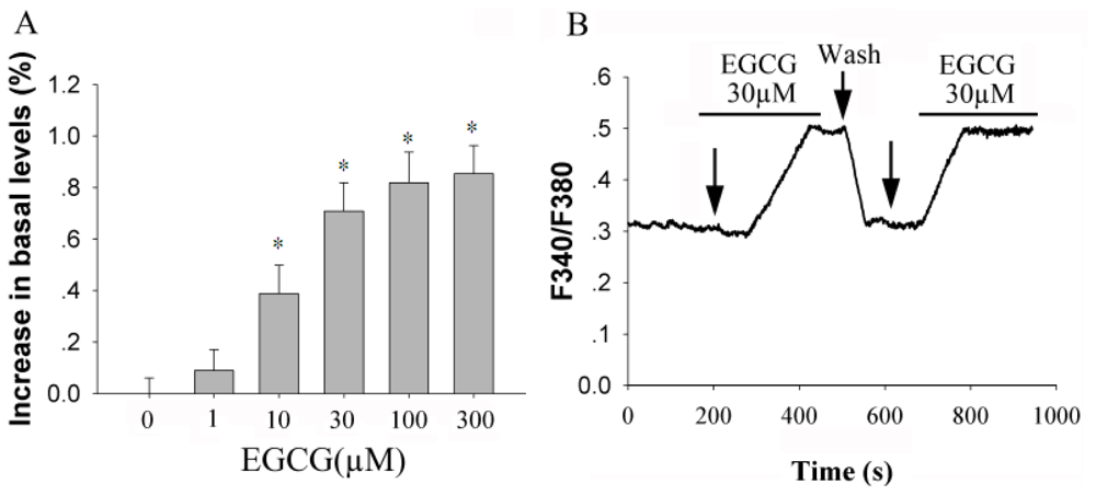
2.1. EGCG Elevates [Ca2+]i in Rat Hippocampal Neurons in a Dose-Dependent Manner

In the first set of experiments, we employed the [Ca2+]i imaging technique to study the dynamic alteration of intracellular calcium mediated by EGCG in primary culture of rat hippocampal neurons. From the results, the effect of EGCG was significant at 10 μM (P < 0.05, n = 29), and reached a maximal level at a concentration of 100 μM (Figure 1B). The EC50 for potentiation effect was 11.6 ± 1.6 μM (fitted with Sigmaplot version 9.0 to the Hill equation). Calcium mobilization was initially detected 100 s following the application of 30 μM EGCG and rapidly reached a plateau in 236 ± 48 s. Repeated 30 μM EGCG stimulation produced equal response and this response was fully reversed after washout with ACSF. The response were detected in 59% (34 of 58 cells tested) of neurons, resulting in a 70.8 ± 11.0% increase over basal levels.

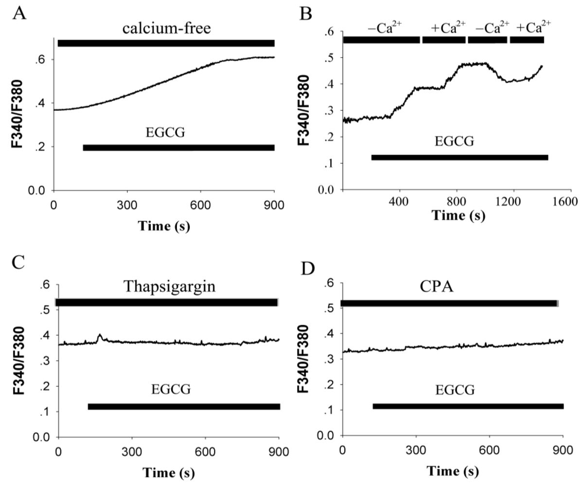
2.2. EGCG-Evoked Enhancement of [Ca2+]i Depends on the Release from Intracellular Calcium Store and Ca2+ Influx

To explore the respective contribution of Ca2+ influx from the extracellular compartment or the release from intracellular Ca2+ stores on EGCG-induced [Ca2+]i elevation in hippocampal neurons, the standard extracellular solution was replaced by Ca2+-free solution. In the absence of extracellular Ca2+, 30 μM EGCG was able to induce a significant increase of basal [Ca2+]i (54 ± 8.1% over unstimulated level, n = 28 ), although the magnitude of elevation (Figure 2A) was less than that in the presence of extracellular Ca2+ (Figure 2B). This suggests that calcium release from intracellular stores was partly responsible for the induced Ca2+ elevation. As shown in Figure 2B, EGCG induced Ca2+ increase was blunt while Ca2+-free medium was applied, but recovered again after reperfusion with Ca2+-containing medium.
The importance of Ca2+ release from intracellular stores in triggering the elevation of [Ca2+]i was further investigated. After pre-incubation with 1 μM thapsigargin to deplete the intracellular calcium store in neurons bathed in Ca2+-containing medium, EGCG failed to evoke a significant increase in [Ca2+]i (n = 31, Figure 2C). Similar results were obtained when cyclopiazonic acid (CPA), an inhibitor of sarcoplasmic/endoplasmic reticulum Ca2+-ATPase, was applied (n = 27, Figure 2D). These experiments indicated that Ca2+ release from intracellular stores was necessary and sufficient for EGCG-induced [Ca2+]i rise.

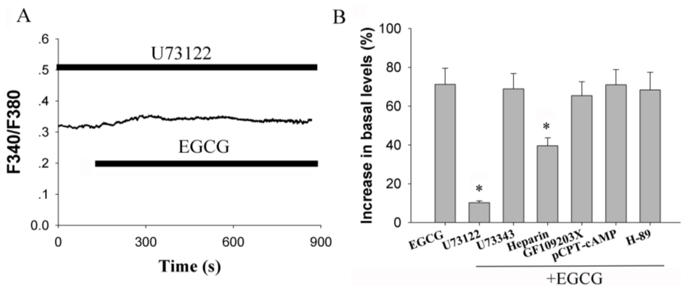
2.3. Activation of Phospholipase C (PLC) Signaling Pathways is Essential for EGCG-Stimulated [Ca2+]i Elevation

To ascertain which intracellular signaling pathway is activated in EGCG-induced Ca2+ release from intracellular calcium stores, cells were treated with 10 μM U73122, a PLC inhibitor. Notably, the increase in intracellular calcium levels by EGCG was prevented by pre-treatment with U73122 (n = 20, Figure 3A), whereas U73343, an inactive analog of U73122 (without inhibitory activity), did not alter EGCG-stimulated [Ca2+]i elevation (n = 21, P > 0.05).
Activation of PLC is known to stimulated PI hydrolysis, IP3 and 1,2-diacylglycerol (DAG) production. Subsequently, IP3 stimulates the Ca2+ release from endoplasmic reticulum (ER) stores; DAG leads to activation of protein kinase C (PKC). To test which pathway was involved in EGCG-mediated elevation of [Ca2+]i, primary cultured hippocampal neurons were pretreated with heparin (20 mg/mL, a competitive antagonist of IP3), EGCG-evoked responses were attenuated to 39.6 ± 4.1% over basal levels (n = 12, P < 0.05 vs. EGCG alone). When cells were treated with PKC inhibitor GF109203X (2 μM), the stimulation effects of EGCG were not noticeably altered. The results suggested that the potentiation effect of EGCG was partially dependent on the activation of IP3. To determine whether coupling to the AC signaling pathway was affected by EGCG, cells treated with pCPT-cAMP, a membrane permeable cAMP analog, was examined. In contrast, pretreatment with 200 μM pCPT-cAMP for 3 min had no effect on the increase in EGCG-mediated intracellular calcium levels (n = 19, P > 0.05). Meanwhile, H-89, a permeable PKA inhibitor, did not alter EGCG-stimulated elevation (n = 23, P > 0.05). These data suggested that EGCG mediated calcium signaling was not dependent on PKA pathway, but a PLC dependent pathway.

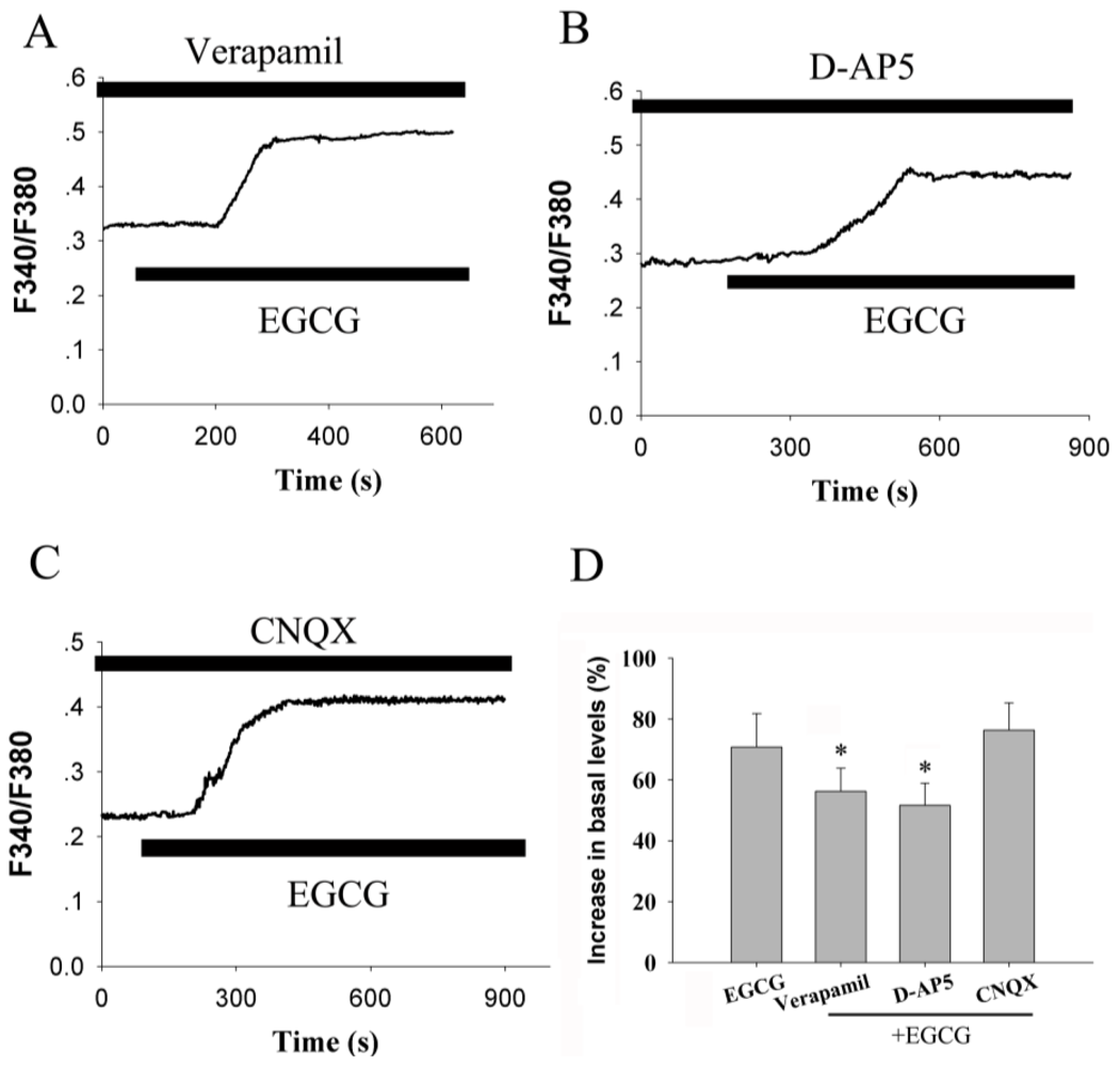
2.4. Voltage-Gated Calcium Channel and Receptor-Operated Calcium Channel Contribute to the Increase of [Ca2+]i

Although we demonstrated that Ca2+ influx from the extracellular compartment contributed to the EGCG induced [Ca2+]i elevation, the exact pathway involved in this regulation is unclear. To address this issue, we first employed verapamil to block voltage-gated calcium channels (VGCC). As shown in Figure 4A, 10 μM verapamil prevented the potentiating effect of 30 μM EGCG on intracellular calcium levels from 70.8 ± 11.0% to 56.3 ± 7.5% (n = 21, P < 0.05).
Next, the role of receptor-operated calcium channels (ROCC) in this process was checked. As depicted in Figure 4B, 50 μM DL-2-amino-5 phonovaleric acid (D-AP5) attenuated the EGCG-induced elevation of [Ca2+]i. The maximal increases in [Ca2+]i over basal levels were 51.7 ± 7.2% (n = 19, P < 0.05). However, 10 μM 6-cyano-7-nitroquinoxaline-2,3-dione (CNQX), a selective AMPA/kainite receptor competitive antagonist, did not produce any inhibition on EGCG induced elevation of free calcium in the neurons (Figure 4C). This result indicated that activation of the NMDA receptor contributed to the calcium influx stimulated by EGCG in hippocampal neurons.

2.5. EGCG Suppresses IHVA and INMDA via Elevating Intracellular Ca2+ Concentration

To further determine the roles of VGCC and NMDA receptor in the EGCG induced [Ca2+]i elevation, the whole cell patch-clamp technique was employed to study the effects of EGCG on high-voltage-activated calcium currents (IHVA) and NMDA-induced inward currents (INMDA). IHVA were elicited by a depolarization from holding potential of −80 mV to +10 mV. EGCG 30 μM caused a reduction in IHVA from 63.2 ± 5.9 pA.pF−1 to 46.7 ± 5.3 pA.pF−1(n = 21, P < 0.05). IHVA decreased by EGCG was irreversible after EGCG was washed out (Figure 5A). The cells were further dialyzed with high concentration of fast Ca2+ chelator BAPTA (15 mM), which is sufficient to maintain [Ca2+]i at a nanomolar level. As shown in Figure 5B, BAPTA almost abolished EGCG-induced inhibitory effect completely (n = 8, P < 0.05 vs. EGCG alone). These results revealed that the inhibition induced by EGCG was Ca2+-dependent.
NMDA (100 μM), an agonist of NMDA receptors, generated inward whole-cell membrane currents (INMDA) at a holding potential of −50 mV in cultured hippocampal neurons. The currents were blocked by the NMDA antagonist D-AP5, but not by the non-NMDA glutamate receptor antagonist CNQX (data not shown). As depicted in Figure 5C, with treatment with EGCG (30 μM) for 3 min, the amplitude of INMDA was decreased by 62.9 ± 6.1% (n = 10, P < 0.05), whereas the inhibitory effects of EGCG on INMDA were blocked by addition of BAPTA (n = 9, Figure 5D).

2.6. Discussion

In the present study, we found that EGCG elevated [Ca2+]i in a dose-dependent manner in cultured hippocampal neurons, which was initially driven by Ca2+ release from intracellular stores. Our data further demonstrated that EGCG induced inhibition of IHVA and INMDA was mediated by elevation of intracellular calcium through activation of a PLC-IP3 pathway. This biochemical cascade induced by EGCG is thought to be causative for the neuroprotective/neurotoxic effects of EGCG exposure.
EGCG, as the main catechin polyphenols of tea, has been investigated experimentally. Attention has already been paid to the effects of EGCG on the nervous system in previous investigations. It has been reported that EGCG exerts neuroprotective effects in various models of toxicity induced by ischemia, glutamate, N-methyl-4-phenyl-1,2,3,6-tetrahydropyridine(MPTP), oxidative stress and Aβ peptides [13,1618]. In contrast to the neuroprotective effects of the EGCG, increasing evidence indicates that there is a concentration-dependent window of pharmacological action, in which, at high concentrations, EGCG has pro-oxidant/pro-apoptotic activity [19,20]. EGCG showed some maternal toxicity and reduced the growth rate of offspring in company with a slight increase in pup loss at high dose in genotoxity and teratogenicity studies [21,22]. And EGCG increased DNA strand breakage in purified blood lymphocytes at high doses [23]. More similarly, EGCG was reported to decrease the neuronal activity of medial vestibular nuclear neurons by playing a role in decreasing the neuronal activity of contralateral vestibular nuclei [24]. More recently, EGCG was found to block the voltage-gated sodium channel currents (INa) at the concentration of 400 μM and higher in hippocampal neurons [25].
[Ca2+]i. is central to multiple signal transduction pathways to accomplish a variety of biological functions. Our result is in agreement with a previous report that EGCG could cause a [Ca2+]i elevation in a dose-dependent way [14]. Two major intracellular sources contribute to [Ca2+]i mobilization: an intracellular release through plasma membrane channels and a internal reservoir in the ER and mitochondria [26]. Our data showed that EGCG could still induce an increase in [Ca2+]i in the absence of extracellular Ca2+ and was abolished in cells that were depleted of intracellular calcium stores by thapsigargin and CPA, which suggested that the mobilization of Ca2+ in the ER was necessary and sufficient for this process. However, inhibition of EGCG-induced [Ca2+]i increase by removal of extracellular Ca2+ recovered again after reperfusion with Ca2+-containing medium. These results suggest that Ca2+ influx is partly involved in the EGCG induced calcium elevation. In our present study, a large proportion of EGCG-induced elevation of [Ca2+]i was still apparent in the presence of VGCC and ROCC blockers, which suggested that these entry pathways did not represent the principal mode of elevation of [Ca2+]i. Verapamil and D-AP5 slightly reduced EGCG-induced increase in [Ca2+]i in hippocampal neurons. However, EGCG inhibited IHVA and INMDA, suggesting that the EGCG-induced Ca2+ influx occurred mainly though other pathways. Capacitative calcium entry (CCE) via store-operated calcium channel is considered to be the major mechanism for influx of Ca2+ in a nonexcitable cell system. It is generally believed that CCE operation is nonexistent in neurons, although there were some disagreements [27]. Although our data did not find the occurrence of CCE in cultured hippocampal neurons, this pathway could not be excluded to be involved in EGCG-induced Ca2+ influx.
Excessive elevation of [Ca2+]i would result in intracellular calcium overload, which triggers a cascade of events leading to cell death. Calcium-buffering mechanism is essential for cells to reduce [Ca2+]i. One target of intracellular calcium is voltage-dependent and ligand-gated ion channels [28]. Intracellular calcium can inactivate these channels by binding directly to ion channels or via Ca2+-dependent enzymes, such as calmodulin and calcineurin [29]. Ca2+-dependent inactivation of HVA Ca2+ currents restricts the entry of Ca2+ into the cytoplasm, which is a negative feedback mechanism between Ca2+ entry and the intracellular Ca2+ concentration [30]. This mechanism was reported to be involved in nicotine-induced neuroprotection [31]. The present study demonstrated that EGCG reversibly inhibited IHVA and INMDA in primary cultured hippocampal neurons. Chelation of intraculluar Ca2+ with BAPTA reduced the effects of EGCG, which suggested that EGCG induced inhibition of IHVA and INMDA was dependent on Ca2+ release from the intracellular stores. We argue that this type of coordinated modulation in signaling processes might protect neurons from Ca2+ overload.
EGCG is known to bind to several proteins and affects the activity of enzymes and receptors. It has been reported to act through phosphoinositide 3-kinase (PI 3-kinase), Akt/protein kinase B (Akt/PKB), tyrosine kinases, protein kinase C (PKC), and mitogen activated protein kinase (MAP kinase) signaling cascades [32]. Inhibitory or stimulatory actions at these pathways are likely to influence cellular function profoundly. However, in our experiments, the limb of these signaling cascades did not participate in the EGCG-induced elevation of [Ca2+]i. A clear understanding of the signal transduction mechanisms is a key to the evaluation of action of EGCG, either as a neuroprotective agent or neurotoxic agent [33]. In our study, inhibitors of PKA and PKC did not alter the effects of EGCG. In contrast, blockade of IP3 receptors with heparin or depletion of intracellular calcium store with thapsigargin and CPA disrupted the EGCG-induced increase in [Ca2+]i. These data indicated that EGCG stimulated [Ca2+]i via PLC-IP3 pathway in hippocampal neurons.

3. Experimental Section

3.1. Materials

EGCG, U-73122,U-73343, p-CPT-cAMP, CPA, cyclopiazonic acid, NMDA, H-89, DL-2-amino-5-phos-phonovaleric acid (AP-5), 6-cyano-7-nitroquinoxaline-2,3-dione (CNQX), heparin, GF109203X, verapamil, and thapsigargin were purchased from Sigma (St. Louis, MO, USA). Fura-2/AM was obtained from Biotium (Hayward, CA, USA). DMEM/F12 and B27 supplement were obtained from Gibco Invitrogen Corporation (Carlsbad, CA, USA). Other general agents were available commercially. Other agents were purchased from commercial suppliers. Agents were prepared as stock solutions with sterile water except difedipine, U-73122, U-73343 and Fura-2/AM, which were dissolved in dimethylsulfoxide (DMSO) and stored at −20 °C. They were diluted to the final concentrations before application. The final concentration of DMSO was <0.05%. No detectable effect of the vehicles was found in our experiments.

3.2. Cell Culture

Neonatal SD (Sprague-Dawley) rats (day 0–3) of both sexes were obtained from the Center for Disease Control of Hubei Province, China. All experiments were conducted in accordance with the National Institutes of Health Guide for the Care and Use of Laboratory Animals. All experiments conformed to named local guidelines on the ethical use of animals and all efforts were exerted to minimize the number of animals used and their suffering. Neurons were isolated as previously described with some modification [25]. Briefly, hippocampi of newborn rats were dissected and rinsed in ice cold dissection buffer. Blood vessels and white matter were removed and tissues were incubated in 0.125% trypsin for 25 min at 37 °C. Neurons were collected by centrifugation and resuspended in Dulbecco’s modified Eagle’s medium (DMEM) and F-12 supplement (1:1) (Gibco Invitrogen corporation) with 10% fetal bovine serum (heat-inactivated, Hyclone), 2 mM L-glutamine (Sigma), and Penicillin (100 U/mL)-streptomycin (100 U/mL). Cells were plated at a density of 104–105 per 35 mm2 on coverslides precoated with poly-L-lysine and kept at a 37 °C in a 5% CO2 incubator. After 24 h, the culture medium was changed to DMEM medium containing 2% B27 and 2 mmol/L glutamine. Astrocytes were minimized by treating the culture with cytarabine (10 μM) on day 3. The medium was replaced with fresh medium every 3 days. Experiments were performed on day 5–7.

3.3. Calcium Imaging Experiment

Digital calcium imaging was performed as described by Ming et al. [34]. Hippocampal neurons were washed three times with 1 μmol/L Fura-2/AM in artificial cerebrospinal fluid (ACSF, containing 140 mM NaCl, 5 mM KCl, 1 mM MgCl2, 2 mM CaCl2, 10 mM glucose and 10 mM HEPES, pH 7.3) then incubated in the same solution for 30 min at 37 °C. In calcium-free experiments, EGTA (100 μM) was substituted for CaCl2. Before each experiment, the coverslides were mounted on a chamber positioned on the movable stage of an inverted Olympus IX-70 microscope equipped with a calcium imaging system (TILL Photonics GmbH, Gräfelfing, Germany), and superfused by ACSF for 10 min. Fura-2/AM loaded cells were illuminated at 340 nm for 150 ms and 380 nm for 50 ms at 1-s intervals using a TILL Polychrome monochromator. Fura-2 fluorescence emission was imaged at 510 nm by an intensified cooled charge coupled device (TILL Photonic GmbH) through an X-70 fluor oil immersion lens (Olympus) and a 460 nm long-pass barrier filter. F340/F380 fluorescence ratios were generated by TILLvision 4.0 software. Paired F340/F380 fluorescence ratio images were acquired every second for [Ca2+]i. The intracellular free calcium concentration is presented as the ratio of the fluorescence signals obtained (340/380 nm). All experiments were repeated at least three times using different batches of cells.

3.4. Whole Cell Patch Clamp Recording

The procedure for whole cell patch-clamp recording was described in our previous study [35]. Currents were recorded with whole-cell patch-clamp technique, using an EPC-10 amplifier (HEKA, Lambrecht, Germany) controlled by the Pulse/PulseFit software (HEKA, Southboro, Germany). Data were acquired at a sampling rate of 10 kHz and filtered at 3 kHz. The pipette tips were heat-polished and have resistances of 2–4 MΩ when filled with intracellular buffer. The pipette solution used to record calcium currents consisted of (in mM) CsCl 100, MgCl2 2.0, Na2ATP 5.0, egtazic acid 11, HEPES 10, TEA-Cl 20, creatine phosphate 20, and the pH was adjusted to 7.2 with CsOH. The extracellular solution composed of (in mM) NaCl 110, KCl 5.0, CaCl2 5.0, MgCl2 1.0, HEPES 10, Glucose 11, 4-AP 5.0, TEA-Cl 25, TTX 0.5 × 10⁻³ and the pH was adjusted to 7.4 with NaOH. NMDA currents were recorded using electrodes filled with (concentration in mM) cesium gluconate 135, CsCl5, CaCl2 5.0, MgCl2 1.0, HEPES 10, Glucose 11, pH 7.2, with Tris base, and the bath solution contained (in mM) NaCl 140, KCl 5, CaCl2 2.5, HEPES 10, Glucose 10, TTX 0.5 × 10⁻³, pH 7.4 with Tris base. 100 μM NMDA was applied to cells for 15 s using a modified Y-tube. Drug actions were measured only after steady-state conditions reached, which were judged by the amplitudes and time courses of currents remaining constant.

3.5. Statistical Analysis

The amplitudes of [Ca2+]i elevation over the basal were represented as the difference between baseline level [(F340/F380)B] and the peak amplitude [(F340/F380)S] response to the stimulation, which was shown as [(F340/F380)S-F340/F380)B]/(F340/F380)B. Data are presented as mean ± SEM. Data were analyzed using SPSS 10.0 software. Student’s t-test or one-way ANOVA was used to test for significance. Differences were considered significant at P < 0.05.

4. Conclusions

Results presented here suggested that EGCG triggered a cascade of events: it activated phospholipase C, mobilized intracellular calcium stores, raised the cytosolic Ca2+ levels, and inhibited the VGCC and NMDA receptor-mediated Ca2+ influxes. Although the mechanisms by which EGCG raised [Ca2+]i and inhibited IHVA and INMDA in cultured hippocampal neurons remain a matter of conjecture, our results may help to understand the neurotoxicity and officinal value of EGCG.

Acknowledgements

This work was supported by the National Nature Science Foundation of China (NSFC) Grant 31000249 to J.-H. Wang, and NSFC Grant 81000576, Natural Science Foundation of Hubei Province of China 2010CDZ067 to F. Cai.

References

  1. Shi, X; Ye, J; Leonard, S; Ding, M; Vallyathan, V; Castranova, V; Rojanasakul, Y; Dong, Z. Antioxidant properties of (–)-epicatechin-3-gallate and its inhibition of Cr (VI)-induced DNA damage and Cr (IV)- or TPA-stimulated NF-κB activation. Mol. Cell. Biochem 2000, 206, 125–132. [Google Scholar]
  2. Valcic, S; Burr, JA; Timmermann, BN; Liebler, DC. Antioxidant chemistry of green tea catechins. New oxidation products of (–)-epicatechin-3-gallate from their reactions with perosyl radicals. Chem. Res. Toxicol 2000, 13, 801–810. [Google Scholar]
  3. Komori, A; Yatsunami, J; Okabe, S; Abe, S; Hara, K; Suganuma, M; Kim, SJ; Fujiki, H. Anticarcinogenic activity of green tea polyphenols. Jpn. J. Clin. Oncol 1993, 23, 186–190. [Google Scholar]
  4. Kuroda, Y; Hara, Y. Antimutagenic and anticarcinogenic activity of tea polyphenols. Mutat. Res 1999, 436, 69–97. [Google Scholar]
  5. Wand, SL; Mukhtar, H. Gene expression profile in human prostate LNCaP cancer cells by (–)-epicatechin-3-gallate. Cancer Lett 2002, 182, 43–51. [Google Scholar]
  6. Dona, M; Dell’Aica, I; Calabrese, F; Benelli, R; Morini, M; Albini, A; Garbisa, S. Neutrophil restraint by green tea: inhibiton of inflammation, associated angiogenesis, and pulmonary fibrosis. J. Immunol 2003, 170, 4335–4341. [Google Scholar]
  7. Nakagawa, K; Miyazawa, T. Absorption and distribution of tea catechin, (–)-epicatechin-3-gallate, in the rat. J. Nutr. Sci. Vitaminol 1997, 43, 679–684. [Google Scholar]
  8. Berridge, MJ; Lipp, P; Bootman, MD. The versatility and universality of calcium signaling. Nat. Rev. Mol. Cell Biol 2000, 1, 11–21. [Google Scholar]
  9. Shieh, PB; Hu, SC; Bobb, K; Timmusk, T; Ghosh, A. Identification of a signaling pathway involved in calcium regulation of BDNF expression. Neuron 1998, 20, 727–740. [Google Scholar]
  10. Shoop, RD; Esquenazi, E; Yamada, N; Ellisman, MH; Berg, DK. Ultra-structure of a somatic spine mat for nicotinic signaling in neurons. J. Neurosci 2002, 22, 748–756. [Google Scholar]
  11. Tao, X; Finkbeiner, S; Arnold, DB; Shaywitz, AJ; Greenberg, ME. Ca2+ influx regulates BDNF transcription by a CREB family transcription factor-dependent mechanism. Neuron 1998, 20, 709–726. [Google Scholar]
  12. Bae, JH; Mun, KC; Park, WK; Lee, SR; Suh, SI; Baek, WK; Yim, MB; Kwon, TK; Song, DK. EGCG attenuates AMPA-induced intracellular calcium increase in hippocampal neurons. Biochem. Biophys. Res. Commun 2002, 290, 1506–1512. [Google Scholar]
  13. Lee, H; Bae, JH; Lee, SR. Protective effect of green tea polyphenol EGCG against neuronal damage and brain edema after unilateral cerebral ischemia in gerbils. J. Neurosci. Res 2004, 77, 892–900. [Google Scholar]
  14. Yin, ST; Tang, ML; Deng, HM; Xing, TR; Chen, JT; Wang, HL; Ruan, DY. Epigallocatechin-3-gallate induced primary cultures of rat hippocampal neurons death linked to calcium overload and oxidative stress. Naunyn-Schmiedeberg’s Arch. Pharmacol 2009, 379, 551–564. [Google Scholar]
  15. Tedford, HW; Zamponi, GW. Direct G protein modulation of Cav2 calcium channels. Pharmacol. Rev 2006, 58, 837–862. [Google Scholar]
  16. Levites, Y; Amit, T; Youdim, MBH; Mandel, S. Involvement of protein kinase C activation and cell survival/cell cycle genes in green tea polyphenol (–)-epigallocatechin-3-gallate neuroprotective action. J. Biol. Chem 2002, 277, 30574–30580. [Google Scholar]
  17. Choi, YB; Kim, YI; Lee, KS; Kim, BS; Kim, DJ. Protective effect of epigallocatechin gallate on brain damage after transient middle cerebral artery occlusion in rats. Brain Res 2004, 1019, 47–54. [Google Scholar]
  18. Levites, Y; Amit, T; Mandel, S; Youdim, MB. Neuroprotection and neurorescue against Abeta toxicity and PKC-dependent release of non-amyloidogenic soluble precursor protein by green tea polyphenol (–)-epigallocatechin-3-gallate. FASEB J 2003, 17, 952–954. [Google Scholar]
  19. Weinreb, O; Mandel, S; Youdim, MBH. cDNA gene expression profile homology of antioxidants and their anti-apoptotic and pro-apoptotic activities in human neuroblastoma cells. FASEB J 2003, 17, 935–937. [Google Scholar]
  20. Schroeter, H; Williams, RJ; Matin, R; Iversen, L; Rice-Evans, CA. Phenolic antioxidants attenuate neuronal cell death following uptake of oxidized low-density lipoprotein. Free Radic Biol Med 2000, 29, 1222–1233. [Google Scholar]
  21. Isbrucer, RA; Bausch, J; Edwards, JA; Wolz, E. Safety studies on epigallocatechin gallate (EGCG) preparations. Part 1. Genotoxicity. Food Chem. Toxicol 2006, 44, 626–635. [Google Scholar]
  22. Isbruker, RA; Edwards, JA; Wolz, E; Davidocich, A; Bausch, J. Safety studyies on epigallocatechin gallate (EGCG) preparations. Part 3. Teratogenicity and reproductive toxicity studies in rats. Food Chem. Toxicol 2006, 44, 651–661. [Google Scholar]
  23. Kanadzu, M; Lu, Y; Morimoto, K. Dual function of (–)-epigallocatechin gallate (EGCG) in healthy human lymphocytes. Cancer Lett 2006, 241, 250–255. [Google Scholar]
  24. Jeong, HS; Kim, YS; Park, JS. Modulation of neuronal activity by EGCG. Brain Res 2005, 1047, 267–270. [Google Scholar]
  25. Deng, HM; Yin, ST; Yan, D; Tang, ML; Li, CC; Chen, JT; Wang, M; Ruan, DY. Effects of EGCG on voltage-gated sodium channels in primary cultures of rat hippocampal CA1 neurons. Toxicology 2008, 252, 1–8. [Google Scholar]
  26. Bootman, MD; Collins, TJ; Peppiatt, CM; Prothero, LS; Mac-Kenzaie, L; de Smet, P; Travers, M; Tovey, SC; Seo, JT; Berridge, MJ; Ciccolini, F; Lipp, P. Calcium signaling—An overview. Semin. Cell Dev. Biol 2001, 12, 3–10. [Google Scholar]
  27. Putney, JW, Jr. Capacitative calcium entry in the nervous system. Cell Calcium 2003, 34, 339–344. [Google Scholar]
  28. Marchenko, SM. Mechanism of modulation of GABA-activated current by internal calcium in rat central neurons. Brain Res 1991, 546, 355–357. [Google Scholar]
  29. Lukyanetz, EA; Piper, TP; Sihra, T. Calcineurin involvement in the regulation of high-threshold Ca2+ channels in NG108-15 (rodent neuroblastoma × glioma hybrid) cells. J. Physiol 1998, 510, 371–385. [Google Scholar]
  30. Brehm, P; Eckert, R. Calcium entry leads to inactivation of calcium channel in paramecium. Science 1978, 202, 1203–1206. [Google Scholar]
  31. Stevens, TR; Krueger, SR; Fitzsimonds, RM; Picciotto, MR. Neuroprotection by nicotine in mouse primary cortical cultures involves activation of calcineurin and L-type calcium channel inactivation. J. Neurosci 2003, 23, 10093–10099. [Google Scholar]
  32. Mandel, SA; Avramovich-Tirosh, Y; Reznichenko, L; Zheng, H; Weinreb, O; Amit, T; Youdim, MBH. Multifunctional Activities of Green Tea Catechins in Neuroprotection. Neurosignals 2005, 14, 46–60. [Google Scholar]
  33. Williams, RJ; Spencer, JPE; Rice-Evans, C. Flavonoids: Antioxidants or signaling molecules? Free Radic. Biol. Med 2004, 36, 838–849. [Google Scholar]
  34. Ming, Y; Zhang, H; Long, L; Wang, F; Chen, J; Zhen, X. Modulation of Ca2+ signals by phosphatidylinositol-linked novel D1 dopamine receptor in hippocampal neurons. J. Neurochem 2006, 98, 1316–1323. [Google Scholar]
  35. Wang, JH; Wang, F; Yang, MJ; Yu, DF; Wu, WN; Liu, J; Ma, LQ; Cai, F; Chen, JG. Leptin regulated calcium channels of neuropeptide Y and proopiomelanocortin neurons by activation of different signal pathways. Neuroscience 2008, 156, 89–98. [Google Scholar]
Figure 1. EGCG induced increase of intracellular calcium levels in hippocampal neurons. (A) Summary data of increases in [Ca2+]i for each dose of EGCG from three independent experiments. [Ca2+]i increase was recorded as described in the Experimental section and expressed as [(F340/F380)S-F340/F380)B]/(F340/F380)B. * P < 0.05 vs. corresponding value in control cultures; (B) Hippocampal neurons were treated with 30 μM EGCG for the designated time. The arrows indicate the time of EGCG application or washout.
Ijms 12 00742f1
Figure 2. EGCG-mediated increase in [Ca2+]i depends on release of Ca2+ from intracellular calcium stores and Ca2+ influx. (A) Superfusion of cultured neurons was switched to a Ca2+-free solution when 30 μM EGCG was added; (B) [Ca2+]i increase was halted while Ca2+-free solution was applied and restored after switching back to Ca2+-containing solution during EGCG-induced increase in [Ca2+]i. +Ca2+, Ca2+-containing ACSF applied. –Ca, Ca2+-free ACSF applied; (C) Application of 1 μM thapsigargin to delete the intracellular calcium store completely blocked EGCG-induced [Ca2+]i increase in hippocampal neurons; (D) Application of 10 μM CPA to deplete the intracellular calcium store, EGCG failed to evoke a significant increase in [Ca2+]i. The experiments were repeated at least three times and representative data are shown for each treatment.
Ijms 12 00742f2
Figure 3. Activation of phospholipase C (PLC) signaling pathways mediates EGCG-induced [Ca2+]i elevation. (A) Pretreatment with 10 μM U73122 blocked EGCG-induced [Ca2+]i increase in hippocampal neurons (n = 20). Experiments were repeated at least 3 times; (B) Summary data of the results with U73122, U73343 (10 μM), heparin (20 mg/mL), GF109203X (2 μM), p-CPT-cAMP (200 μM) and H-89 (30 μM) preincubation on EGCG-mediated elevation in hippocampal neurons. * P < 0.05 compared with EGCG alone.
Ijms 12 00742f3
Figure 4. Voltage-gated calcium channels and receptor-operated calcium channels contribute to EGCG-induced Ca2+ influx. (A) Application of 10 μM verapamil for 3 min prior to the addition of 30 μM EGCG reduced the drug-stimulated [Ca2+]i elevation (n = 21); (B) Pretreatment with 50 μM D-AP5 for 3 min before the addition of 30 μM EGCG reduced the drug-stimulated [Ca2+]i elevation (n = 19); (C). 30 μM CNQX failed to have an effect on EGCG-stimulated [Ca2+]i elevation (n = 23); (D) Summary data of the results with verapamil, D-AP5 and CNQX preincubation on EGCG-mediated elevation in hippocampal neurons. *P < 0.05 compared with EGCG alone.
Ijms 12 00742f4
Figure 5. The EGCG-induced inhibitory effect on IHVA and INMDA are Ca2+-dependent. (A) 30 μM EGCG reversibly decreased high-voltage-activated (HVA) Ca2+ currents in primary cultured hippocampal neurons. Voltage protocol is shown on the top; (B) Representative trace showing dialysis with 15 mM bis-(o-aminophenoxy)-N,N,N’,N’-tetraacetic acid (BAPTA) significantly reduced the inhibitory modulation of EGCG on IHVA; (C) 30 μM EGCG reversibly decreased NMDA activated currents in primary cultured hippocampal neurons; (D) Representative traces showing dialysis with 15 mM BAPTA significantly attenuated the inhibitory effects of EGCG.
Ijms 12 00742f5

