Article

DNA Sequence Analysis of South African Helicobacter pylori Vacuolating Cytotoxin Gene (vacA)

1
Microbial Pathogenicity and Molecular Epidemiology Research Group, Department of Biochemistry and Microbiology, Faculty of Science and Agriculture, University of Fort Hare, P/Bag X1314, Alice 5700, South Africa
2
Department of Biochemistry and Microbiology, Faculty of Science, University of Buea, Box 63, Buea, Cameroon
*
Author to whom correspondence should be addressed.
Int. J. Mol. Sci. 2011, 12(11), 7459-7468; https://doi.org/10.3390/ijms12117459
Submission received: 22 August 2011 / Accepted: 28 October 2011 / Published: 31 October 2011
(This article belongs to the Section Molecular Pathology, Diagnostics, and Therapeutics)

Abstract

:
Sequence diversity and population structures can vary widely among pathogenic bacteria species. In some species, all isolates are highly similar, whereas in others most of the isolates are distinguished easily. H. pylori is known for its wide genetic diversity amongst the various strains most especially in the genes involved in virulence. The aim of this study was to evaluate by PCR and sequence analysis, the genetic profile of H. pylori vacA gene (s1, s2, m1 and m2). We sequenced small DNA segments from 13 vacAs1, 10 vacAm2, 6 vacAm1 and 6 vacAs2 strains which were amplified with amplicon size of 259/286 bp, 290 bp and 352 bp for vacAs1/s2, m1 and m2 respectively. Based on similarities among our strains accession numbers were provided for seven vacAs1 (HQ709109–HQ709115), six vacAs2 (JN848463–JN848468), six vacAm1 (JN848469–JN848474) and six vacAm2 (HQ650801–HQ650806) strains. Amongst the strains studied, 98.07%, 98.58%, 97.38% and 95.41% of vacAs1, vacAs2, vacAm1 and vacAm2 of the strains were conserved respectively. Findings of this study underscores the importance of understanding the virulence composition and diversity of H. pylori in South Africa for enhanced clinico-epidemiological monitoring and pathophysiology of disease.

1. Introduction

H. pylori is a gastric pathogen that infects more than 50% of the world’s population; is the major cause of a number of gastro duodenal pathologies in infected patients [1,2], however, only a small subset of infected people experience H. pylori-associated illnesses such as chronic gastritis, peptic ulcer disease, gastric carcinoma and mucosa-associated lymphoid tissue (MALT) lymphoma [3,4]. Strains of H. pylori are renowned for tremendous variation in their genes [57]; in some species, all isolates are highly similar, whereas in other species, any two unrelated isolates can be easily distinguished from most others by DNA fingerprinting or sequencing of the representative gene segments [5,8].
H. pylori is frequently considered panmictic in population structure in that genetic recombination is so frequent in its DNA sequences and generates linkage equilibrium [9]. Typically some 3 to 5% DNA sequence divergence exist between isolates even in essential genes [7]. This diversity probably reflects a combination of factors including: mutation, recombination among divergent lineages, gene transfer from unrelated species and preferential transmission among family members and people in close contact which is propagated as a result of lack of selection for just one or a few genotype that might be best adapted for all humans [6,10]. Also, the knowledge that humans differ in traits that could be important to individual strains such as specificity, strength of the immune system, inflammatory responses and availability and distribution of adhesion molecules for the organism would select for divergence among H. pylori strains [7].
Clinically, the outcome of infection is variable and is considered to relate to bacterial virulence factors which may vary with sequence type, host and the environment [6,11]. VacA is secreted as vacuolating cytotoxin thought to be involved in ulcerogenesis whose sequence variability seems to reflect mosaicism (vacAs and m) [6]. Direct sequencing, multilocus sequence typing (MLST) and other high throughput typing schemes has indicated that, different H. pylori genotypes predominate in different parts of the world and human ethnic groups [6,8]. In particular, African strains seem to be distinct from those of other continents [6,12]. Mukhopadhyay et al. [8] reported tremendous diversities amongst strains of India, ethnic European and East Asia in their study. Also, DNA sequence motifs predominating in two virulence-associated genes vacA (vacuolating cytotoxin) and cagA (cytotoxin associated gene), in strains from the United States and Europe were found to differ from those predominating in southern coastal China and Japan [7].
Colonization with H. pylori is very common in South Africa, as in other developing countries [1316]. Most studies of H. pylori have concentrated on virulence factors determination rather than sequence analysis of these factors. This is because a good knowledge of the burden of a particular genotype will enhance better understanding of its molecular epidemiology and foster drug design in that regard. Limitations in resources also make it difficult to embark on highly informative technique like sequencing. However, sequence typing provides intricate information on the nucleotide diversity which cannot be accomplished by polymerase chain reaction alone (virulence factors determination).
Recently, we genotyped strains of H. pylori in the Eastern Cape Province of South Africa and found different combinations of genotypes with s1 and m2 being the most prevalent genotype combination [17]. However, there is a dearth of information on comparative sequence analysis of H. pylori strains in this part of the world. Suerbaum et al. [18] analyzed for diversity and linkage equilibrium between South Africa, Canada and Germany. Their study was based on H. pylori strains isolated from colored patients with gastroduodenal disease in Cape Town which is anthropologically distinct (from native South African populations) with ancestry from Western Europeans, South East Asians and South Africans (Hottentots). Also, Kersulyte et al. [7] studied South African strains from native black Africans that are residents of Soweto. In the current study we compared sequences of vacA (vacAs1, s2, m1 and m2 alleles) derived from strains of H. pylori in different racial groups (white, colored, blacks) in the Eastern Cape province of South Africa with one another and emphatically we further compared vacAs1 and m2 from our study with those from other regions because the combination s1m2 was the most prevalent in this study. Our data provide overwhelming evidence of diversity in H. pylori strains.

2. Results and Discussion

2.1. Results

Agarose gel electrophoresis of the samples that were detected by PCR revealed a single band corresponding to the expected amplicon size 259/286 bp, 290 bp and 352 bp for vacAs1/s2, m1 and m2 respectively as previously detailed (17).

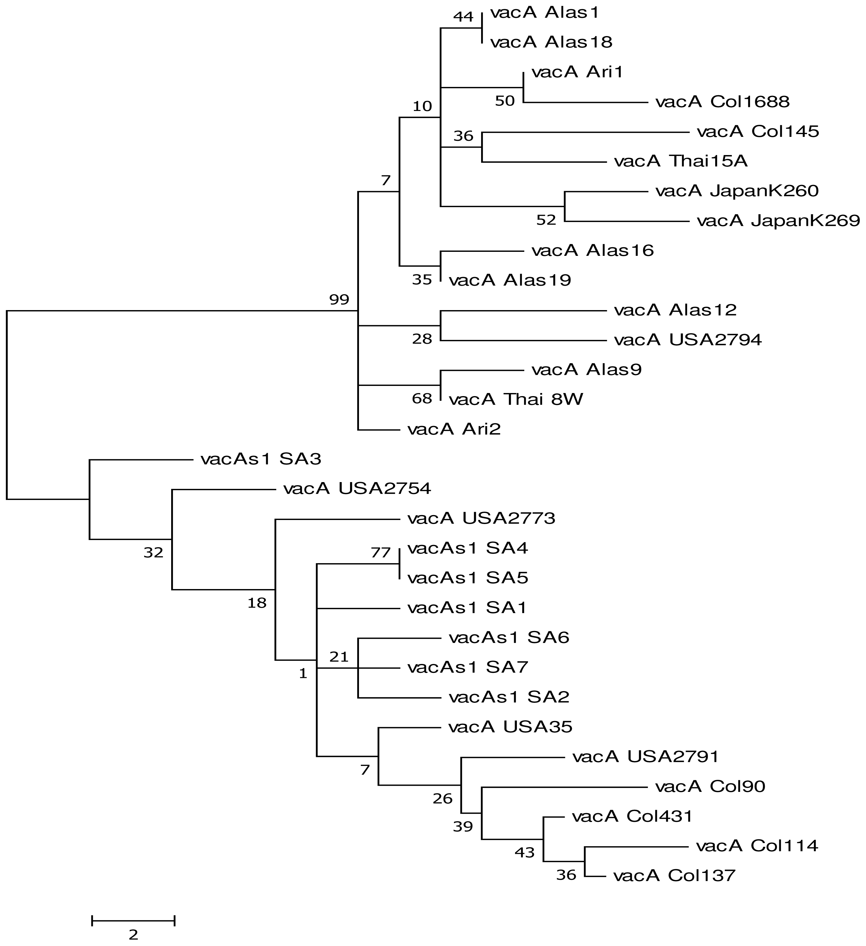
Genetic Diversity of vacA

We compared DNA sequences through blasting and alignments of vacAs1, s2, m1 and m2 nucleotides found in H. pylori strains in our study area with those already deposited in the gene bank from different study areas. Sequenced amplicons for vacAs1 was 92–98% identical to each other and to those submitted already in the genebank while for vacAs2, vacAm1 and vacAm2, it was 90–98%, 87–99% and 89–98% respectively. Six of our vacAs1 sequences were homologous to one or more of the thirteen sequences in our collection for vacAs1 while four of vacAm2 sequences also presented with homology amongst our sequences. All six strains sequenced for both vacAs2 and vacAm1 were different from each other. In this regard, we submitted seven vacAs1 [3 colored (vacAs1 SA1, vacAs1 SA4, vacAs1 SA7), 3 blacks (vacAs1 SA2, vacAs1 SA3, vacAs1 SA6) and 1 white patient (vacAs1 SA5)] and six vacAm2 [3 colored (vacAm2 SA1, vacAm2 SA4, vacAm2 SA5) and 3 black patients (vacAm2 SA2, vacAm2 SA3, vacAm2 SA6), six vacAs2 (4 blacks and 2 colored), and six vacAm1 (3 blacks, 2 colored and 1 white)] sequences in the genebank. The various strains were coded as SA = South Africa and numerical to denote strain number. H. pylori vacAs1 obtained in our study were deposited in the GenBank and assigned accession numbers (HQ709109–HQ709115) while those for vacAm2, vacAs2 and vacAm1 were (HQ650801–HQ650806), (JN848463-JN848468) and (JN848469–JN848474) respectively. Nucleotides for the sequenced genes in this study were conserved except for the mutations (missense and silence) which we observed.
We compared our sequences with other partial coding sequences existing in the genebank for both vacAs1 and m2 (as the combination s1m2 was most prevalent in our study area ) from the following regions: South Africa, USA, Colombia, Thailand, Vietnam, Alaska, Arizona, Japan, Hongkong and Shi (Figures 1 and 2). Results from comparison with other sequences depict clustering of the sequences which generally depicts similarities between the strains employed though with some differences.

2.2. Discussion

H. pylori is a slow growing microaerophilic spiral bacterium of medical importance; determination of its complete genome sequence [1,2,19], has enhanced understanding of its pathogenecity [19]. Specific genes and their sequences have been shown to be involved in the pathogenicity of H. pylori leading to a specific disease condition. Hence understanding nucleotide or protein variation that exists within a gene in H. pylori will help accelerate understanding of possible disease outcome. A significant level of variation at the nucleotide level is seen across the genome providing an explanation why the nucleotide base typing techniques offer high discriminatory power among independent H. pylori isolates [6,20]. We used direct sequencing to generate sequences from small DNA segments of vacA (s1, s2, m1 and vacAm2) genes which are highly associated with disease in H. pylori infection [20]. These sequences were compared to one another and to other strains already deposited in the gene bank. Nucleotide sequence diversity of H. pylori exceeds that of many other bacterial species. The observed differences in sequence diversity between different bacterial species can readily be explained by differences of population structure and population size. Furthermore, almost every nucleotide sequence from unrelated isolates is unique, an unprecedented situation [6]. Nucleotide sequences generated from this study from vacAs1, s2, m1 and vacAm2 exhibited some degree of divergence of 1.93%, 2.62%, 1.42% and 4.59% respectively. Our results are similar to the report of other authors who indicated high genetic diversity in H. pylori strains which renders it impossible to isolate identical strains from different or the same individual [18,19,21].
Divergence in alleles could in theory reflect high mutation rates or frequent recombination or a combination of both [6]. We found mutations among the nucleotide sequences in this study. Missense and silent mutations were the most frequently observed mutations, and based on the unique differences observed for the various strains, different Gene bank accession numbers were assigned to vacAs1 (HQ709109-HQ709115), vacAs2 (JN848463-JN848468), vacAm1 (JN848469-JN848474) and vacAm2 (HQ650801–HQ650806). Almost all polymorphism were synonymous substitutions (which did not affect the amino acid sequence), insertions and deletions. This data is in accord with the findings of Maiden et al. [22] who indicated that extensive DNA sequence data available for H. pylori genes show that transition mutations account for most of the inter-strain micro diversity. Also, mutation is considered as the key for phenotypic variation as well as ability of cellular adaptations to stress [9]. Great similarity was observed between our vacAs1 strains when compared to the standard strain U07145; also nucleotides of vacAs1 strains in our study area were 98.07% identical to each other while 1.93% were different. Also, high level of conservation was reported of the nucleotides in vacAm2 (95.41%) strains as opposed to 4.59% dissimilarity. Furthermore, we had 98.58% identity for vacAm1 and 97.38% identity for vacAs1 amongst our isolates while 2.62% and 1.42% difference was observed respectively. This finding is in line with that of Alm and Trust [20] who reported that significant variation is known to exist in vacA H. pylori sequence to an extend that every isolate seem to poses a unique sequence. However, we found six and four vacAs1 and vacAm2 sequences respectively to be identical to one or more of the sequences of the strains we studied. Our results accords the findings of Suerbaum and Achtman, [6] who found Cape Colored isolates to possess identical flaB and vacA sequences in their studies amongst South African strains. Meanwhile, it contradicts those of other investigators who found all strains from Canada and Germany to be unique, suggesting that the pool of different alleles may be smaller in some geographic regions than in Canada and Europe [6].
Our dissimilarity in results ties with the findings of Mukhopadhyay et al. [8] and Cover et al. [23] that reported high divergence of vacAm2 strains and suggested that there may be considerable sequence diversity among strains in the middle region of the vacA gene.. Also, evolutionary consideration and high rates of transmission favor the emergence of more virulent strains of the organism [24]. The differences observed are thought to have resulted due to selection pressures in different ancestral animal host rather than in humans [8]. More to that, Wirth et al. [24] reported wide genetic diversity in H. pylori strains from different region and even between ethnic groups in the same geographical region.
Sequence analysis has shown that different genotypes of H. pylori prevail in different geographical locations [8]. We compared the sequences derived from this study (s1m2) with those already deposited in the genebank using phylogenetic analysis by maximum parsimony (MP) (Figure 1 and 2). Our results reveal clustering of the strains employed which generally depicts close similarity between the strains though different. Figure 1 shows clustering of strains of vacAs1: SA1, SA2, SA4, SA5 and SA6 except for vacA SA3 which is far apart. The same applies to vacAm2 strains where SA1, SA2 and SA6 cluster around each other while SA3, SA4 and SA5 cluster. This clustering is irrespective of racial backgrounds be it colored, black or white. This is in line with the findings of Suerbaum and Achtman [6] who showed that 20 strains of H. pylori from 12 countries differed according to their sources. They added that six isolates from East Asians differed from the remainder of the world and were called the “Asian” clone. Three isolates from two Africans and one African American formed a second distinct group “Clone 2” and the other eleven isolates from Europe, America and Australia formed a heterogeneous third group. These results tie with the fact that isolates from a single individual, family member and a particular geographical region are frequently clonal; however the overall population structure of H. pylori is panmictic [25]. Comparison of our sequences (vacA) with the three African strains which have been completely sequenced and deposited in the gene bank [(J99 from Caucasian in Pulaski TN, but widely regarded as of the African strain type GenBank: AE001439.1; strain 908 (from West African immigrant in Bordeaux, France GenBank: CP002184.1 and Gambia94/24 GenBank: CP002332.1)] was impracticable because there were several protein identities for vacA in these whole genomes. This study however has as short coming small sample size. Large sample screening would have provided a better understanding of the various possible strains that would exist in this population.

3. Materials and Methods

3.1. Bacterial Strains

H. pylori strains employed in this study were isolated and identified from patients who underwent a complete physical examination, and history was taken by a resident gastro-enterologist. Endoscopic diagnoses were made by a single experienced endoscopist after informed consent and ethical clearance (protocol number EcDoH-Res 0002). Antral and corpus biopsy specimens each were obtained at endoscopy, and immediately placed in sterile bijou bottles containing 0.2 g/L of cysteine and 20% glycerol in brain-heart infusion (BHI) broth and transported in ice to the laboratory within 2 h of collection. Gastric biopsies were collected from patients and H. pylori was isolated on Columbia agar base supplemented with 7% sheep’s blood and Skirrow’s supplement containing trimethoprim (2.5 mg), vancomycin (5 mg) and cefsulodin(2.5 mg). Amphotericin (2.5 mg) was added to the medium. Recovered isolates were identified following standard microbiology and biochemical techniques [26]. A reference strain of H. pylori (NCTC 11638) was included as a positive control. Confirmed isolates were suspended in 20% glycerol and stored at −80 °C in a freezer (Sanyo, Japan) for future experiments.

3.2. Molecular Characterization

DNA extraction and PCR amplification were as reported in our previous study [17]. Briefly, DNA was extracted from a hundred strains. PCR analysis of the targeted genes was performed using Thermo-stat Taq DNA polymerase (ABgene, UK) and manufacturer-provided reaction buffer. The primers used to amplify the targeted genes were 5′-ATGGAAATACAACAAACACAC-3′ and 3′-CTGCTTGAATGCGCCAAAC-5′ for vacAs1/s2 while for vacAm1 and vacAm2 the primers were 5′-GTCAAAATGCGGTCATGG-3′ and 3′-CCATTGGTACCTGTAGAAAC-5′ and 5GGAGCCCCAGGAAACATTG-3′ and 3′- CATAACTAGCGCCTTGCAC-5′ respectively [2].

3.3. Sequence Analysis

We sequenced thirteen small DNA segment from vacAs1 (Seven from coloured patients, five from blacks and one white), ten vacAm2 strains (five colored and five blacks), six vacAs2 (4 blacks, 2 colored) and six vacAs1 (3 blacks, 2 colored and 1 white) (Iqaba, Pretoria, SA). Mutations and genetic diversity were analyzed by sequencing using a Big Dye Terminator DNA sequencing kit v3.1 (Applied Biosystems, UK) after purification of products using shrimp alkaline phosphatase. DNA sequence editing and analysis was performed with the programs Bioedit and EMBL- EBI- Clustalw2. Sequences were aligned using DNAMAN. Maximum parsimony, a non-parametric statistical method commonly used in computational phylogenetics for estimating phylogenies was used to generate trees for this study.

4. Conclusion

Our finding underscores the need of understanding the virulence composition and diversity of H. pylori in South Africa for enhanced clinico-epidemiological monitoring and pathophysiology of disease.

Acknowledgments

This study received financial support from the National Research Foundation (NRF) (grant reference UID 69816) South Africa; the Govan Mbeki Research and Development Centre, University of Fort Hare, South Africa. We are also grateful to the staff of the GIT Unit, Livingston Hospital, Port Elizabeth and Mr. Clarke B for technical assistance.

References

  1. Andreson, H.; Lõivukene, K.; Sillakivi, T.; Maaroos, H.-I.; Ustav, M.; Peetsalu, A.; Mikelsaa, M. Association of cagA and vacA genotypes of Helicobacter pylori with gastric diseases in Estonia. J. Clin. Microbiol 2002, 40, 298–300. [Google Scholar]
  2. Smith, S.I.; Kirsch, C.; Oyedeji, K.S.; Arigbabu, A.O.; Coker, A.O.; Bayerdöffer, E.; Miehlke, S. Prevalence of Helicobacter pylori vacA, cagA and iceA genotypes in Nigerian patients with duodenal ulcer disease. J. Med. Microbiol 2002, 51, 851–854. [Google Scholar]
  3. Matsuhisa, T.M.; Yamada, N.Y.; Kato, S.K.; Matsukura, N.M. Helicobacter pylori infection, mucosal atrophy and intestinal metaplasia in Asian populations: A comparative study in age-, gender- and endoscopic diagnosis-matched subjects. Helicobacter 2003, 8, 29–35. [Google Scholar]
  4. Devi, S.M.; Ahmed, I.; Khan, A.A.; Rahman, S.A.; Alvi, A.; Sechi, L.A.; Ahmed, N. Genomes of Helicobacter pylori from native Peruvians suggest a mixture of ancestral and modern lineages and reveal a western type cag-pathogenicity island. BMC Genomics 2006, 7, 191. [Google Scholar]
  5. Akopyanz, N.; Baranov, N.O.; Westblom, TUlf.; Kresovich, S.; Berg, D.E. DNA diversity among clinical isolates of Helicobacter pylori detected by PCR-based RAPD fingerprinting. Nucl. Acids Res. 1992, 20, 5137–5142. [Google Scholar]
  6. Suerbaum, S.; Achtman, M. Population Genetics—Helicobacter pylori. In Helicobacter pylori: Physiology and Genetics; Mobley, H.L.T., Mendz, G.L., Hazell, S.L., Eds.; ASM Press: Washington, DC, USA, 2001; Volume Chapter 32. [Google Scholar]
  7. Kersulyte, D.; Mukhopadhyay, A.K.; Velapatiño, B.; Su, W.; Pan, Z.; Garcia, C.; Hernandez, V.; Valdez, Y.; Mistry, R.S.; Gilman, R.H.; et al. Differences in Genotypes of Helicobacter pylori from Different Human Populations. J. Bacteriol 2000, 182, 3210–3218. [Google Scholar]
  8. Mukhopadhyay, A.K.; Kersulyte, D.; Jeong, J.Y.; Datta, S.; Ito, Y.; Chowdhury, A.; Chowdhury, S.; Santra, A.; Bhattacharya, S.K.; Azuma, T.; Nair, G.B.; Berg, D.E. Distinctiveness of genotypes of Helicobacter pylori in Calcutta, India. J. Bacteriol 2000, 182, 3219–3227. [Google Scholar]
  9. Singh, V.; Mishra, S.; Maurya, P.; Rao, G.; Jain, A.K.; Dixt, V.K.; Gulati, A.K.; Nath, G. Drug resistance pattern and clonality in H. pylori. J. Infect. Dev. Ctries 2009, 3, 130–136. [Google Scholar]
  10. Akada, K.J.; Ogura, K.; Dailidiene, D.; Dailide, G.; Cheverud, M.J.; Berg, E.D. Helicobacter pylori tissue tropism: Mouse-colonizing strains can target different gastric niches. J. Microbiol 2003, 149, 1901–1909. [Google Scholar]
  11. Kidd, M.; Peek, R.M.; Lastovica, A.J.; Israel, D.A.; Kummer, A.F.; Louw, J.A. Analysis of iceA genotypes in South African Helicobacter pylori strains and relationship to clinically significant disease. Gut 2001, 49, 629–635. [Google Scholar]
  12. Falush, D.; Stephens, M.; Pritchard, J.K. Inference of population structure using multilocus genotype data: Linked loci and correlated allele frequencies. Genetics 2003, 164, 1567–1587. [Google Scholar]
  13. Ally, R.; Mitchell, H.M.; Segal, I. Cag—A positive H. pylori aplenty in South Africa: The first systemic study of H. pylori infection in asymptomatic children in Soweto. Gut 1999, 45, A97–A98. [Google Scholar]
  14. Mosane, T.W.; Malope, B.I.; Ratshikhopha, M.E.; Hiss, D.C.; Sitas, F. Seroprevalence of Helicobacter pylori IgG antibodies in South African mothers and their children. Eur. J. Gastroenterol. Hepathol 2004, 16, 113–114. [Google Scholar]
  15. Samie, A.; Ramalivhana, J.; Igumbor, E.O.; Obi, C.L. Prevalence, haemolytic and haemagglutination activities and antibiotic susceptibility profiles of Campylobacter spp. isolated from human diarrhoeal stools in Vhembe District, South Africa. J. Health Popul. Nutr 2007, 25, 406–413. [Google Scholar]
  16. Dube, C.; Nkosi, T.C.; Clarke, A.M.; Mkwetshana, N.; Green, E.; Ndip, R.N. Helicobacter pylori antigenemia in an asymptomatic population of the Eastern Cape Province, South Africa: Public health implications. Rev. Environ. Health 2009, 24, 249–255. [Google Scholar]
  17. Tanih, N.F.; McMillan, M.; Naidoo, N.; Ndip, L.M.; Weaver, L.T.; Ndip, R.N. Prevalence of Helicobacter pylori vacA, cagA and iceA genotypes in South African patients with upper gastrointestinal diseases. Acta Trop 2010, 116, 68–73. [Google Scholar]
  18. Suerbaum, S.; Maynard, S.J.; Bapumia, K.; Morelli, G.; Smith, N.H.; Kunstmann, E.; Dyrek, I.; Achtman, M. Free recombination within Helicobacter pylori. Proc. Natl. Acad. Sci. USA 1998, 95, 12619–12624. [Google Scholar]
  19. Alm, R.A.; Ling, L.S.; Moir, D.T.; King, B.L.; Brown, E.D.; Doig, P.C.; Smith, D.R.; Noonan, B.; Guild, B.C.; deJonge, B.L.; et al. Genomic sequence comparison of two unrelated isolates of the human gastric pathogen Helicobacter pylori. Nature 1999, 397, 176–180. [Google Scholar]
  20. Alm, R.A.; Trust, J.T. Analysis of the genetic diversity of Helicobacter pylori: The tale of two genomes. J. Mol. Med 1999, 77, 834–846. [Google Scholar]
  21. Blaser, M.J.; Berg, D.E. Helicobacter pylori genetic diversity and risk of human disease. J. Clin. Invest 2001, 107, 767–773. [Google Scholar]
  22. Maiden, M.C.J.; Malorny, B.; Achtman, M.A. Global gene pool in the Neisseriae [letter]. Mol. Microbiol 1996, 21, 1297–1298. [Google Scholar]
  23. Cover, L.T.; Tummuru, K.R.M.; Cao, P.; Thompson, A.S.; Blaser, J.M. Divergence of genetic sequence of the vacuolating cytotoxin among Helicobacter pylori strains. J. Biol. Chem 1993, 269, 10566–10573. [Google Scholar]
  24. Wirth, T.; Wang, X.; Linz, B.; Novick, R.P.; Lum, J.K.; Blaser, M.J.; Morelli, G.; Falush, D.; Achtman, M. Distinguishing human ethnic groups via sequences from Helicobacter pylori: Lessons from Ladakh. Proc. Natl. Acad. Sci. USA 2004, 101, 4746–4751. [Google Scholar]
  25. Israel, D.A.; Salama, N.; Krishna, U.; Rieger, U.M.; Atherton, J.C.; Falkow, S.; Peek, R.M., Jr. Helicobacter pylori genetic diversity within the gastric niche of a single human host. Proc. Natl. Acad. Sci. USA 2001, 98, 14625–14630. [Google Scholar]
  26. Tanih, N.F.; Okeleye, B.I.; Green, E.; Mkwetshana, N.; Clarke, A.M.; Ndip, L.M.; Ndip, R.N. Helicobacter pylori prevalence in dyspeptic patients in Eastern Cape Province: Race and disease status. South Afr. Med. J 2010, 100, 734–737. [Google Scholar]
Figure 1. Maximum parsimony analysis of vacAs1 sequences from clinical isolates showing that phylogenetic analysis between strains is not distinct. There is clustering between the strains. Branches with significant bootstrap support are indicated; origins of H. pylori strains are labelled with a code: SA—South Africa, USA—USA, Col—Colombia, Thai—Thailand, Alas—Alaska, Ari—Arizona, Japan-Japan; Bar indicates 2 nucleotide substitution per site. Our vacAs1 sequences were compared with (AB057218.1, AB057223.1, AB057222.1, AB057221.1 AB057219.1, AB057214.1, AB057216.1, AB057217.1, AB057211.1, AB057215.1, AB057190.1, AB057188.1, AB057187.1, AB057185.1, AB057178.1, AB057175.1, AB057184.1, AB057182.1, AB057167.1, AB057164.1, AB057166.1, AB057140.1, AB057139.1).
Ijms 12 07459f1
Figure 2. Maximum parsimony analysis of vacAm2 sequences from clinical isolates showing that phylogenetic analysis between strains is not distinct. There is clustering between the strains. Branches with significant bootstrap Support are indicated; origins of H. pylori strains are labelled with a code: SA—South Africa, USA—USA, Col—Colombia, Thailand—Thailand, Vietnam—Vietnam, Alas—Alaska, and Hongkong—Hongkong, Shi—Shi; Bar indicates 2 nucleotide substitutions per site. vacAm2 sequences were compared with the following sequences with accession numbers (AB057325.1 AB057323.1, AB057328.1, AB057327.1, AB057326.1, AB057334.1, AB057301.1, AB057290.1, AB057295.1, AB057302.1, AB057292.1, AB057307.1, AB057277.1, AB057276.1, AB057284.1, AB057286.1, AB057280.1, AB057281.1, AB057279.1, AB057282.1, GU064493.1, GU064499.1, GU064498.1, GU064495.1, GU064496.1, GU064497.1) derived from the gene bank.
Ijms 12 07459f2

