Article

In Vivo Anti-Tumor Activity of Polypeptide HM-3 Modified by Different Polyethylene Glycols (PEG)

Department of Marine Pharmacy, College of Life Science and Technology, China Pharmaceutical University, Nanjing 210009, China
*
Author to whom correspondence should be addressed.
Int. J. Mol. Sci. 2011, 12(4), 2650-2663; https://doi.org/10.3390/ijms12042650
Submission received: 12 February 2011 / Revised: 8 March 2011 / Accepted: 1 April 2011 / Published: 19 April 2011
(This article belongs to the Special Issue Conjugated Polymers)

Abstract

:
HM-3, designed by our laboratory, is a polypeptide composed of 18 amino acids. Pharmacodynamic studies in vivo and in vitro indicated that HM-3 could inhibit endothelial cell migration and angiogenesis, thereby inhibiting tumor growth. However, the half-life of HM-3 is short. In this study, we modified HM-3 with different polyethylene glycols (PEG) in order to reduce the plasma clearance rate, extend the half-life in the body, maintain a high concentration of HM-3 in the blood and increase the therapeutic efficiency. HM-3 was modified with four different types of PEG with different molecular weights (ALD-mPEG5k, ALD-mPEG10k, SC-mPEG10k and SC-mPEG20k), resulting in four modified products (ALD-mPEG5k-HM-3, ALD-mPEG10k-HM-3, SC-mPEG10k-HM-3 and SC-mPEG20k-HM-3, respectively). Anti-tumor activity of these four modified HM-3 was determined in BALB/c mice with Taxol as a positive control and normal saline as a negative control. Tumor weight inhibition rates of mice treated with Taxol, HM-3, ALD-mPEG5k-HM-3, ALD-mPEG10k-HM-3, SC-mPEG10k-HM-3 and SC-mPEG20k-HM-3 were 44.50%, 43.92%, 37.95%, 31.64%, 20.27% and 50.23%, respectively. Tumor inhibition rates in the Taxol, HM-3 and SC-mPEG20k-HM-3 groups were significantly higher than that in the negative control group. The efficiency of tumor inhibition in the SC-mPEG20k-HM-3 group (drug treatment frequency: once per two days) was better than that in the HM-3 group (drug treatment frequency: twice per day). In addition, tumor inhibition rate in the SC-mPEG20k-HM-3 group was higher than that in the taxol group. We conclude that SC-mPEG20k-HM-3 had a low plasma clearance rate and long half-life, resulting in high anti-tumor therapeutic efficacy in vivo. Therefore, SC-mPEG20k-HM-3 could be potentially developed as new anti-tumor drugs.
Abbreviations::
ALD-mPEG (methoxy-polyethylene glycol propionaldehyde); molecular weight 10 kDa; named ALD-mPEG10k; SC-mPEG (α-methoxy-polyethylene glycol-ω-Succinimidyl Carbonate); molecular weight 20 kDa, named SC-mPEG20k

1. Introduction

HM-3 is a new anti-tumor polypeptide developed in our laboratory, which is composed of 18 amino acids and previous studies showed that the target of HM-3 is integrin (αvβ3) [1]. In vivo and in vitro studies have shown that HM-3 could inhibit endothelial cell migration and angiogenesis, thereby inhibiting tumor growth. HM-3 is expected to be developed as therapeutic drugs for the treatment of solid tumors including gastric and liver cancer [2]. HM-3 has been authorized as a national invention patent and declared for an international patent [3].
However, similar to other peptide drugs, the half-life of HM-3 is short (approximately 25 min). HM-3 is easily degraded by proteases [1,4]. Animal studies suggested that the optimal therapeutic efficiency could be obtained by administration of the drug twice a day.
An effective approach to solve this problem is to modify peptide drugs with polymers such as polyethylene glycol (PEG) and dextran [5]. Currently, most studies are focusing on PEG modification [68]. PEG is a class of large polymers with unique physical and chemical properties. PEG is non-toxic, non-antigenic and has good biocompatibility [9,10]. The main biological function of PEG-modified protein or peptide drugs remains unchanged. More importantly, PEG modification increases the circulatory stability, extends the half-life, reduces the immunogenicity and decreases the toxcity of protein or peptide drugs [1114].
However, PEG modification still exhibits several problems in application. Based on the literature [9,10], the main problems associated with PEG modification were an abnormal increase of molecular weight of the modified products and a decrease or loss of activity during the process of modification. Loss of activity is essentially the primary problem that needs to be solved for PEG modification of peptide drugs [15,16].
Selection of the appropriate molecular size of PEG is key for the modification of peptide drugs. Two main considerations for selecting the type of PEG are the residual in vivo biological activity and the renal elimination rate of the modified products. The molecular size of PEG is negatively related to the residual biological activity of the peptide drugs [17]. The relationship between the size of PEG and renal elimination rate is that a larger size of PEG normally results in a lower renal elimination rate. Studies have shown that the renal elimination rate when using PEG with a molecular weight of 20 kDa is approximately 10%, which plays a significant role in increasing the half-life, extending drug administration frequency and maintaining the blood drug concentration at a high level [18,19]. Currently, a variety of PEG-modified protein and peptide drugs have been developed and approved by the FDA for biomedical purposes [20].
ALD-mPEG and SC-mPEG are two different types of PEG, which are currently the most commonly used and relatively mature with active groups to modify polypeptide drugs. The active groups of ALD-mPEG and SC-mPEG are propionaldehyde and succinimidyl carbonate, respectively, which are specificity reactive groups of the peptide’s end amino. Physical properties, reaction activity with the peptide’s end amino of ALD-mPEG and SC-mPEG were different, and the remaining bioactivity of the same peptide PEGylated by the different two PEG were different too. A previous study [1] reported that HM-3 modified with ALD-mPEG10k showed improved half-life and biological activity in vitro, but the half-life improvement was not significant.
In order to solve the problem of short half-life and rapid plasma clearance, our laboratory utilized four different types of PEG (ALD-mPEG and SC-mPEG) with different molecular weights to modify the N-terminus of HM-3. We expected that the modified products would have a long half-life while maintaining the anti-tumor activity. Our results showed that the modified product, SC-mPEG20k-HM-3, had a low plasma clearance rate, long half-life and retained its anti-tumor activity excellently. These results provide a fundamental basis for the development of new anti-tumor drugs.

2. Materials and Methods

2.1. Materials

2.1.1. Cell Line and Animals

Human hepatic carcinoma SMMC-7721 cell line was purchased from American type Cell Culture (ATCC, Shanghai, China) and maintained in Dulbecco’s modified Eagle’s medium (DMEM) with 10% fetal bovine serum and antibiotics. BALB/c nude female mouse (SPF grade, 4–5 w, 20.9 ± 0.492 g) were purchased from the Shanghai Laboratory Animal Center of the Chinese Academy of Sciences.

2.1.2. Main Reagents and Drugs

ALD-mPEG5k (98% purity), ALD-mPEG10k (100% purity), SC-mPEG10k (98% purity), and SC-mPEG20k (100% purity) were purchased from Beijing Kaizheng Biotech Development Co. Ltd. Batch No. were KZ-M05ALD-091207, KZ-M10ALD-091208, KZ-M10SC-091205, and KZ-M20SC-091206, respectively.
Trifluoroacetate (TFA), acetonitrile (ACN), and sodium cyanoborohydride were purchased from TEDIA, ROE Scientific Inc., and Shanghai Darui Fine Chemicals Co. Ltd, respectively.
HM-3 was chemically synthesized by GL Biochem (Shanghai), Ltd. The purity of the products was more than 99% by analytical high-performance liquid chromatography (HPLC), Batch No. 091020.
Mouse anti-HM-3 monoclonal antibody was prepared in our laboratory (density: 1.19 mg/mL, tite: 2,048,000). Horseradish peroxidase-labeled goat anti-mouse secondary antibody was purchased from Boster Biological Company of China.
Docetaxel was purchased from Hengrui Medicine Co. Ltd, Jiangsu province of China, Batch No. 10012812.

2.2. Methods

2.2.1. PEG Modification of HM-3 and Purification

The forms of PEG modified HM-3 and the optimal conditions have been systematically studied [1,4,21]. As to the modification of HM-3 with ALD-mPEG5k and ALD-mPEG10k, the optimal reaction time was over night at 4 °C; whereas the optimal time for SC-mPEG10k and SC-mPEG20k was 3 h at 4 °C. After the modification experiments, the products were purified by a semipreparative RP-HPLC and the purity was detected with analytical RP-HPLC. The reaction buffers and molar ratios (PEG:HM-3) are shown in Table 1.

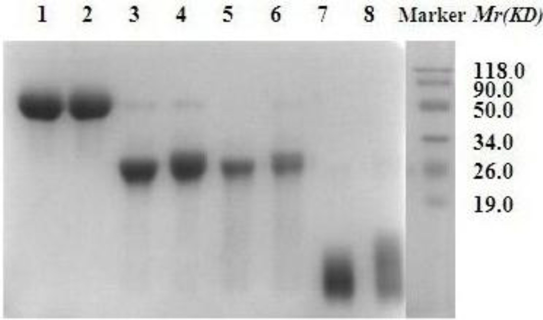
2.2.2. SDS-PAGE Electrophoresis and Western Blot Analysis of the Modified Products

Reaction mixtures were separated on a SDS-PAGE gel which was comprised of a 5% stacking gel and a 10% separation gel. The lane of the marker was stained with Coomassie brilliant blue and the lanes for ALD-mPEG5k-HM-3, ALD-mPEG10k-HM-3, SC-mPEG10k-HM-3 and SC-mPEG20k-HM-3 were stained with BaI2.
After SDS-PAGE electrophoresis, ALD-mPEG5k-HM-3, ALD-mPEG10k-HM-3, SC-mPEG10k-HM-3 and SC-mPEG20k-HM-3 were further identified by Western blot analysis using nitrocellulose membranes. The immunological reaction of ALD-mPEG5k-HM-3, ALD-mPEG10k-HM-3, SC-mPEG10k-HM-3 and SC-mPEG20k-HM-3 were performed on sections using a specific mouse anti-HM-3 monoclonal antibody of high titer and specificity and subsequently with horseradish peroxidase-labeled goat anti-mouse secondary antibody. Immunodetection was visualized using enhanced chemiluminescence.

2.2.3. Inhibitory Effect of the Modified Products on the Growth of Human Hepatoma Cells in Vivo

SMMC-7721 cells were cultured in DMEM medium under 5% CO2/saturated humidified air at 37 °C. Cells were digested with the mixture of the trypsin (0.25%) and EDTA (0.02%) at logarithmic growth phase and passaged every 3–4 day. Then SMMC-7721 cells at logarithmic growth phase were digested to make a cell suspension (1.0 × 10⁷/mL), and 0.1 mL was injected into axilla of the left arm of BALB/c nude mouse. When tumor volume grew to 50 mm3–60 mm3, the mice were divided into 7 groups with 6 in each group. The doses and frequencies of drug administration are presented in Table 2 for the groups of paclitaxel, HM-3, ALD-mPEG5k-HM-3, ALD-mPEG10k-HM-3, SC-mPEG10k-HM-3 and SC-mPEG20k-HM-3.
All the mice (20 g) were administrated intravenously for 21 days and the tumor volumes were measured every two days. The solid tumor tissues were taken out the next day after the administration menu and the tissues were weighed to determine the inhibitory effects of the modified products. The calculation formula of tumor volume (TV) was as follows [22]: TV = 0.52 × a × b2, a and b indicate tumor length and width, respectively. Antitumor ratio determined by the tumor volumes is (VVt)/V (V is the average tumor volume of the control group, and Vt is the average tumor volume of the treated group). The antitumor ratio by the tumor weights was also calculated as (WWt)/W (W is the average tumor weight of the control group, and Wt is the average tumor weight of the treatment group).

2.2.4. Statistical Methods

The data was analyzed using the statistics software SPSS13.0, which are expressed as mean ± SD. Statistical significance was assessed using the Student t test. For all statistical comparisons, treated groups were compared with normal sodium treated controls, and P < 0.05 was considered statistically significant; P < 0.01 was considered statistically very significant.

3. Results

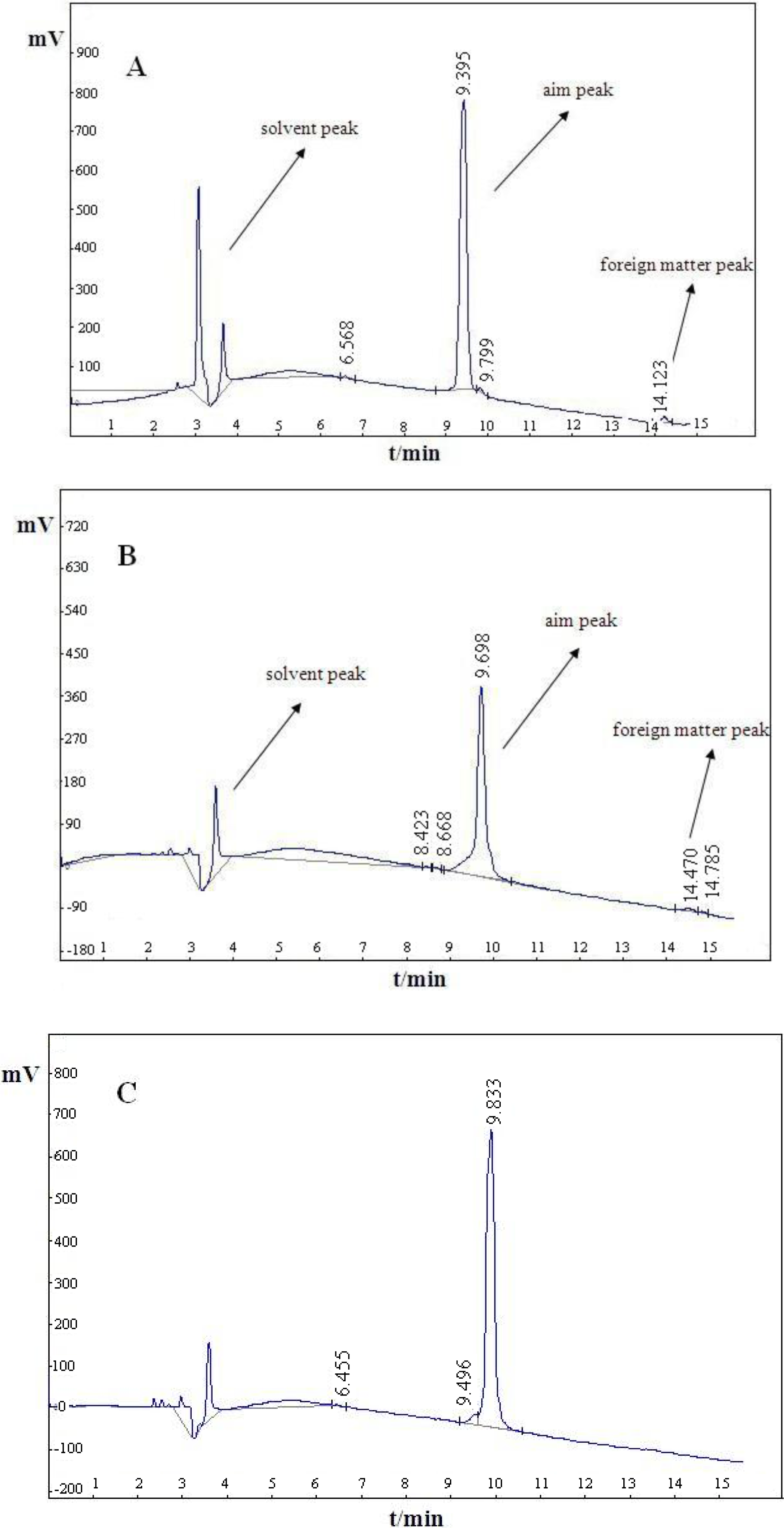
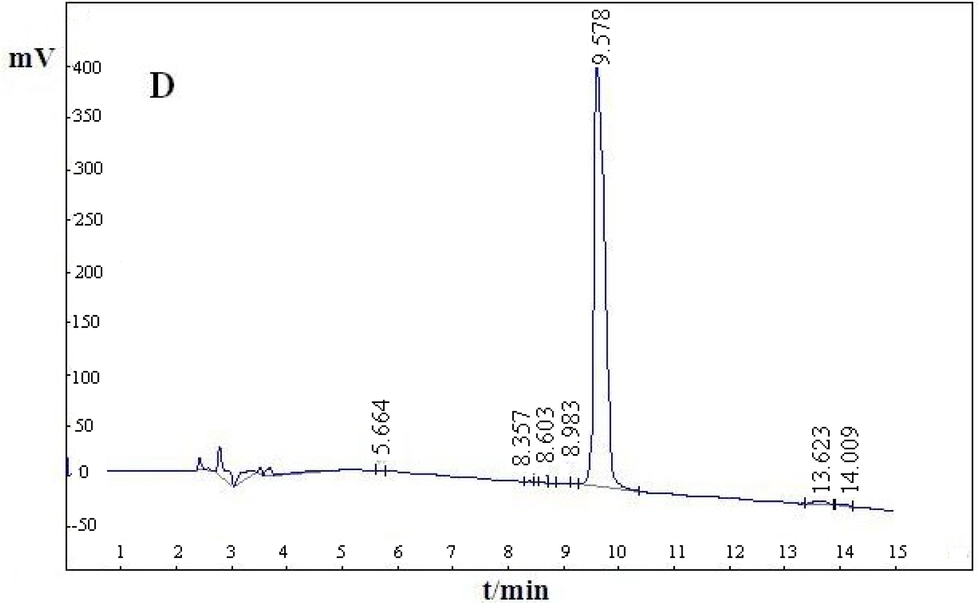
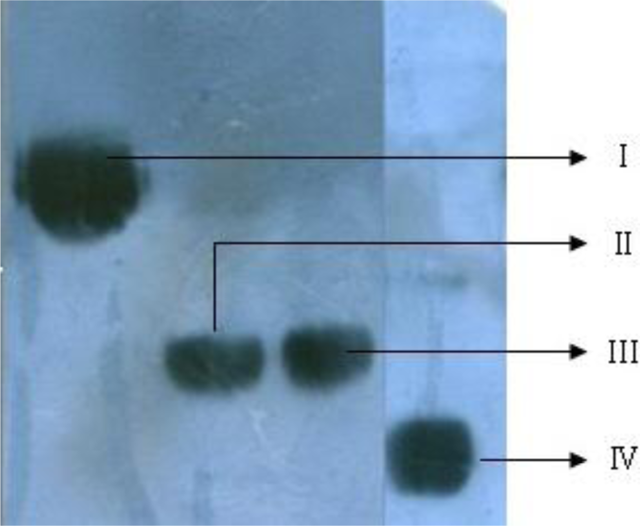
3.1. Purity of the Four Modified HM-3 Products

RP-HPLC analysis showed that the purity of the four PEG-modified HM-3 products (ALD-mPEG5k-HM-3, ALD-mPEG10k-HM-3, SC-mPEG10k-HM-3 and SC-mPEG20k-HM-3) was over 97% (Figure 1).

3.2. SDS-PAGE and Western Blot Analysis of the Modified HM-3 Products

Modification of HM-3 with mPEG resulted in mPEG-HM-3 with relatively high purity. SDS-PAGE analysis showed that the Mr of the modified product mPEG-HM-3 was much higher than that of the sum of HM-3 and mPEG (Figure 2). This could be due to the following two reasons. Firstly, linear PEG could extend on the surface of the protein, reducing the protein migration rate. Secondly, PEG had a very high degree of hydration, which increased its molecular size and reduced the migration rate in SDS-PAGE. We also found that monoclonal antibody binding site of the modified HM-3 were not covered by mPEG, which may be favorable for the maintenance of anti-tumor activity (Figure 3).

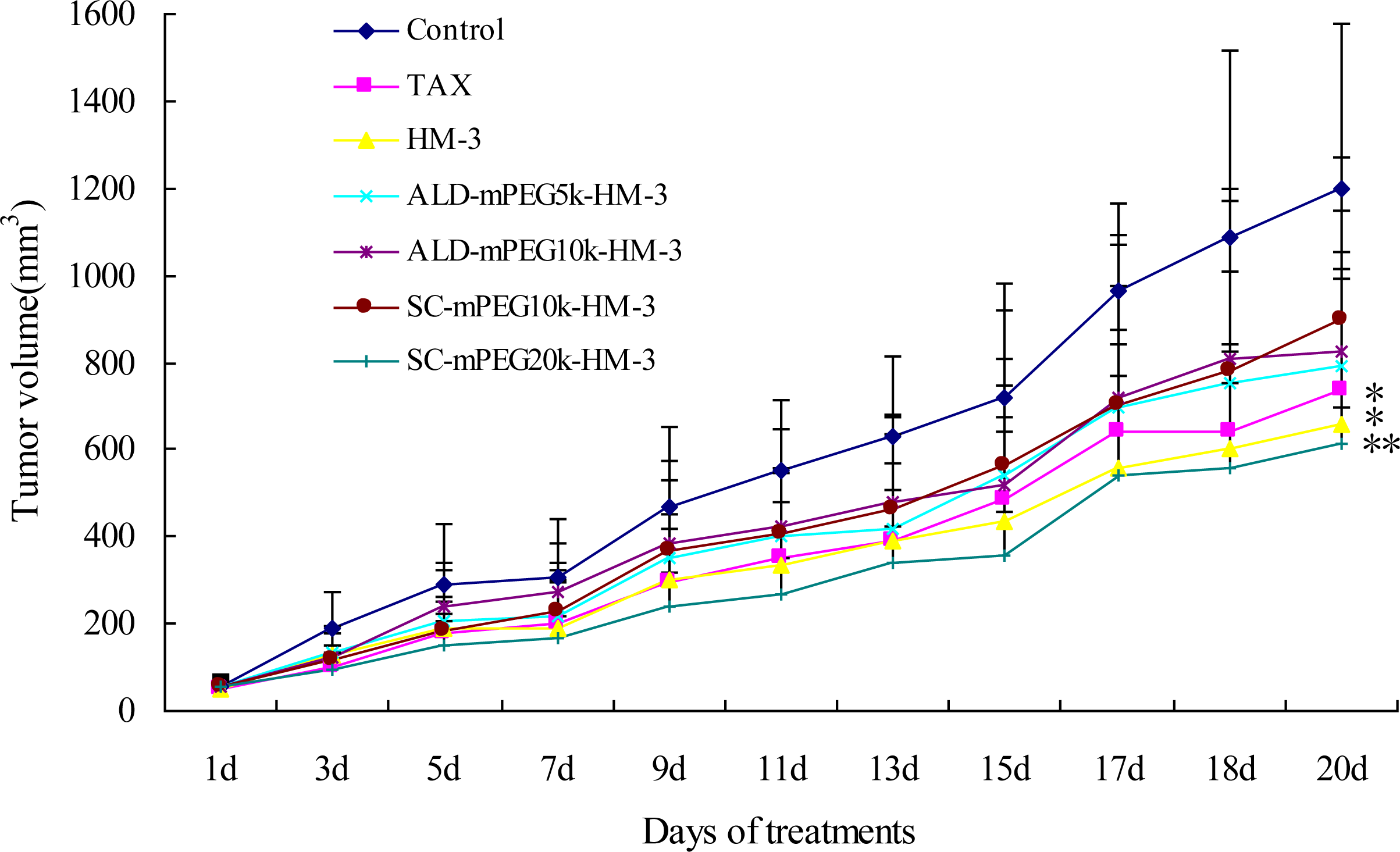
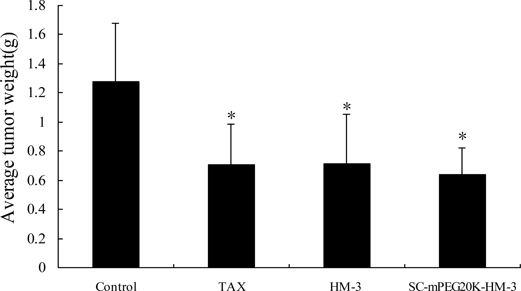
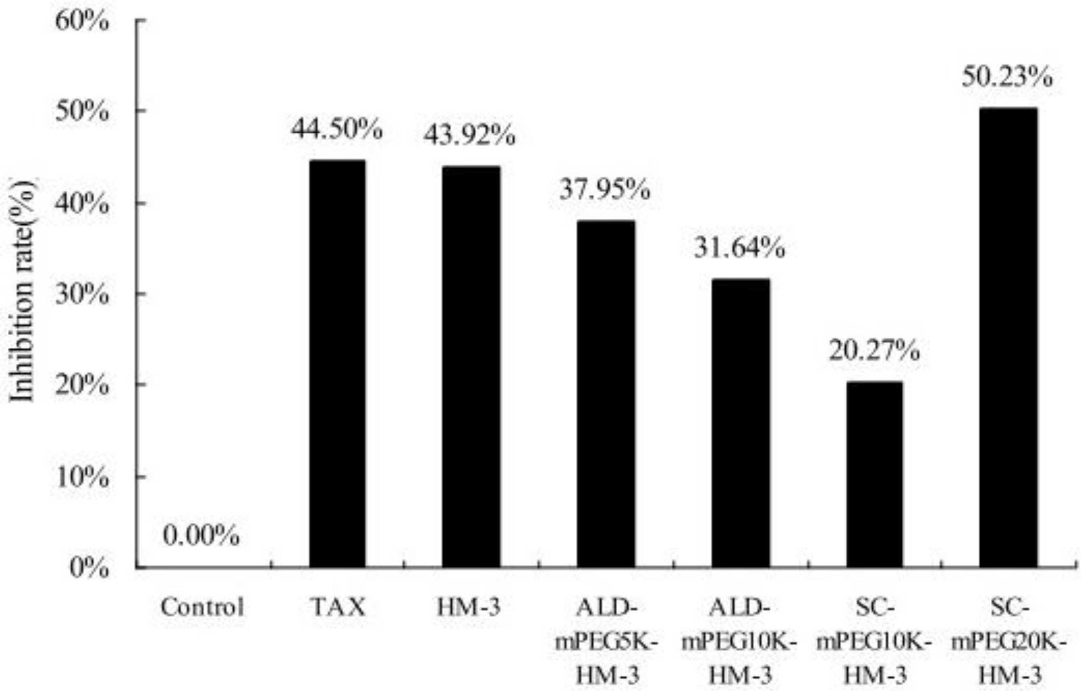
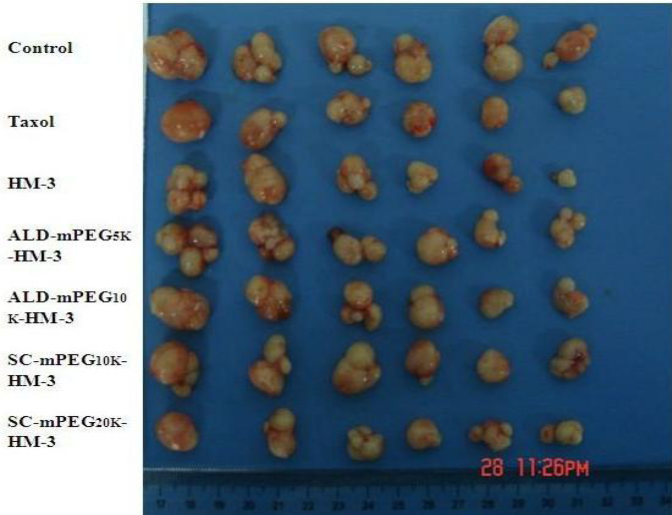
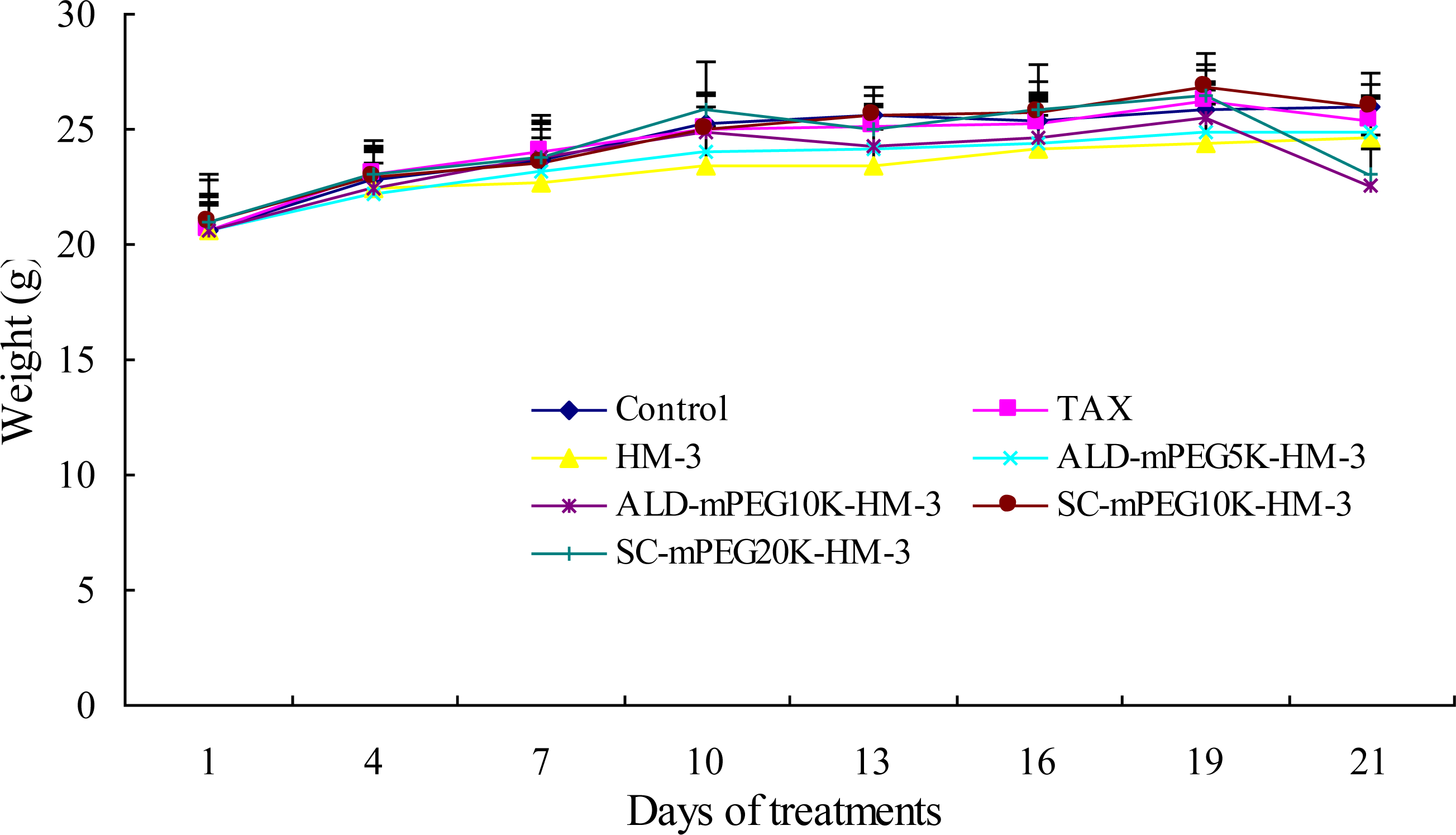
3.3. Anti-Tumor Activity of the Different PEG-Modified HM-3

We treated nude mouse with HM-3, ALD-mPEG5k-HM-3, ALD-mPEG10k-HM-3, SC-mPEG10k-HM-3 and SC-mPEG20k-HM-3 and determined the tumor growth. Taxol was used as a positive control. We found that tumor growth in the HM-3 and SC-mPEG20k-HM-3 groups was significantly slower than that in the Taxol group. In addition, tumor growth in the HM-3 group and SC-mPEG20k-HM-3 group was significantly different from that in the negative control group (P < 0.05 and P < 0.01, respectively) (Figure 4).
Tumor inhibitory rate in the SC-mPEG20k-HM-3 group (50.23%) and HM-3 group (43.92%) were similar to that in the Taxol group (44.50%), indicating that SC-mPEG20k-HM-3 maintained its anti-tumor activity. Tumor inhibitory rate in the ALD-mPEG5k-HM-3 and ALD-mPEG10k-HM-3 groups was 37.95% and 31.64%, respectively. Tumor weight in SC-mPEG20k-HM-3, HM-3 or Taxol groups was significantly different from that in the negative group (P < 0.05) (Figures 5 and 6). Photographs of the tumors at the end of the study are shown in Figure 7, tumors in the SC-mPEG20k-HM-3, HM-3 or Taxol groups were significantly different from that in the negative group. The body weight of nude mice in each group was stable (20–25 g), and was similar between different groups. Rapid increase or decrease of body weight did not occur (Figure 8).

4. Discussion

It is well known that attachment of polyethylene glycol (PEG) to therapeutic peptides or proteins (referred to as PEGylation) effectively prolongs their half-lives, reduces antigenicity and immunogenicity, and improves their pharmacokinetic and pharmacodynamic properties, rendering the drugs significantly prolonged circulating half-lives [23,24].
SC-mPEG (mPEG-succinimidyl carbonate) and ALD-mPEG (mPEG-propionaldehyde) have been the most commonly used chain-shaped PEG with active groups. Urethane bonds generated after coupling between SC-mPEG and peptides, and secondary amine generated after combining ALD-mPEG and peptides, are more stable and have a higher hydrolytic stability than ester bonds [25,26]. The SC-mPEG was extremely reactive, with reactions with peptides or proteins requiring higher pH in PBS, while the ALD-mPEG required mild reaction conditions, and could combine with peptides or proteins in mild pH in PBS [27]. Yet, the reactivity of the latter was lower than the former using the same peptides or proteins.
In PEGylation of pharmaceuticals, the design of an appropriate conjugation strategy is essential to confer the desired properties to the parent peptide or protein. The random conjugation of PEG would result in significant decrease, even loss of bioactivities, which showed inconsistent therapeutic effects and was undesired in PEGylated pharmaceuticals [20,28,29].
Therefore, when we modify a molecule with PEG, we should choose PEGs that have optimal balance between the activity maintaining rate and their half-life in vivo. According to previous studies, the in vitro activity of the PEGylated HM-3 products (especially SC-mPEG20k-HM-3) were high, but the in vivo activity was not measured. Thus, it is necessary to study the in vivo activity of the PEGylated HM-3 [1,4].
Figure 6 shows that the activity maintaining rate of modification by SC-mPEG10k and ALD-mPEG10k were not high, and the tumor inhibition rate of SC-mPEG10k-HM-3 (20.27%) was lower than ALD-mPEG10k-HM-3 (31.64%), while the modified product of SC-mPEG20k-HM-3 (50.23%) was excellent, displaying similar tumor inhibitory activity in vivo to the Taxol and unmodified HM-3 groups.
Generally speaking, modification with large molecule PEG may result in reduced activity. The activity of modified products of anti-tumor peptides and proteins is closely associated with organism resistance, PEG mass and shape, stability of PEG-peptide or PEG-protein bond, and changes in the spatial structure of modified products [27,30].
In our study, the activity of SC-mPEG20k-HM-3 did not change. This might be due to the fact that the loss of biological activity was compensated for by the significantly prolonged half-life and blood plasma residence time in vivo, as a result of the increased stability and higher hydrodynamic volume after modification. The activity of ALD-mPEG5k-HM-3, ALD-mPEG10k-HM-3, and SC-mPEG10k-HM-3 would decline with different degrees after modification, while the half-life and blood plasma residence time of these in vivo were low, which were not obviously prolonged in the previous study. Perhaps these factors cause the anti-tumor activity of ALD-mPEG5k-HM-3, ALD-mPEG10k-HM-3, and SC-mPEG10k-HM-3 to be lower than SC-mPEG20k-HM-3 and unmodified HM-3 in vivo. Besides, the active groups of SC-mPEG and ALD-mPEG are different, coupling between SC-mPEG and peptides with urethane bond, and in the presence of sodium cyanoborohydride, a secondary amine would be generated after combination of ALD-mPEG and peptides. The latter stability was higher than the former in vivo [31,32]. Maybe this led to the different activity of SC-mPEG10k-HM-3 and ALD-mPEG10k-HM-3 in vivo.
SC-mPEG20k-HM-3 retained the original biological activity of HM-3. Tumor inhibition of SC-mPEG20k-HM-3 (50.23%) was slightly higher than that of Taxol in vivo. These data suggest that the half-life of HM-3 can be improved by modification with SC-mPEG20k. For instance, drug administration frequency was extended from twice per day to once per two days after modification by SC-mPEG20k. The anti-tumor therapeutic efficacy of SC-mPEG20k-HM-3 was more efficient than that of HM-3. The body weight of mice in each group was maintained at a stable level, indicating that the effective dosage of drugs is non-toxic or the toxicity is low.
Therefore, this study demonstrated that SC-mPEG20k can be used for modification of HM-3 and further determined in vivo biological activity of the modified product SC-mPEG20k-HM-3. We will further characterize the physio-chemical properties, in vivo pharmacokinetics, immunogenicity and the antitumor target of SC-mPEG20k-HM-3.
Our preliminary results found that although the tumor grew slowly and the tumor formation rate was low in nude mice inoculated with 5 × 10⁵ tumor cells/mice, administration of drugs had a good effect in inhibiting the tumors. The tumor inhibition for small tumors (20 mm3–30 mm3) was more efficient than for large tumors (>80 mm3). Taxol administered once per two days (10 mg/kg/time) had better tumor inhibition than that administered twice per week. However, Taxol is highly toxic and frequent administration of taxol may result in weight loss or even death. Thus, in this study, we treated the mice with Taxol twice per week. In addition, if the tumor cell line was transferred in culture too many times, the property of the cells may be changed, which may affect the evaluation of the therapeutic efficiency. These results provide references for other related studies.

5. Conclusion

SC-mPEG20k-HM-3 retained the anti-tumor activity of HM-3 better than other PEG-modified products. In addition, SC-mPEG20k modified peptide HM-3 had a low plasma clearance rate, long half-life and retained its anti-tumor activity. Therefore, SC-mPEG20k-HM-3 has a very high value for development of new anti-tumor drugs.

Acknowledgments

The authors want to thank National Science Foundation of China (NSFC, No. 30472085,30873073) and thank China scientific and technological major special project—“significant creation of new drugs” (grant No. 2009ZX09102).

References

  1. Zhou, K; Zheng, X; Xu, HM; Zhang, J; Chen, Y; Xi, T; Feng, T. Studies of poly (ethylene glycol) modification of HM-3 polypeptides. Bioconjug. Chem 2009, 20, 932–936. [Google Scholar]
  2. Xu, HM; Yin, R; Chen, L; Siraj, S; Huang, X; Wang, M; Fang, H; Wang, Y. An RGD-modified endostatin-derived synthetic peptide shows antitumor activity in vivo. Bioconjug. Chem 2008, 19, 1980–1986. [Google Scholar]
  3. Xu, HM. HM-3, Angiogenesis Inhibitor Preparative Method and Application. China Patent 1830487, 13 September 2006. [Google Scholar]
  4. Zhu, B; Xu, HM; Zhao, L; Huang, X; Zhang, F. Site-specific modification of anti-angiogenesis peptide HM-3 by polyethylene glycol molecular weight of 20 kDa. J. Biochem 2010, 148, 341–347. [Google Scholar]
  5. Cox, GN; Rosendahl, MS; Chlipala, EA; Smith, DJ; Carlson, SJ; Doherty, DH. A long-acting, mono-PEGylated human growth hormone analog is a potent stimulator of weight gain and bone growth in hypophysectomized rats. Endocrinology 2007, 148, 1590–1597. [Google Scholar]
  6. Kim, MY; Kwon, JS; Kim, HJ; Lee, EK. In vitro refolding of PEGylated lipase. J. Biotechnol 2007, 131, 177–179. [Google Scholar]
  7. Molineux, G. Pegylation: Engineering improved biopharmaceuticals for oncology. Pharmacotherapy 2003, 23, 3S–8S. [Google Scholar]
  8. Tsubery, H; Mironchik, M; Fridkin, M; Shechter, Y. Prolonging the action of protein and peptide drugs by a novel approach of reversible polyethylene glycol modification. J. Biol. Chem 2004, 279, 38118–38124. [Google Scholar]
  9. Roberts, MJ; Bentley, MD; Harris, JM. Chemistry for peptide and protein PEGylation. Adv. Drug Deliv. Rev 2002, 54, 459–476. [Google Scholar]
  10. Veronese, FM. Peptide and protein PEGylation: a review of problems and solutions. Biomaterials 2001, 22, 405–417. [Google Scholar]
  11. Chilukuri, N; Sun, W; Naik, RS; Parikh, K; Tang, L; Doctor, BP; Saxena, A. Effect of polyethylene glycol modification on the circulatory stability and immunogenicity of recombinant human butyrylcholinesterase. Chem. Biol. Interact 2008, 175, 255–260. [Google Scholar]
  12. Kang, JS; Deluca, PP; Lee, KC. Emerging PEGylated drugs. Expert Opin. Emerg. Drugs 2009, 14, 363–380. [Google Scholar]
  13. Ryan, SM; Mantovani, G; Wang, X; Haddleton, DM; Brayden, DJ. Advances in PEGylation of important biotech molecules: delivery aspects. Expert Opin. Drug Deliv 2008, 5, 371–383. [Google Scholar]
  14. So, T; Ito, HO; Tsujihata, Y; Hirata, M; Ueda, T; Imoto, T. The molecular weight ratio of monomethoxypolyethylene glycol (mPEG) to protein determines the immunotolerogenicity of mPEG proteins. Protein Eng. Des. Sel 1999, 12, 701–705. [Google Scholar]
  15. Cohen, O; Kronman, C; Lazar, A; Velan, B; Shafferman, A. Controlled concealment of exposed clearance and immunogenic domains by site-specific polyethylene glycol attachment to acetylcholinesterase hypolysine mutants. J. Biol. Chem 2007, 282, 35491–35501. [Google Scholar]
  16. Jain, A; Jain, SK. PEGylation: An approach for drug delivery. A review. Crit. Rev. Ther. Drug Carrier Syst 2008, 25, 403–447. [Google Scholar]
  17. Kubetzko, S; Sarkar, CA; Pluckthun, A. Protein PEGylation decreases observed target association rates via a dual blocking mechanism. Mol. Pharmacol 2005, 68, 1439–1454. [Google Scholar]
  18. Zalipsky, S. Hydrazided derivatives of poly (ethyleneglycol) and their conjugates. ACS Symp. Ser 1997, 12, 318–341. [Google Scholar]
  19. Yang, Z; Wang, J; Lu, Q; Xu, J; Kobayashi, Y; Takakura, T; Takimoto, A; Yoshioka, T; Lian, C; Chen, C; et al. PEGylation confers greatly extended half-life and attenuated immunogenicity to recombinant methioninase in primates. Cancer Res 2004, 64, 6673–6678. [Google Scholar]
  20. Veronese, FM; Pasut, G. PEGylation, successful approach to drug delivery. Drug Discov. Today 2005, 10, 1451–1458. [Google Scholar]
  21. Fontana, A; Spolaore, B; Mero, A; Veronese, FM. Site-specific modification and PEGylation of pharmaceutical proteins mediated by transglutaminase. Adv. Drug Deliv. Rev 2008, 60, 13–28. [Google Scholar]
  22. State Food and Drug Administration, P.R. China. Guiding principles of non-clinical research technique of cytotoxic antitumor drugs. Chin. J. New Drugs Clin. Rem 2008, 27, 462–465. [Google Scholar]
  23. Harris, JM; Chess, RB. Effect of pegylation on pharmaceuticals. Nat. Rev. Drug Discov 2003, 2, 214–221. [Google Scholar]
  24. Molineux, G. Pegylation: Engineering improved pharmaceuticals for enhanced therapy. Cancer Treat. Rev 2002, 28, 13–16. [Google Scholar]
  25. Veronese, FM; Mero, A. The impact of PEGylation on biological therapies. Biol. Drugs 2008, 22, 315–329. [Google Scholar]
  26. Zalipsky, S; Seltzer, R; Menon-Rudolph, S. Evaluation of a new reagent for covalent attachment of polyethylene glycol to proteins. Biotechnol. Appl. Biochem 1992, 15, 100–114. [Google Scholar]
  27. Yu, PZ; Zheng, CY; Chen, J; Zhang, GF; Liu, YD; Suo, XY; Zhang, GC; Su, ZG. Investigation on PEGylation strategy of recombinant human interleukin-1 receptor antagonist. Bioorg. Med. Chem 2007, 15, 5396–5405. [Google Scholar]
  28. Lee, S; Greenwald, RB; McGuire, J; Yang, K; Shi, C. Drug delivery systems employing 1,6-elimination: releasable poly(ethylene glycol) conjugates of proteins. Bioconjugate. Chem 2001, 12, 163–169. [Google Scholar]
  29. Pettit, DK; Bonnert, TP; Eisenman, J; Srinivasan, S; Paxton, R; Beers, C; Lynch, D; Miller, B; Yost, J; Grabstein, KH; Gombotz, WR. Structure-function studies of interleukin 15 using site-specific mutagenesis, polyethylene glycol conjugation, and homology modeling. J. Biol. Chem 1997, 272, 2312–2318. [Google Scholar]
  30. Zhai, YQ; Zhao, YJ; Lei, JD; Su, ZG; Ma, GH. Enhanced circulation half-life of site-specific PEGylated rhG-CSF: Optimization of PEG molecular weight. J. Biotechnol 2009, 142, 259–266. [Google Scholar]
  31. Van Arnum, SD; Niemczyk, HJ; Chang, CF. HPLC method validation studies on a specific assay for monomethoxypoly(ethylene glycol) succinimido carbonate (mPEG-SC). J. Pharm. Biomed. Anal 2009, 50, 138–143. [Google Scholar]
  32. Yu, PZ; Li, XQ; Li, XN; Lu, XL; Ma, GH; Su, ZG. Preparative purification of polyethylene glycol derivatives with polystyrene-divinylbenzene beads as chromatographic packing. Bioorg. Med. Chem. Lett 2007, 17, 5605–5609. [Google Scholar]
Figure 1. RP-HPLC analysis of PEGylated HM-3. Analysis was performed on a C18 column with water/ACN/TFA as eluent and a flow rate of 1 mL/min, and peaks were monitored at 220 nm. (A) ALD-mPEG5k-HM-3 purity: 97.39%, T: 9.395 min; (B) ALD-mPEG10k-HM-3 purity: 98.23%, T: 9.698 min; (C) SC-mPEG10k-HM-3 purity: 97.02%, T: 9.883 min; (D) SC-mPEG20k-HM-3 purity: 98.52%, T: 9.578 min.
Ijms 12 02650f1aIjms 12 02650f1b
Figure 2. SDS-PAGE analysis of PEGylated HM-3. Lane Marker, Molecular weight markers; Lane 1, SDS-PAGE of SC-mPEG20k; Lane 2, SDS-PAGE of SC-mPEG20k-HM-3; Lane 3, SDS-PAGE of SC-mPEG10k; Lane 4, SDS-PAGE of SC-mPEG10k-HM-3; Lane 5, SDS-PAGE of ALD-mPEG10k; Lane 6, SDS-PAGE of ALD-mPEG10k-HM-3; Lane 7, SDS-PAGE of ALD-mPEG5k; Lane 8, SDS-PAGE of ALD-mPEG5k-HM-3.
Ijms 12 02650f2
Figure 3. Western Blot analysis of PEGylated HM-3. (I) SC-mPEG20k-HM-3; (II) SC-mPEG10k-HM-3; (III) ALD-mPEG10k-HM-3; (IV) ALD-mPEG5k-HM-3.
Ijms 12 02650f3
Figure 4. Therapeutic effects of peptides on the growth of tumors in BALB/c nude mice. Each point represents mean ±SD of each group (mean ± SD, n = 6, *P < 0.05; **P < 0.01 versus control). Control, once a day; The positive control drug Taxol at the dose of 10 mg/kg twice a week; HM-3 at the dose of 3 mg/kg twice a day; ALD-mPEG5k-HM-3 at the dose of 11.4 mg/kg once a day; ALD-mPEG10k-HM-3 at the dose of 19.9 mg/kg once every 2 day; SC-mPEG10k-HM-3 at the dose of 19.9 mg/kg once every 2 days; SC-mPEG20k-HM-3 at the dose of 36.7 mg/kg once every 2 day.
Ijms 12 02650f4
Figure 5. Average tumor weight in BALB/c nude mice. The data was analyzed using the statistics software SPSS13.0. Columns represent mean ± SD of each group (mean ± SD, n = 6, *P < 0.05 versus control).
Ijms 12 02650f5
Figure 6. Inhibition rate of average tumor weight in BALB/c nude mice (n = 6).
Ijms 12 02650f6
Figure 7. Photos of tumors at the end of the study indicating the therapeutic effects of peptides in BALB/c nude mice (n = 6).
Ijms 12 02650f7
Figure 8. Weight change of BALB/c nude mice. Results are expressed as mean ±SD per group (mean ± SD, n = 6).
Ijms 12 02650f8
Table 1. HM-3, conditions of polyethylene glycol (PEG) modification.
Phosphate Buffers, PBS (pH)PEGmol Ratio (PEG:HM-3)Modification Rate
PBS 5.0ALD-mPEG5k2:169.61%
PBS 5.0ALD-mPEG10k2.5:188.41%
PBS 8.0SC-mPEG10k1.5:195.94%
PBS 8.5SC-mPEG20k1.5:196.84%
Table 2. Dosage and frequency of drugs.
GroupsDrugsFrequencyDosage
FirstControlonce a day0.2 mL/d
SecondTaxoltwice a week10 mg/kg
ThirdHM-3twice a day3 mg/(kg·d)
FourthALD-mPEG5k-HM-3once a day11.4 mg/(kg·d)
FifthALD-mPEG10k-HM-3every two days19.9 mg/(kg·2d)
SixthSC-mPEG10k-HM-3every two days19.9 mg/(kg·2d)
SeventhSC-mPEG20k-HM-3every two days36.7 mg/(kg·2d)
Annotation: PEG-HM-3 dosage is total PEG conjugate.

