Proteomic Analysis Identified DJ-1 as a Cisplatin Resistant Marker in Non-Small Cell Lung Cancer

Abstract
:1. Introduction
2. Results and Discussion
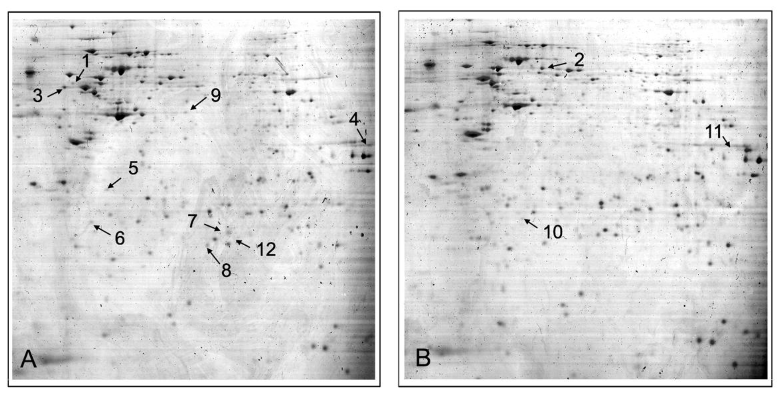
2.1. Proteomic Analysis Identified DJ-1 Up-Regulated in Drug-Resistant A549/DDP Compared with Drug-Sensitive A549 Cells
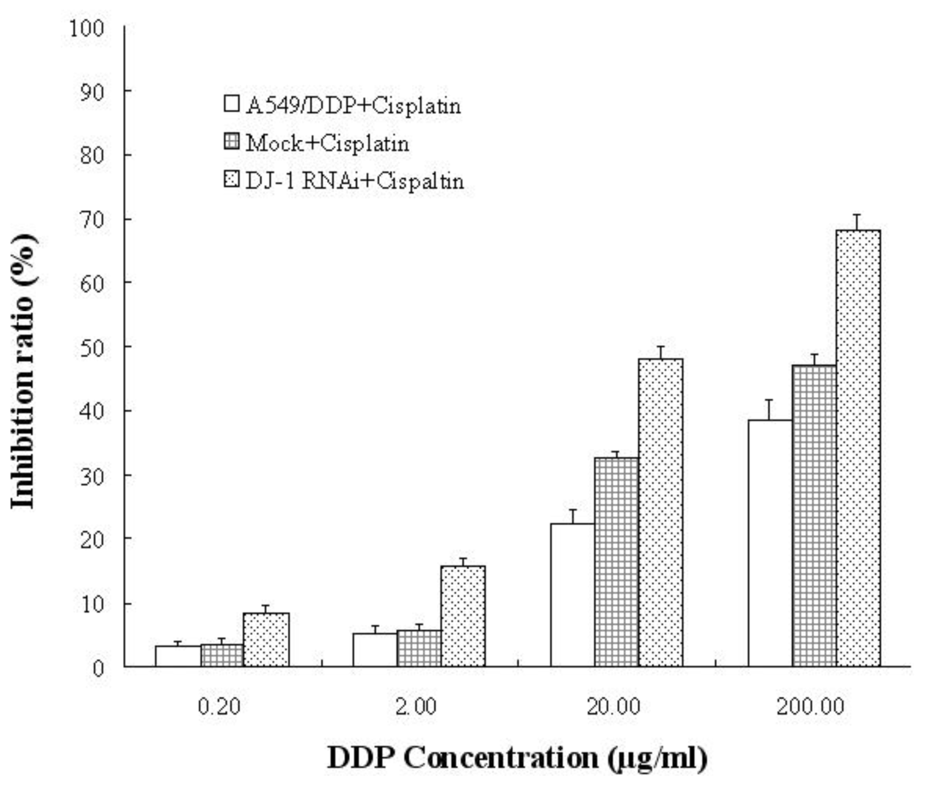
2.3. Silencing of DJ-1 Partially Reversed Cisplatin Resistance in Drug-Resistant A549/DDP Cells
2.4. Discussion
3. Experimental Section
3.1. Cell Lines and Patients
3.2. Two-Dimensional Gel Electrophoresis (2-DE) and Mass Spectrometry Analysis
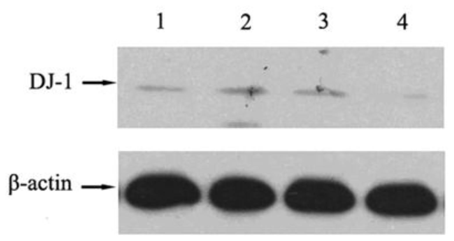
3.3. Western-Blot
3.4. Immunohistochemistry
3.5. RNA Interference and Cisplatin Sensitivity Assay
3.6. Statistical Analysis
4. Conclusions
References
- Jemal, A; Thomas, A; Murray, T; Thun, M. Cancer statistics. CA Cancer J. Clin 2002, 52, 23–47. [Google Scholar]
- Sève, P; Dumontet, C. Chemoresistance in non-small cell lung cancer. Curr. Med. Chem. Anticancer Agents 2005, 5, 73–88. [Google Scholar]
- Cobo, M; Isla, D; Massuti, B; Montes, A; Sanchez, JM; Provencio, M; Viñolas, N; Paz-Ares, L; Lopez-Vivanco, G; Muñoz, MA; et al. Customizing cisplatin based on quantitative excision repair cross-complementing 1 mRNA expression: A phase III trial in non-small-cell lung cancer. J. Clin. Oncol 2007, 25, 2747–2754. [Google Scholar]
- Kim, HT; Lee, JE; Shin, ES; Yoo, YK; Cho, JH; Yun, MH; Kim, YH; Kim, SK; Kim, HJ; Jang, TW; et al. Effect of BRCA1 haplotype on survival of non-small-cell lung cancer patients treated with platinum-based chemotherapy. J. Clin. Oncol 2008, 26, 5972–5979. [Google Scholar]
- Park, KS; Kim, HK; Lee, JH; Choi, YB; Park, SY; Yang, SH; Kim, SY; Hong, KM. Transglutaminase 2 as a cisplatin resistance marker in non-small cell lung cancer. J. Cancer Res. Clin. Oncol 2010, 136, 493–502. [Google Scholar]
- Gong, F; Peng, X; Zeng, Z; Yu, M; Zhao, Y; Tong, A. Proteomic analysis of cisplatin resistance in human ovarian cancer using 2-DE method. Mol. Cell. Biochem 2011, 348, 141–147. [Google Scholar]
- Li, SL; Ye, F; Cai, WJ; Hu, HD; Hu, P; Ren, H; Zhu, FF; Zhang, DZ. Quantitative proteome analysis of multidrug resistance in human ovarian cancer cell line. J. Cell. Biochem 2010, 109, 625–633. [Google Scholar]
- Yang, Y; Gehrke, S; Haque, ME; Imai, Y; Kosek, J; Yang, L; Beal, MF; Nishimura, I; Wakamatsu, K; Ito, S; Takahashi, R; Lu, B. Inactivation of Drosophila DJ-1 leads to impairments of oxidative stress response and phosphatidylinositol 3-kinase/Akt signaling. Proc. Natl. Acad. Sci. USA 2005, 102, 13670–13675. [Google Scholar]
- Vasseur, S; Afzal, S; Tardivel-Lacombe, J; Park, DS; Iovanna, JL; Mak, TW. DJ-1/PARK7 is an important mediator of hypoxia-induced cellular responses. Proc. Natl. Acad. Sci. USA 2009, 106, 1111–1116. [Google Scholar]
- Shinbo, Y; Taira, T; Niki, T; lguchi-Ariga, SM; Ariga, H. DJ-1 restores p53 transcription activity inhibited by Topors/p53BP3. Int. J. Oncol 2005, 26, 641–648. [Google Scholar]
- Kim, RH; Peters, M; Jang, Y; Shi, W; Pintilie, M; Fletcher, GC; DeLuca, C; Liepa, J; Zhou, L; Snow, B; et al. DJ-1, a novel regulator of the tumor suppressor PTEN. Cancer Cell 2005, 7, 263–273. [Google Scholar]
- Asechi, H; Hatano, E; Nitta, T; Tada, M; Iwaisako, K; Tamaki, N; Nagata, H; Narita, M; Yanagida, A; Ikai, I; Uemoto, S. Resistance to cisplatin-induced apoptosis via PI3K-dependent survivin expression in a rat hepatoma cell line. Int. J. Oncol 2010, 37, 89–96. [Google Scholar]
- Peng, DJ; Wang, J; Zhou, JY; Wu, GS. Role of the Akt/mTOR survival pathway in cisplatin resistance in ovarian cancer cells. Biochem. Biophys. Res. Commun 2010, 394, 600–605. [Google Scholar]
- Hao, J; Song, X; Song, B; Liu, Y; Wei, L; Wang, X; Yu, J. Effects of lentivirus-mediated HIF-1alpha knockdown on hypoxia-related cisplatin resistance and their dependence on p53 status in fibrosarcoma cells. Cancer Gene Ther 2008, 15, 449–455. [Google Scholar]
- Huang, XZ; Wang, J; Huang, C; Chen, YY; Shi, GY; Hu, QS; Yi, J. Emodin enhances cytotoxicity of chemotherapeutic drugs in prostate cancer cells: the mechanisms involve ROS-mediated suppression of multidrug resistance and hypoxia inducible factor-1. Cancer Biol. Ther 2008, 7, 468–475. [Google Scholar]
- Jeong, SH; Jung, JH; Han, JH; Kim, JH; Choi, YW; Lee, HW; Kang, SY; Hwang, YH; Ahn, MS; Choi, JH; et al. Expression of Bcl-2 predicts outcome in locally advanced non-small cell lung cancer patients treated with cisplatin-based concurrent chemoradiotherapy. Lung Cancer 2010, 68, 288–294. [Google Scholar]


| Spot | Gene Symbol | Protein Name | Swiss-Prot Accession | Fold * | Function |
|---|---|---|---|---|---|
| 1 | VIM | Vimentin | P08670 | 3.76 | structural constituent of cytoskeleton protein C-terminus binding |
| 2 | ALDOA | Fructose-bisphosphate aldolase A | P04075 | −3.09 | actin binding fructose binding fructose-bisphosphate aldolase activity identical protein binding tubulin binding |
| 3 | HNRNPA2B1 | Heterogeneous nuclear ribonucleoproteins A2/B1 | P22626 | 2.82 | RNA binding nucleotide binding protein binding single-stranded telomeric DNA binding |
| 4 | PGK1 | Phosphoglycerate kinase 1 | P00558 | 4.47 | ATP binding phosphoglycerate kinase activity |
| 5 | PRDX4 | Peroxiredoxin 4 | Q13162 | 8.11 | thioredoxin peroxidase activity |
| 6 | ANXA1 | Annexin A2 | P04083 | 5.93 | calcium ion binding calcium-dependent phospholipid binding phospholipase A2 inhibitor activity protein binding, bridging receptor binding structural molecule activity |
| 7 | PARK7 | Protein DJ-1 | Q99497 | 5.43 | protein binding |
| 8 | STMN1 | Stathmin | P16949 | 3.49 | signal transducer activity tubulin binding |
| 9 | PFN1 | Profilin-1 | P07737 | 2.95 | actin binding proline-rich region binding |
| 10 | P07437 | Tubulin β chain | TUBB | −5.31 | GTP binding GTPase activity MHC class I protein binding |
| 11 | P09972 | Fructose-bisphosphate aldolase C | ALDOC | −2.37 | cytoskeletal protein binding fructose-bisphosphate aldolase activity |
| 12 | P23528 | Cofilin-1 | CFL1 | 3.94 | actin binding |
| Characteristics | Total (n = 67) | DJ-1 Low Expression (n = 34) | DJ-1 High Expression (n = 33) | P Value |
|---|---|---|---|---|
| Gender | ||||
| Male | 48 | 26 | 22 | 0.373 |
| Female | 19 | 8 | 11 | |
| Age | ||||
| <60 year | 29 | 17 | 12 | 0.260 |
| ≥60 year | 38 | 17 | 21 | |
| Histology | ||||
| Squamous cell carcinoma | 37 | 18 | 19 | 0.703 |
| Adenocarcinoma | 30 | 16 | 14 | |
| Pathologic stage | ||||
| IIIA | 9 | 3 | 6 | 0.261 |
| IIIB | 56 | 31 | 27 | |
| Chemotherapy regimens | ||||
| NVB + DDP | 29 | 17 | 12 | |
| Taxol + DDP | 23 | 10 | 13 | 0.521 |
| Gemzar + DDP | 15 | 7 | 8 | |
| Response to cisplatin-based chemotherapy | ||||
| Resistant | 29 | 10 | 19 | 0.020 |
| Sensitive | 38 | 24 | 14 |
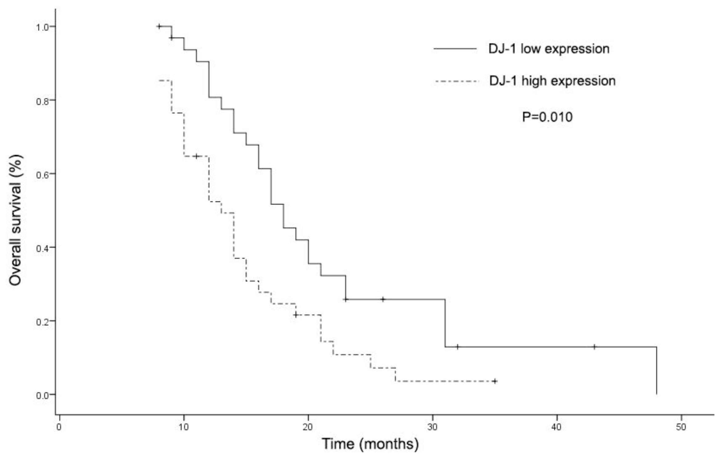
| Characteristics | Univariate Analysis
| |
|---|---|---|
| HR (95% CI) | P | |
| Age | 1.082 (0.641–1.825) | 0.768 |
| Gender | 1.434 (0.800–2.571) | 0.226 |
| Histology | 0.922 (0.547–1.556) | 0.762 |
| Pathologic stage | 0.691 (0.313–1.525) | 0.360 |
| Chemotherapy regimens | 0.609 (0.611–2.322) | 0.609 |
| Response to cisplatin-based chemotherapy | 0.688 (0.403–1.174) | 0.170 |
| DJ-1 expression level | 0.495 (0.291–0.843) | 0.010 |
© 2011 by the authors; licensee Molecular Diversity Preservation International, Basel, Switzerland. This article is an open-access article distributed under the terms and conditions of the Creative Commons Attribution license (http://creativecommons.org/licenses/by/3.0/).
Share and Cite
Zeng, H.-Z.; Qu, Y.-Q.; Zhang, W.-J.; Xiu, B.; Deng, A.-M.; Liang, A.-B. Proteomic Analysis Identified DJ-1 as a Cisplatin Resistant Marker in Non-Small Cell Lung Cancer. Int. J. Mol. Sci. 2011, 12, 3489-3499. https://doi.org/10.3390/ijms12063489
Zeng H-Z, Qu Y-Q, Zhang W-J, Xiu B, Deng A-M, Liang A-B. Proteomic Analysis Identified DJ-1 as a Cisplatin Resistant Marker in Non-Small Cell Lung Cancer. International Journal of Molecular Sciences. 2011; 12(6):3489-3499. https://doi.org/10.3390/ijms12063489
Chicago/Turabian StyleZeng, Hua-Zong, Yi-Qing Qu, Wen-Jun Zhang, Bing Xiu, An-Mei Deng, and Ai-Bin Liang. 2011. "Proteomic Analysis Identified DJ-1 as a Cisplatin Resistant Marker in Non-Small Cell Lung Cancer" International Journal of Molecular Sciences 12, no. 6: 3489-3499. https://doi.org/10.3390/ijms12063489