Article

Preparation and Scintillating Properties of Sol-Gel Eu3+, Tb3+ Co-Doped Lu2O3 Nanopowders

by
Ángel de Jesús Morales Ramírez
1,
Antonieta García Murillo
1,*,
Felipe de Jesús Carrillo Romo
1,
Margarita García Hernández
1,
Joel Moreno Palmerin
2 and
Rosario Ruiz Guerrero
1
1
Instituto Politécnico Nacional, CIITEC IPN, Cerrada de Cecati S/N. Col. Santa Catarina, Azcapotzalco Mexico D.F. C.P. 02250, Mexico
2
CINVESTAV Querétaro, Libramiento Norponiente #2000, Fracc. Real de Juriquilla, C.P. 76230, Querétaro, Mexico
*
Author to whom correspondence should be addressed.
Int. J. Mol. Sci. 2011, 12(9), 6240-6254; https://doi.org/10.3390/ijms12096240
Submission received: 11 August 2011 / Revised: 14 September 2011 / Accepted: 20 September 2011 / Published: 23 September 2011
(This article belongs to the Section Materials Science)

Abstract

:
Nanocrystalline Eu3+, Tb3+ co-doped Lu2O3 powders with a maximum size of 25.5 nm were prepared by the sol-gel process, using lutetium, europium and terbium nitrates as precursors, and ethanol as a solvent. Differential thermal analysis (DTA) and infrared spectroscopy (IR) were used to study the chemical changes during the xerogel annealing. After the sol evaporation at 100 °C, the formed gel was annealed from 300 to 900 °C for 30 min under a rich O2 atmosphere, and the yielded product was analyzed by X-ray diffraction (XRD) to characterize the microstructural behavior and confirm the crystalline structure. The results showed that Lu2O3 nanopowders start to crystallize at 400 °C and that the crystallite size increases along with the annealing temperature. A transmission electron microscopy (TEM) study of samples annealed at 700 and 900 °C was carried out in order to analyze the microstructure, as well as the size, of crystallites. Finally, in regard to scintillating properties, Eu3+ dopant (5 mol%), Tb3+ codoped Lu2O3 exhibited a typical red emission at 611 nm (D°7F2), furthermore, the effect of Tb3+ molar content (0.01, 0.015 and 0.02% mol) on the Eu3+ radioluminiscence was analyzed and it was found that the higher emission intensity corresponds to the lower Tb3+ content.

1. Introduction

Since Lu2O3:Eu3+ first attracted attention as a potential X-ray phosphor [1], many efforts have been conducted in the last few decades to process it, due to a growing need for new materials to be employed in high-resolution X-ray imaging systems, including high-definition X-ray radiographers, positron emission tomography (PET) scanners as well as many others industrial measuring systems [25]. What makes lutetia attractive for such applications is its high efficiency in absorbing any kind of ionizing radiation [6]. Indeed, the combination of its high density (9.42 gcm−3) and its high atomic number of Lu (Z = 71) [7], along with its good absorption in the diagnostic medical energy range (15–150 keV) makes it a very interesting materials for the mentioned applications [8]. Furthermore, it is a convenient host lattice for activators forming scintillating materials [9], especially for Eu3+ and Tb3+, due to the fact that its band gap is large enough to accommodate the energy levels of the ions [10]. On the other hand, lutetia systems doped with Eu3+ are expected to replace the typical CsI:Tl scintillators in digital imaging, since, along with their stated properties, they present a reddish emission wavelength (~611 nm) that matches the spectral sensitivity of CCD detectors [11,12], and, moreover, CsI:Tl suffer from radiation damage at high doses, probably due to its low density (4.51 gcm−3) and is slightly hygroscopic [13,14]. Another common material, the bismuth orthogermanate (BGO), presents a low density (7.13 gcm−3) and emits less than 10 photons/keV [15], compared to 30 photons/keV for Lu2O3:Eu3+ [16]. Finally, the well known Gd2O2S:Tb3+ (GOS), tends to be replaced in scintillating devices due to its chemical instability and sensitivity to moisture [17,18]. Furthermore, it has been demonstrated that Lu2O3:Eu3+ presents a light yield similar to that of GOS [19] and that a scintillating screen made of this material can produce images of better quality than those obtained by the standard GOS [20]. On the other hand, it has been noted that different methods of synthesis lead to particles with diverse morphologies and specific surface areas, and, therefore, different luminescent properties [21,22]. Because it is essential for scintillating applications to yield ultrafine, mono-sized, low-aggregated and spherical powders [23], many soft chemical routes have been used to produce lutetia nanopowders, such as co-precipitation [2426], solvothermal reaction [2731], molten salts synthesis [32], and the combustion method [3337]. Oxalic acid and urea have been used extensively in some of these methods, but these present some drawbacks for rare earth compounds, particularly due to the fact that rare earth oxalates produce particles that grow rapidly, along with severe agglomeration, whereas urea limits the yield from the homogeneous precipitation process [38]. Therefore, the sol-gel process, due to its unique advantages, like high chemical homogeneity, the possibility of achieving several compositions by simples changes in the process, and the ability to vary the nature and concentration of doping ions [39], it is ideal to produce rare earth doped Lu2O3 nanopowders [4043]. Finally, in order to improve the light yield of these materials, the Tb3+ ion has been proposed for co-doping Lu2O3:Eu3+ [44]. In Y2O3:Eu3+,Tb3+, it has been found that there is an increment of the Eu3+ luminescence, due to an energy transfer from Tb+3+ to Eu3+ ions [45,46]; furthermore in previous work [47], it has been determined that, in Gd2O3:Eu3+ nanopowders, the radioluminescence emission is enhanced by the incorporation of Tb3+ ions.
The aim of the current work is to synthesize for first time Eu3+,Tb3+ co-doped Lu2O3 scintillating nanopowders by a simple sol-gel process, starting with nitrates as precursors and without the use of urea or oxalic acid, and instead, diethyleneglycol (DEG) C4H10O3 as polymerization agent. It was studied the chemical changes during the sol gel process of the nanopowders by means of a Thermogravimetric (TGA), differential thermal analyses (DTA) and infrared spectroscopy (IR). The structural evolution during the annealing process was analyzed by X-ray diffraction (XRD), the morphology by means of TEM observations, and their scintillating properties under X-ray (30 keV) radiation as a function of the heat treatment temperature and the Tb3+ content.

2. Results and Discussion

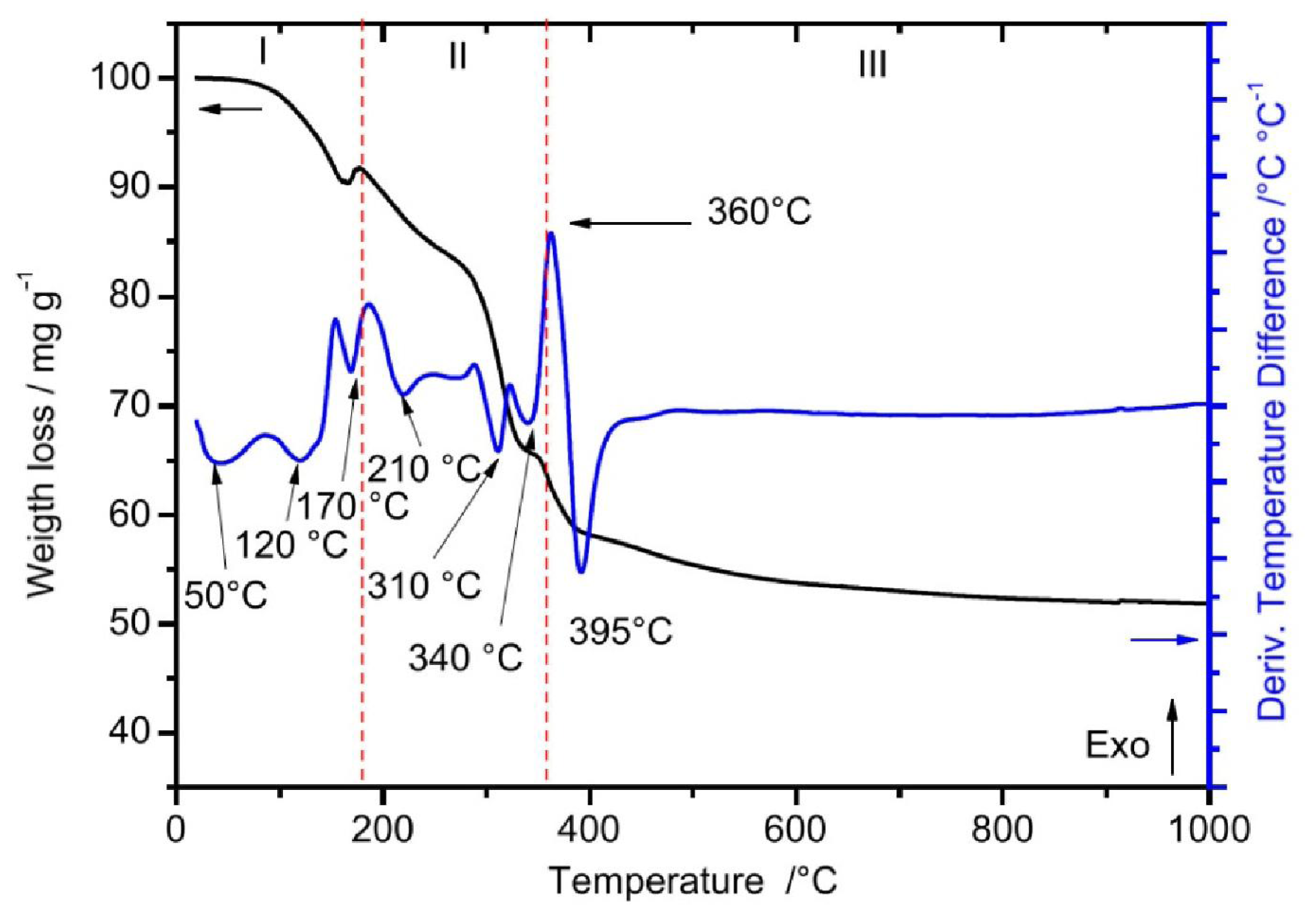
2.1. Thermal Analysis

Figure 1 shows the TGA-DTA curves of the as-prepared Lu2O3 xerogel 5 mol % Eu3+, 0.01 mol % Tb3+. As observed, the TGA curve shows three stages of weight loss. The first stage (I), from 100–175 °C, shows a weight loss of 9% and two endothermic events, at 120 and 170 °C. The first event can be attributed to the release of water of hydration and OH, and the second one to the decomposition of the organic matter, this event suggest the formation of lutetium hydrated species or even carbonate species to form precipitates with Lu3+ ions [48]. The second stage (II), from 175–360 °C, corresponding to a weight loss of 25%, involves two types of endothermic events, occurring at 210, 310 and 340°C. The first can be ascribed to the evaporation of DEG (b. p. 240 °C), whereas the last two correspond to the elimination of carbonyl groups. The last stage (III) exhibits a weight loss of 14% and includes two major events: a strong exothermic peak at 360 °C, which can be associated with the crystallization process of the ceramic sample into the cubic phase, and a strong endothermic peak at 390 °C, which was related to the pyrolisis of the remnant’s carbon groups.

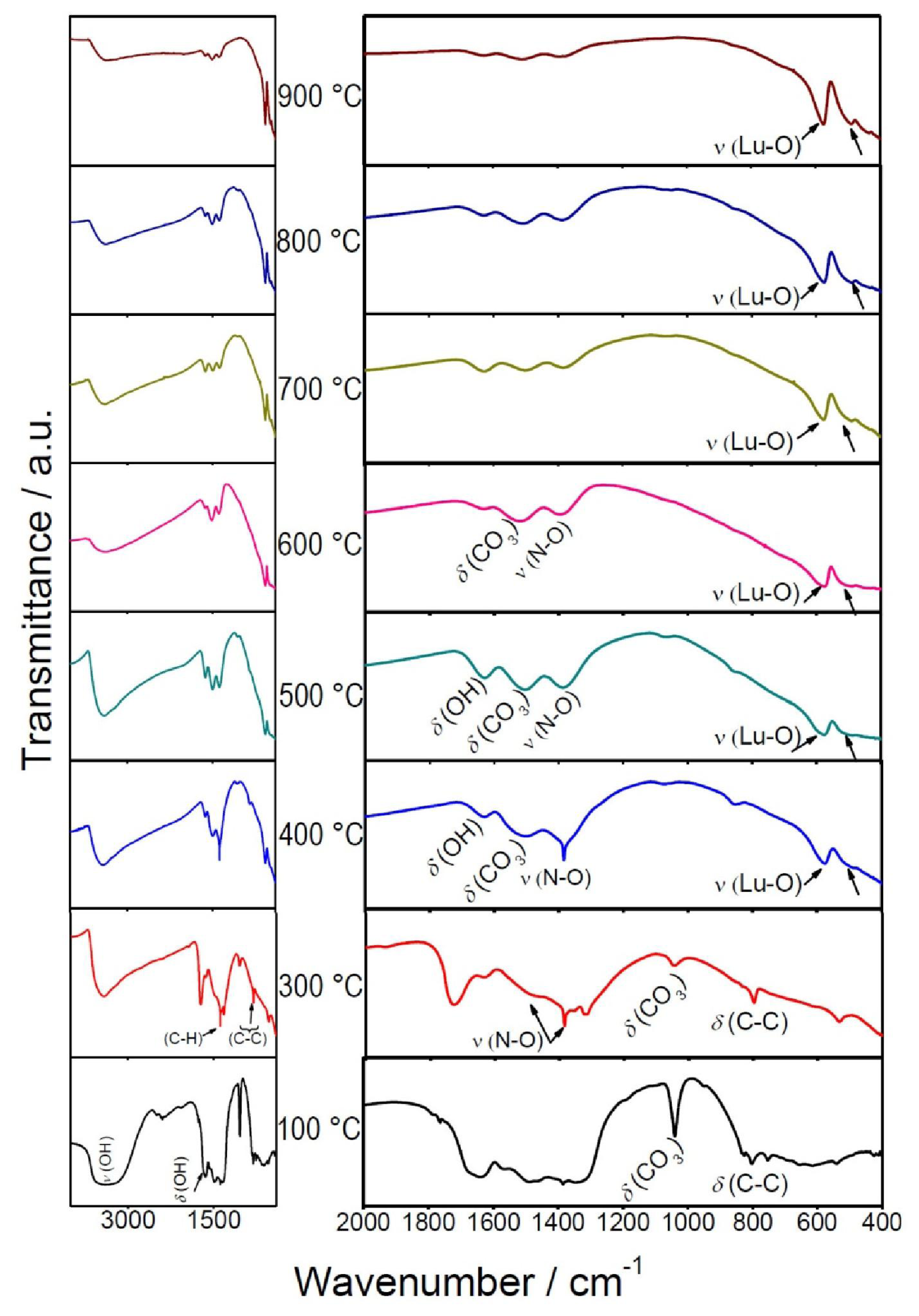
2.2. Infrared Analysis

The Fourier transformed infrared spectra of Lu2O3: Eu3+ 5 mol %, Tb3+ 0.01 mol% are depicted in Figure 2. This study was carried out in the range 4000–400 cm−1 on the dried precursor sol, thermally treated at different temperatures, in order to determine the evolution of the decomposed products of the xerogel powders calcined up to the crystallization process. For the xerogel at 100 °C, bands observed at 3400 cm−1 (ν), 1650 cm−1 (δ) and 750 cm−1 (δ) can be ascribed to O-H stretching (ν) and deformation (δ) vibrations, due the presence of water and alcohol groups. Since the heating at 400 °C was not enough to remove these species, we have to conclude that they were structurally built into the host and not merely adsorbed on the surface. After a 600 °C heat treatment, these O-H vibrations were less intense than those observed at lower temperatures, exerting an influence on the powder’s microstructure, as demonstrated by XRD analysis. While the heating of the powders at much higher temperatures (necessarily to stimulate the growth of the crystallites and their partial sintering), the vibrations related with the O-H impurities [49] are almost absent, i.e., these vibrations are nearly missing at 900 °C. The absorption peak around 1380 cm−1 indicates the N-O stretching vibration [50] of NO3 , which remains present until 500 °C. The peak situated at 1530 cm−1 can be attributed to the asymmetrical stretching of C-O, while the absorption bands at 1090 cm−1 and 850 cm−1 are due to the symmetrical stretching of C-O and deformation vibrations of C-O in CO3 2−. These absorption peaks indicate the presence of carbonate groups. Bands of C-O-H and CH2- corresponding to bending vibrations appear around 1410 cm−1 and 1460 cm−1, respectively, arising from the decomposition of DEG [51], and one is observed at 820–880 cm−1, characteristic of the C-C bond [52]. The intensity of all these bands decreased with the annealing temperature, as has been observed for lutetia ceramics fabricated by nitrate sources [53]. However, after 800 °C sintering, the N-O vibrations were still present, suggesting that some NO3 residues were adsorbed in the sample, which could be removed after 900 °C thermal treatment. Finally, the bands at 580 and 489 cm−1, observed from 400 °C, and attributed to the Lu-O stretching vibrations of cubic Lu2O3 (a Lu2O3 host lattice vibration) [5456], indicate that the crystallization was just beginning at 400 °C, which was confirmed by XRD and TEM observations.

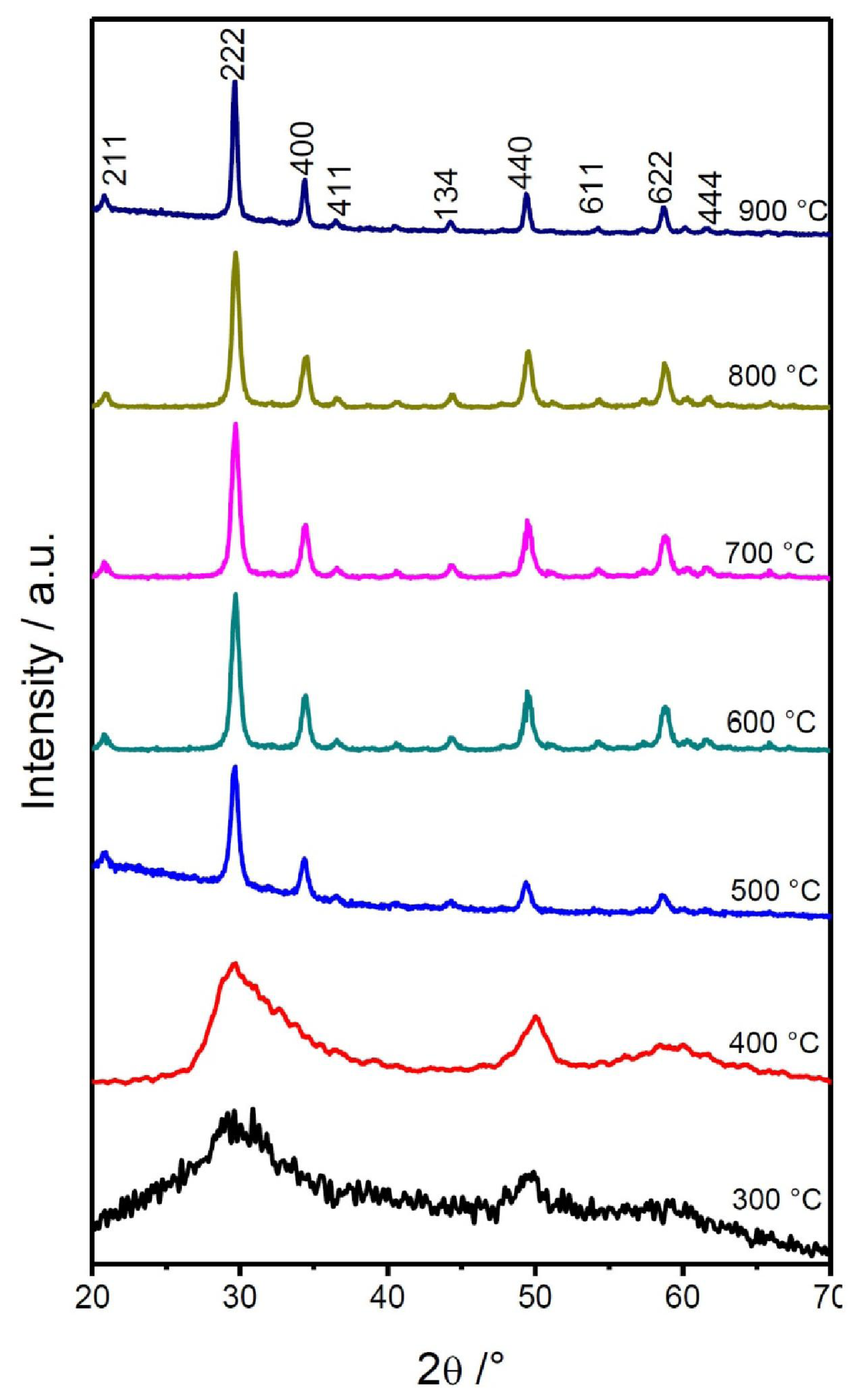
2.3. Structural Properties

Figure 3 shows the evolution of the X-ray diffraction patterns of the Lu2O3: Eu3+ 5 mol %, Tb3+ 0.01 mol% nanopowders annealed in air at temperatures ranging from 300 to 900 °C. At 300 °C, the xerogel exhibits an almost amorphous behavior; however, at 400 °C, the system possesses an aspect of an amorphous phase characterized by broad diffraction peaks, which also indicates that the crystallites are very small, lower than 5 nm.
This result is in good agreement with the DTA observations, since the crystallization process began between 300 and 400 °C. As the annealing temperature reaches 500 °C, it becomes evident that the crystallization process has ended in a cubic Lu2O3 structure (JCPDS 431021) with a spatial group Iā3 (lattice parameter 10.391 Å). With an increments the annealing temperature, the diffraction peaks become narrower, which reflects an increase in the size of the Lu2O3 crystallites. Table 1 shows the calculated crystal sizes according to Scherer’s formula D = 0.9λ/β cosθ [57], taking into account the broadening line of the diffracted peak, due to the effect of crystal size, where D is the crystal size of the powder, λ (0.15406 nm) is the wavelength of the diffracted X-ray, β is the full-width radiation at half-maximum (FWHM) of the peak, and θ is the Bragg angle of the diffracted X-ray. The crystallite size ranges from 5.3 nm at 500 °C to 25.5 nm at 900 °C; these observations were confirmed by TEM observations for the 700 and 900 °C co-doped lutetia nanopowders.
Figure 4a shows a TEM bright-field micrograph of selected Lu2O3: Eu3+ 5 mol %, Tb3+ 0.01 mol % scintillating nanopowders, annealed at 700 °C. As observed, the morphology of the powders is mainly angular; however, some of the faces are rounded and the particles are highly agglomerated. Figure 4b shows the particles’ indexed diffraction pattern, which exhibits typical nanometric ring-type behavior and confirms the cubic structure. Figure 4c shows a dark field micrograph of the area at the (2 2 2) direction. The average size determined from these observations was ≈ 17 nm, in good accord with XRD results, and presents a normal centered distribution in the range of 5–40 nm (Figure 4d).
Bright field, diffraction pattern and dark field micrographs, as well as particle size distribution of the sample annealed at 900 °C are shown in Figure 5a–b. As noted, similar observations can be made for this sample, whose average size was ≈ 27 nm. The kinetics growing of the sample can be related to the sintering and agglomeration processes activated by the thermal process.

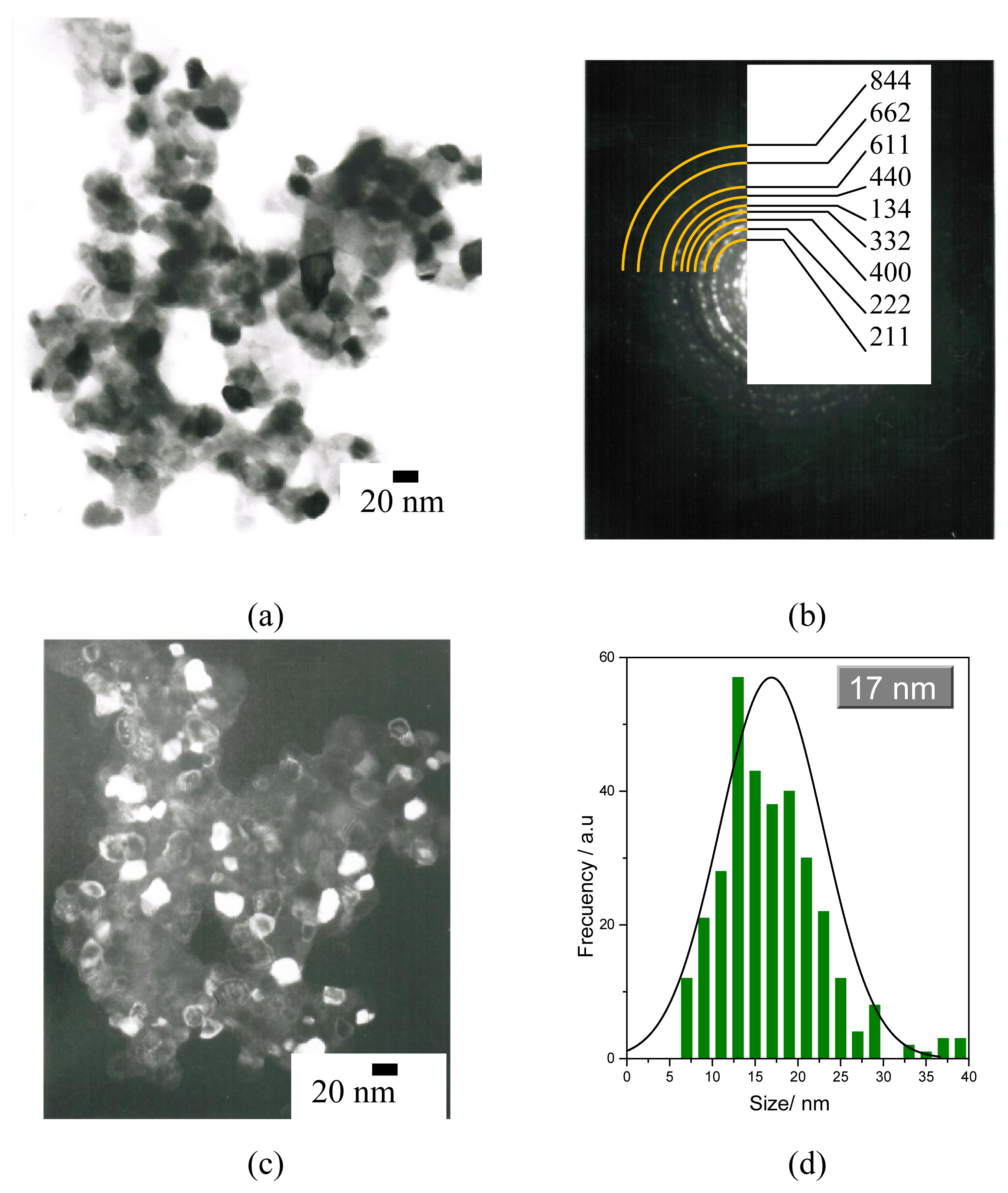
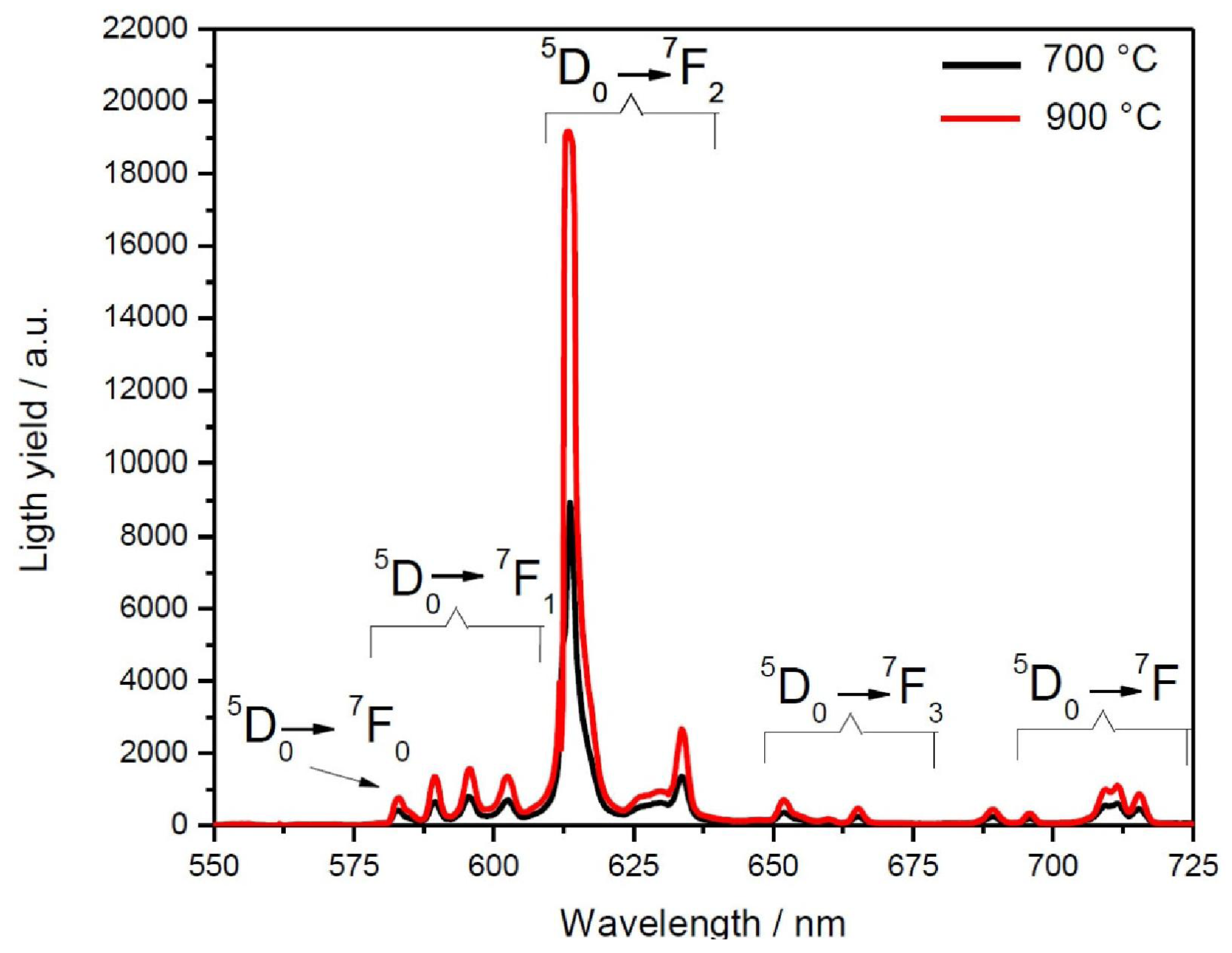
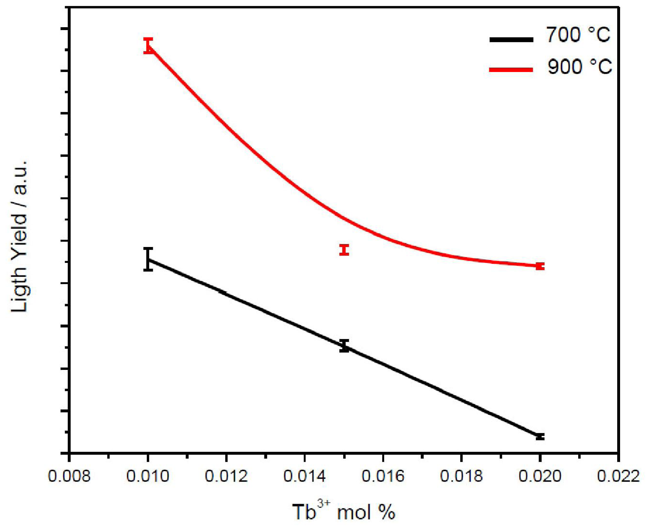
2.4. Scintillating Properties

Figure 6 presents the scintillating properties of Lu2O3: Eu3+ 5 mol%, Tb3+ 0.01 mol% nanopowders annealed at 700 and 900 °C, under X-ray excitation of 30 kV and 40 mA. The scintillating spectra of the sample display a group of emission lines situated in the 575- to 725-nm spectral region, corresponding the Eu3+ transitions from the excited 5D0 level to 7Fj (J = 0,1,2,3,4) levels, with no evidence of any Tb3+ emission (545 nm). The maximum emission line at 611 nm, which matches well the spectral sensitivity range of CCD cameras, corresponds to the 5D07F2 electric dipole transition, and is a product of the Eu3+ ion located at C2 sites in the Lu2O3 host cubic lattice [58], and its intensity is evidence of an efficient channel of energy transfer from the lutetia matrix to Eu3+ emission centers according to a recombination mechanism [59]. The less intense emission lines at 532, 580, 630 and 665 nm, correspond to the 5D07F0, 5D07F1, 5D07F3 and 5D07F4 transitions, respectively, and are in good agreement with the energy levels described elsewhere [60]. The difference in the light yield output between the two samples, of about ~110%, can be explained in terms of two effects. First, it is well known that the crystallinity achieved at higher temperatures is related to a more efficient activation of the Eu3+ ion in the nanopowder sample. Furthermore, it has been demonstrated that until the complete elimination of residual OH contamination, the non-radiative multiphonon relaxation process can be stimulated with high-energy phonons introduced by this impurity [33]. In the current work, as can be established by IR experiments, at 900 °C the residual OH impurity has been almost completely eliminated, which is a temperature considerably lower to the 1300 °C range that has been previously reported [61].
Figure 7 shows the light yield variation of the Eu3+ 5D07F2 (611 nm) emission as a function of the Tb3+ concentration for nanopowders annealed at 700 and 900 °C. In both cases, the lutetia co-doped at 0.01 mol % Tb3+ presents the highest light yield, and for reasons explained earlier, the emission of the sample heat-treated at 900 °C is higher than that of the 700 °C one. The enhancement of the Eu3+ emission in the nanopowders is due to a non-radiative energy transfer from Tb3+ to Eu3+, as Tb can absorb more X-ray radiation. At higher Tb3+ concentrations, a self-quenching mechanism [45] exists, a product of the fact that the Tb-Tb energy transfer mechanism is more efficient than the Tb-Eu one, and, therefore, presents a higher possibility that a Tb ion is closer than another sort, resulting in a drop in Eu3+ emission. Similar results have been observed for Gd2O3 co-doped Eu, Tb nanopowders [47].

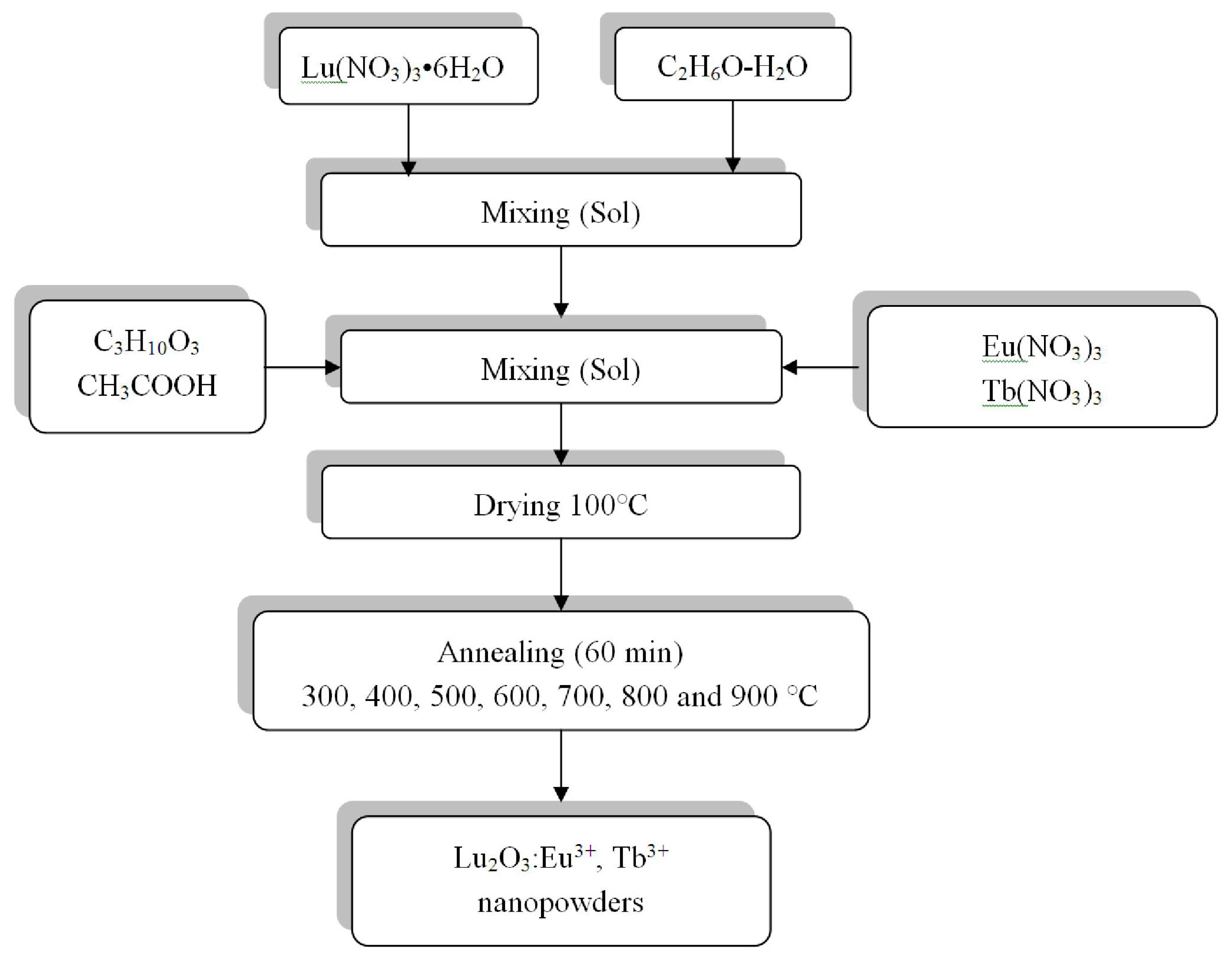
3. Experimental Section

Lu2O3:Eu3+, Tb3+ nanopowders were prepared by a simple sol-gel process method. Figure 8 shows the flow scheme for this process. The starting materials used were lutetium nitrate, Lu(NO3)3•6H2O (Alfa Aesar, 99.96%), europium nitrate Eu(NO3)3, terbium nitrate Tb(NO3)3 (99.5% and 99.6% respectively, Alfa Aesar), acetic acid CH3COOH (Fermont 98%) used as a catalyst, and diethyleneglycol (DEG) C4H10O3 (Alfa Aesar, 99%) used as a polymerization agent. Initially, the lutetium nitrate was dissolved in an ethanol-deionized water solution (95-5 vol%) using a hot stirrer at 40 °C for 3 hours to obtain 25 mL of a 0.95 M Lu sol. Thereafter, the solution pH was adjusted by incorporating acetic acid (0.17 M), and the DEG was added (0.42 M). Finally, the co-doping elements, previously dissolved in ethanol, were incorporated the initial lutetium sol. The sol obtained was mixed for 2 hours to produce a transparent solution, stable for more than 3 months. Subsequently, the sol was dried at 100 °C for 24 hours, and the yielded gel was calcined at different temperatures for 1 hour to obtain the Lu2O3:Eu3+, Tb3+ scintillating nanopowders.
Thermogravimetric (TGA) and differential thermal analyses (DTA) of dried gel were conducted using a SDT Q600 TA instrument; the studies were performed in air with a heating rate of 10 °C min−1. The IR spectra of the samples were recorded in the range of 4000–400 cm−1 using Fourier transform infrared spectroscopy (FTIR 2000, Perkin Elmer) and the KBr pelleting technique. The phase composition of the powders was identified by X-ray diffraction at room temperature on a powder diffractometer (Bruker D8Advance) using Cu Kα radiation (1.5418 Å). The morphology and particle size of the powders were observed with a transmission electronic microscope (JEOL 2200) operating at 200 keV. The scintillating properties were recorded employing an X-ray generator (PW-1830 Phillips) operating at 30 kV and 40 mA, using W Kα radiation (0.2086 Å) and a photon flux of 1012 ph s−1.

4. Conclusions

Lu2O3:Eu3+, Tb3+ nanopowders have been prepared by a simple sol-gel method, using lutetium, europium and terbium nitrate as precursors, and DEG as a polymerization agent. This nanopowder crystallizes into a cubic system from 400 °C, and the process is completed at 500 °C, resulting in particles composed of crystallites ranging in size from 5.3 to 25.5 nm. The increments in crystallite sizes depend on the heat treatment temperatures. The powder exhibits interesting scintillating properties, and at 611 nm presents a reddish emission corresponding with the Eu3+ 5D07F2 transition, which makes it a promising material for X-ray detection systems, since this emission matches the maximum efficiency of the CCD cameras. The light yield of the nanopowders was analyzed at two different annealing temperatures: 700 and 900 °C. It was established that at the sample heat treated at the higher temperature, presents an enhanced light output, presumably due to better crystallinity and to the complete removal of OH. Finally, it was determined that with the co-doping of Tb3+ at a 0.01 mol% level, the light yield was enhanced compared to trials with higher Tb contents, presumably due to the effect of a self-quenching process.

Acknowledgments

The authors gratefully acknowledge the financial support of this work by the SEP-CONACYT projects 100764, 136219 and SIP-IPN projects 20110816, 20111209, 20110863. The authors would like to thank Henry Jankiewicz for his editing work on this paper. The authors would also like to thank M. García Murillo for her assistance.

References

  1. Zych, E; Hreniak, D; Stark, W. Lu2O3:Eu, a new X-ray phosphor. Mater Sci 2002, 20, 111–122. [Google Scholar]
  2. Van Eijik, CWE. Inorganic-scintillator development. Nucl Intrum Meth A 2001, 460, 1–14. [Google Scholar]
  3. Greskovich, C; Duclos, S. Ceramic scintillators. Annu Rev Mater Sci 1997, 27, 69–88. [Google Scholar]
  4. Zych, E; Meijerink, A; Mello Donega, C. Quantum efficiency of europium emission from nanocrystalline powders of Lu2O3:Eu. J Phys Condens Mat 2003, 15, 5145–5155. [Google Scholar]
  5. Liaparinos, PF; Kandarakis, IS. The imaging performance of compact Lu2O3:Eu powdered phosphor screens: Monte Carlo simulation for applications in mammography. Med Phys 2009, 36, 1985–1997. [Google Scholar]
  6. Zych, E. Lumininscence and scintillation of inorganic phosphor materials. In Handbook of Luminiscence Dysplay Materials and Devices, 1st ed; Halwa, HS, Rohwer, LS, Eds.; American Scientific Publishers: Stevenson Ranch, CA, USA, 2003; Volume 2, pp. 251–300. [Google Scholar]
  7. Garcia-Murillo, A; Le Luyer, C; Dujardin, C; Martin, T; Garapon, C; Pedrini, C. Elaboration and scintillation properties of Eu3+-doped Gd2O3 and Lu2O3 sol-gel films. Nucl Instrum Meth A 2002, 486, 181–185. [Google Scholar]
  8. Zych, E; Trojan-Piegza, J; Dorenbos, P. Radioluminescence of Lu2O3:Eu nanocrystalline powder and vacuum-sintered ceramic. Radiat Meas 2004, 38, 471–474. [Google Scholar]
  9. Nagarkar, VV; Miller, SR; Tipnis, SV; Lempikchi, A; Brecher, C; Lingertat, H. A new large area scintillator screen for X-ray imaging. Nucl Instrum Meth B 2004, 213, 250–254. [Google Scholar]
  10. Zych, E; Hreniak, D; Strek, W. Spectroscopic properties of Lu2O3/Eu3+ nanocrystalline powders and sintered ceramics. J Phys Chem B 2002, 106, 3805–3812. [Google Scholar]
  11. Liu, XJ; Lio, HL; Xie, RJ; Hirosaki, N; Xu, X; Huang, LP. Synthesis, characterization, and luminescent properties of Lu2O3:Eu phosphors. J Lumin 2007, 127, 469–473. [Google Scholar]
  12. Lempicky, A; Brecher, C; Szupryczynski, P; Lingertat, H; Nagarkar, VV; Tipnis, SV; Miller, SR. A new lutetia-based ceramic scintillator for X-ray imaging. Nucl Instrum Meth A 2002, 488, 579–590. [Google Scholar]
  13. Quaranta, A; Gramegna, F; Kravchuk, V; Scian, C. Radiation damage mechanism in CsI Tl studied by ion beam induced luminiscence. Nucl Instrum Meth B 2008, 266, 2123–2731. [Google Scholar]
  14. Nik, M; Yoshikawa, A; Vedda, A; Fakuda, T. Development of novel scintillator crystals. J Cryst Growth 2006, 488, 579–590. [Google Scholar]
  15. Lalic, MV; Souza, SO. The fist principles study of electronic and optical properties of BGO and BSO scintillators. Opt Mater 2008, 30, 1189–1192. [Google Scholar]
  16. Sthephen, G; Topping, V; Sarin, K. CVD Lu2O3:Eu3+ coatings for advanced scintillators. Int J Refract Metals Hard Mater 2009, 27, 498–501. [Google Scholar]
  17. Jones, SL; Kumar, D; Sing, PK; Hollouway, PH. Luminescence of pulsed laser deposited Eu doped yttrium oxide films. Appl Phys Lett 1997, 71, 404–406. [Google Scholar]
  18. Kumar, D; Sankar, J; Cho, KG; Cracium, V; Singh, RK. Enhancement of cathodoluminescent and photoluminescent properties of Eu:Y2O3 luminescent films by vacuum cooling. Appl Phys Lett 2000, 77, 2518–2520. [Google Scholar]
  19. Cho, S; Lee, H; Moon, C; Kim, J; Park, J; Jeon, G; Lee, R; Nam, S. Synthesis and characterization of Eu3+ doped Lu2O3 nanophosphor using a solution-combustion method. J Sol-Gel Sci Tech 2010, 53, 171–175. [Google Scholar]
  20. Farman, TT; Gakenheimer, DC; Lempicki, A; Miller, SR; Scheetz, JP; Shafie, A; Farman, AG. Computer-aided maxillofacial radiographic diagnosis: Impact of variations in scintillator and acquisition mode. Int Congr Ser 2003, 1256, 1212–1218. [Google Scholar]
  21. Antic-Findacev, E; Hölsä, J; Lastusaari, M. Crystal field energy levels of Eu3+ and Yb3+ in the C2 and S6 sites of the cubic C-type R2O3. J Phys Condes Matter 2003, 15, 863–872. [Google Scholar]
  22. Daldosso, M; Sokolnicki, J; Kepinski, L; Legendziewicz, J; Speghini, A; Bettinelli, M. Preparation and optical properties of nanocrystalline Lu2O3:Eu3+ phosphors. J Lumin 2007, 122–123, 858–861. [Google Scholar]
  23. Wang, Z; Zhang, W; Lin, L; You, B; Fu, Y; Yin, M. Preparation and spectroscopic characterization of Lu2O3:Eu3+ nanopowders and ceramics. Opt Mater 2008, 30, 1484–1488. [Google Scholar]
  24. Jia, G; You, H; Zheng, Y; Liu, K; Guo, N; Zhang, H. Synthesis and characterization of highly uniform Lu2O3:Ln3+ (Ln = Eu, Er, Yb) luminescent hollow microspheres. Cryst Eng Comm 2010, 12, 2943–2948. [Google Scholar]
  25. Lu, Z; Chen, L; Tang, Y; Li, Y. Facile synthesis and characterization of sheet-like Y2O3:Eu3+ microcrystals. J Cryst Growth 2005, 276, 513–518. [Google Scholar]
  26. Dulina, NA; Yermolayeva, YV; Tolmachev, AV; Sergienko, ZP; Vovk, OM; Vovk, EA; Matveevskaya, NA; Mateychenko, PV. Synthesis and characterization of the crystalline powders on the basis of Lu2O3:Eu3+ spherical submicron-sized particles. J Eur Ceram Soc 2010, 30, 1717–1724. [Google Scholar]
  27. Yin, S; Akita, S; Shinozaki, M; Li, R; Sato, T. Synthesis and morphological control of rare earth oxide nanoparticles by solvothermal reaction. J Mater Sci 2008, 43, 2234–2239. [Google Scholar]
  28. Li, Y; Zhang, J; Luo, Y; Zhang, X; Hao, Z; Wang, X. Color control and white light generation of upconversion luminescence by operating dopant concentrations and pump densities in Yb3+, Er3+ and Tm3+ tri-doped Lu2O3 nanocrystals. J Mater Chem 2011, 21, 2895–2900. [Google Scholar]
  29. Qiu, HJ; Jun, QH; Xie, JJ; Ji, X; Lin, X; Xu, FF. Hydrothermal route to Eu doped LuO(OH) and Lu2O3 nanorods. Sci Chiba Tech Sci 2010, 53, 1576–1582. [Google Scholar]
  30. Wang, J; Liu, Q; Liu, Q. Synthesis and luminescence properties of Eu or Tb doped Lu2O3 square nanosheets. Opt Mater 2007, 29, 593–597. [Google Scholar]
  31. Li, L; Yang, HK; Moon, BK; Choi, BCh; Jeong, JH; Kim, KH. Photoluminescent properties of Ln2O3:Eu3+ (Ln = Y, Lu and Gd) prepared by hydrothermal process and sol-gel method. Mat Chem Phys 2010, 119, 471–477. [Google Scholar]
  32. Trojan-Piegza, J; Zych, E. Preparation of nanocrystalline Lu2O3:Eu phosphor via a molten salts route. J Alloy Compd 2004, 380, 118–122. [Google Scholar]
  33. Zych, E; Trojan-Piegza, J; Kepinsky, L. Homogeneously precipitated Lu2O3:Eu nanocrystalline phosphor for X-ray detection. Sensor Actuat B Chem 2005, 109, 112–118. [Google Scholar]
  34. Chen, QW; Shia, Y; Chena, JY; Shia, JL. Photoluminescence of Lu2O3:Eu3+ phosphors obtained by glycine-nitrate combustion synthesis. J Mater Res 2005, 20, 1409–1414. [Google Scholar]
  35. Qi, Z; Liu, M; Chen, Y; Zhang, G; Xu, M; Shi, C; Zhang, W; Yin, M; Xie, Y. Local structure of nanocrystalline Lu2O3:Eu studied by X-ray absorption spectroscopy. J Phys Chem C 2007, 111, 1945–1950. [Google Scholar]
  36. William Barrera, E; Cinta Pujol, M; Cascales, C; Carvajal, JJ; Mateos, X; Aguiló, M; Diaz, F. Synthesis and structural characterization of Tm:Lu2O3 nanocrystals. An approach towards new laser ceramics. Opt Mat 2011, 33, 722–727. [Google Scholar]
  37. Sokolnicky, J. Photoluminescence and structural characteristics of Lu2O3:Eu3+ nanocrystallites in silica matrix. J Solid State Chem 2007, 180, 2400–2408. [Google Scholar]
  38. Chen, Q; Shi, Y; An, L; Wang, S; Chen, J; Shi, J. A novel co-precipitation synthesis of a new phosphor Lu2O3:Eu3+. J Eur Ceram Soc 2007, 27, 191–197. [Google Scholar]
  39. Nedelec, JM. Sol-gel processing of nanostructured inorganic scintillating materials. J Nanomater 2007, 2007, 1–8. [Google Scholar]
  40. Hreniak, J; Zych, E; Kepinsky, L; Strek, W. Structural and spectroscopic studies of Lu2O3/Eu3+ nanocrystallites embedded in SiO2 sol-gel ceramics. J Phys Chem Solids 2003, 64, 111–119. [Google Scholar]
  41. Yan, J; Li, J. Sol-gel synthesis of nanocrystalline Yb3+/Ho3+-doped Lu2O3 as an efficient green phosphor. J Electrochem Soc 2010, 157, 273–278. [Google Scholar]
  42. García-Murillo, A; Carrillo-Romo, FJ; Le Luyer, C; Morales-Ramírez, AJ; García-Hernández, M; Moreno-Palmerin, J. Sol-gel elaboration and structural investigations of Lu2O3 planar waveguides. J Sol-Gel Sci Tech 2009, 50, 359–367. [Google Scholar]
  43. Guo, H; Yin, M; Dong, N; Xu, M; Lou, L; Zhang, W. Effect of heat-treatment temperature on the luminescent properties of Lu2O3:Eu film prepared by Pechini sol-gel method. Appl Surf Sci 2005, 243, 245–250. [Google Scholar]
  44. Liu, Y; Yang, Y; Qian, G; Wang, Z; Wang, M. Energy transfer processes from Tb3+ to Eu3+ in ternary chelate doped in gel glasses via in situ technique. Mat Sci Eng B 2007, 137, 74–79. [Google Scholar]
  45. Mukherjee, S; Sudarsan, V; Vatsa, RK; Godbole, SV; Kadam, RM; Bhatta, UM; Tyagi, AK. Effect of structure, particle size and relative concentration of Eu3+ and Tb3+ ions on the luminescence properties of Eu3+ co-doped Y2O3:Tb nanoparticles. Nanotechnology 2008, 19, 325704–325711. [Google Scholar]
  46. Liu, Z; Yu, L; Wang, Q; Tao, Y; Yang, H. Effect of Eu,Tb codoping on the luminescent properties of Y2O3 nanorods. J Lumin 2011, 131, 12–16. [Google Scholar]
  47. Morales-Ramírez, A. de J., García-Murillo, A., Carrillo-Romo, F. de J., García-Hernández, M., Jaramillo-Vigueras, D., Chaderyron G., Boyer, D. Properties of Gd2O3:Eu3+, Tb3+ nanopowders obtained by sol-gel process. Mater Res Bull 2010, 45, 40–45. [Google Scholar]
  48. Chen, Q; Ying, S; An, L; Wang, S; Chen, J; Shi, J. A novel co-precipitation synthesis of a new phosphor Lu2O3:Eu3+. J Eur Ceram Soc 2007, 27, 191–197. [Google Scholar]
  49. Zych, E. On the reasons for low luminescence efficiency in combustion-made Lu2O3:Tb. Opt Mater 2001, 16, 445–452. [Google Scholar]
  50. Chi, Y; Chuang, S. Infrared and TPD studies of nitrates adsorbed on Tb4O7, La2O3, BaO, and MgO/γ-Al2O3. J Phys Chem B 2000, 104, 4673–4683. [Google Scholar]
  51. Stefanescu, M; Stoia, M; Stefanescu, O. Thermal and FT-IR study of the hybrid ethylene-glycol-silica matrix. J Sol-Gel Sci Tech 2007, 41, 71–78. [Google Scholar]
  52. Ksapabutr, B; Gulari, E; Wongkasemjit, S. One-pot synthesis and characterization of novel sodium tris(glycozirconate) and cerium glycolate precursors and their pyrolisis. Mater Chem Phys 2004, 83, 34–42. [Google Scholar]
  53. Shi, Y; Chen, QW; Shi, JL. Processing and scintillation properties of Eu3+ doped Lu2O3 transparent ceramics. Opt Mater 2009, 31, 729–733. [Google Scholar]
  54. Socrates, G. Infrared Characteristic Grooup Frequencies. In Table and Charts, 3rd ed; John Wiley and Sons LTD: West Sussex, UK, 2001; pp. 117–118. [Google Scholar]
  55. McDevitt, NT; Baun, WL. Infrared absorption study of metal oxides in the low frequency region (700–240 cm−1). Spectrochim Acta 1964, 20, 799–808. [Google Scholar]
  56. Garcia-Murillo, A; Le Luyer, C; Pedrini, C; Mugnier, J. Synthesis and properties of Lu2O3 sol-gel films. J Alloys Compd 2001, 323, 74–77. [Google Scholar]
  57. Cullity, BD. Elements of X-Ray Diffraction, 2nd ed; Addison-Wesley: Reading, MA, USA, 1978; p. 99. [Google Scholar]
  58. Zych, E. Concentration dependence of energy transfer between Eu3+ ions occupying two symmetry sites in Lu2O3. J Phys Condens Mater 2002, 14, 5637–5650. [Google Scholar]
  59. Zych, E; Trojan-Piegza, J. Low-temperature luminescence of Lu2O3:Eu ceramics upon excitation with synchrotron radiation in the vicinity of band gap energy. Chem Mater 2006, 18, 2194–2203. [Google Scholar]
  60. Karbowiak, M; Zych, E; Holsa, J. Crystal-field analysis of Eu3+ in Lu2O3. J Phys Condens Matter 2003, 15, 2169–2181. [Google Scholar]
  61. Zych, E; Deren, PJ; Strek, W; Meijerink, A; Mielcarek, W; Dmagala, K. Preparation, X-ray analysis and spectroscopic investigation of nanostructured Lu2O3:Tb. J Alloy Compd 2001, 323, 8–12. [Google Scholar]
Figure 1. Differential Thermal Gravimetry (DTG) and differential thermal analyses (DTA) profiles of Lu2O3: 5 mol % Eu3+, 0.01 mol % Tb3+ xerogel powders.
Ijms 12 06240f1
Figure 2. Infrared spectra of Lu2O3: 5 mol% Eu3+, 0.01 mol% Tb3+ powders at different annealing temperatures.
Ijms 12 06240f2
Figure 3. Structural evolution of Lu2O3: 5 mol % Eu3+, 0.01 mol % Tb3+ powders as function of thermal treatment.
Ijms 12 06240f3
Figure 4. (a) TEM bright field micrograph of Lu2O3 5 mol %Eu3+, 0.01 mol % Tb3+ powders annealed at 700 °C; (b) XRD diffraction pattern; (c) TEM dark field at (222) plane; (d) crystal size distribution.
Ijms 12 06240f4
Figure 5. (a) TEM bright field micrograph of Lu2O3 5 mol%Eu3+, 0.01 mol % Tb3+ powders annealed at 900 °C; (b) XRD diffraction pattern; (c) TEM dark field at (222) plane; (d) crystal size distribution.
Ijms 12 06240f5
Figure 6. Emission spectra of Lu2O3: 5 mol % Eu3+, 0.01 mol % Tb3+ powders annealed at 700 and 900 °C under X-ray excitation.
Ijms 12 06240f6
Figure 7. Evolution of the scintillating light yield of Lu2O3: (5 mol % Eu3+, X mol % Tb3+ powders annealed at 700 and 900 °C as a function of Tb3+.
Ijms 12 06240f7
Figure 8. Sol-gel process of Lu2O3:Eu3+, Tb3+ nanopowders synthesis scheme.
Ijms 12 06240f8
Table 1. Crystallite size as function of annealing temperature.
Temperature/°C500600700800900
FWHM/Degree1.940.750.570.490.38
Crystal Size/nm514181926

