Article

Screening of Crude Plant Extracts with Anti-Obesity Activity

Radiation Research Division for Biotechnology, Advanced Radiation Technology Institute (ARTI), Korea Atomic Energy Research Institute (KAERI), 1266, Shinjeong-dong, Jeongeup, Jeonbuk 580-185, Korea
*
Authors to whom correspondence should be addressed.
Int. J. Mol. Sci. 2012, 13(2), 1710-1719; https://doi.org/10.3390/ijms13021710
Submission received: 15 November 2011 / Revised: 20 December 2011 / Accepted: 17 January 2012 / Published: 6 February 2012
(This article belongs to the Section Biochemistry)

Abstract

:
Obesity is a global health problem. It is also known to be a risk factor for the development of metabolic disorders, type 2 diabetes, systemic hypertension, cardiovascular disease, dyslipidemia, and atherosclerosis. In this study, we screened crude extracts from 400 plants to test their anti-obesity activity using porcine pancreatic lipase assay (PPL; triacylglycerol lipase, EC 3.1.1.3) in vitro activity. Among the 400 plants species examined, 44 extracts from plants, showed high anti-lipase activity using 2,4-dinitrophenylbutyrate as a substrate in porcine pancreatic lipase assay. Furthermore, 44 plant extracts were investigated for their inhibition of lipid accumulation in 3T3-L1 cells. Among these 44 extracts examined, crude extracts from 4 natural plant species were active. Salicis Radicis Cortex had the highest fat inhibitory activity, whereas Rubi Fructus, Corni Fructus, and Geranium nepalense exhibited fat inhibitory capacity higher than 30% at 100 μg/mL in 3T3-L1 adipocytes, suggesting anti-obesity activity. These results suggest that four potent plant extracts might be of therapeutic interest with respect to the treatment of obesity.

1. Introduction

Obesity is a chronic metabolic disorder caused by an imbalance between energy intake and expenditure. Overweight and obesity are defined as abnormal or excessive fat accumulation that presents a risk to health [14]. Many scientific communities have become increasingly interested in the molecular regulation of triglyceride synthesis and in pharmaceutical approaches to reduce fat absorption and storage due to phytochemicals, presenting an exciting opportunity for the discovery of newer anti-obesity agents [57]. The regulation of fatty acid and triglyceride availability in biological responses depends on the activity of lipolytic enzymes present in fatty acid metabolism in adipose tissue [810].
The characterization and identification of several genes involved in lipid metabolism have yielded a rich pool of potential targets for drugs to treat obesity and other metabolic syndromes [1114]. Pancreatic lipase, the main lipid digesting enzyme, removes fatty acids from the α and α′ positions of dietary triglycerides, which yield the lipolytic product β-monoglyceride and long chain saturated and polyunsaturated fatty acids. Inhibition of pancreatic lipase is an attractive targeted approach for the discovery of potent anti-obesity agents for obesity treatment [15,16].
One of the screening strategies used in the discovery of anti-obesity drugs is to search for potent lipase inhibitors from plant extracts. Plants have been used as traditional natural medicines for healing many diseases. In particular, various oriental medicinal plants are reported to have biological activity [17]. In this study, we screened crude extracts from natural sources as potential anti-obesity agents by monitoring their anti-lipase activity. We also elucidated anti-obesity effects on lipid accumulation in cultured 3T3-L1 adipocytes by measuring Oil Red O staining and triglyceride (TG) contents as indicators of lipid accumulation. Among the plant extracts screened, the four most promising extracts (Rubi Fructus, Corni Fructus, Salicis Radicis Cortex, and Geranium nepalense) might be of therapeutic interest with respect to the treatment of obesity. To the best of our knowledge, these plant extracts have not been previously screened for their lipid inhibitory activity.

2. Experimental Methods

2.1. Chemicals

Four-hundred kinds of plants were purchased from a plant extract bank at Korea Research Institute of Bioscience & Biotechnology (KRIBB), and were authenticated by H.K. Lee. A collection of voucher specimens is available for confirmation in the Plant Extract Bank, Korea Research Institute of Bioscience and Biotechnology, Daejeon, Republic of Korea. The plant extracts were dissolved in dimethylsulfoxide (DMSO) and used as samples for screening tests. Orlistat, p-nitrophenyl butyrate (NPB), and lipase (Type II: from Porcine pancreas) were purchased from Sigma-Aldrich Chemical Co. (St. Louis, MO, USA). All reagents were of the highest grade available.

2.2. Preparation of Natural Extracts

The plants were extracted three times with ethanol, and extracts were obtained through the removal of the solvent during evaporation. The concentrated samples were stored at −20 °C for further study. Extracts were dissolved in DMSO at a final concentration that did not affect enzyme activity within the total volume (1%).

2.3. Pancreatic Lipase Inhibition Assay

Porcine pancreatic lipase (PPL, type II) activity was measured using p-nitrophenyl butyrate (p-NPB) as a substrate. The method used for measuring the pancreatic lipase activity was modified from that previously described by Kim, et al. and Zheng, et al. [18,19]. PPL stock solutions (1 mg/mL) were prepared in a 0.1 mM potassium phosphate buffer (pH 6.0) and the solutions were stored at −20 °C. To determine the lipase inhibitory activity, the extracts (final concentrations 100, 50, 25, 10, 5, 2.5, 1.25 μg/mL) or Orlistat (at same concentrations) as a positive control were pre-incubated with PPL for 1 h in a potassium phosphate buffer (0.1 mM, pH 7.2, 0.1% Tween 80) at 30 °C before assaying the PPL activity. The reaction was then started by adding 0.1 μL NPB as a substrate, all in a final volume of 100 μL. After incubation at 30 °C for 5 min, the amount of p-nitrophenol released in the reaction was measured at 405 nm using a UV-Visible spectrophotometer (BioTek Synergy HT, Winooski, VT, USA). The activity of the negative control was also examined with and without an inhibitor. The inhibitory activity (I) was calculated according to the following formula:
Inhibitory activity  ( I % ) = 100 - ( ( B - b ) / ( A - a ) × 100 )
where A is the activity without inhibitor; a is the negative control without inhibitor; B is the activity with inhibitor; and b is the negative control with inhibitor. DMSO was used as negative control and its activity was also examined.

2.4. Cell Culture and Differentiation

3T3-L1 preadipocytes were obtained from ATCC (Manassas, VA, USA). 3T3-L1 preadipocytes were grown in DMEM supplemented with 10% (v/v) heat-inactivated FBS at 37 °C in an atmosphere containing 5% CO2. To induce adipocyte differentiation, 2-day post-confluent 3T3-L1 preadipocytes (day 0) were stimulated for 48 h (day 2) with an inducer (10 μg/mL insulin, 2.5 μM dexamethasone, and 0.5 mM 3-isobutyl-1-methylxanthine) including natural extracts, and then maintained for 6 days (day 8) in DMEM supplemented with 10% FBS and 10 μg/mL insulin including natural extracts. 3T3-L1 cells were treated with natural extracts in DMEM supplemented with 10% FBS for 2 days (day 10). To examine the effect of natural extracts on adipocyte differentiation in 3T3-L1 cells, the media and natural extracts were changed every 2 days until the end of the experiment at day 10.

2.5. Cell Viability and Oil Red O Staining Intracellular Triglycerides

Cell viability was determined colorimetrically using an MTT assay [20]. Cells cultured in DMEM medium were treated with natural extracts at a final concentration of 100 μg/mL for 2 days, and then incubated with a 5 mg/mL MTT (3-(4,5-dimetyl-2-thiazolyl)-2,5-diphenyltetrazoliumbromide) solution (Sigma) for 3 h. After the cells were dissolved in 0.04 N HCl (in isopropanol), the formazane level was analyzed by measuring the optical density (OD) at 570 nm (against OD at 630 nm) [18]. 3T3-L1 adipocytes were washed with PBS and fixed with 10% formalin for 30 min. After two washes with distilled water, the cells were stained for at least 1 h at room temperature in a freshly diluted Oil Red O solution (Oil Red O stock solution used is 0.5% Oil Red O in isopropanol). Finally, the dye retained in the 3T3-L1 cells was eluted with isopropanol and quantified by measuring the absorbance at 500 nm.

2.6. Measurement of Triglyceride (TG) and Glycerol

Cellular TG contents were measured using a commercial TG assay kit (Asan Pharm. Co., Seoul, Republic of Korea) according to the manufacturer’s instructions. Cells were treated with plant extracts at concentrations of 100 μg/mL in 6-well plates during adipocyte differentiation for 6 days. The cells were washed twice with phosphate-buffered saline (PBS), scraped in 75 μL of a homogenizing solution (154 mM KCl, 1 mM EDTA and 50 mM Tris, pH 7.4), and sonicated to homogenize cell suspension. The residual cell lysate was centrifuged at 3000× g for 5 min at 25 °C to remove fat layers. The supernatants were assayed for TG and protein contents. TG was normalized to the protein concentration determined by the Bradford assay using bovine serum albumin (BSA) as standard. The results were expressed in milligrams of TG per milligram of cellular protein. Lipolysis was assessed through the measurement of glycerol released (Free glycerol reagent, Sigma, St. Louis, MO, USA) into the medium, according to the manufacturer’s instructions [21]. Briefly, differentiated 3T3-L1 adipocytes were treated with plant extracts for 24 h. After incubation, 50 μL of the medium was incubated with 200 μL of a Free Glycerol Reagent for 15 min at room temperature. The glycerol was quantified by measuring absorbance at 540 nm.

2.7. Statistics

Statistical analysis was done by using one way analysis of variance using the SPSS program [22]. Values of p < 0.05 were considered to be statistically significant.

3. Results and Discussion

3.1. Anti-Lipase Activity of Crude Natural Source Extracts

Four-hundred crude extracts were prepared from natural plant species found in Korea or Asia and their anti-lipase activity was investigated at a concentration of 100 μg/mL for PPL inhibition. The inhibitory activities towards pancreatic lipase are reported in Table 1. Among the 400 plant extracts examined, 44 crude extracts from natural plant species, at a concentration of 100 μg/mL, significantly inhibited PPL in vitro activity when using a 2,4-dinitrophenylbutirate-based assay. Among those examined, four of the extracts showed a relatively high anti-lipase activity of more than 30%. The significant inhibition of PPL was observed up to 32.5% with Rubi Fructus fruit, 34.8% with Corni Fructus fruit, 38% with Salicis Radicis Cortex bark and 31.4% with Geranium nepalense whole grass, respectively. Treatment with Orlistat (at final concentration 100 μg/mL) as a positive control, a well-known anti-lipase agent, significantly inhibited the PPL activity up to 42%. Orlistat, a hydrogenated derivative of lipstatin, is the only pancreatic lipase inhibitor currently approved for a long-term treatment of obesity. Crude extracts of Rubi Fructus, Corni Fructus, Salicis Radicis Cortex, and Geranium nepalense were further investigated for their PPL inhibitory effects at different concentrations, and a dose-response curve was obtained, as shown in Figure 1.

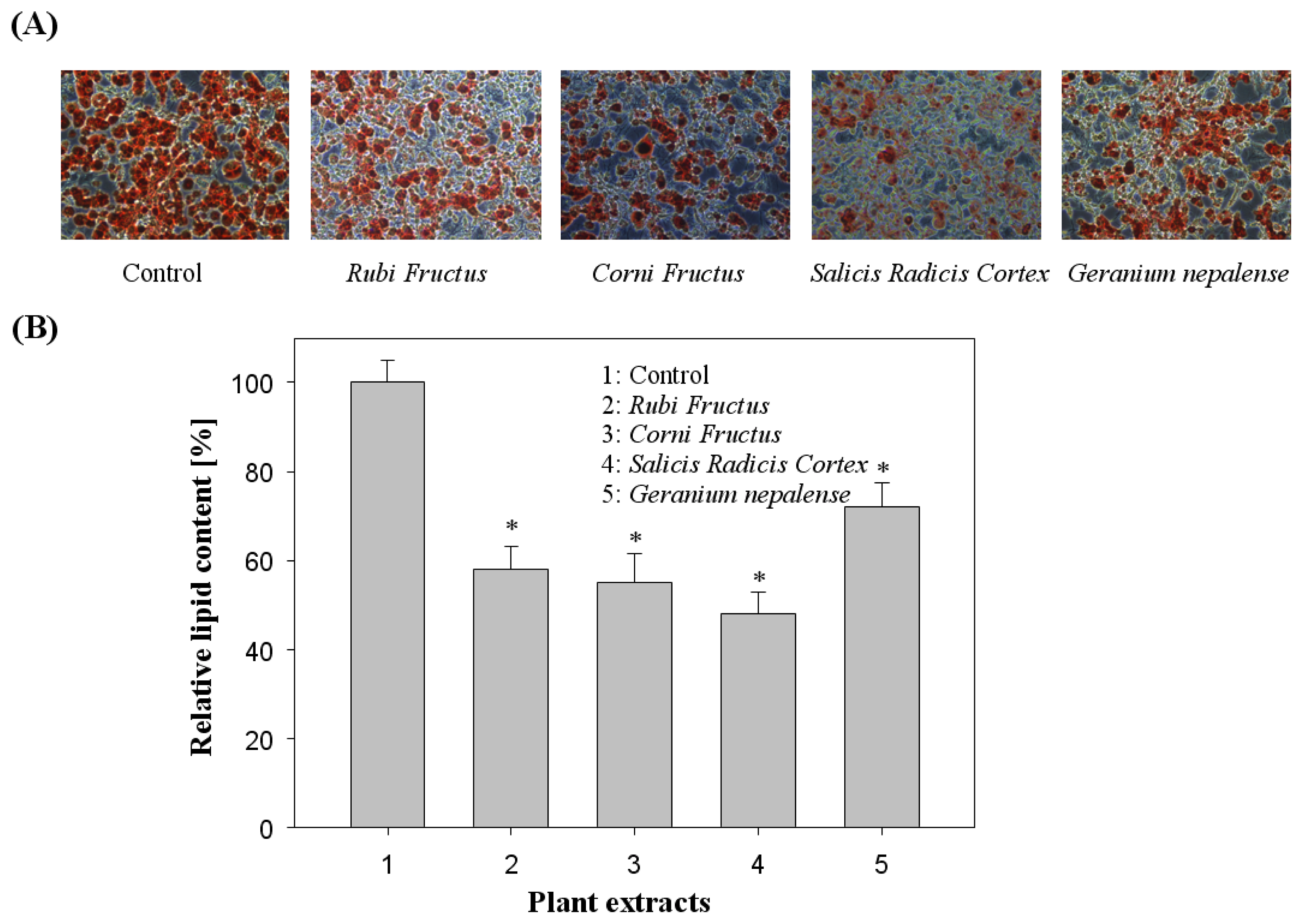
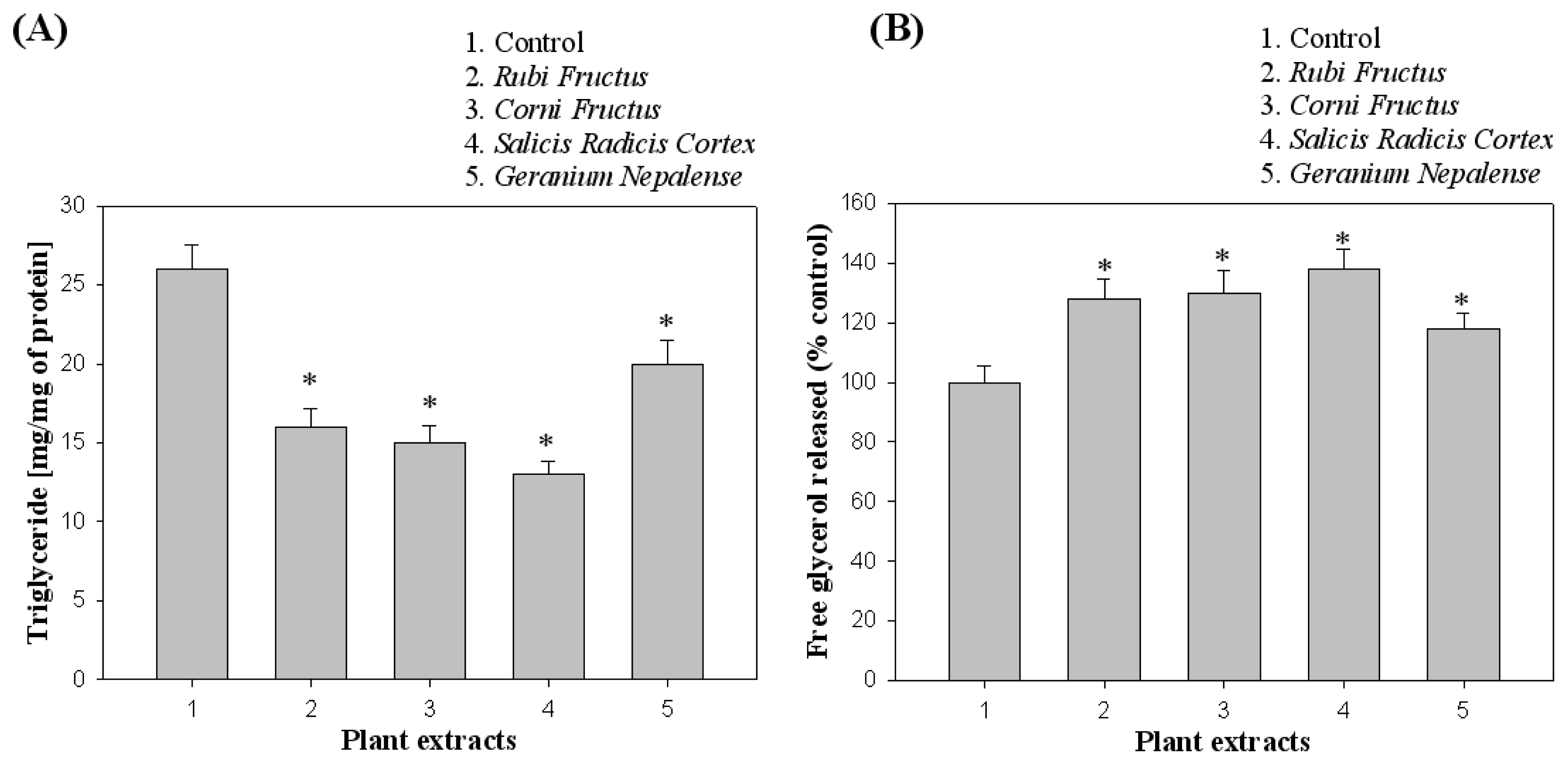
3.2. Effect of Cell Viability and Lipid Inhibition in 3T3-L1 Cells

The 3T3-L1 adipocytes were cultured and differentiated in a Dulbecco Modified Eagle Medium containing 10% fetal bovine serum for 6 to 8 days in the absence and presence of 44 plant extracts (at a final concentration, 100 μg/mL) according to differentiating protocols. Extracts were dissolved in DMSO at a final concentration that did not affect cell activity within the total volume (1%). As shown in Figure 2, the 44 plant extracts at 100 μg/mL attenuated lipid accumulation in differentiated adipocytes as evidenced by Oil Red O staining. Among the natural extracts examined, the four potent natural extracts (Rubi Fructus, Corni Fructus, Salicis Radicis Cortex, and Geranium nepalense) were found to significantly reduce lipid accumulation in 3T3-L1 adipocytes, suggesting anti-obesity activity. In Figure 2A,B, the effects of plant extracts on fat droplet formation in 3T3-L1 cells, and inhibition through the quantification method of Oil Red O staining, were presented. To examine the effect of plant extracts on cell viability of 3T3-L1 preadipocytes, we performed an MTT assay, which assesses cell viability by measuring mitochondrial activity in 3T3-L1 cells treated with concentrations of crude extracts. The potent crude extracts (Rubi Fructus, Corni Fructus, Salicis Radicis Cortex, and Geranium nepalense) screened have relatively low cytotoxicity to 3T3-L1 preadipocyte cells. The cell cytotoxicity remained approximately 90–100% (data not shown).
As shown in Figure 3A, lipid accumulation was measured based on the TG contents of 3T3-L1 cells differentiated in the presence of natural extracts. Furthermore, the lipolysis was assessed through the measurement of glycerol released in culture medium for 24 h incubation, as shown in Figure 3B. The four natural extracts that exhibited inhibitory activity towards pancreatic lipase (Rubi Fructus, Corni Fructus, Salicis Radicis Cortex, and Geranium nepalense) were found to inhibit triglyceride accumulation in 3T3-L1 adipocytes and trigger lipid metabolism process to glycerol release.
Rubi Fructus, the unripe fruit of Rubus Chingii Hu belonging to the Rosaceae family, can be found in many parts of the Asia, especially in China. In general, Rubi Fructus has estrogenic effect, promote lymphocyte proliferation and elevate testosterone level by promoting the activity of steroid synthesizing enzymes and by inhibiting their degradation [23]. Corni Fructus, the pulp of Cornus officinalis sieb. belonging to the Cornaceae in family, can be found in China, Japan and Korea. The biological activity of Corni Fructus was reported to relieve cyclophosphamid-induced leukopenia and to have antibacterial effects [24]. Salicis Radicis Cortex belonging to the Ulmaceae in family was reported to have antioxidant, antitumor, antimetastatic effects [25], to increase NO synthase activity [26], to lower LDL cholesterol levels [27], to prevent cancer development and metastasis [28] and to be effective in wound healing, angiogenesis, and cardiovascular disease [29]. Geranium Nepalense belonging to the Geraniaceae in family was reported to have antibacterial and antifungal effects [30]. Even though these four plants have reported to have various biological activities, there was no report indicating them to have lipid inhibitory activity.

4. Conclusions

Obesity is a risk factor for metabolic syndromes, and a flexible approach for the treatment of obesity is to promote early adipogenesis in adipose tissue, thereby leading to the replacement of enlarged adipocytes that secrete inflammatory factors with small adipocytes. In this study, we screened crude anti-obesity drugs from four-hundred plant extracts on in vitro enzymatic lipase activity. Among 400 plant extracts examined, 44 extracts from plant extracts significantly inhibited against in vitro anti-lipase activity. Among 44 natural extracts examined, the four plant extracts were active to inhibit lipid formation in 3T3-L1 adipocytes, suggesting their use as crude anti-obesity agents. Among them, Salicis Radicis Cortex had highest fat inhibition activity. Therefore, these results suggest that these four active plant extracts could be useful for prevention or treatment of obesity.

Acknowledgments

This work was supported by grant No. 2007-2000091 from Nuclear R & D Program of Ministry of Education, Science and Technology, Republic of Korea.

References

  1. Spiegelman, B.M.; Flier, J.S. Obesity and the regulation of energy balance. Cell 2001, 104, 531–543. [Google Scholar]
  2. Kopelman, P.G. Obesity as a medical problem. Nature 2000, 404, 635–643. [Google Scholar]
  3. Panico, S.; Lannuzzi, A. Dietary fat composition and the metabolic syndrome. Eur. J. Lipid Sci. Technol. 2004, 106, 61–67. [Google Scholar]
  4. Wakil, S.J.; Abu-Elheiga, L.A. Fatty acid metabolism: Target for metabolic syndrome. J. Lipid Res 2009, S139–143. [Google Scholar]
  5. Bray, G.A.; Tartaglia, L.A. Medicinal strategies in the treatment of obesity. Nature 2000, 404, 672–677. [Google Scholar]
  6. Sharma, N.; Sharma, V.K.; Seo, S.Y. Screening of some medicinal plants for anti-lipase activity. J. Ethnopharmacol 2005, 97, 453–456. [Google Scholar]
  7. Hino, K.; Nagata, H.; Shimonishi, M.; Ido, M. High-throughput screening for small-molecule adiponectin secretion modulators. J. Biomol. Screen 2011, 16, 628–636. [Google Scholar]
  8. Vázquez-Vela, M.E.F.; Torres, N.; Tovar, A.R. White adipose tissue as endocrine organ and its role in obesity. Arch. Med. Res 2008, 39, 715–728. [Google Scholar]
  9. Tanzi, M.C.; Fare, S. Adipose tissue engineering: State of the art, recent advances and innovative approaches. Expert Rev. Med. Devices 2009, 6, 533–551. [Google Scholar]
  10. Miyoshi, H.; Perfield, J.W., II; Obin, M.S.; Greenberg, A.S. Adipose triglyceride lipase regulates basal lipolysis and lipid droplet size in adipocytes. J. Cell. Biochem. 2008, 105, 1430–1436. [Google Scholar]
  11. Shi, Y.; Burn, P. Lipid metabolism enzymes: Emerging drug targets for the treatment of obesity. Nat. Rev. Drug Discov 2004, 3, 695–710. [Google Scholar]
  12. Hong, Y.H.; Hishikawa, D.; Miyahara, H.; Tsuzuki, H.; Nishimura, Y.; Gotoh, C.; Choi, K.C.; Hokari, Y.; Takagi, Y.; Lee, H.G.; et al. Up-regulation of adipogenin, an adipocyte plasma transmembrane protein, during adipogenesis. Mol. Cell. Biochem 2005, 276, 133–141. [Google Scholar]
  13. Reilly, S.M.; Lee, C.H. PPARδ as a therapeutic target in metabolic disease. FEBS Lett 2008, 582, 26–31. [Google Scholar]
  14. Kos, K.; Wilding, J.P.H. SPARC: A key player in the pathologies associated with obesity and diabetes. Nat. Rev. Endocrinol 2010, 6, 225–235. [Google Scholar]
  15. Thomson, A.B.; de Pover, A.; Keelan, M.; Jarocka-Cyrta, E.; Clandinin, M.T. Inhibition of lipid absorption as an approach to the treatment of obesity. Meth. Enzymol 1997, 286, 3–41. [Google Scholar]
  16. Tsujita, T.; Ninomiya, H.; Okuda, H. p-Nitrophenyl butyrate hydrolyzing activity of hormone-sensitive lipase from bovine adipose tissue. J. Lipid Res 1989, 30, 997–1004. [Google Scholar]
  17. Shizhen, L.; Xiwen, L. Compendium of Materia Medica; Foreign Languages Press: Beijing, China, 2003; Volume 6. [Google Scholar]
  18. Kim, Y.S.; Lee, Y.M.; Kim, H.; Kim, J.; Jang, D.K.; Kim, J.H.; Kim, J.S. Anti-obesity effect of Morus bombycis root extract: Anti-lipase activity and lipolytic effect. J. Ethnopharmacol 2010, 130, 621–624. [Google Scholar]
  19. Zheng, C.D.; Duan, Y.Q.; Gao, J.M.; Ruan, Z.G. Screening of anti-lipase properties of 37 traditional Chinese medicinal herbs. J. Chin. Med. Assoc 2010, 73, 319–324. [Google Scholar]
  20. Yang, Z.; Tu, Y.; Xia, H.; Jie, G.; Chen, X.; He, P. Suppression of free-radicals and protection against H2O2-induced oxidative damage in HPF-1 cell by oxidized phenolic compounds present in black tea. Food Chem 2007, 105, 1349–1356. [Google Scholar]
  21. Mcgowan, M.W.; Artiss, J.D.; Strandbergh, D.R.; Zak, B. A peroxidase-coupled method for the colorimetric determination of serum triglycerides. Clin. Chem 1983, 29, 538–542. [Google Scholar]
  22. SPSS, version 19.0; IBM: Chicago, IL, USA, 2010.
  23. Kim, J.S.; Jeon, W.J.; You, H.J.; Park, M.S.; Ji, G.E. Inhibitory activities of Rubi fructus on digestive enzymes. Food Sci. Biotechnol 2010, 19, 1165–1170. [Google Scholar]
  24. Park, C.H.; Cho, E.J.; Yokozawa, T. Protection against hypercholesterolemia by Corni fructus extract and its related protective mechanism. J. Med. Food 2009, 12, 973–981. [Google Scholar]
  25. Bonfili, L.; Cecarini, V.; Amici, M.; Cuccioloni, M.; Angeletti, M.; Keller, J.N.; Eleuteri, A.M. Natural polyphenols as proteasome modulators and their role as anti-cancer compounds. FEBS J 2008, 275, 5512–5526. [Google Scholar]
  26. Tunon, M.J.; Garcia-Mediavilla, M.V.; Sanchez-Campos, S.; Gonzalez-Gallego, J. Potential of flavonoids as antiinflammatory agents: Modulation of pro-inflammatory gene expression and signal transduction pathways. Curr. Drug Metab 2009, 10, 256–271. [Google Scholar]
  27. Lugasi, A. Foods fortified with phytosterins: Their role in decreasing serum cholesterol level, their European Community authorization and requirements for placing them on the market. Orv. Hetil 2009, 150, 483–496. [Google Scholar]
  28. Ovesna, Z.; Vachalkova, A.; Horvathova, K. Taraxasterol and beta-sitosterol: New naturally compounds with chemoprotective/chemopreventive effects. Neoplasma 2004, 51, 407–414. [Google Scholar]
  29. Plat, J.; Mensink, R.P. Effects of plant sterols and stanols on lipid metabolism and cardiovascular risk. Nutr. Metab. Cardiovasc. Dis 2001, 11, 31–40. [Google Scholar]
  30. Uprety, Y.; Asselin, H.; Boon, E.K.; Yadav, S.; Shrestha, K.K. Indigenous use and bio-efficacy of medicinal plants in the Rasuwa District, Central Nepal. J. Ethnobiol. Ethnomed 2010, 6, 3–9. [Google Scholar]
Figure 1. Porcine pancreatic lipase (PPL) inhibitory activities of Rubi Fructus, Corni Fructus, Salicis Radicis Cortex, Geranium nepalense. Orlistat was used as a positive control. Experiments have been performed in triplicate.
Ijms 13 01710f1
Figure 2. Effects of plant extracts on Oil Red O staining in cultured 3T3-L1 adipocytes. (A) Effects of plant extracts on fat droplet formation in 3T3-L1 cells. It was stained with Oil Red O dye and examined using a light microscope; (B) Relative lipid content by quantification method of Oil Red O staining. Data are presented as average ± SD (n = 3). * indicates p < 0.05.
Ijms 13 01710f2
Figure 3. Effects of plant extracts on adipocyte differentiation in 3T3-L1 cells. (A) Triglyceride (TG) accumulation was measured by TG contents of 3T3-L1 cells differentiated in the presence of natural extracts; (B) Lipolysis was assessed by the measurement of glycerol released into the medium. Data are presented as average ± SD (n = 3). * indicates p < 0.05.
Ijms 13 01710f3
Table 1. Forty four plant extracts that inhibit porcine pancreatic lipase (PPL).
No.Latin NameScientific NameFamily NamePlant PartInhibition (%) *
1Platycodi RadixPlatycodon grandiflorum A. De CandolleCampanulaceaeRoot3.5 ± 0.7
2Aconiti TuberAconitum carmichaeli DebeauxRanunculaceaeRoot12.1 ± 1.1
3Cannabis SemenCannabis sativa LinneMoraceaeSeed7.7 ± 0.2
4Chaenomelis FructusChaenomeles sinensis (Thouin) KoehneRosaceaeFruit7.1 ± 0.7
5Actinidiae FructusActinidia chinensisActinidiaceaeFruit3.2 ± 0.2
6Tribuli SemenTribulus terrestrisZygophyllaceaeSeed2.5 ± 0.6
7Lilie BulbusLilium brownii var. viridulun BakerLiliaceaeStem6.5 ± 1.2
8Luffae Fructus RetinervusLuffa cylindrica RoemerCucurbitaceaeFruit8.2 ± 1.4
9Crataegi FructusCrataegus pinnatifida Bunge var. typica SchneiderRosaceaeFruit5.5 ± 1.2
10Puerariae RadixPueraria thunbergiana BenthamLeguminosaeRoot3.2 ± 0.1
11Nardostachyos RhizomaNardostachys chinensis BatalValerianaceaeRhizoma6.4 ± 0.5
12Zizyphi FructusZizyphus jujuba Miller var. inermis RehderRhamnaceaeFruit6.6 ± 1.5
13Akebiae CaulisAkebia quinata DecaisneLardizabalaceaeStem11.5 ± 0.1
14Quisqalis FructusQuisqualis indica LinnéCombretaceaeFruit11.8 ± 0.3
15Loranthi RamulusLoranthus parasticus Merr.LoranthaceaeWhole grass13.5 ± 0.7
16Schizandrae FructusSchizandra chinensis BaillonSchizandraceaeFruit5.8 ± 0.2
17Lonicerae FoliumLonicera japonica ThunbergCaprifoliaceaeStem6.4 ± 0.5
18Rehmaniae Radix PreparataRehmannia glutinosaScrophulariaceaeRoot6.8 ± 1.1
19Dipsaci RadixDipsacus asperoides C. Y. Cheng et T. M. AiDipsacaceaeRoot12.5 ± 0.3
20Morindae RadixMorinda officinalis HowRubiaceaeRoot3.9 ± 0.7
21Perillae SemenPerilla sikokiana NakaiLabiataeSeed8.4 ± 0.3
22Pruni Nakaii SemenPrunus nakaii LeveilleRosaceaeSeed7.7 ± 0.6
23Meliae FructusMelia azedarach Linné var. japonica MakinoMeliaceaeFruit8.6 ± 0.8
24Bletillae RhizomaBletilla striata (Thunberg) Reichenbach fil.OrchidaceaeRhizoma12.1 ± 0.4
25Hedyotidis Diffusae HerbaOldenlandia diffusa (Willd.) RoxburghRubiaceaeWhole grass1.80 ± 0.4
26Hoelen rubraPoria cocos wolfPolyporaceaeBark12.7 ± 0.5
27Gastrodiae RhizomaGastrodia BlumeOrochidaceaeRhizoma8.4 ± 0.8
28Gentianae Scabrae RadixGentiana scabraSympetalaeRoot7.5 ± 1.1
29Cuscutae Semen crudusCuscuta chinensis LamarkConvolvulaceaeSeed5.8 ± 0.4
30Tetrapanacis MedullaTetrapanax papyriferus K. KochAraliaceaeStem6.6 ± 0.7
31Fritillariae BulbusFritillaria thunbergii MiquelLiliaceaeStem8.7 ± 0.5
32Patriniae RadixPatrinia villosa JussieuValerianaceaeRoot7.7 ± 1.4
33Scutellariae RadixScutellaria baicalenis GeorgiLabiataeRoot8.7 ± 0.4
34Astragali RadixAstragalus membranaceus BungeLeguminosaeRoot7.4 ± 1.4
35Phellodendri CortexPhellodendron amurense RuprechtRutaceaeBark11.5 ± 0.2
36Drynariae RhizomaDrynaria fortunei Smith.PolypodiaceaeRhizoma10.5 ± 0.4
37Rubi FructusRubus coreanus MiquelRosaceaeFruit32.5 ± 1.1
38Eriobotriae FoliumEriobotrya japonica LindleyRosaceaeLeaf9.8 ± 0.3
39Corni FructusCornus officinalis Siebold et ZuccariniCornaceaeFruit34.8 ± 2.3
40Forsythiae FructusForsythia koreana NakaiOleaceaeFruit5.7 ± 1.2
41Salicis Radicis CortexUlmus darvidian for. SuberoseUlmaceaeBark38.0 ± 1.9
42Ammomi Tsao-ko FrucutsAmomum tsao-ko Crevost et LemaireZingiberaceaeFruit7.5 ± 0.6
43Polygoni Avicularis HerbaPolygonum aviculare LinnéPolygonaceaeWhole grass21.2 ± 1.7
44Geranium NepalenseGeranium thunbergii Siebold et ZuccariniGeraniaceaeWhole grass31.4 ± 0.7
45Orlistat42.0 ± 2.5
*The inhibition of lipase activity by crude natural extracts was compared to the one observed with the positive control (Orlistat). Data are presented as average ± standard deviation (n = 3) and the anti-lipase activity was investigated at a concentration of 100 μg/mL for PPL inhibition.

