Tannins, Peptic Ulcers and Related Mechanisms

Abstract
:1. Introduction
2. Pathophysiology of the Peptic Ulcer
3. Plants with Peptic Anti-Ulcer Activity
4. Purified Tannins and Peptic Antiulcer Activity
5. Material and Methods
6. Conclusions
Acknowledgements
References
- Karamaæ, M. Chelation of Cu(II), Zn(II), and Fe(II) by tannin constituents of selected edible nuts.
- Amarowicz, R.; Estrella, I.; Hernández, T.; Dueñas, M.; Troszyñska, A.; Kosiñska, A.; Pegg, R.B. Antioxidant activity of a red lentil extract and its fractions. Int. J. Mol. Sci 2009, 10, 5513–5527. [Google Scholar]
- Yoshida, T.; Amakura, Y.; Yosh, M. Structural features and biological properties of ellagitannins in some plant families of the order Myrtales. Int. J. Mol. Sci 2010, 11, 79–106. [Google Scholar]
- Politi, F.A.S.; Mello, J.C.P.; Migliato, K.F.; Nepomuceno, A.L.A.; Moreira, R.R.D.; Pietro, R.C.L.R. Antimicrobial, cytotoxic and antioxidant activities and determination of the total tannin content of bark extracts Endopleura uchi. Int. J. Mol. Sci 2011, 12, 2757–2768. [Google Scholar]
- Okuda, T.; Ito, H. Tannins of constant structure in medicinal and food plants-hydrolysable tannins and polyphenols related to tannins. Molecules 2011, 16, 2191–2217. [Google Scholar]
- Blenn, C.; Wyrsch, P.; Althaus, F.R. The ups and downs of tannins as inhibitors of poly(ADP-ribose)glycohydrolase. Molecules 2011, 16, 1854–1877. [Google Scholar]
- Zhao, S.; Liu, J.Y.; Chen, S.Y.; Shi, L.L.; Liu, Y.J.; Ma, C. Antioxidant potential of polyphenols and tannins from burs of Castanea mollissima Blume. Molecules 2011, 16, 8590–8600. [Google Scholar]
- Albuquerque, U.P.; Monteiro, J.M.; Araújo, E.L. Taninos: uma abordagem da química à ecologia. Quim. Nova 2005, 28, 892–896. [Google Scholar]
- Simões, C.M.O.; Schenkel, E.P.; Gosmann, G.; Mello, J.C.P.; Mentz, L.A. Farmacognosia da Planta ao Medicamento, 5th ed; Editora da UFRGS: Porto Alegre, Brasil, 2003; p. 424. [Google Scholar]
- Heil, M.; Delsinne, T.; Hilpert, A.; Schürkens, S.; Andary, C.; Linsenmair, E.K.; Sousa, M.; Mckey, D. Reduced chemical defense in ant-plants? A critical re-evaluation of a widely accepted hypothesis. Oikos 2002, 99, 457–468. [Google Scholar]
- Moura, M.D.; Torres, A.R.; Oliveira, R.A.G.; Diniz, M.F.F.M.; Barbosa-Filho, J.M. Natural products as inhibitors of models of mammary neoplasia. Br. J. Phytother 2001, 5, 124–145. [Google Scholar]
- Moura, M.D.; Silva, J.S.; Oliveira, R.A.G.; Diniz, M.F.F.M.; Barbosa-Filho, J.M. Natural products reported as potential inhibitors of uterine cervical neoplasia. Acta Farm. Bonaerense 2002, 21, 67–74. [Google Scholar]
- Silva, J.S.; Moura, M.D.; Oliveira, R.A.G.; Diniz, M.F.F.; Barbosa-Filho, J.M. Natural product inhibitors of ovarian neoplasia. Phytomedicine 2003, 10, 221–232. [Google Scholar]
- Gonçalves, M.C.R.; Moura, L.S.A.; Rabelo, L.A.; Barbosa-Filho, J.M.; Cruz, H.M.M.; Cruz, J. Natural products inhibitors of HMG CoA reductase. Rev. Bras. Farm. 2000, 81, 63–71. [Google Scholar]
- Barbosa-Filho, J.M.; Martins, V.K.M.; Rabelo, L.A.; Moura, M.D.; Silva, M.S.; Cunha, E.V.L.; Souza, M.F.V.; Almeida, R.N.; Medeiros, I.A. Natural products inhibitors of the angiotensin converting enzyme (ACE). A review between 1980–2000. Rev. Bras. Farmacogn 2006, 16, 421–446. [Google Scholar]
- Barbosa-Filho, J.M.; Medeiros, K.C.P.; Diniz, M.F.F.M.; Batista, L.M.; Athayde-Filho, P.F.; Silva, M.S.; Cunha, E.V.L.; Almeida, J.R.G.S.; Quintans-Júnior, L.J. Natural products inhibitors of the enzyme acetylcholinesterase. Rev. Bras. Farmacogn 2006, 16, 258–285. [Google Scholar]
- Quintans-Júnior, L.J.; Almeida, J.R.G.S.; Lima, J.T.; Nunes, X.P.; Siqueira, J.S.; Oliveira, L.E.G.; Almeida, R.N.; Athayde-Filho, P.F.; Barbosa-Filho, J.M. Plants with anticonvulsant properties— a review. Rev. Bras. Farmacogn 2008, 18, 798–819. [Google Scholar]
- Sousa, F.C.F.; Melo, C.T.V.; Citó, M.C.O.; Félix, F.H.C.; Vasconcelos, S.M.M.; Fonteles, M.M.F.; Barbosa-Filho, J.M.; Viana, G.S.B. Plantas medicinais e seus constituintes bioativos: Uma revisão da bioatividade e potenciais benefícios nos distúrbios da ansiedade em modelos animais. Rev. Bras. Farmacogn 2008, 18, 642–654. [Google Scholar]
- Almeida, R.N.; Navarro, D.S.; Barbosa-Filho, J.M. Plants with central analgesic activity. Phytomedicine 2001, 8, 310–322. [Google Scholar]
- Morais, L.C.S.L.; Barbosa-Filho, J.M.; Almeida, R.N. Plants and bioactive compounds for the treatment of Parkinson’s disease. Arq. Bras. Fitomed. Cient 2003, 1, 127–132. [Google Scholar]
- Pereira, J.V.; Modesto-Filho, J.; Agra, M.F.; Barbosa-Filho, J.M. Plant and plant-derived compounds employed in prevention of the osteoporosis. Acta Farm. Bonaerense 2002, 21, 223–234. [Google Scholar]
- Rocha, L.G.; Almeida, J.R.G.S.; Macedo, R.O.; Barbosa-Filho, J.M. A review of natural products with antileishmanial activity. Phytomedicine 2005, 12, 514–535. [Google Scholar]
- Amaral, F.M.M.; Ribeiro, M.N.S.; Barbosa-Filho, J.M.; Reis, A.S.; Nascimento, F.R.F.; Macedo, R.O. Plants and chemical constituents with giardicidal activity. Rev. Bras. Farmacogn 2006, 16, 696–720. [Google Scholar]
- Barbosa-Filho, J.M.; Nascimento-Júnior, F.A.; Tomaz, A.C.A.; Athayde-Filho, P.F.; Silva, M.S.; Cunha, E.V.L; Souza, M.F.V.; Batista, L.M.; Diniz, M.F.F.M. Natural products with antileprotic activity. Rev. Bras. Farmacogn. 2007, 17, 141–148. [Google Scholar]
- Barbosa-Filho, J.M.; Vasconcelos, T.H.C.; Alencar, A.A.; Batista, L.M.; Oliveira, R.A.G.; Guedes, D.N.; Falcão, H.S.; Moura, M.D.; Diniz, M.F.F.M.; Modesto-Filho, J. Plants and their active constituents from South, Central, and North America with hypoglycemic activity. Rev. Bras. Farmacogn 2005, 15, 392–413. [Google Scholar]
- Falcão, H.S.; Lima, I.O.; Santos, V.L.; Dantas, H.F.; Diniz, M.F.F.M.; Barbosa-Filho, J.M.; Batista, L.M. Review of the plants with anti-inflammatory activity studied in Brazil. Rev. Bras. Farmacogn 2005, 15, 381–391. [Google Scholar]
- Barbosa-Filho, J.M.; Piuvezam, M.R.; Moura, M.D.; Silva, M.S.; Lima, K.V.B.; Cunha, E.V.L.; Fechine, I.M.; Takemura, O.S. Anti-inflammatory activity of alkaloids: A twenty century review. Rev. Bras. Farmacogn. 2006, 16, 109–139. [Google Scholar]
- Lima, G.R.M.; Montenegro, C.A.; Almeida, C.L.F.; Athayde-Filho, P.F.; Barbosa-Filho, J.M.; Batista, L.M. Database survey of anti-inflammatory plants in South America: A review. Int. J. Mol. Sci 2011, 12, 2692–2749. [Google Scholar]
- Souto, A.L.; Tavares, J.F.; Silva, M.S.; Diniz, M.F.F.M.; Athayde-Filho, P.F.; Barbosa Filho, J.M. Anti-inflammatory activity of alkaloids: An update from 2000 to 2010. Molecules 2011, 16, 8515–8534. [Google Scholar]
- Mariath, I.R.; Falcão, H.S.; Barbosa-Filho, J.M.; Sousa, L.C.F.; Tomaz, A.C.A.; Batista, L.M.; Diniz, M.F.F.M.; Athayde-Filho, P.F.; Tavares, J.F.; Silva, M.S.; et al. Plants of the American continent with antimalarial activity. Rev. Bras. Farmacogn 2009, 19, 158–192. [Google Scholar]
- Falcão, H.S.; Mariath, I.R.; Diniz, M.F.F.M.; Batista, L.M.; Barbosa-Filho, J.M. Plants of the American continent with antiulcer activity. Phytomedicine 2008, 15, 132–146. [Google Scholar]
- Ribeiro Filho, J.; Falcão, H.S.; Batista, L.M.; Barbosa Filho, J.M.; Piuvezam, M.R. Effects of plant extracts on HIV-1 protease. Curr. HIV Res 2010, 8, 531–544. [Google Scholar]
- Agra, M.F.; Freitas, P.F.; Barbosa-Filho, J.M. Synopsis of the plants known as medicinal and poisonous in Northeast of Brazil. Rev. Bras. Farmacogn 2007, 17, 114–140. [Google Scholar]
- Agra, M.F.; Silva, K.N.; Basílio, I.J.L.D.; Freitas, P.F.; Barbosa-Filho, J.M. Survey of medicinal plants used in the region Northeast of Brazil. Rev. Bras. Farmacogn 2008, 18, 472–508. [Google Scholar]
- Silva, F.L.; Fischer, D.C.H.; Tavares, J.F.; Silva, M.S.; Athayde-Filho, P.F.; Barbosa-Filho, J.M. Compilation of secondary metabolites from Bidens pilosa L. Molecules 2011, 16, 1070–1102. [Google Scholar]
- Gonçalves, M.C.R.; Diniz, M.F.F.M.; Borba, J.D.C.; Nunes, X.P.; Barbosa-Filho, J.M. Berinjela (Solanum melongena L.): mito ou realidade no combate as dislipidemias? Rev. Bras. Farmacogn 2006, 16, 252–257. [Google Scholar]
- Barbosa-Filho, J.M.; Alencar, A.A.; Nunes, X.P.; Tomaz, A.C.A.; Sena-Filho, J.G.; Athayde-Filho, P.F.; Silva, M.S.; Souza, M.F.V.; Cunha, E.V.L. Sources of alpha-, beta-, gamma-, delta- and epsilon-carotenes: A twentieth century review. Rev. Bras. Farmacogn 2008, 18, 135–154. [Google Scholar]
- Alves, J.S.; Castro, J.C.M.; Freire, M.O.; Cunha, E.V.L.; Barbosa-Filho, J.M.; Silva, M.S. Complete assignment of the 1H and 13C spectra of four triterpenes of the ursane, artane, lupane and friedelane groups. Magn. Reson. Chem 2000, 38, 201–206. [Google Scholar]
- Sena-Filho, J.G.; Duringer, J.M.; Maia, G.L.A.; Tavares, J.F.; Xavier, H.S.; Silva, M.S.; Cunha, E.V.L.; Barbosa-Filho, J.M. Ecdysteroids from Vitex species: Distribution and compilation of their 13C-NMR spectral data. Chem. Biodivers 2008, 5, 707–713. [Google Scholar]
- Oliveira, S.L.; Silva, M.S.; Tavares, J.F.; Sena-Filho, J.G.; Lucena, H.F.S.; Romero, M.A.V.; Barbosa-Filho, J.M. Tropane alkaloids from genus Erythroxylum: Distribution and compilation of 13C-NMR spectral data. Chem. Biodivers 2010, 7, 302–326. [Google Scholar]
- Palmeira-Junior, S.F.; Conserva, L.M.; Barbosa Filho, J.M. Clerodane diterpenes from Croton species: Distribution and a compilation of their and 13C NMR. Nat. Prod. Commun 2006, 1, 319–344. [Google Scholar]
- Sena Filho, J.G.; Duringer, J.M.; Uchoa, D.E.A.; Xavier, H.S.; Barbosa Filho, J.M.; Braz Filho, R. Distribution of iridoid glucosides in plants from the genus Lippia (Verbenaceae): An investigation of Lippia alba (Mill.) N.E. Brown. Nat. Prod. Commun 2007, 2, 715–716. [Google Scholar]
- Almeida, C.L.F.; Falcão, H.S.; Lima, G.R.M.; Montenegro, C.A.; Lira, N.S.; Athayde-Filho, P.F.; Rodrigues, L.C.; Souza, M.F.V.; Barbosa-Filho, J.M.; Batista, L.M. Bioactivities from marine algae of the genus Gracilaria. Int. J. Mol. Sci 2011, 12, 4550–4573. [Google Scholar]
- Lira, N.S.; Montes, R.C.; Tavares, J.F.; Silva, M.S.; Cunha, E.V.L.; Athayde-Filho, P.F.; Rodrigues, L.C.; Dias, C.S.; Barbosa-Filho, J.M. Brominated compounds from marine sponges of the genus Aplysina and a compilation of their 13C NMR spectral data. Mar. Drugs 2011, 9, 2316–2368. [Google Scholar]
- Honório, J.E.R., Júnior; Soares, P.M.; Melo, C.L.; Arruda Filho, A.C.V.; Sena Filho, J.G.; Barbosa Filho, J.M.; Sousa, F.C.F.; Fonteles, M.M.F.; Leal, L.K.A.; Queiroz, M.G.R.; et al. Atividade farmacológica da monocrotalina isolada de plantas do gênero Crotalaria. Rev. Bras. Farmacogn 2010, 20, 453–458. [Google Scholar]
- Vasconcelos, S.M.M.; Honório-Júnior, J.E.R.; Abreu, R.N.D.C.; Silva, M.C.C.; Barbosa-Filho, J.M.; Lobato, R.F.G. Pharmacologic study of some plant species from the Brazilian Northeast: Calotropis procera, Agava sisalana, Solanum paludosum, Dioscorea cayenensis and Crotalaria retusa. In Medicinal Plants: Classification, Biosynthesis and Pharmacology; Varela, A., Ibanez, J., Eds.; Nova Science Publishers: Hauppauge, NY, USA, 2009; Volume 4, pp. 189–202. [Google Scholar]
- Vasconcelos, S.M.M.; Pereira, E.C.; Chaves, E.M.C.; Lobato, R.F.G.; Barbosa-Filho, J.M.; Patrocínio, M.C.A. Pharmacologic study of Amburana cearensis and Aniba genus. In Recent Progress in Medicinal Plants Volume 30: Drug Plant IV; Singh, V.K., Govil, J.N., Eds.; Studium Press: Houston, TX, USA, 2010; Volume 30–51, p. 64. [Google Scholar]
- Barbosa-Filho, J.M.; Sette, I.M.F.; Cunha, E.V.L.; Guedes, D.N.; Silva, M.S. Protoberberine alkaloids. In The Alkaloids; Cordell, G.A., Ed.; Elsevier Amsterdam: The Netherlands, 2005; Volume 62–1, p. 75. [Google Scholar]
- Conserva, L.M.; Pereira, C.A.B.; Barbosa-Filho, J.M. Alkaloids of the Hernandiaceae: Occurrence and a compilation of their biological activities. Alkaloids Chem. Biol 2005, 62, 175–243. [Google Scholar]
- Barbosa-Filho, J.M.; da-Cunha, E.V.L.; Gray, A.I. Alkaloids of the Menispermaceae. Alkaloids Chem. Biol 2000, 54, 1–190. [Google Scholar]
- Andrade, N.C.; Cunha, E.V.L.; Silva, M.S.; Agra, M.F.; Barbosa-Filho, J.M. Terpenoids of the Annonaceae: Distribution and compilation of 13C NMR data. In Recent Research Developments in Phytochemistry; Gayathri, A., Ed.; Research Signpost: Kerala, India, 2003; Volume 7, pp. 1–85. [Google Scholar]
- Almeida, R.N.; Barbosa-Filho, J.M. Drogas psicotrópicas. In Psicofarmacologia—Fundamentos Práticos, 1st ed; Almeida, R.N., Ed.; Guanabara Koogan: Rio de Janeiro, Brasil, 2006; pp. 3–24. [Google Scholar]
- Barbosa-Filho, J.M.; Silva, T.M.S.; Sette, I.M.F.; Franca, F.; Agra, M.F.; Soares, F.P.; Costa, J.F.O.; Santos, R.R. Plantas da Caatinga: Perfil Botânico, Fitoquímica e Atividade Biológica, 1st ed; Giulietti, A.M., de Queiroz, L.P., Eds.; IMSEAR: Brasília, Brasil, 2006; Volume 4, pp. 19–497. [Google Scholar]
- Agra, M.F.; Camara, C.A.; Barbosa-Filho, J.M.; Silva, T.M.S.; Almeida, R.N.; Medeiros, I.A.; Nurit, K.; Oliveira, F.S.; Freire, K.R.L.; Morais, L.C.S.L. Medicinais e Produtoras de Princípios Ativos. In Espécies da Flora Nordestina de Importância Econômica Potencial; Everaldo, V.S.B., Sampaio, E.V.S.B., Eds.; Associação de Plantas do Nordeste: Recife, Brasil, 2005; Volume 1, pp. 135–198. [Google Scholar]
- Mota, K.S.L.; Dias, G.E.N.; Pinto, M.E.F.; Luiz-Ferreira, A.; Souza-Brito, A.R.M.; Hiruma-Lima, C.A.; Barbosa-Filho, J.M.; Batista, L.M. Flavonoids with gastroprotective activity. Molecules 2009, 14, 979–1012. [Google Scholar]
- Falcão, H.S.; Leite, J.A.; Barbosa-Filho, J.M.; Athayde-Filho, P.F.; Chaves, M.C.O.; Moura, M.D.; Ferreira, A.L.; Almeida, A.B.A.; Souza-Brito, A.R.M.; Diniz, M.F.F.M.; et al. Gastric and duodenal antiulcer activity of alkaloids: A review. Molecules 2008, 13, 3198–3223. [Google Scholar]
- Zapata-Colindres, J.C.; Zepeda-Gomez, S.; Montano-Loza, A.; Vazquez-Ballesteros, E.; Jesus-Villalobos, J.; Valdovinos-Andraca, F. The association of Helicobacter pylori infection and nonsteroidal anti-inflammatory drugs in peptic ulcer disease. Can. J. Gastroenterol 2006, 20, 277–280. [Google Scholar]
- Jain, K.S.; Shah, A.K.; Bariwal, J.; Shelke, S.M.; Kale, A.P.; Jagtap, J.R.; Bhosale, A.V. Recent advances in proton pump inhibitors and management of acid-peptic disorders. Bioorg. Med. Chem 2007, 15, 1181–1205. [Google Scholar]
- Malfertheiner, P.; Chan, F.K.L.; McColl, K.E.L. Peptic ulcer disease. Lancet 2009, 374, 1449–1461. [Google Scholar]
- Suerbaum, S.; Michetti, P. Helicobacter pylori infection. N. Engl. J. Med 2002, 347, 1175–1186. [Google Scholar]
- Tombola, F.; Campello, S.; De Luca, L.; Ruggiero, P.; Del Giudice, G.; Papini, E.; Zoratti, M. Plant polyphenols inhibit VacA, a toxin secreted by the gastric phathogen Helicobacter pylori. FEBS Lett 2003, 543, 184–189. [Google Scholar]
- Backert, S.; Naumann, M. What a disorder: Proinflammatory signaling pathways induced by Helicobacter pylori. Trends Microbiol 2010, 18, 479–486. [Google Scholar]
- Repetto, M.G.; Llessuy, S.F. Antioxidant properties of natural compounds used in popular medicine for gastric ulcer. Braz. J. Med. Biol. Res 2002, 35, 523–534. [Google Scholar]
- Wada, K.; Kamisaki, Y.; Kentaro, N.; Kishimoto, Y.; Ashida, K.; Itoh, T. Effect of plaunotol on gastric injury induced by ischaemia-reperfusion in rats. J. Pharm. Pharmacol 1997, 49, 903–907. [Google Scholar]
- Kwieciente, S.; Brzozowski, T.; Konturek, S.J. Effect of reactive oxygen species action on gastric mucosa in various models of mucosal injury. J. Physiol. Pharmacol 2002, 53, 761–773. [Google Scholar]
- Odabasoglu, F.; Cakir, A.; Suleyman, H.; Aslan, A.; Bayr, Y.; Halici, M.; Kaza, C. Gastroprotective and antioxidant effects of usnic acid on indomethacin-induced gastric ulcer in rats. J. Ethnopharmacol 2006, 103, 59–65. [Google Scholar]
- Chattophadhyay, I.; Bandyopadhyay, U.; Biswas, K.; Maity, P.; Banerjee, R.K. Indomethacin inactivates gastric peroxidase to induced reactive oxygen mediated gastric mucosal injury and curcumin protects it by preventing peroxidase inactivation and scavenging reactive oxygen. Free Rad. Biol. Med 2006, 40, 1397–1408. [Google Scholar]
- Cartea, M.A.; Francisco, M.; Soengas, P.; Velasco, P. Phenolic compounds in Brassica vegetables. Molecules 2011, 16, 251–280. [Google Scholar]
- Konturek, P.C.; Brzozowski, T.; Walter, B.; Burnat, G.; Hess, T.; Hahn, E.G.; Konturek, S.J. Gherlin induced gastroprotective against ischemia reperfusion injury involves an activation of sensory afferent nerves and hyperemia mediated by nitric oxide. Eur. J. Pharmacol 2006, 536, 171–181. [Google Scholar]
- Kim, H.; Kim, K.H. Role of nitric oxide and muçus in isquemia/reperfusion induced gastric mucosal injury in rats. Pharmacology 2001, 62, 200–207. [Google Scholar]
- Wallace, J.L. Gastric resistance to acid: Is the mucus-bicarbonate barrier functionally redundant? Am. J. Physiol 1989, 256, G31–G38. [Google Scholar]
- Komori, M.; Tsuji, S.; Sun, W.; Tsujii, M.; Kawai, N.; Yasumaru, M.; Kakiuchi, Y.; Kimura, A.; Sasaki, Y.; Higashiyama, S.; et al. Gastrin enhances gastric mucosal integrity through cyclooxygenase 2 upregulation in rats. Am. J. Physiol. Gastrointest. Liver Physiol 2002, 283, 1368–1378. [Google Scholar]
- Parente, L.; Parretti, M. Advances in the pathophysiology of constitutive and inducible cyclooxygenases: Two enzymes in the spolight. Biochem. Pharmacol 2003, 65, 153–159. [Google Scholar]
- Abdel Salam, O.M.; Czimmer, J.; Debreceni, A.; Szolcsanyi, J.; Mozsik, G. Gastric mucosal integrity: gastric mucosal blood flow and microcirculation. J. Physiol 2001, 95, 105–127. [Google Scholar]
- Annuk, H.; Hirmo, S.; Türi, E.; Mikelsaar, M.; Arak, E.; Wadström, T. Effect on cell surface hydrophobicity and susceptibility of Helicobacter pylori to medicinal plant extracts. FEMS Microbiol. Lett 1999, 172, 41–45. [Google Scholar]
- Perera, L.M.S.; Ruedas, D.; Gómez, B.C. Gastric antiulcer effects of Rhizophora mangle L. J. Ethnopharmacol 2001, 77, 1–3. [Google Scholar]
- Berenguer, B.; Sánchez, L.M.; Quílez, A.; López-Barreiro, M.; Haro, O.; De Gálvez, J.; Martín, M.J. Protective and antioxidant effects of Rizophora mangle L. against NSAID-induced gastric ulcers. J. Ethnopharmacol 2006, 103, 194–200. [Google Scholar]
- Gonzales, F.G.; Portela, T.Y.; Stipp, E.J.; Di Stasi, L.C. Antiulcerogenic and analgesic effects of Maytenus aquifolium, Soroceae bomplandii and Zolernia ilicifolia. J. Ethnopharmacol 2001, 77, 41–47. [Google Scholar]
- Martins, D.T.O.; Lima, J.C.S.; Rao, V.S.N. The acetone soluble fraction from bark extract of Stryphynodendron adstringeens (Mart.) Coville inhibits gastric acid secretion and experimental gastric ulceration in rats. Phytother. Res 2002, 16, 427–431. [Google Scholar]
- Rafhael, K.R.; Kuttan, R. Inhibition of experimental gastric lesion and inflammation by Phyllanthus amarus extract. J. Ethnopharmacol 2003, 87, 193–197. [Google Scholar]
- Birdane, F.M.; Cemek, M.; Birdane, Y.O.; Gülçin, I.; Büyükokuroglu, M.E. Benefical effects of Foeniculum vulgare on ethanol-induced acute gastric mucosal injury in rats. Word J. Gastoenterol 2007, 13, 607–611. [Google Scholar]
- Khennouf, S.; Benabdallah, H.; Gharzouli, K.; Amira, S.; Ito, H.; Kim, T.; Yoshida, T.; Gharzouli, A. Effect of tannins from Quercus suber and Quercus coccifera leaves on ethanol-induced gastric lesions in mice. J. Agric. Food Chem 2003, 51, 1469–1473. [Google Scholar]
- Hiruma-Lima, C.A.; Santos, L.C.; Pellizzon, C.H.; Silveira, G.G.; Vasconcelos, P.C.P.; Vilegas, W.; Souza Brito, A.R.M. Qualea grandiflora, a Brasilian cerrado medicinal plant presentes an important antiulcer activity. J. Ethnopharmacol 2006, 104, 207–214. [Google Scholar]
- Shay, H.; Komarov, S.A.; Fels, S.S.; Meranze, D.; Gruestein, M.; Siplet, H. A simple method for the uniform production of gastric ulceration in the rat. Gastroenterology 1945, 5, 43–61. [Google Scholar]
- Andreo, M.A.; Ballesteros, K.V.R.; Hiruma-Lima, C.A.; Rocha, L.R.M.; Souza Brito, A.R.M.; Vilegas, W. Effect of Mouriri pusa extracts on experimentally induced gastric lesions in rodents: Role of endogenous sulfhydryls compouds and nitric oxide in gastroprotection. J. Ethnopharmacol 2006, 107, 431–441. [Google Scholar]
- Vasconcelos, P.C.P.; Kushima, H.; Andreo, M.; Hiruma-Lima, C.A.; Vilegas, W.; Takahira, R.K.; Pellizon, C.H. Studies of gastric mucosa regeneration and safety promoted by Mouriri pusa treatment in acetic acid ulcer model. J. Ethnopharmacol 2008, 115, 293–301. [Google Scholar]
- Vasconcelos, P.C.P.; Andreo, M.A.; Vilegas, W.; Pellizzon, C.H. Effect of Mouriri pusa tannins and flavonoids on prevention and treatment against experimental gastric ulcer. J. Etnopharmacol 2010, 131, 146–153. [Google Scholar]
- Zayachkivska, O.S.; Gzhegotsky, M.R.; Terletska, O.I.; Lutsyk, D.A.; Yaschenko, A.M.; Dzhura, O.R. Influence of Viburnum opulus proanthocyanidins on stress-induced gastrointestinal mucosal damage. J. Physiol. Pharmacol 2006, 57, 155–167. [Google Scholar]
- Baliga, M.S.; Dsouza, J.J. Amla (Emblica officinalis Gaertn), a wonder berry in the treatment and prevention of cancer. Eur. J. Cancer Prev 2011, 20, 225–239. [Google Scholar]
- Murakami, S.; Isobe, Y.; Kijima, H.; Nagai, H.; Muramatu, M.; Otomu, S. Inhibition of gastric H+, K+-ATPase and acid secretion by ellagic acid. Planta Med 1991, 57, 305–308. [Google Scholar]
- Murakami, S.; Muramatsu, M.; Otomo, S. Inhibitory effect of tannic acid on gastric H+, K+-ATPase. J. Nat. Prod 1992, 55, 513–516. [Google Scholar]
- Funatogawa, K.; Hayashi, S.; Shimomura, H.; Yoshida, T.; Hatano, T.; Ito, H.; Hirai, Y. Antibacterial activity of hydrolysable tannins derived from medicinal plants against Helicobacter pylori. Microbiol. Immunol 2004, 48, 251–261. [Google Scholar]
- Okuda, T.; Yoshida, T.; Hatano, T. Correlation of oxidative transformations of hydrolysable tannins and plant evolution. Phytochemistry 2000, 55, 513–529. [Google Scholar]
- Kang, J.; Liu, Y.; Xie, M.X.; Lia, S.; Jianga, M.; Wanga, Y.D. Interactions of human serum albumin with chlorogenic acid and ferulic acid. Biochim. Biophys. Acta 2004, 1674, 205–214. [Google Scholar]
- Lampire, O.; Mila, I.; Raminosoa, M.; Michon, V.; Catherine, H.; Penhoat, D.; Faucheur, N.; Laprevote, O.; Scalbert, A. Polyphenols isolated from the bark of Castanea sativa Mill. Chemical structures and auto association. Phytochemistry 1998, 49, 623–631. [Google Scholar]
- Yoshida, T.; Ito, H.; Hipolito, I.J. Pentameric ellagitannin oligomers in melastomataceous plants-chemotaxonomic significance. Phytochemistry 2005, 66, 1972–1983. [Google Scholar]
- Yoshimura, M.; Ito, H.; Miyashita, K.; Hatano, T.; Taniguchi, S.; Amakura, Y.; Yoshida, T. Flavonol glucuronides and C-glucosidic ellagitannins from Melaleuca squarrosa. Phytochemistry 2008, 69, 3062–3069. [Google Scholar]
- Zheng, G.; Xu, L.; Wu, P; Xie, H.; Jiang, Y.; Chen, F.; Wei, X. Polyphenols from longan seeds and their radical-scavenging activity. Food Chem. 2009, 116, 433–436. [Google Scholar]
- Ito, H.; Kobayashi, E.; Tamakatsu, Y.; Li, S.H.; Hatano, T.; Sakagami, H.; Kusama, K.; Satoh, K.; Sugita, D.; Shimura, S.; et al. Polyphenols from Eriobotrya japonica and their cytotoxicity against human oral tumor cell lines. Chem. Pharm. Bull 2000, 48, 687–693. [Google Scholar]
- Ito, H.; Miyake, M.; Nishitani, E.; Mori, K.; Hatano, T.; Okuda, T.; Konoshima, T.; Takasaki, M.; Kozuka, M.; Mukainaka, T.; Tokuda, H.; Nishino, H.; Yoshida, T. Anti-tumor promoting activity of polyphenols from Cowania mexicana and Coleogyne ramosissima. Cancer Lett 1999, 143, 5–13. [Google Scholar]
- Silva, H.R.; Silva, C.C.M.; Caland Neto, L.B.; Lopes, J.A.D.; Citó, A.M.G.L.; Chaves, M.H. Constituintes químicos das cascas do caule de Cenostigma macrophyllus: Ocorrência de colesterol. Quim. Nova 2007, 30, 1877–1881. [Google Scholar]
- Babich, H.; Krupka, M.E.; Nissim, H.A.; Zuckerbraun, H.L. Differential in vitro cytotoxicity of (−)-epicatechin gallate (ECG) to cancer and normal cells from the human oral cavity. Toxicol. In Vitro 2005, 19, 231–242. [Google Scholar]
- Fielman, K.S. Recent progress in ellagitannin chemistry. Phytochemistry 2005, 66, 1984–2000. [Google Scholar]
- Jin, Z.X.; Ito, H.; Yoshida, T. Dimeric and trimeric ellagitannins from Corylus heterophylla. Phytochemistry 1998, 48, 333–338. [Google Scholar]
- Champavier, Y.; Comte, G.; Vercauteren, J.; Allais, D.P.; Chulia, A.J. Norterpenoid and sesquiterpenoid glucosides from Juniperus phoeniceae and Galega officinalis. Phytochemistry 1999, 50, 1219–1223. [Google Scholar]
- Shu, P.; Hong, J.L.; Wu, G.; Yu, B.Y.; Qin, M.J. Analysis of flavonoids and phenolic acids in Iris tectorum by HPLC-DAD-ESI-MS. Chin. J. Nat. Med 2010, 8, 202–207. [Google Scholar]
- Taniguchi, S.; Nakamura, N.; Nose, M.; Takeda, S.; Uchi, R.Y.; Ito, H.; Yoshida, T.; Yazaki, K. Production of macrocyclic ellagitannin oligomers by Oenothera laciniata callus cultures. Phytochemistry 1998, 48, 981–985. [Google Scholar]
- Yokozawa, T.; Chen, C.P.; Dong, E.; Tanaka, T.; Nonaka, G.; Nishioka, I. Study on the inhibitory effect of tannins and flavonoids against the 1,1-diphenyl-2-picrylhydrazyl radical. Biochem. Pharmacol 1998, 56, 213–222. [Google Scholar]
- Heijmen, F.H.; Pont, J.S.; Middelkoop, E.; Kreis, R.W.; Hoekstra, M.J. Cross-linking of dermal sheep collagen with tannic acid. Biomaterials 1997, 18, 749–754. [Google Scholar]
- Tani, S. Effects of tannic acid and tannic acid-starch on the experimental gastric ulcer in rat. Yakugaku Zasshi 1976, 96, 648–652. [Google Scholar]
- Tani, S.; Sakurai, Y.; Kondo, Y. Effects of tannins and some related compounds on ethanol-induced gastric lesions in the rat. Yakugaku Zasshi 1986, 106, 347–349. [Google Scholar]
- Piao, M.J.; Kang, K.A.; Zhang, R.; Ko, D.O.; Wang, Z.H.; Lee, K.H.; Chang, W.Y.; Chae, S.; Jee, Y.; Shin, T.; et al. Antioxidant properties of 1,2,3,4,6-penta-O-galloyl-β-d-glucose from Elaeocarpus sylvestris var. ellipticus. Food Chem 2009, 115, 412–418. [Google Scholar]
- Li, J.H.; Nesumi, A.; Shimizu, K.; Sakata, Y.; Liang, M.Z.; He, Q.Y.; Zhou, H.J.; Hashimoto, F. Chemosystematics of tea trees based on tea leaf polyphenols as phenetic markers. Phytochemistry 2010, 71, 1342–1349. [Google Scholar]
- Hung, C.R.; Cheng, J.T.; Neu, S.L. Prophylactic effects of sucralfate and geraniin on ethanol-induced gastric mucosal damage in rats. Chin. J. Physiol 1995, 38, 211–217. [Google Scholar]
- Guo, Q.; Zhao, B. Electron spin resonance study of free radicals formed from a procyanidin-rich pine (Pinus maritima) bark extract, pycnogenol. Free Rad. Biol. Med 1999, 27, 1308–1312. [Google Scholar]
- Spencer, J.P.E.; Schroeter, H.; Shenoy, B.; Srai, S.K.S.; Debnam, E.S.; Evans, C.R. Epicatechin is the primary bioavailable form of the procyanidin dimers B2 and B5 after transfer across the small intestine. Biochem. Biophys. Res. Commun 2001, 285, 588–593. [Google Scholar]
- Takahashi, T.; Kamiya, T.; Hasegawa, A.; Yokoo, Y. Procyanidin oligomers selectively and intensively promote proliferation of mouse hair epithelial cells in vitro and activate hair follicle growth in vivo. J. Invest. Dermatol 1999, 112, 310–316. [Google Scholar]
- Walters, D.; Meurer-Grimes, B.; Rovira, I. Antifungal activity of three spermidine conjugates. FEMS Microbiol. Lett 2001, 201, 255–258. [Google Scholar]

| Tannin | Chemical structure | Model assay/way of route/dose | Organism tested | Activity | Ref. |
|---|---|---|---|---|---|
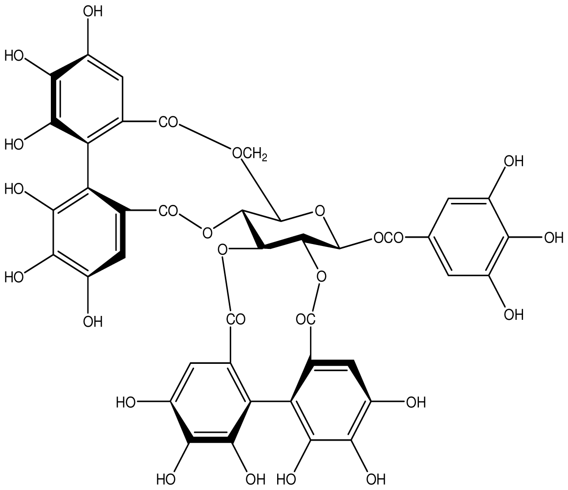
| Acutissimmin |  | Ethanol-induced ulcers/Intragastric/50.0 mg/kg | Mouse | Active | [82] |
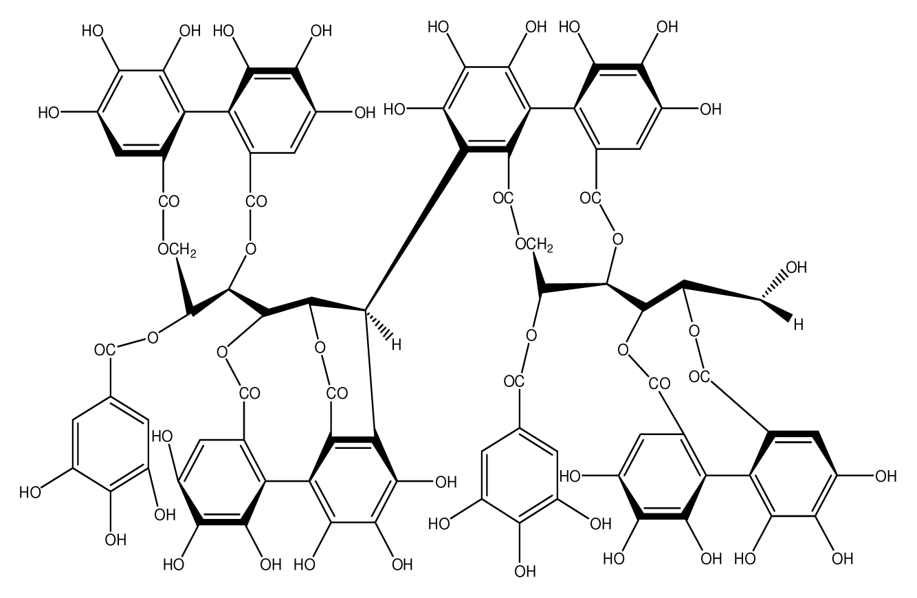
| Agrimoniin |  | Helicobacter pylori-MIC (25 μg/mL) | In vitro | Active | [92] |
| Alienanin B |  | Helicbacter pylori-MIC (25 μg/mL) | In vitro | Active | [92,93] |
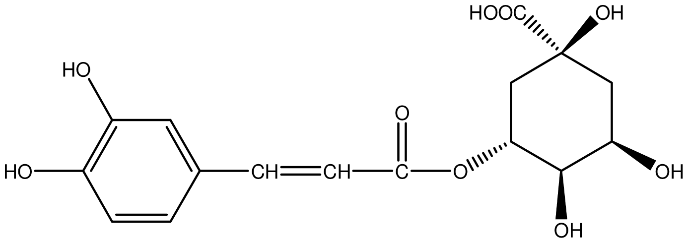
| Chlorogenic acid |  | Helicobacter pylori-MIC (>100 μg/mL) | In vitro | Inactive | [92,94] |
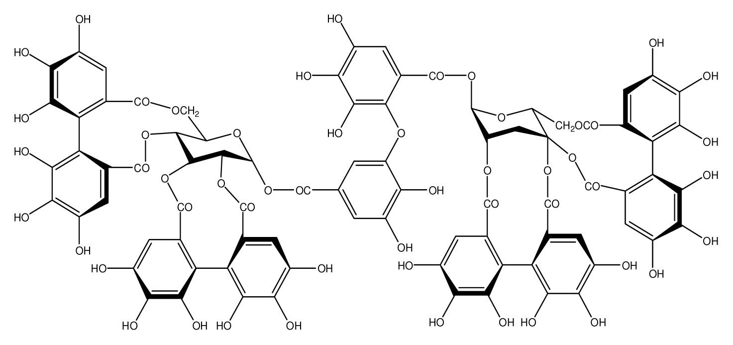
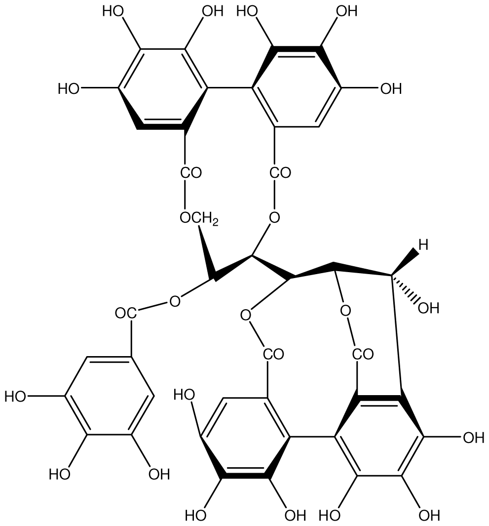
| Castalagin |  | Ethanol-induced ulcers/Intragastric/50.0 mg/kg | Mouse | Active | [82,95] |
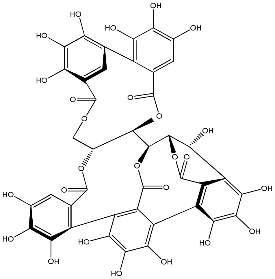
| Casuarictin |  | Helicobacter pylori-MIC (12.5 μg/mL) | In vitro | Active | [92,96] |
| Casuarinin |  | Helicobacter pylori-MIC (12.5 μg/mL) | In vitro | Active | [92,97] |
| Corilagin |  | Helicobacter pylori-MIC (6.25 μg/mL) | In vitro | Active | [92,98] |
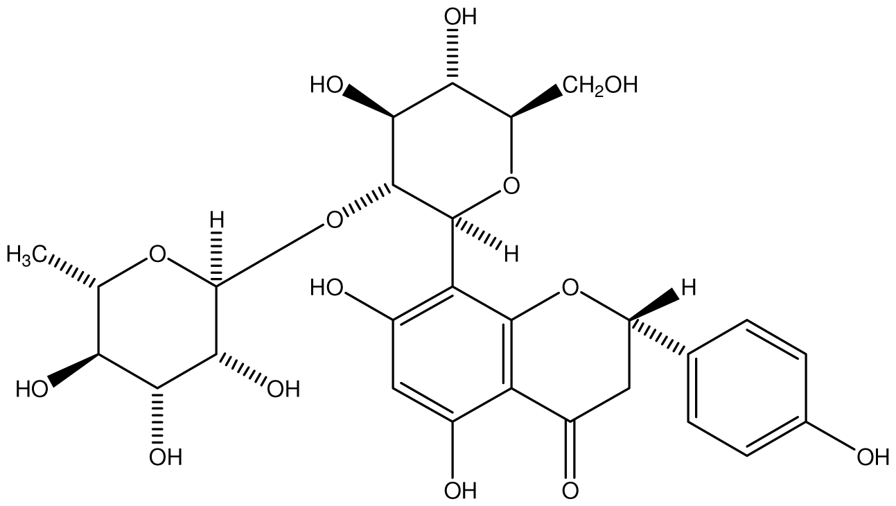
| 8-CRHA-Glc naringenin |  | Helicobacter pylori-MIC (>100 μg/mL) | In vitro | Inactive | [92,99] |
| Elaeagnatin A |  | Helicobacter pylori-MIC (25 μg/mL) | In vitro | Active | [92,100] |
| Elagic acid |  | Stress-induced ulcers(water immersion)/intraperitoneal/5, 10 and 25 mg/kg | Rat | Active | [90,101] |
| Pylorus-ligated animals/Intraperitoneal/5, 10 and 25 mg/kg | Rat | Active | [90,101] | ||
| Inhibition of gastric H+, K+-ATPase | Hog gastric mucosal | Active | [90,101] | ||
| Epicatechin |  | Helicobacter pylori-MIC (>100 μg/mL) | In vitro | Inactive | [92,102] |
| Epicatechin gallate |  | Helicobacter pylori-MIC (50 μg/mL) | In vitro | Active (Less) | [92,102] |
| Epigallocatechin gallate |  | Helicobacter pylori-MIC (25 μg/mL) | In vitro | Active | [92,102] |
| Geraniin |  | Helicobacter pylori-MIC (12.5 μg/mL) | In vitro | Active | [92] |
| Stress induced ulcer | Mouse | Active | [92,103] | ||
| Heterophylliin G |  | Helicobacter pylori-MIC (12.5 μg/mL) | In vitro | Active | [92,104] |
| Hippophenin A |  | Helicobacter pylori-MIC (12.5 μg/mL) | In vitro | Active | [92,100] |
| Iridin |  | Helicobacter pylori-MIC (>100 μg/mL) | In vitro | Inactive | [92,105] |
| Isorhamnetin 3-O-rutinoside |  | Helicobacter pylori-MIC (>100 μg/mL) | In vitro | Inactive | [92,106] |
| Nobotanin B |  | Helicobacter pylori-MIC (12.5 μg/mL) | In vitro | Active | [92,96] |
| Oenothein A |  | Helicobacter pylori-MIC (12.5 μg/mL) | In vitro | Active | [92] |
| Oenothein B |  | Helicobacter pylori-MIC (12.5 μg/mL) | In vitro | Active | [92,107] |
| Pedunculagin |  | Ethanol-induced ulcers/Intragastric/50.0 mg/kg | Mouse | Active | [82,108] |
| Penta-O-galloyl-β-d-glucose |  | Helicobacter pylori-MIC (12.5 μg/mL) | In vitro | Active | [92,109] |
| Phillyraeoidin A |  | Ethanol-induced ulcers/Intragastric/50.0 mg/kg | Mouse | Active | [82,110] |
| Procyanidin B1 |  | Helicobacter pylori-MIC (>100 μg/mL) | In vitro | Inactive | [92,111] |
| Procyanidin B3 |  | Helicobacter pylori-MIC (50 μg/mL) | In vitro | Minimal activity | [92,111] |
| Procyanidin B4 |  | Helicobacter pylori-MIC (50 μg/mL) | In vitro | Minimal activity | [92,111] |
| Procyanidin B5 |  | Helicobacter pylori-MIC (25 μg/mL) | In vitro | active | [92,112] |
| Procyanidin C1 |  | Helicobacter pylori-MIC (>100 μg/mL) | In vitro | Inactive | [92,113] |
| Procyanidin polymer |  | Helicobacter pylori-MIC (>100 μg/mL) | In vitro | Inactive | [92,99] |
| Rugosin D |  | Helicobacter pylori-MIC (25 μg/mL) | In vitro | Active | [92] |
| Strictinin |  | Helicobacter pylori-MIC (6.25 μg/mL) | In vitro | Active | [114] |
| Tannic acid |  | Shay ulcer/oral/50.0 mg/kg | Rat | Active | [115,116] |
| Acetic acid-induced ulcer/oral/200.0 mg/kg | Rat | Active | [116] | ||
| Pylorus-ligated animals/oral/50.0, 100.0 and 500 mg/kg | Rat | Active | [116] | ||
| Ethanol induced gastric lesions/gastric intubation/100.0 mg/kg | Rat | Active | [117] | ||
| Inhibition of gastric H+, K+-ATPase | Hog gastric mucosal | Active | [118] | ||
| Tellimagrandin I |  | Helicobacter pylori-MIC (12.5 μg/mL) | In vitro | Active | [92] |
| Tellimagrandin II |  | Helicobacter pylori-MIC (6.25 μg/mL) | In vitro | Active | [92] |
| Tri-N-coumaroyl-spermidine |  | Helicobacter pylori-MIC (>100 μg/mL) | In vitro | Inactive | [92,118] |
© 2012 by the authors; licensee Molecular Diversity Preservation International, Basel, Switzerland. This article is an open-access article distributed under the terms and conditions of the Creative Commons Attribution license (http://creativecommons.org/licenses/by/3.0/).
Share and Cite
De Jesus, N.Z.T.; Falcão, H.d.S.; Gomes, I.F.; Leite, T.J.d.A.; Lima, G.R.d.M.; Barbosa-Filho, J.M.; Tavares, J.F.; Silva, M.S.d.; Athayde-Filho, P.F.d.; Batista, L.M. Tannins, Peptic Ulcers and Related Mechanisms. Int. J. Mol. Sci. 2012, 13, 3203-3228. https://doi.org/10.3390/ijms13033203
De Jesus NZT, Falcão HdS, Gomes IF, Leite TJdA, Lima GRdM, Barbosa-Filho JM, Tavares JF, Silva MSd, Athayde-Filho PFd, Batista LM. Tannins, Peptic Ulcers and Related Mechanisms. International Journal of Molecular Sciences. 2012; 13(3):3203-3228. https://doi.org/10.3390/ijms13033203
Chicago/Turabian StyleDe Jesus, Neyres Zinia Taveira, Heloina de Souza Falcão, Isis Fernandes Gomes, Thiago Jose de Almeida Leite, Gedson Rodrigues de Morais Lima, Jose Maria Barbosa-Filho, Josean Fechine Tavares, Marcelo Sobral da Silva, Petrônio Filgueiras de Athayde-Filho, and Leonia Maria Batista. 2012. "Tannins, Peptic Ulcers and Related Mechanisms" International Journal of Molecular Sciences 13, no. 3: 3203-3228. https://doi.org/10.3390/ijms13033203