Article

Loss of p16Ink4a Function Rescues Cellular Senescence Induced by Telomere Dysfunction

1
Faculty of Environmental Science and Engineering, Kunming University of Science & Technology, Kunming 650224, China
2
Lab of Molecular Genetics of Aging & Tumor, Faculty of Life Science and Technology, Kunming University of Science & Technology, Kunming 650224, China
*
Authors to whom correspondence should be addressed.
Int. J. Mol. Sci. 2012, 13(5), 5866-5877; https://doi.org/10.3390/ijms13055866
Submission received: 11 February 2012 / Revised: 3 May 2012 / Accepted: 10 May 2012 / Published: 16 May 2012
(This article belongs to the Special Issue Oxidative Stress and Ageing)

Abstract

:
p16Ink4a is a tumor suppressor and a marker for cellular senescence. Previous studies have shown that p16Ink4a plays an important role in the response to DNA damage signals caused by telomere dysfunction. In this study, we crossed Wrn−/− and p16Ink4a−/− mice to knock out the p16Ink4a function in a Wrn null background. Growth curves showed that loss of p16Ink4a could rescue the growth barriers that are observed in Wrn−/− mouse embryonic fibroblasts (MEFs). By challenging the MEFs with the global genotoxin doxorubicin, we showed that loss of p16Ink4a did not dramatically affect the global DNA damage response of Wrn−/− MEFs induced by doxorubicin. However, in response to telomere dysfunction initiated by the telomere damaging protein TRF2ΔBΔM, loss of p16Ink4a could partially overcome the DNA damage response by disabling p16Ink4a up-regulation and reducing the accumulation of γ-H2AX that is observed in Wrn−/− MEFs. Furthermore, in response to TRF2ΔBΔM overexpression, Wrn−/− MEFs senesced within several passages. In contrast, p16Ink4a−/− and p16Ink4a−/−Wrn−/− MEFs could continuously grow and lose expression of the exogenous TRF2ΔBΔM in their late passages. In summary, our data suggest that in the context of telomere dysfunction, loss of p16Ink4a function could prevent cells from senescence. These results shed light on the anti-aging strategy through regulation of p16Ink4a expression.

1. Introduction

Extensive studies have demonstrated that telomere dysfunction could induce cellular senescence, ultimately resulting in either aging or cancer [14]. Telomere dysfunction, including telomere shortening due to a lack of telomerase function, telomere uncapping and structural damage due to defects in proteins maintaining telomere structure, could be recognized as a DNA damage signal that triggers the DNA damage response (DDR) in mammalian cells [5,6]. Depending on the intactness of the DDR system, for example, the status of the ATM-p53-p21 pathway, telomere dysfunction could either prevent or promote tumorigenesis [79].
The role of the Rb-p16Ink4a pathway in response to DNA damage induced by telomere dysfunction has been controversial. p16Ink4a regulates cell cycle progression by specifically binding to and inhibiting the cyclin-dependent kinases Cdk4 and Cdk6. It has previously been demonstrated that p16Ink4a accumulates in conjunction with p38 activation in ras-induced senescent cells and that this accumulation is essential for senescence [10]. It has also been shown that p16Ink4a accumulates in both human and mouse cells as a function of replicative senescence [11,12]. In a previous study, telomere dysfunction was induced by inhibiting the telomere binding protein TRF2 with the dominant negative protein TRF2ΔBΔM. This study demonstrated that the telomere damage induced by TRF2ΔBΔM induces p16Ink4a accumulation. Inhibition of p16Ink4a with shRNA or overexpression of BMI1 (a p16Ink4a transcription regulator) showed a nearly complete bypass of the telomere-directed senescence in p53-deficient cells [13]. These data demonstrated that in human cells, p16Ink4a plays an important role along with the p53 pathway in the DDR to telomere dysfunction. This same study demonstrated that in wild type MEFs, the loss of p53 function was sufficient to completely abrogate the senescence arrest, indicating that in the absence of p53, the loss of p16Ink4a function is not necessary for mouse cells to bypass senescence [13]. In addition, previous studies have shown that inhibition of TRF2 results in apoptosis in MEFs and that this response is mediated by ATM and p53 [14].
Wrn, a member of the RecQ family of DNA helicases, is deficient in patients with Werner syndrome (WS) [15]. WS is an autosomal recessive progeric disease that is characterized by premature atherosclerosis, ischemic heart disease, osteoporosis, and cataracts [16]. A previous study showed that inhibition of the stress-induced p38 MAP kinase could prevent the senescence of WS cells [17]. Fibroblasts from WS patients have accelerated telomere erosion and senescence that can be rescued by the overexpression of telomerase, suggesting that telomere dysfunction may play an essential role in the manifestation of the WS phenotype [18]. To address this hypothesis, a mouse model of WS with a double knockout of mTerc and Wrn was generated, which faithfully manifested the human phenotype of WS [19,20]. The Wrn protein is necessary for efficient replication of G-rich telomeric DNA, preventing telomere dysfunction and consequent genomic instability [21]. In addition, the function of Wrn in telomere regulation is regulated by TRF1 and TFR2 [22]. Thus, a mouse model deficient in Wrn function (Wrn−/−) provides a very useful tool for investigating the factors involved in aging in response to telomere dysfunction.
By crossing mice deficient in p16Ink4a function (p16Ink4a−/−) with mice deficient in Wrn function (Wrn−/−), we generated mouse embryonic fibroblasts (MEFs) with the following genotypes: p16Ink4a−/− only, Wrn−/− only, and p16Ink4a−/−Wrn−/−. Furthermore, we challenged these cells with either the global genomic DNA damage reagent doxorubicin or the telomere specific damaging protein TRF2ΔBΔM. By comparing their growth rates, DDR, senescence status, and genomic instability, we investigated the role of p16Ink4a in the induction of senescence by Wrn-related telomere dysfunction.

2. Results and Discussion

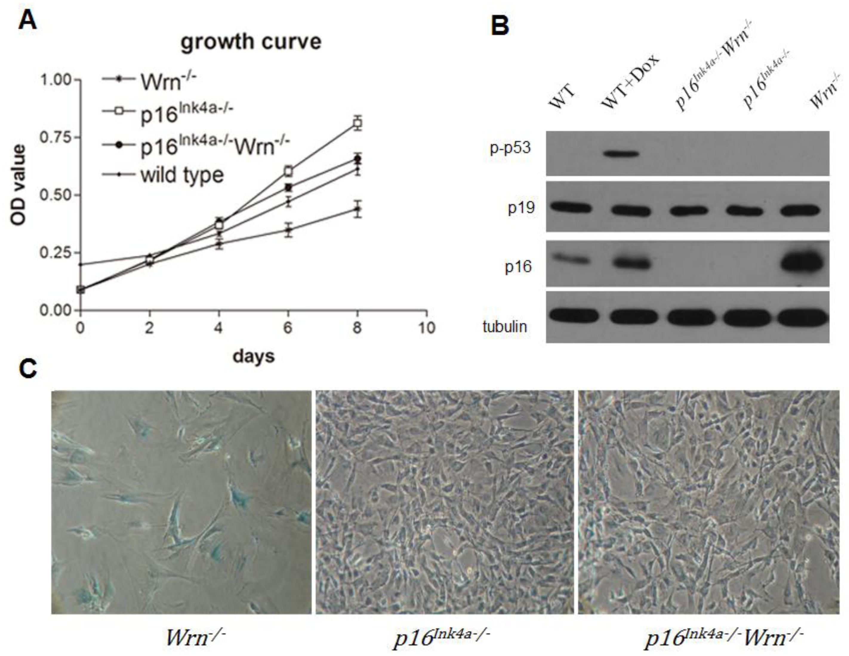
2.1. Loss of p16Ink4a Function Overcame the Growth Barrier Caused by Dysfunction of Wrn

To investigate the growth potential of MEFs with the genotypes p16Ink4a−/−, Wrn−/−, and p16Ink4a−/−Wrn−/−, we performed a growth curve analysis using wild type MEFs as a control. We found that Wrn−/− MEFs grew slower than p16Ink4a−/− MEFs; however, knock out of p16Ink4a function (p16Ink4a−/−Wrn−/−) rescued the growth potential of Wrn−/− MEFs (Figure 1A). These data suggest that the growth barrier caused by dysfunction of Wrn is overcome by the loss of p16Ink4a function. Furthermore, western blot analysis revealed that compared to wild type MEFs (Figure 1B, WT), Wrn−/− MEFs have dramatically increased p16Ink4a expression (Figure 1B, Wrn−/−). This result suggests that loss of Wrn function stimulates the expression of p16Ink4a, which might cause the growth barrier of Wrn−/− MEFs due to negative regulation of the cell cycle. Thus, knockout of p16Ink4a function could overcome the growth barrier caused by a defect in Wrn. Surprisingly, we did not detect an up-regulation of phosphorylated p53 in Wrn−/− MEFs, suggesting that unlike doxorubicin treatment, the loss of Wrn did not cause sufficient DNA damage to stimulate the p53 pathway (Figure 1B, p-p53). In addition, p19Arf, a response protein to oncogenic stimulation, did not show up-regulation in the absence of Wrn (Figure 1B, p19). Furthermore, we performed SA-β-Gal staining to detect cellular senescence in passage 13 of p16Ink4a−/−, Wrn−/−, and p16Ink4a−/−Wrn−/− MEFs. The results indicated that cellular senescence occurred in passage 13 of Wrn−/− MEFs (57.31 ± 7.19% of cells were positive for SA-β-Gal staining), but not in p16Ink4a−/− MEFs (1.40 ± 0.53% cells were positive for SA-β-Gal staining) or p16Ink4a−/−Wrn−/− MEFs (2.23 ± 1.86% cells were positive for SA-β-Gal staining) (Figure 1C).

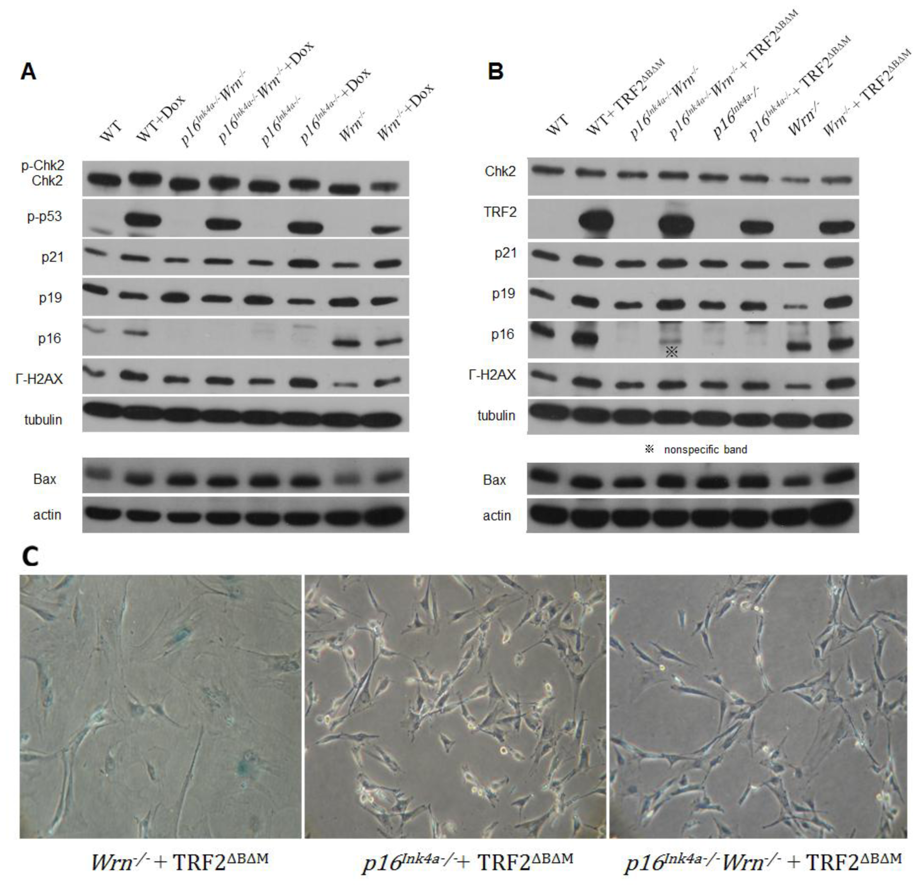
2.2. DNA Damage Response to Genotoxin or Telomere Dysfunction in Cells Lacking Wrn or p16Ink4a Function

To further understand cellular senescence caused by telomere dysfunction in Werner syndrome and the role of Wrn and p16Ink4a in the DNA damage response to telomere dysfunction, a well defined telomere damaging protein TRF2ΔBΔM [13] was introduced into p16Ink4a−/−, Wrn−/− and p16Ink4a−/−Wrn−/− MEFs. The global genotoxin doxorubicin was used in parallel to investigate the distinct stimulation of pathways involved in the DNA damage response.
Western blot analysis demonstrated that in response to the global DNA damage caused by doxorubicin, Chk2, p53, and H2AX proteins were phosphorylated in wild type, p16Ink4a−/−, Wrn−/− and p16Ink4a−/−Wrn−/− MEFs (Figure 2A, Chk2, p-p53, γ-H2AX). These data confirm that the regular DNA damage responses were stimulated in MEFs lacking Wrn or p16Ink4a (Figure 2A, Wrn−/−). Interestingly, in response to telomere dysfunction induced by the overexpression of TRF2ΔBΔM (Figure 2B, TRF2), Chk2 was not phosphorylated (Figure 2B, Chk2). In addition, no phosphorylated p53 could be detected (data not shown), suggesting a lack of response of Chk2-p53 to telomere dysfunction induced by TRF2ΔBΔM in the genetic context of these MEFs. However, p21 proteins were slightly up-regulated despite the p53 phosphorylation status (Figure 2B, p21). This result could be due to a p53-independent activation of p21 or a low level of p53 activation that is not detectable by western blot. H2AX proteins were phosphorylated significantly in response to TRF2ΔBΔM overexpression in wild type, p16Ink4a−/−, Wrn−/−, and p16Ink4a−/−Wrn−/− MEFs (Figure 2B, γ-H2AX), suggesting that TRF2ΔBΔM induces the activation of the DNA damage response. p16Ink4a proteins were remarkably up-regulated in response to TRF2ΔBΔM overexpression in wild type MEFs, as well as Wrn−/− MEFs (Figure 2B, p16). These data suggest that in p16Ink4a−/− and p16Ink4a−/−Wrn−/− MEFs, the loss of p16Ink4a function prevents cells from undergoing TRF2ΔBΔM-induced senescence.
Surprisingly, p19Arf proteins were up-regulated in response to TRF2ΔBΔM overexpression in wild type, p16Ink4a−/−, Wrn−/− and p16Ink4a−/−Wrn−/− MEFs (Figure 2B, p19), which was contrary to the response of MEFs to doxorubicin in which p19Arf proteins were slightly down-regulated (Figure 2A, p19). These western blots were quantified (supplementary data 1).
Interestingly, the background expression levels of the apoptotic marker Bax were up-regulated in p16Ink4a−/− and p16Ink4a−/−Wrn−/− MEFs, but not in Wrn−/− MEFs. Upon doxorubicin treatment or TRF2ΔBΔM overexpression, Bax was not up-regulated (Figure 2A,B, Bax, actin was used as the loading control). SA-β-Gal staining of p16Ink4a−/−, Wrn−/− and p16Ink4a−/−Wrn−/− MEFs after TRF2ΔBΔM overexpression showed that Wrn−/− MEFs senesced rapidly after TRF2ΔBΔM overexpression, while p16Ink4a−/−Wrn−/− MEFs did not appear to senesce (Figure 2C). These data further support the conclusion that loss of p16Ink4a function prevents cells from TRF2ΔBΔM-induced senescence.
Telomere dysfunction could induce chromosomal aberrations, which further enhance the destructiveness of telomere dysfunction, ultimately resulting in tumorigenesis.
To investigate the chromosomal aberrations induced by TRF2ΔBΔM overexpression in p16Ink4a−/−, Wrn−/− and p16Ink4a−/−Wrn−/− MEFs, we attempted to harvest chromosomes and perform cytogenetic analysis. However, TRF2ΔBΔM overexpression leads to cellular senescence in wild type and Wrn−/− MEFs, thereby preventing us from harvesting chromosomes from these cells for analysis. In contrast, despite the overexpression of TRF2ΔBΔM, p16Ink4a−/− and p16Ink4a−/−Wrn−/− MEFs continuously grow. We harvested the chromosomes from early passaged (p4), medium passaged (p18), and late passaged (p27) cells. Chromosome analysis (Table 1) showed that very few chromosomal breakages or fusions occurred in early and medium passages of MEFs, late passages of p16Ink4a−/−Wrn−/− MEFs that were overexpressing TRF2ΔBΔM accumulated chromosomal fusions (12 of 30 cells in metaphase), and only a few fusions were found in late passages of p16Ink4a−/− MEFs (2 of 30 cells in metaphase). We observed an increase of aneuploidy that correlated with increasing numbers of cell passages in both types of MEFs, which might be due to the cell passaging process itself. These data suggest that loss of p16Ink4a function in a Wrn-deficient background rescues cellular senescence, but with the price of accumulating chromosomal aberrations.

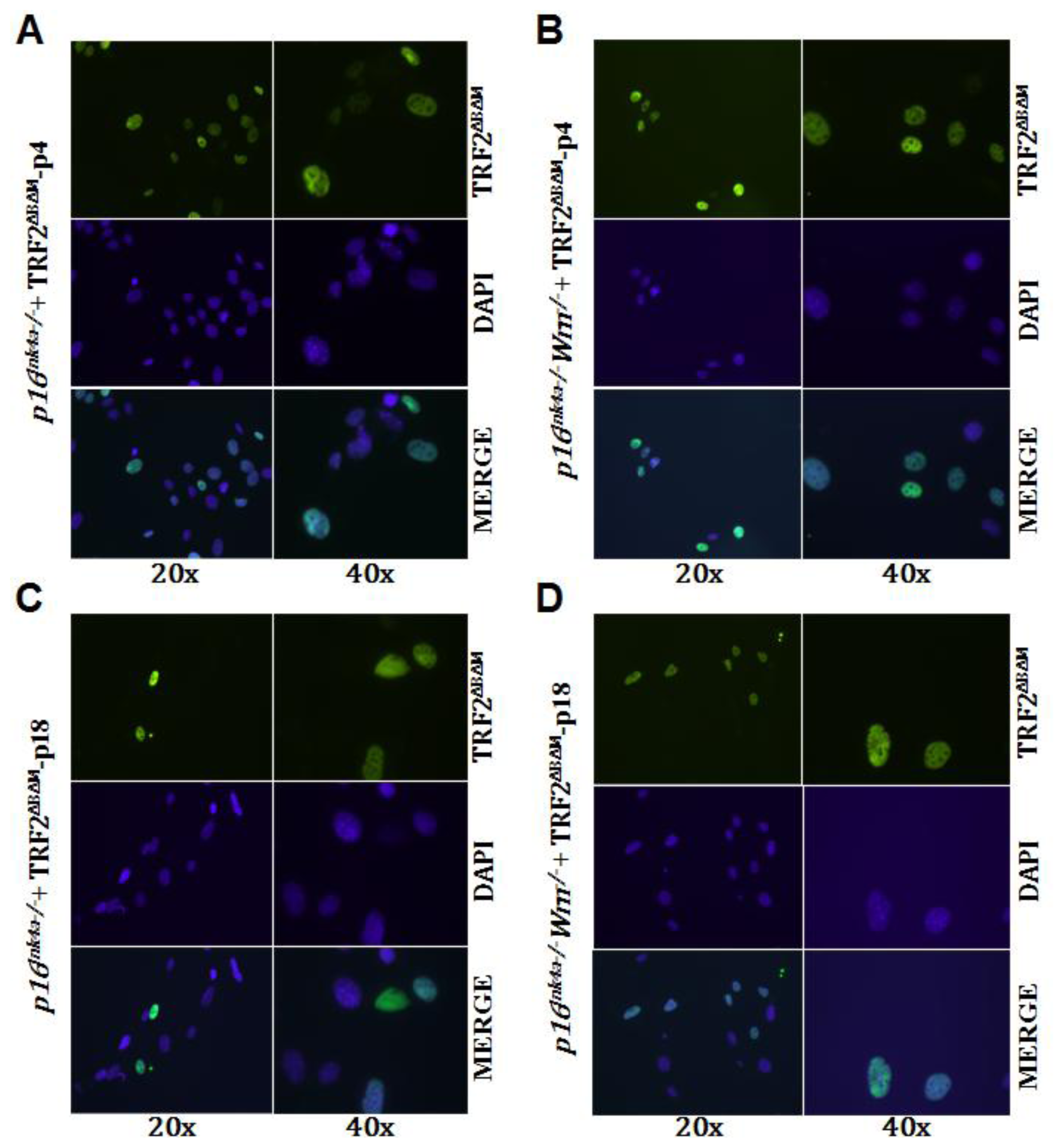
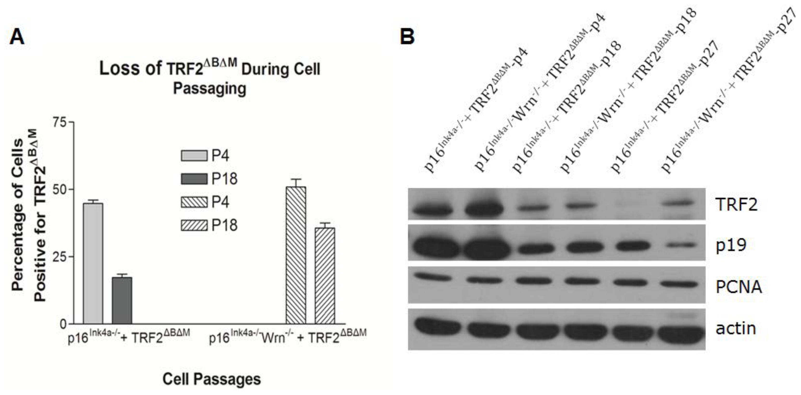
2.3. Loss of p16Ink4a Function Facilitated the Elimination of Exogenous TRF2ΔBΔM in MEFs

The above data raise the question of how p16Ink4a−/− and p16Ink4a−/−Wrn−/− MEFs could continuously grow while maintaining the overexpression of TRF2ΔBΔM. First, we confirmed the overexpression of TRF2ΔBΔM in early and medium passages of p16Ink4a−/− and p16Ink4a−/−Wrn−/− MEFs using immunostaining. As expected, we could detect expression of TRF2ΔBΔM in the nuclei of early passages (p4) of p16Ink4a−/− and p16Ink4a−/−Wrn−/− MEFs (Figure 3A,B). However, in passage 18 of p16Ink4a−/− and p16Ink4a−/−Wrn−/− MEFs, the number of cells with positive staining for TRF2ΔBΔM decreased dramatically (Figure 3C,D).
The percentage of P4 p16Ink4a−/− MEFs with positive staining for TRF2ΔBΔM was around 45%, while positive staining of P18 p16Ink4a−/− MEFs decreased to 20% (Figure 4A). Similarly, positive staining of P4 p16Ink4a−/−Wrn−/− MEFs was around 56%, while positive staining of P18 p16Ink4a−/−Wrn−/− MEFs decreased to 39% (Figure 4A). Western blotting further confirmed the decrease of TRF2ΔBΔM expression in MEFs with the increasing cell passage number (Figure 4B, supplementary data 2). These data suggest that in the absence of p16Ink4a, MEFs are able to lose expression of telomere dysfunction factors and, therefore, bypass senescence and sustain cell growth.
When the mouse model of Werner Syndrome (Wrn−/−) was established by deleting a segment of the Wrn gene, the premature aging phenotype of human Werner Syndrome was not observed [23]. It is now clear that this observation was due to the endogenous telomerase activity in mouse somatic cells [19,20]. While Wrn−/− mice display reduced embryonic survival, live-born mice appear normal during their first year of life. However, Wrn−/− MEFs display a prematurely reduced growth rate as a function of passage number in culture, demonstrating that Wrn−/− MEFs grow more slowly and have a lower saturation density in culture than their heterozygous and wild type counterparts [23].
Using Wrn−/− MEFs, we also observed the low growth potential of Wrn−/− MEFs, which may be due to the up-regulation of the cell cycle inhibitor p16Ink4a. Knockout of p16Ink4a rescued the growth potential of Wrn−/− MEFs, further supporting this point.
Furthermore, the loss of either Wrn or p16Ink4a function did not dramatically affect the cellular DNA damage response to the global genotoxin doxorubicin. However, when the dominant negative inhibitor of TRF2 (TRF2ΔBΔM) was introduced into the cells and induced cellular senescence, the up-regulation response of p16Ink4a was disabled in p16Ink4a-deficient cells, which resulted in distinct cellular consequences between Wrn−/− MEFs and p16Ink4a−/−Wrn−/− MEFs. In addition, the up-regulation of γ-H2AX in response to TRF2ΔBΔM is reduced in p16Ink4a−/−Wrn−/− MEFs. Together these regulations in p16Ink4a−/−Wrn−/− MEFs overcame the cellular senescence induced by telomere dysfunction and sustained the continuous growth of cells with the consequence of the accumulation of chromosomal aberrations. Eventually p16Ink4a−/−Wrn−/− MEFs and p16Ink4a−/− MEFs could lose expression of TRF2ΔBΔM, which might further stabilize the chromosomes and stop the accumulation of chromosomal aberrations. However, we found that in p16Ink4a−/−Wrn−/− MEFs the chromosomal aberrations still accumulated slowly due to the existing overexpression of TRF2ΔBΔM. In this case, the reduction of TRF2ΔBΔM levels slowed the accumulation of chromosomal aberrations. The mechanism responsible for the loss of TRF2ΔBΔM expression is not clear; however, the loss of p16Ink4a function played a permissive role in this process because the Wrn−/− MEFs could not survive TRF2ΔBΔM overexpression. In contrast to a previous report [24], we did not observe p53 activation in response to TRF2ΔBΔM overexpression in these MEFs. A previous study also indicated that in normal human epidermal keratinocytes, TRF2ΔBΔM overexpression caused only a two fold increase in both the phosphorylation of p53 at serine 15 and 53BP1 DNA damage foci and no detectable increase in p21. Despite the weak DNA damage response, the keratinocytes demonstrated growth arrest, reduced colony formation and senescence [25]. Another study demonstrated that uncapping telomeres with TRF2ΔBΔM resulted in apoptosis, not senescence. These authors found a more dramatic activation of p53, as well as an up-regulation of Bax [14]. In our study, however, we did not observe activation of Chk2 or p53 or up-regulation of Bax. It is possible that a higher level of TRF2ΔBΔM results in apoptosis rather than senescence. It has also been shown that in human fibroblasts, telomere dysfunction induced senescence and signaled through ATM-p53-p21, but not p16Ink4a, suggesting that distinct senescence programs could progress in parallel, resulting in mosaic cultures as well as individual cells responding to multiple signals [26].
Together, these data suggest different roles of p53 and p16Ink4a in response to distinct DNA damages and shed light on anti-aging strategies by the regulation p16Ink4a expression.

3. Experimental Section

3.1. Cell Lines, Constructs and Antibodies

MEFs were prepared and harvested from individual day 13.5 mouse embryos from designed parental mice with p16Ink4+/− or Wrn+/− genotypes (heterozygous knockouts of either p16Ink4 or Wrn) and the harvested MEFs were cultured and genotyped. MEFs with wild type, p16Ink4a−/−, Wrn−/− or p16Ink4a−/−Wrn−/− genotypes were used for experiments. Wild type MEFs were used for experiments before passage 10 and the cells were passaged at a ratio of 1:2 or 1:3 so that the cells were not excessively diluted and senesced too quickly.
All cell lines were cultured in DMEM medium supplemented with 10% fetal bovine serum (Hyclone, CA, USA) in a 3% oxygen and 5% CO2 incubator at 37 °C.
The pBabe-TRF2ΔBΔM (with a cMyc tag) construct, a gift from Dr. de Lange’s lab (as described in [27]), was introduced into MEFs. For transient transfection, cells were used 48 h after transfection. For stable expression, antibiotic selection was applied for 1–2 months and expression of TRF2ΔBΔM was analyzed by both western blot and immunostaining.
Antibodies used for western blot were anti-p16Ink4a (M-156) (1:500, Santa Cruz, CA, USA), anti-Chk2 (1:500, BD Transduction Laboratories, CA, USA), anti-phospho-p53 (Ser15) (1:500, Cell Signaling, MA, USA), anti-cMyc (9E10) (1:500, Santa Cruz, CA, USA), anti-p19Arf (1:1250, Upstate, NY, USA), anti-γ-H2AX (1:1000, Upstate, NY, USA), and anti-γ-tubulin (1:5000, Upstate, NY, USA).

3.2. Growth Curve Assay

A total of 2.5 × 10⁴ cells were seeded per well in 12 well plates and triplicates were prepared for each sample. At each time point, cells were fixed for 20 min at room temperature in 10% buffered formalin and stained with 1 mL 0.1% crystal violet for 20 min at room temperature. After the plates were washed and dried, cells were treated with 2 mL 10% acetic acid and the OD of the extracted dye was measured at 595 nm. Statistical analysis was performed to compare the difference between cell lines (n = 3).

3.3. Immunostaining Assay

Cells were fixed with 2% paraformaldehyde and 2% sucrose in 1× PBS (pH 7.6) for 10 min and then permeabilized with 1% NP-40. After pre-incubation with 5% BSA/PBS, cells were incubated first with the primary antibody and then with the secondary antibody in 1% BSA/PBS for 1 h at room temperature. After DAPI staining, the slides were mounted with ProLong mounting medium (Invitrogen, CA, USA). Fluorescence microscopy was performed using a Nikon Eclipse 90i fluorescence microscope and a Nikon Digital Sight CCD (controlled by NIS-Elements 3.0 software).

3.4. SA-β-Gal Staining

SA-β-Gal staining was performed as described previously [28]. Briefly, cultured cells were washed in 1× PBS and fixed for 3–5 min (room temperature) in 2% formaldehyde, 0.2% glutaraldehyde. Fixed cells were stained with fresh stain solution for SA-β-galactosidase activity at 37 °C for 4 h. The percentage of cells positive for SA-β-Gal staining were quantified and statistically analyzed (n = 3).

4. Conclusions

Our data suggest that, in the context of Wrn deficiency-related telomere dysfunction, loss of p16Ink4a function could prevent cells from senescence. These results shed light on the anti-aging strategy by regulation of p16Ink4a expression.

Supplementary Information

ijms-13-05866-s001.pdf

Acknowledgments

This work was supported by The New Century Excellent Scholarship to Y.L, NSFC grants to Y.L. (30771194, 30970598, 31170735) and to W.T. (30960152), and the Yunnan Provincial Science Foundation to W.T. (20080043M). We thank Sandy Chang from Yale University and Ronald A Depinho from MD Anderson Cancer Center for providing the transgenic mice used in this study.

References

  1. Wright, W.E.; Shay, J.W. Telomere positional effects and the regulation of cellular senescence. Trends Genet 1992, 8, 193–197. [Google Scholar]
  2. Wright, W.E.; Shay, J.W. Time, telomeres and tumours: Is cellular senescence more than an anticancer mechanism? Trends Cell Biol 1995, 5, 293–297. [Google Scholar]
  3. Rudolph, K.L.; Chang, S.; Lee, H.W.; Blasco, M.; Gottlieb, G.J.; Greider, C.; DePinho, R.A. Longevity, stress response, and cancer in aging telomerase-deficient mice. Cell 1999, 96, 701–712. [Google Scholar]
  4. Stewart, S.A.; Weinberg, R.A. Telomeres: Cancer to human aging. Annu. Rev. Cell Dev. Biol 2006, 22, 531–557. [Google Scholar]
  5. De Lange, T. Protection of mammalian telomeres. Oncogene 2002, 21, 532–540. [Google Scholar]
  6. De Lange, T. Shelterin: The protein complex that shapes and safeguards human telomeres. Gene Dev 2005, 19, 2100–2110. [Google Scholar]
  7. Artandi, S.E.; DePinho, R.A. Mice without telomerase: What can they teach us about human cancer? Nat. Med 2000, 6, 852–855. [Google Scholar]
  8. Chin, L.; Artandi, S.E.; Shen, Q.; Tam, A.; Lee, S.L.; Gottlieb, G.J.; Greider, C.W.; DePinho, R.A. P53 deficiency rescues the adverse effects of telomere loss and cooperates with telomere dysfunction to accelerate carcinogenesis. Cell 1999, 97, 527–538. [Google Scholar]
  9. Zhang, X.F.; Tang, W.R.; Luo, Y. Aging or tumor: The crosstalk between telomerase and p53 (in Chinese). Yi Chuan 2009, 31, 451–456. [Google Scholar]
  10. Wang, W.; Chen, J.X.; Liao, R.; Deng, Q.; Zhou, J.J.; Huang, S.; Sun, P. Sequential activation of the mek-extracellular signal-regulated kinase and mkk3/6-p38 mitogen-activated protein kinase pathways mediates oncogenic ras-induced premature senescence. Mol. Cell Biol 2002, 22, 3389–3403. [Google Scholar]
  11. Palmero, I.; McConnell, B.; Parry, D.; Brookes, S.; Hara, E.; Bates, S.; Jat, P.; Peters, G. Accumulation of p16ink4a in mouse fibroblasts as a function of replicative senescence and not of retinoblastoma gene status. Oncogene 1997, 15, 495–503. [Google Scholar]
  12. Hara, E.; Smith, R.; Parry, D.; Tahara, H.; Stone, S.; Peters, G. Regulation of p16cdkn2 expression and its implications for cell immortalization and senescence. Mol. Cell Biol 1996, 16, 859–867. [Google Scholar]
  13. Jacobs, J.J.; de Lange, T. Significant role for p16ink4a in p53-independent telomere-directed senescence. Curr. Biol 2004, 14, 2302–2308. [Google Scholar]
  14. Karlseder, J.; Broccoli, D.; Dai, Y.; Hardy, S.; de Lange, T. P53- and atm-dependent apoptosis induced by telomeres lacking trf2. Science 1999, 283, 1321–1325. [Google Scholar]
  15. Gray, M.D.; Shen, J.C.; Kamath-Loeb, A.S.; Blank, A.; Sopher, B.L.; Martin, G.M.; Oshima, J.; Loeb, L.A. The Werner Syndrome protein is a DNA helicase. Nat. Genet 1997, 17, 100–103. [Google Scholar]
  16. Epstein, C.J.; Martin, G.M.; Schultz, A.L.; Motulsky, A.G. Werner’s syndrome a review of its symptomatology, natural history, pathologic features, genetics and relationship to the natural aging process. Medicine (Baltimore) 1966, 45, 177–221. [Google Scholar]
  17. Davis, T.; Bachler, M.A.; Wyllie, F.S.; Bagley, M.C.; Kipling, D. Evaluating the role of p38 map kinase in growth of Werner Syndrome fibroblasts. Ann. N. Y. Acad. Sci 2010, 1197, 45–48. [Google Scholar]
  18. Wyllie, F.S.; Jones, C.J.; Skinner, J.W.; Haughton, M.F.; Wallis, C.; Wynford-Thomas, D.; Faragher, R.G.; Kipling, D. Telomerase prevents the accelerated cell ageing of Werner Syndrome fibroblasts. Nat. Genet 2000, 24, 16–17. [Google Scholar]
  19. Chang, S.; Multani, A.S.; Cabrera, N.G.; Naylor, M.L.; Laud, P.; Lombard, D.; Pathak, S.; Guarente, L.; DePinho, R.A. Essential role of limiting telomeres in the pathogenesis of Werner Syndrome. Nat. Genet 2004, 36, 877–882. [Google Scholar]
  20. Du, X.; Shen, J.; Kugan, N.; Furth, E.E.; Lombard, D.B.; Cheung, C.; Pak, S.; Luo, G.; Pignolo, R.J.; DePinho, R.A.; et al. Telomere shortening exposes functions for the mouse werner and bloom syndrome genes. Mol. Cell Biol 2004, 24, 8437–8446. [Google Scholar]
  21. Crabbe, L.; Verdun, R.E.; Haggblom, C.I.; Karlseder, J. Defective telomere lagging strand synthesis in cells lacking wrn helicase activity. Science 2004, 306, 1951–1953. [Google Scholar]
  22. Opresko, P.L.; Otterlei, M.; Graakjaer, J.; Bruheim, P.; Dawut, L.; Kolvraa, S.; May, A.; Seidman, M.M.; Bohr, V.A. The Werner Syndrome helicase and exonuclease cooperate to resolve telomeric d-loops in a manner regulated by trf1 and trf2. Mol. Cell 2004, 14, 763–774. [Google Scholar]
  23. Lebel, M.; Leder, P. A deletion within the murine Werner Syndrome helicase induces sensitivity to inhibitors of topoisomerase and loss of cellular proliferative capacity. Proc. Natl. Acad. Sci. USA 1998, 95, 13097–13102. [Google Scholar]
  24. Jacobs, J.J.; de Lange, T. P16ink4a as a second effector of the telomere damage pathway. Cell Cycle 2005, 4, 1364–1368. [Google Scholar]
  25. Minty, F.; Thurlow, J.K.; Harrison, P.R.; Parkinson, E.K. Telomere dysfunction in human keratinocytes elicits senescence and a novel transcription profile. Exp. Cell Res 2008, 314, 2434–2447. [Google Scholar]
  26. Herbig, U.; Jobling, W.A.; Chen, B.P.; Chen, D.J.; Sedivy, J.M. Telomere shortening triggers senescence of human cells through a pathway involving atm, p53, and p21(cip1), but not p16(ink4a). Mol. Cell 2004, 14, 501–513. [Google Scholar]
  27. Smogorzewska, A.; de Lange, T. Different telomere damage signaling pathways in human and mouse cells. EMBO J 2002, 21, 4338–4348. [Google Scholar]
  28. Dimri, G.P.; Lee, X.; Basile, G.; Acosta, M.; Scott, G.; Roskelley, C.; Medrano, E.E.; Linskens, M.; Rubelj, I.; Pereira-Smith, O.; et al. A biomarker that identifies senescent human cells in culture and in aging skin in vivo. Proc. Natl. Acad. Sci. USA 1995, 92, 9363–9367. [Google Scholar]
Figure 1. Wrn−/− induced up-regulation of p16Ink4a and caused a growth barrier in MEFs. (A) Growth curves of wild type, p16Ink4a−/−, Wrn−/− and p16Ink4a−/−Wrn−/− MEFs; (B) Western blot analysis of p16Ink4a−/−, Wrn−/− and p16Ink4a−/−Wrn−/− MEFs. Wild type MEFs (WT) with or without doxorubicin treatment were used as controls. (C) SA-β-Gal staining of p16Ink4a−/−, Wrn−/− and p16Ink4a−/−Wrn−/− MEFs at passage 13.
Ijms 13 05866f1
Figure 2. DNA damage response to genotoxin or telomere dysfunction in cells lacking either Wrn or p16Ink4a function. (A) Western blot analysis to determine the response of MEFs to doxorubicin treatment; (B) Western blot analysis to determine the response of MEFs to TRF2ΔBΔM overexpression. Wild type MEFs (WT) were used as controls; (C) SA-β-Gal staining of p16Ink4a−/−, Wrn−/− and p16Ink4a−/−Wrn−/− MEFs following TRF2ΔBΔM overexpression.
Ijms 13 05866f2
Figure 3. Overexpression of TRF2ΔBΔM in early and medium passages of p16Ink4a−/− and p16Ink4a−/−Wrn−/− MEFs revealed by immunostaining (20× and 40× refer to the amplification power of the objective lens, DAPI was used to show the nuclear localization). (A) Early passage (P4) of p16Ink4a−/− MEFs; (B) Early passage (P4) of p16Ink4a−/−Wrn−/− MEFs; (C) Late passage (P18) of p16Ink4a−/− MEFs; (D) Late passage (P18) of p16Ink4a−/−Wrn−/− MEFs.
Ijms 13 05866f3
Figure 4. Elimination of TRF2ΔBΔM expression in the late passages of p16Ink4a−/− and p16Ink4a−/−Wrn−/− MEFs. (A) The percentage of cells positive for TRF2ΔBΔM measured by immunostaining; (B) Western blot showing the decrease of TRF2ΔBΔM expression with increasing cell passages.
Ijms 13 05866f4
Table 1. Chromosome Analysis in MEFs overexpressing TRF2ΔBΔM.
MEFs’ Genotypemetaphases countedbreakagefusionDiploid 2N = 40Aneuploid 2N > 40Aneuploid 2N < 40
p16Ink4a−/− + TRF2ΔBΔM-p43011514
p16Ink4a−/−Wrn−/− + TRF2ΔBΔM-p43014142
p16Ink4a−/− + TRF2ΔBΔM-p183017202
p16Ink4a−/−Wrn−/− + TRF2ΔBΔM-p183015132
p16Ink4a−/− + TRF2ΔBΔM-p27302127
p16Ink4a−/−Wrn−/− + TRF2ΔBΔM-p273012116

