Article

Infrared Study of Er3+/Yb3+ Co-Doped GeO2-PbO-Bi2O3 Glass

by
Hamid-Reza Bahari
1,*,
Hj. A. A. Sidek
1,*,
Faisal Rafiq M. Adikan
2,
Wan M. M. Yunus
1 and
Mohamed K. Halimah
1
1
Department of Physics, Faculty of Science, Universiti Putra Malaysia, Serdang, Selangor, UPM 43400, Malaysia
2
Department of Electrical Engineering, Faculty of Engineering, University of Malaya, Kuala Lumpur, 50603, Malaysia
*
Authors to whom correspondence should be addressed.
Int. J. Mol. Sci. 2012, 13(7), 8609-8614; https://doi.org/10.3390/ijms13078609
Submission received: 21 May 2012 / Revised: 20 June 2012 / Accepted: 25 June 2012 / Published: 10 July 2012
(This article belongs to the Section Materials Science)

Abstract

:
Heavy metal oxide glasses, containing bismuth and/or lead in their glass structure are new alternatives for rare eart (RE) doped hosts. Hence, the study of the structure of these vitreous systems is of great interest for science and technology. In this research work, GeO2-PbO-Bi2O3 glass host doped with Er3+/Yb3+ ions was synthesized by a conventional melt quenching method. The Fourier transform infrared (FTIR) results showed that PbO and Bi2O3 participate with PbO4 tetragonal pyramids and strongly distort BiO6 octahedral units in the glass network, which subsequently act as modifiers in glass structure. These results also confirmed the existence of both four and six coordination of germanium oxide in glass matrix.

1. Introduction

The optical fibers using heavy metal oxide glasses have good environmental stability and medium optical loss in comparison with fluorides, which makes them suitable for photonic applications in short distances [1]. Low cut-off optical phonon energy, which has appeared in lead-germanate glasses, decreases the nonradiative relaxation rate of erbium excited states leading to excellent upconversion efficiency [2]. In addition, bismuth-germanate glasses are increasingly used for applications in non-linear optics, optical switching and second harmonic generation (SHG) [3] especially because of their high linear and nonlinear refractive index [4], high thermal expansion, low transition temperature, and excellent infrared transmission [2].
To design a glass for specific applications, a basic understanding of host material is necessary and, therefore, analysis of their structure is useful. Recent infrared (IR) and Raman studies on manganese doped (100 − x)GeO2-xBi2O3 system showed that Bi3+ ions incorporate in glass structure with deformed BiO6 octahedral units [5]. The distorted BiO6 octahedral groups participate in glass network by formation of non-bridging oxygens to modify the glass network. According to recent extended X-ray absorption fine structure (EXAFS) and vibrational study on xPbO-(1 − x)GeO2 system [6], at low lead content, lead ions act as modifiers in the germanate network, however, in PbO compositions higher than 40%, lead ions increasingly play a network former role in glass structure. Infrared spectroscopies are powerful techniques from which to infer structural information about glass material and hence many research groups are motivated to study different vitreous networks by IR spectroscopies [7,8]. In the present work, GeO2-PbO and GeO2-Bi2O3 binary and GeO2-PbO-Bi2O3 ternary systems as heavy metal oxide glass hosts with Er3+/Yb3+ dopants were studied. Fourier transform infrared (FTIR) spectroscopy was used to probe into the structure of the prepared glass samples.

2. Results and Discussion
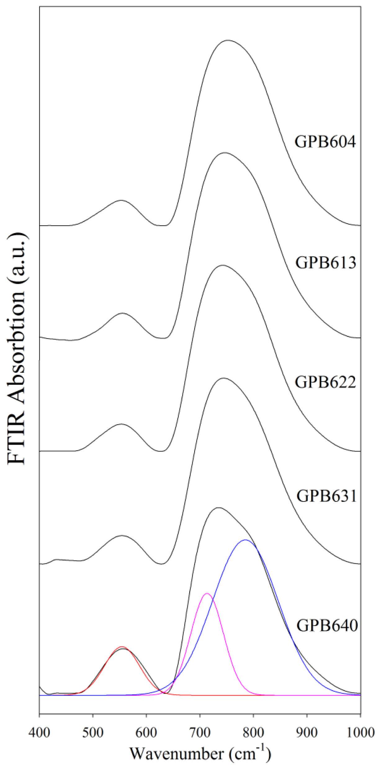
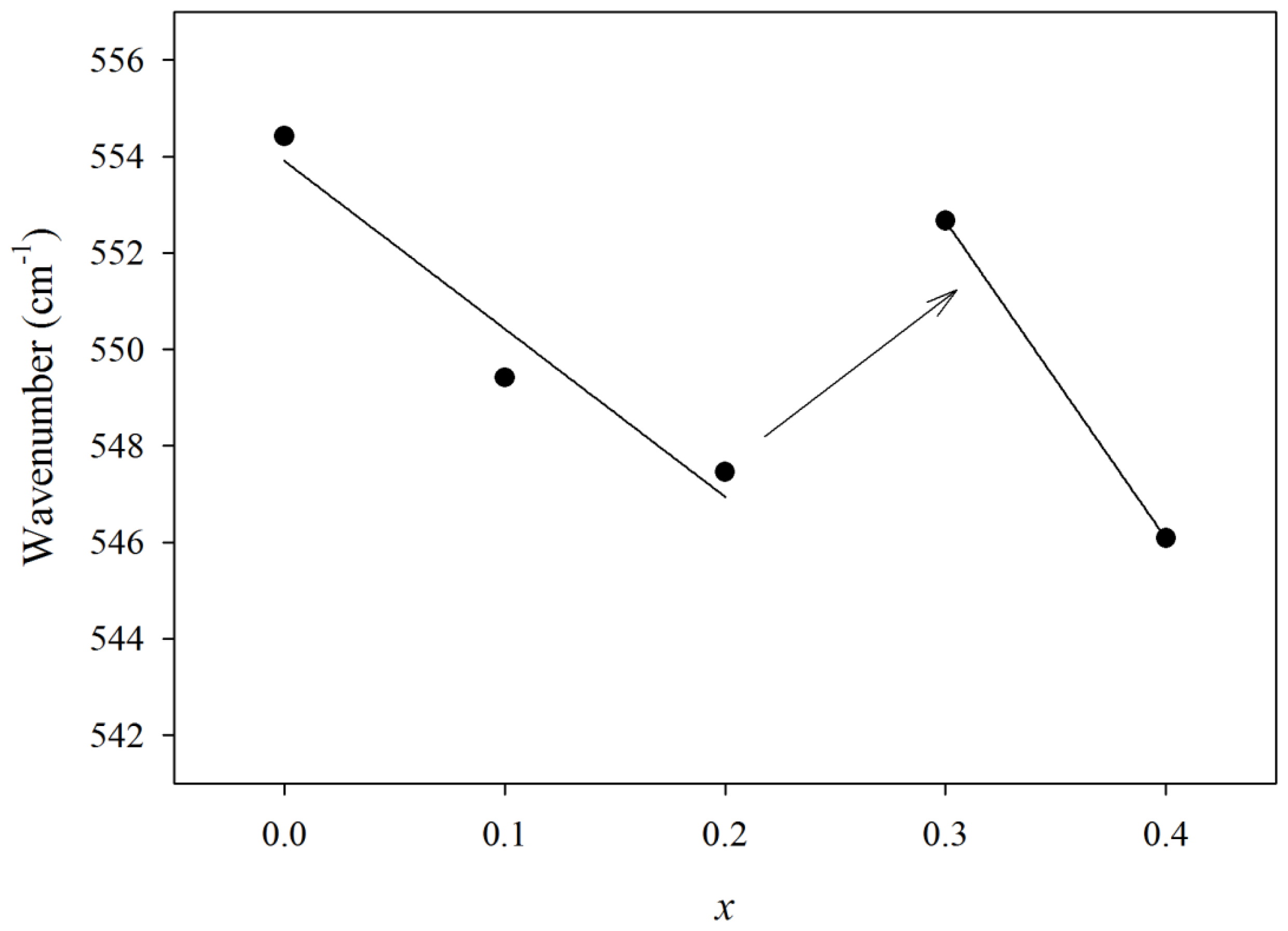
FTIR

Figure 1 shows FTIR absorption spectra of GeO2-PbO-Bi2O3 glass where two envelops at around 500–600 and 700–800 cm−1 wavenumbers are present. Table 1 provides the wavenumbers of the deconvoluted bands. Absorption at 550 cm−1 indicates bending and symmetric stretching vibration of the Ge–O–Ge of GeO4 tetrahedral unit [9], Bi–O(−) vibration bond of BiO6 unit [10] and antisymmetric bending vibration of Pb-O-Pb bond [11]. The band at 700–800 cm−1 wavenumbers was deconvoluted to peaks at 715 cm−1 and 785 cm−1. The first peak is related to stretching vibration mode of the Ge–O–Ge bond in GeO6 octahedral units of rutile germanium oxide [12]; the second is related to antisymmetric stretching vibration mode of the Ge–O–Ge bond in GeO4 tetrahedral units [9].
FTIR absorption at 830 cm−1 is characteristic of the pyramidal BiO3 group [13]. Absence of this peak indicates that only BiO6 units participate in the glass structure. The peak at 470 cm−1 is related to symmetric bending vibration of Pb–O in PbO4 tetragonal pyramid (PbO covalent bond) and Bi–O bond bending of strongly distorted BiO6 octahedral units formed when both Pb and Bi act as network former in a glass matrix. Absence of this peak clearly indicates that the modification role of both lead and bismuth is predominant. Pb2+ ions in the germanate-lead oxide system act as a modifier in low PbO content with an increase of non-bridging oxygen, yet, participate in the glass network in high composition as PbO chains [6]. Based on Figure 2, the wavenumber at 550 cm−1 decreases gradually with increasing x. This is attributed to replacement of lead with bismuth ions where Bi3+ is surrounded by more oxygen than lead. Figure 2 shows two linear decreases of wavenumber in both Pb-rich and Bi-rich regions, but a deviation from the linear interpolation is observed when x changes from 0.2 to 0.3. This can be related to the appearance of Bi2+ and/or Bi1+ which have less oxygen in their surroundings in comparison with Bi3+ ions. Bi3+ is shared with three oxygens but Bi2+ and/or Bi1+ ions are surrounded with less oxygens. So Bi–O bonding should be stronger for Bi2+ or Bi+ ions in contrast to weaker bonding for the Bi3+ ion and appears in the deviation shown in Figure 2 to greater wavenumbers for GPB631 and GPB640.
The 700–800 cm−1 band was deconvoluted into two peaks at 715 and 785 cm−1 which are related to GeO6 and GeO4 units, respectively, and show the existence of both four and six coordination. Coordination number (CN), proposed by the Dachille and Roy study [14], relates the coordination number of cation “T” to the wavelength of the strongest infrared peak of the TX stretching bond in TX2 compounds like SiO2, GeO2 and BeF2:
K = C N · μ ( A T + A X ) 1 / 3 Z T Z X λ 2
where AT and AX are the atomic number of atoms T and X, respectively; ZT and ZX are valance of atoms T and X, respectively; μ is reduced mass and K is a constant. With μ in atomic mass units and λ in μm, Dachille and Roy [14] proposed the average value of K = 0.168. In this study, crystalline GeO2 precursor was used to determine the K value, supposing that all germanium atoms are 4-fold coordinated. The FTIR peak in 853 cm−1, substituted in the Dachille and Roy relation, yielded K = 0.163 which is in accordance with previous results [10,11].
Figure 3 shows the average coordination number of Ge atoms in 6-fold coordination that was evaluated with Equation 1. The average coordination number decreases from 5.38 to 5.14 by increasing bismuth content, with a step-up between GPB631 and GPB622 showing the existence of CN values of both six and four, and a gradual change in germanium coordination from six to four with the sudden coordination change in the intermediate region.

3. Experimental Section

A series of GeO2-PbO-Bi2O3 glass samples were prepared by conventional melt-quenching method. High purity (more than 99%) GeO2, PbO, Bi2O3, Er2O3 and Yb2O3 precursors were used to synthesize [GeO2]60-[ PbO](40 − x)-[½Bi2O3]x with x = 0, 10, 20, 30, and 40 mol% and 0.5 and 1.5 wt% of Er2O3 and Yb2O3, respectively. After mixing and grinding the precursors with the above concentrations and drying the mixture at about 300 °C, the mixture was heated at 1100 °C for 1 h. Then, the melt was quenched into a preheated cylindrical metal mold to obtain a transparent glass sample, and annealed at 420 °C. The samples were cut and polished for measurements.
FTIR spectroscopy was used to study the vibrational properties and structure of glass. The results were extracted from the Perkin-Elmer Spectrum-100 spectrometer, with a UATR accessory. Spectra were taken from 400 cm−1 to 1000 cm−1 and deconvoluted into Gaussian component bands.

4. Conclusions

The structure of glass host was studied by peak-deconvolution of FTIR spectra, which showed the existence of ionic Pb–O bonds in PbO4 tetragonal pyramids and Bi–O(−) bonds in distorted BiO6 octahedral groups implying that Bi and Pb behave as modifier in glass. Deconvoluted spectra showed the existence of both four and six germanium coordination (GeO4 and GeO6 units) in all of the samples with variation of coordination number from 5.38 to 5.14 by increasing of bismuth content.

References

  1. Lezal, D. Chalcogenide glasses-survey and progress. J. Optoelectron. Adv. Mater. 2003, 5, 23–34. [Google Scholar]
  2. Li, X.; Nie, Q.; Dai, S.; Xu, T.; Lu, L.; Zhang, X. Energy transfer and frequency upconversion in Ho3+/Yb3+ co-doped bismuth-germanate glasses. J. Alloy Compd 2008, 454, 510–514. [Google Scholar]
  3. Kassab, L.; Hora, W.; Lozano, W.; Oliveira, M.; Maciel, G. Optical properties of Er3+ doped GeO2–PbO glass: Effect of doping with Bi2O3. Opt. Commun 2007, 269, 356–361. [Google Scholar]
  4. Cacho, V.; Kassab, L.; Oliveira, S.; Mansano, R.; Verdonck, P. Blue cooperative luminescence properties in Yb3+ doped GeO2 PbO Bi2O3 vitreous system for the production of thin films. Thin Solid Films 2006, 515, 764–767. [Google Scholar]
  5. Baia, L.; Iliescu, T.; Simon, S.; Kiefer, W. Raman and IR spectroscopic studies of manganese doped GeO2-Bi2O3 glass. J. Mol. Struct 2001, 599, 9–13. [Google Scholar]
  6. Ribeiro, J.L.; Dexpert-Ghy, J.; Piriou, B.; Mastelaro, R. Structural studies in lead germanate glasses: EXAFS and vibrational spectroscopy. J. Non-Cryst. Solids 1993, 159, 213–221. [Google Scholar]
  7. Vasantharani, P.; Shailajha, S. Acoustic properties of borate glasses doped with Ag2O. Glob. J. Mol. Sci 2009, 4, 29–33. [Google Scholar]
  8. Hwa, L.G.; Chao, W.C.; Szu, S.P. Temperature dependence of elastic moduli of lanthanum gallogermanate glasses. J. Mater. Sci 2002, 37, 3423–3427. [Google Scholar] [Green Version]
  9. Kim, Y.; Saienga, J.; Martin, S.W. Glass formation in and structural investigation of Li2S + GeS2 + GeO2 composition using Raman and IR spectroscopy. J. Non-Cryst. Solids 2005, 351, 3716–3724. [Google Scholar]
  10. Saddeek, Y.B.; Abousehly, A.M.; Hussien, S.I. Synthesis and several features of the Na2O-B2O3-Bi2O3-MoO3 glasses. J. Phys. D Appl. Phys 2007, 40, 4674–4681. [Google Scholar]
  11. Rath, S.; Kabiraj, D.; Avasthi, D.K.; Tripathi, A.; Jain, K.P.; Kumar, M.; Mavi, H.S.; Shukla, A.K. Evidence of nanostructure formation in Ge oxide by crystallization induced by swift heavy ion irradiation. Nucl. Instrum. Meth. Phys. Res. B 2007, 263, 419–423. [Google Scholar]
  12. Pascuta, P.; Culea, E. FTIR spectroscopic study of some bismuth germanate glasses containing gadolinium ions. Mater. Lett 2008, 62, 4127–4129. [Google Scholar]
  13. Baia, L.; Stefan, R.; Popp, J.; Simon, S.; Kiefer, W. Vibrational investigations of highly iron doped B2O3–Bi2O3 glass systems. J. Non-Cryst. Solids 2003, 324, 109–117. [Google Scholar]
  14. Dachille, F.; Roy, R. The use of infra-red absorption and molar refractivities to check coordination. Z. Kristallogr 1959, 111, 462–470. [Google Scholar]
Figure 1. Fourier transform infrared (FTIR) spectrum of (GeO2)0.6(PbO)0.4 − x(1/2Bi2O3)x glass of different compositions.
Ijms 13 08609f1
Figure 2. Dependence of FTIR wavenumber, located at about 550 cm−1, to bismuth composition of (GeO2)0.6(PbO)0.4 − x(1/2Bi2O3)x glass. Bi-rich samples deviate to higher values.
Ijms 13 08609f2
Figure 3. Dependence of coordination number of germanium to bismuth composition of (GeO2)0.6(PbO)0.4 − x(1/2Bi2O3)x glass.
Ijms 13 08609f3
Table 1. Deconvoluted wavenumbers and coordination number (CN) extracted from the Fourier transform infrared (FTIR) spectrum of (GeO2)0.6(PbO)0.4 − x(1/2Bi2O3)x glass.
xComposition (mol%)Composition (wt%)Sample Codef1 (cm−1)f2 (cm−1)f3 (cm−1)Coordination number


GeO2PbOBi2O3Er2O3Yb2O3
0604000.51.5GPB6405547147855.38
0.16030100.51.5GPB6315497207915.25
0.26020200.51.5GPB6225477147855.28
0.36010300.51.5GPB6135537177885.21
0.4600400.51.5GPB6045467197905.14

