Article

Studies on the Interactions of Copper and Zinc Ions with β-Amyloid Peptides by a Surface Plasmon Resonance Biosensor

1
College of Chemistry and Chemical Engineering, Graduate University, Chinese Academy of Sciences, Beijing, 100049, China
2
Institute of Materia Medica, Chinese Academy of Medical Sciences and Peking Union Medical College, Beijing 100050, China
*
Authors to whom correspondence should be addressed.
Int. J. Mol. Sci. 2012, 13(9), 11832-11843; https://doi.org/10.3390/ijms130911832
Submission received: 18 July 2012 / Revised: 7 September 2012 / Accepted: 7 September 2012 / Published: 19 September 2012
(This article belongs to the Section Molecular Recognition)

Abstract

:
The aggregation of β-amyloid peptide (Aβ) into fibrils plays an important role in the pathogenesis of Alzheimer’s disease (AD). Metal ions including copper and zinc are closely connected to the precipitation and toxicity of Aβ. In this study, a surface plasmon resonance (SPR) biosensor was constructed to investigate the interactions between Aβ and metal ions. Aβ peptide was immobilized on the SPR chip surface through a preformed alkanethiol self-assembled monolayer (SAM). Our observations indicate that the immobilized Aβ undergoes a conformational change upon exposure to the metal ions. A difference in metal binding affinity between Aβ1–28 and Aβ1–42 was also detected. The results suggest that SPR is an effective method to characterize the interactions between Aβ and metal ions.

1. Introduction

Alzheimer’s disease (AD), which is characterized by irreversible and progressive neurodegeneration, is the dominant cause of dementia. The morphological hallmarks of AD are extracellular senile plaques and intracellular neurofibrillary tangles. The aggregated β-amyloid peptide (Aβ) containing 39–43 amino acid residues, which is generated from the amyloid precursor protein (APP) through the sequential cleavage by two enzymes, β-secretase and γ-secretase, is the principal component of the senile plaques. According to the amyloid cascade hypothesis, the aggregation of Aβ in AD leads to the formation of neurotoxic oligomers that are purportedly responsible for neuronal dysfunction and cell death [1]. Therefore, conditions that influence aggregation and the formation of oligomers are of great interest.
The aggregation of Aβ peptide is primarily affected by pH [2,3], peptide concentration [2], incubation time, membrane lipids [4] and temperature [5]. Moreover, a large body of evidence suggests that metal ions such as copper, zinc and iron may induce the aggregation of Aβ peptides, and these ligands may act as seeding factors in the formation of amyloid plaques [610]. In fact, elevated levels of zinc and copper have been found in amyloid plaques at concentrations reaching 1 mM and 400 μM, respectively [11]. These metal ions bind at the N-terminus (amino acids 1–16) and influence aggregation behavior. However, experimental evidence has shown that these metal ions in complex with Aβ peptides may have opposite functions with Zn accelerating aggregation, while Cu can reduce or accelerate aggregation [1214]. Furthermore, it has been proposed that copper and iron mediate the production of reactive oxygen species (ROS) and oxidative stress [15,16]. It has been reported that reactive oxygen species (ROS) such as H2O2 are produced during the association of Aβ with Cu2+ through the reduction of Cu2+ to Cu+, which mediates cell toxicity [17]. Therefore, intensive efforts have been made to study the primary interaction of Aβ peptide with metal ions.
Surface Plasmon resonance (SPR) spectroscopy is capable of detecting a mass or conformational change above a metal surface. At a specific angle, the absorption of incident light by a thin metal film causes a collective oscillation of electrons in the film that launches an evanescent wave into the dielectric layer adjacent to the metal film. The propagation of the evanescent wave decays exponentially away from the metal film and is thus significantly perturbed by the adsorption of a species on the metal film or changes in the adlayer structure. Due to the advantages of real-time, label-free and direct detection of molecules in various media, SPR has become a popular and powerful technique to study molecular interactions. In recent years, SPR has been successfully employed to study biomolecular interactions related to AD [18,19]. Various aspects of Aβ oligomerization, fibril formation and extension and Aβ interactions with biomolecules have been investigated by SPR [20,21]. A custom-built flow-injection (FI) SPR instrument equipped with a bicell detector was constructed in our lab and used to investigate the interactions between Aβ1–16 and metal ions [22]. Immobilization of monomeric Aβ was performed using a self-assembled monolayer (SAM) of 11-mercaptoundecanoic acid (MUA) and amino coupling chemistry. To further test the feasibility of this biosensor, the interactions between metal ions and Aβ1–28 or the actual senile plaque component, Aβ1–42, were investigated.

2. Results and Discussion

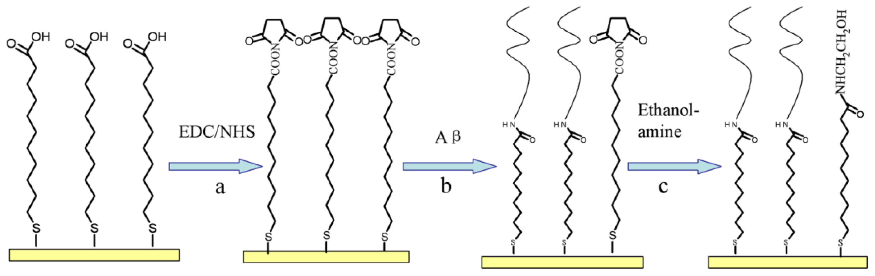
2.1. Aβ Immobilization

The successful immobilization of the Aβ peptide on the sensor chip surface was tested by injecting a Zn2+ ion solution into the flow chamber and pumping it across the Aβ-immobilized sensing surface. In our experiment, the sample loop volume is 20 μL, the flowing rate is 10 μL/min, so that the Zn2+ ions start to flow onto the immobilized Aβ1–28 at about 80 s, the flowing of metal ions ends at about 200 s; the difference in the baseline SPR angles before and after the Zn2+ ion solution injection (termed as the SPR dip shift Δθ) is approximately 0.00246° (Figure 1a), considering the sophisticated interaction between Aβ peptide and metal ions, the detection of the SPR dip shift Δθ is selected at 550 s.
To confirm that the net change is induced by the metal ions, we conducted a control experiment in which Zn2+ ions flowed over an ethanolamine-blocked surface without the immobilization of Aβ peptide. Figure 1b shows that the SPR signal returned to its baseline within a short time. Additionally, the recovery of the original baseline in curve b suggests that the blocking process is efficient because it has been reported that alkanethiol SAM containing carboxylic acid can be used for SPR measurements of heavy metal ions [23]. Furthermore, the conversion of the negatively charged carboxyl groups on the MUA SAM to neutral amide groups during the peptide immobilization and blocking process could reduce the undesirable electrostatic attraction to metal ions in solution.

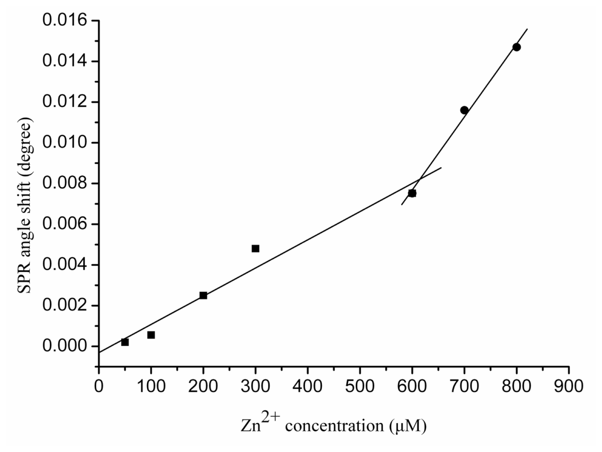
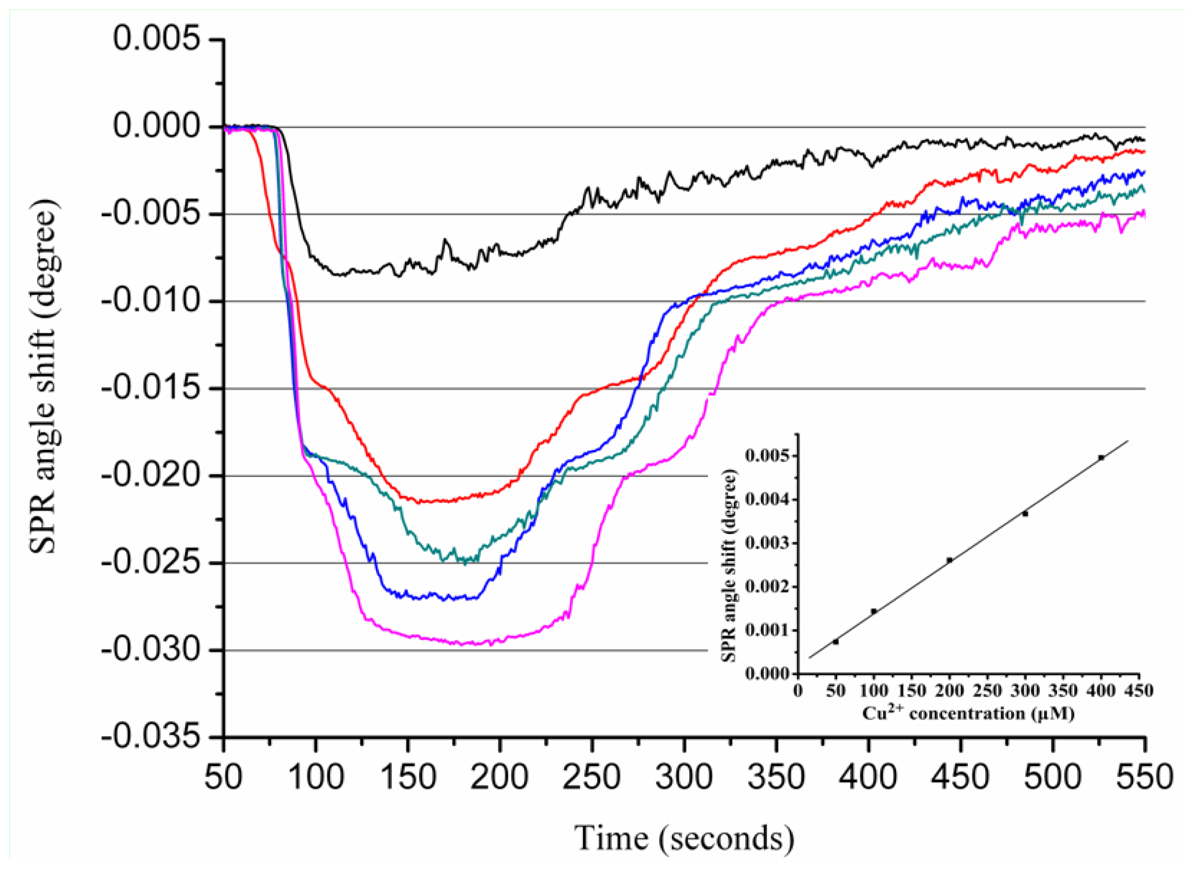
2.2. Real-Time Determination of Aβ1–28 Binding Zn2+ and Cu2+ by SPR

Figure 2 shows the sensorgrams of various Zn2+ concentrations flowing over the Aβ1–28 sensor chip. In these experiments, EDTA (10 mM) was used to regenerate the surface. Therefore, a single chip can be used repeatedly for multiple samples. As shown in Figure 2, increasing Zn2+ concentrations results in a greater SPR dip shift. When 600 μM Zn2+ flowed over the Aβ1–28-covered SPR sensor, the metal ions caused a net change of 0.0075°, which can be attributed to the binding of Zn2+ ions to Aβ1–28 resulting in conformational changes of the peptide molecules.
The SPR angle shift is directly correlated with the Zn2+ concentration ([Zn2+]). In Figure 3, the calibration curve generated from the sensor response to a series of different Zn2+ concentrations is shown. The calibration curve contains two regions; the first is from 50 to 300 μM, and the second from 600 to 800 μM. Linear regression analysis of both regions yielded the following equations:
Δ θ = - 3.07 × 10 - 4 + 1.39 × 10 - 5 [ Zn 2 + ] R 2 = 0.95 ( region  1 )
Δ θ = - 1.39 × 10 - 2 + 3.59 × 10 - 5 [ Zn 2 + ] R 2 = 0.99 ( region  2 )
The slope of the linear regression in the second region is higher than that in the first region, which reveals that the SPR angle shift elicited by Zn2+ ions is greater with increasing Zn2+ ion concentration. These results suggest that the Zn2+-induced Aβ1-28 conformation change is concentration dependent. This finding is in agreement with a previous report indicating that Aβ undergoes a conformational change from a random coil to a regular secondary structure in the presence of Zn2+ ions and forms stable 1:1 and 1:2 (peptide/zinc) complexes [8]. A possible explanation for this phenomenon is that the Aβ1–28 binds Zn2+ intramolecularly at low concentrations. When the Zn2+ ion concentration is sufficiently high, the excess Zn2+ ions may begin to bind with Aβ1–28 intermolecularly, and the Zn2+ ions behave like a bridge connecting two adjacent Aβ1–28 molecules. Our finding is supported by previous studies that have shown that Zn2+ can coordinate to Aβ in an intra- and inter-peptide mode [2426].
A similar trend was not found for the interactions between Cu2+ and Aβ1–28. The calibration curve depicted in Figure 4 only contains one linear region ranging from 100 to 600 μM. The linear regression equation with a correlation coefficient of R2 = 0.99 suggests a linear relationship between the SPR angle shift and the Cu2+ concentration.
Δ θ = - 4.67 × 10 - 5 + 1.49 × 10 - 6 [ Cu 2 + ] R 2 = 0.99
Compared with Zn2+-Aβ complexes having stoichiometry ranging from 1:1 to 3:1 [2729], most studies have demonstrated that the Aβ-peptide forms a 1:1 complex with Cu2+ [25,30,31]. NMR studies have demonstrated that Aβ1–28 forms a 1:1 complex with the Cu2+ ion via histidine residues [30]. We interpret these results to different coordination modes. Unlike Zn2+, which can bind to Aβ in an intra- and inter-peptide coordination mode, Cu2+ is primarily involved in intra-peptide binding in a fairly closed structure that protects the metal from further interactions. In addition, it has been reported that under physiological conditions, the coordination of the Cu2+ equivalent to the Aβ peptides leads to a mononuclear complex Cu1(Aβ)1 and is unlikely to form a Cu2(Aβ)1 complex [32].

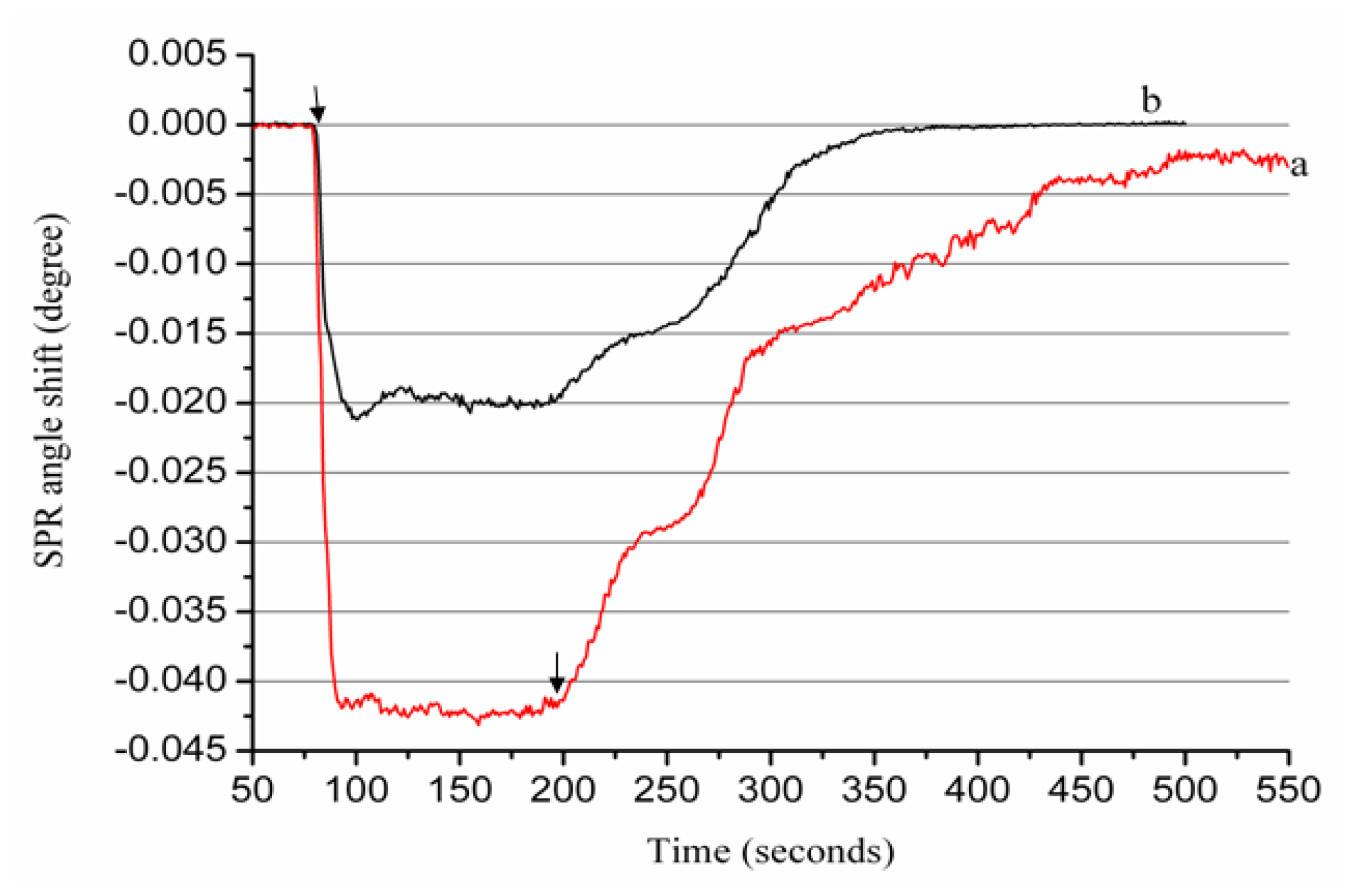
2.3. Real-Time Determination of Aβ1–42 Binding to Zn2+ and Cu2+ by SPR

1–40 and Aβ1–42 are the most prevalent in vivo Aβ forms. In particular, Aβ1–42 has a high propensity to self-assemble and deposit in senile plaques and is highly toxic to neurons [33]. While great progress has been achieved on the coordination chemistry of Zn2+ and Cu2+ with truncated Aβ1–16 and Aβ1–28, it is important to understand whether these peptides, which are missing a large part of the hydrophobic C-terminal residues, coordinate metal ions differently than the full-length peptides. Therefore, to better understand the mechanisms of AD, we continued to study the interactions of Zn2+ and Cu2+ with Aβ1–42 using the FI-SPR biosensor.
Figure 5 depicts SPR sensorgrams obtained from the sensor response to various Zn2+ concentrations. In contrast to the calibration curve of the Zn2+ interactions with Aβ1–28 that contains two regions, the calibration plot of the Zn interactions with Aβ1–42 only contains one region; the linear regression equation is as follows:
Δ θ = - 1.86 × 10 - 3 + 2.27 × 10 - 5 [ Zn 2 + ] R 2 = 0.96
1–42 peptides have 14 additional residues in the hydrophobic tail compared to Aβ1–28, which makes Aβ1–42 more prone to aggregation than the truncated Aβ1–28. A possible explanation for the decreased stoichiometric binding of Aβ1–42 compared to Aβ1–28 may be the interference of the hydrophobic tail in Aβ1–42 with the Zn2+ binding site. Our data are in agreement with a previous report indicating that Zn and Cu form a monomeric complex with Aβ1–42 [34].
Similar results were obtained for the Cu-Aβ1–42 interaction. As shown in Figure 6, the SPR angle shift increased with an increase of Cu2+ bound to the Aβ1–42-immobilized sensor chip. The calibration curve generated from the sensor exposed to different concentrations of Cu2+ is shown in the Figure 6 inset. A strong correlation coefficient (R2 = 0.99) was obtained for the linear regression equation calculated using Cu2+ concentrations ranging from 50 to 400 μM.

3. Experimental Section

3.1. Chemicals

Human amyloid-β peptide (1–28) (Aβ1–28) and human amyloid-β peptide (1–42) (Aβ1–42) were obtained from GL Biochem Ltd. (Shanghai, China). 1-(3-Dimethylaminopropyl)-3-ethylcarbodiimide hydrochloride (EDC), N-hydroxysulfosuccinimide (NHS), 11-mercaptoundecanoic acid (MUA), ARC-grade dimethyl sulfoxide (DMSO, 99%) and ethanolamine were purchased from J & K Chemical Ltd. K2HPO4·3H2O, KH2PO4, NaCl, ZnCl2 and CuCl2 were all of AR grade and purchased from Beijing Chemical Reagent Co. (Beijing, China). Water with a resistivity of 18.25 MΩ·cm−1 was collected from a Millipore Simplicity 185 system. EDC (0.4 M) and NHS (0.1 M) were prepared in a pH 7.4 PBS buffer (10.0 mM K2HPO4·3H2O and KH2PO4 prepared in 1 mM NaCl). MUA (4 mM) was prepared in pure ethanol. Stock solutions of 2.0 mM ZnCl2 and CuCl2 were prepared in water and then diluted to the desired concentrations.

3.2. Preparation of Fresh Aβ Solution

Uniform and nonaggregated monomeric Aβ1–28 and Aβ1–42 peptides were prepared as previously described [35]. Briefly, 0.1 mM of the peptide was dissolved in DMSO, which disrupts the β-sheet structure and renders Aβ monomeric [36]. The freshly dissolved monomeric Aβ was diluted with PBS buffer (pH 7.4) to a concentration of 10 μM.

3.3. Gold Film Preparation

BK7 glass cover slides (Fisher) were cleaned with a piranha solution of 70% concentrated H2SO4 and 30% H2O2 (7:3, v/v) at 80 °C for 30 min. Upon cooling to room temperature, the glass slides were rinsed thoroughly with deionized water. After drying with N2, each glass slide was coated with a 2 nm chromium layer and then covered with 50 nm of gold film using a sputter coater (Model 108, Kert J. Lester Inc., Clariton, PA, USA).

3.4. Aβ Peptide Immobilization

Aβ peptide immobilization was achieved using covalent bonding mediated by a chemical reaction. The N-terminus of the Aβ peptide was reacted with the functional group of the SAM of MUA on the surface of the Au film (as depicted in Figure 7). Briefly, the MUA gold chip was treated with a mixture of 0.4 M EDC and 0.1 M NHS (1:1) for 3 h to ensure that the carboxyl group of the SAM reacted fully with the EDC and NHS. Then, a freshly prepared 10 μM Aβ solution in PBS (pH 7.4) was reacted with the NHS-activated surface for 2 h. Finally, ethanolamine (1 M, pH 8.5) was used to block the remaining activated surface groups. The resulting film was either used immediately or stored at 4 °C for future use.

3.5. SPR Apparatus

SPR measurements were conducted with a custom-built flow injection-SPR equipped with a bicell detector as previously described [22]. The SPR instrument recorded the reflected light on two photodetectors (A and B). The differential (A − B) and sum (A + B) signals were then detected by a PCI-1371 interface card (Advantech, Taiwan) controlled by the Labview program. The resonance angles from the biosensor were measured by the division of the differential and sum signals, (A − B)/(A + B). The inlet of the flow cell was connected to a six-port valve. For each measurement, the sample solution was injected into a 20-μL loop with a microsyringe (Hamilton) and subsequently delivered to the flow cell at a flow rate of 10 μL/min, using a syringe pump (KDS100, KD Scientific Inc., Holliston, MA, USA).

4. Conclusions

In the present study, a SPR-based analytical method was used to investigate the interactions between Aβ peptides and metal ions. The coordination of metal ions with the truncated Aβ1–28 and the full-length Aβ1–42 were compared. The conformational transition of Aβ induced by metal ion binding can be readily detected by this highly sensitive FI-SPR sensor equipped with a bicell detector. At physiological pH, Zn2+ demonstrates different binding affinity for Aβ1–28 and Aβ1–42, while Cu2+ exhibits similar interactions with both Aβ peptides. The interactions between Zn2+ and the Aβ-peptides in an intra- and inter-molecular mode have been previously validated. These studies complement other analytical methods and should help elucidate the role of metal ions during Aβ aggregation.

Acknowledgments

This work was supported by the National Natural Science Foundation of China (21145006), Scientific Research Foundation for the Returned Overseas Chinese Scholars, State Education Ministry and Beijing Municipal Natural Science Foundation (2113046).

References

  1. Roychaudhuri, R.; Yang, M.; Hoshi, M.M.; Teplow, D.B. Amyloid β-protein assembly and Alzheimer disease. J. Biol. Chem 2009, 284, 4749–4753. [Google Scholar]
  2. Burdick, D.; Soreghan, B.; Kwon, M.; Kosmoski, J.; Knauer, M.; Henschen, A.; Yates, J.; Cotman, C.; Glabe, C. Assembly and aggregation properties of synthetic Alzheimer’s A4/β amyloid peptide analogs. J. Biol. Chem 1992, 267, 546–554. [Google Scholar]
  3. Fraser, P.E.; Nguyen, J.T.; Surewicz, W.K.; Kirschner, D.A. pH-dependent structural transitions of Alzheimer amyloid peptides. Biophys. J 1991, 60, 1190–1201. [Google Scholar]
  4. McLaurin, J.; Franklin, T.; Chakrabartty, A.; Fraser, P.E. Phosphatidylinositol and inositol involvement in Alzheimer amyloid-β fibril growth and arrest. J. Mol. Biol 1998, 278, 183–194. [Google Scholar]
  5. Yamaguchi, T.; Matsuzaki, K.; Hoshino, M. Transient formation of intermediate conformational states of amyloid-β peptide revealed by heteronuclear magnetic resonance spectroscopy. FEBS Lett 2011, 585, 1097–1102. [Google Scholar] [Green Version]
  6. Bush, A.I.; Pettingell, W.H.; Multhaup, G.; d Paradis, M.; Vonsattel, J.P.; Gusella, J.F.; Beyreuther, K.; Masters, C.L.; Tanzi, R.E. Rapid induction of Alzheimer Aβ amyloid formation by zinc. Science 1994, 265, 1464–1467. [Google Scholar]
  7. Miura, T.; Suzuki, K.; Kohata, N.; Takeuchi, H. Metal binding modes of Alzheimer’s amyloid β-peptide in insoluble aggregates and soluble complexes. Biochemistry 2000, 39, 7024–7031. [Google Scholar]
  8. Kozin, S.A.; Zirah, S.; Rebuffat, S.; Hoa, G.H.; Debey, P. Zinc binding to Alzheimer’s Aβ(1–16) peptide results in stable soluble complex. Biochem. Biophys. Res. Commun 2001, 285, 959–964. [Google Scholar]
  9. Chen, T.; Wang, X.; He, Y.; Zhang, C.; Wu, Z.; Liao, K.; Wang, J.; Guo, Z. Effects of cyclen and cyclam on zinc(II)- and copper(II)-induced amyloid β-peptide aggregation and neurotoxicity. Inorg. Chem 2009, 48, 5801–5809. [Google Scholar]
  10. Sarell, C.J.; Syme, C.D.; Rigby, S.E.; Viles, J.H. Copper(II) binding to amyloid-beta fibrils of Alzheimer’s disease reveals a picomolar affinity: Stoichiometry and coordination geometry are independent of Aβ oligomeric form. Biochemistry 2009, 48, 4388–4402. [Google Scholar]
  11. Lovell, M.A.; Robertson, J.D.; Teesdale, W.J.; Campbell, J.L.; Markesbery, W.R. Copper, iron and zinc in Alzheimer’s disease senile plaques. J. Neurol. Sci 1998, 158, 47–52. [Google Scholar]
  12. Atwood, C.S.; Moir, R.D.; Huang, X.; Scarpa, R.C.; Bacarra, N.M.; Romano, D.M.; Hartshorn, M.A.; Tanzi, R.E.; Bush, A.I. Dramatic aggregation of Alzheimer Aβ by Cu(II) is induced by conditions representing physiological acidosis. J. Biol. Chem 1998, 273, 12817–12826. [Google Scholar]
  13. Zou, J.; Kajita, K.; Sugimoto, N. Cu2+ inhibits the aggregation of amyloid β-peptide(1–42) in vitro. Angew. Chem. Int. Ed 2001, 40, 2274–2277. [Google Scholar]
  14. Raman, B.; Ban, T.; Yamaguchi, K.; Sakai, M.; Kawai, T.; Naiki, H.; Goto, Y. Metal ion-dependent effects of clioquinol on the fibril growth of an amyloid β peptide. J. Biol. Chem 2005, 280, 16157–16162. [Google Scholar]
  15. Huang, X.; Atwood, C.S.; Hartshorn, M.A.; Multhaup, G.; Goldstein, L.E.; Scarpa, R.C.; Cuajungco, M.P.; Gray, D.N.; Lim, J.; Moir, R.D.; et al. The Aβ peptide of Alzheimer’s disease directly produces hydrogen peroxide through metal ion reduction. Biochemistry 1999, 38, 7609–7616. [Google Scholar]
  16. Kowalik-Jankowska, T.; Ruta, M.; Wisniewska, K.; Lankiewicz, L.; Dyba, M. Products of Cu(II)-catalyzed oxidation in the presence of hydrogen peroxide of the 1–10, 1–16 fragments of human and mouse β-amyloid peptide. J. Inorg. Biochem 2004, 98, 940–950. [Google Scholar]
  17. Ali, F.E.; Separovic, F.; Barrow, C.J.; Cherny, R.A.; Fraser, F.; Bush, A.I.; Masters, C.L.; Barnham, K.J. Methionine regulates copper/hydrogen peroxide oxidation products of Aβ. J. Pept. Sci 2005, 11, 353–360. [Google Scholar]
  18. Ryu, J.; Joung, H.A.; Kim, M.G.; Park, C.B. Surface plasmon resonance analysis of Alzheimer’s β-amyloid aggregation on a solid surface: from monomers to fully-grown fibrils. Anal. Chem 2008, 80, 2400–2407. [Google Scholar]
  19. Xia, N.; Liu, L.; Harrington, M.G.; Wang, J.; Zhou, F. Regenerable and simultaneous surface plasmon resonance detection of Aβ(1–40) and Aβ(1–42) peptides in cerebrospinal fluids with signal amplification by streptavidin conjugated to an N-terminus-specific antibody. Anal. Chem 2010, 82, 10151–10157. [Google Scholar]
  20. Aguilar, M.I.; Small, D.H. Surface plasmon resonance for the analysis of β-amyloid interactions and fibril formation in Alzheimer’s disease research. Neurotox. Res 2005, 7, 17–27. [Google Scholar]
  21. Krazinski, B.E.; Radecki, J.; Radecka, H. Surface plasmon resonance based biosensors for exploring the influence of alkaloids on aggregation of amyloid-β peptide. Sensors 2011, 11, 4030–4042. [Google Scholar]
  22. Yao, F.; He, J.; Li, X.; Zou, H.; Yuan, Z. Studies of interaction of copper and zinc ions with Alzheimer’s Aβ(1–16) using surface plasmon resonance spectrometer. Sens. Actuators B 2012, 161, 886–891. [Google Scholar]
  23. Saber, R.; Piskin, E. Investigation of complexation of immobilized metallothionein with Zn(II) and Cd(II) ions using piezoelectric crystals. Biosens. Bioelectron 2003, 18, 1039–1046. [Google Scholar]
  24. Stellato, F.; Menestrina, G.; Serra, M.D.; Potrich, C.; Tomazzolli, R.; Meyer-Klaucke, W.; Morante, S. Metal binding in amyloid β-peptides shows intra- and inter-peptide coordination modes. Eur. Biophys. J 2006, 35, 340–351. [Google Scholar]
  25. Syme, C.D.; Viles, J.H. Solution 1 H NMR investigation of Zn2+ and Cd2+ binding to amyloid-β peptide (Aβ) of Alzheimer’s disease. Biochim. Biophys. Acta 2006, 1764, 246–256. [Google Scholar]
  26. Miller, Y.; Ma, B.; Nussinov, R. Zinc ions promote Alzheimer Aβ aggregation via population shift of polymorphic states. Proc. Natl. Acad. Sci. USA 2010, 107, 9490–9495. [Google Scholar]
  27. Clements, A.; Allsop, D.; Walsh, D.M.; Williams, C.H. Aggregation and metal-binding properties of mutant forms of the amyloid Aβ peptide of Alzheimer’s disease. J. Neurochem 1996, 66, 740–747. [Google Scholar]
  28. Atwood, C.S.; Scarpa, R.C.; Huang, X.; Moir, R.D.; Jones, W.D.; Fairlie, D.P.; Tanzi, R.E.; Bush, A.I. Characterization of copper interactions with alzheimer amyloid β peptides: Identification of an attomolar-affinity copper binding site on amyloid β1-42. J. Neurochem 2000, 75, 1219–1233. [Google Scholar]
  29. Damante, C.A.; Osz, K.; Nagy, Z.; Pappalardo, G.; Grasso, G.; Impellizzeri, G.; Rizzarelli, E.; Sovago, I. Metal loading capacity of Aβ N-terminus: A combined potentiometric and spectroscopic study of zinc(II) complexes with Aβ(1–16), its short or mutated peptide fragments and its polyethylene glycol-ylated analogue. Inorg. Chem 2009, 48, 10405–10415. [Google Scholar]
  30. Karr, J.W.; Akintoye, H.; Kaupp, L.J.; Szalai, V.A. N-Terminal deletions modify the Cu2+ binding site in amyloid-β. Biochemistry 2005, 44, 5478–5487. [Google Scholar]
  31. Tõugu, V.; Karafin, A.; Palumaa, P. Binding of zinc(II) and copper(II) to the full-length Alzheimer’s amyloid-β peptide. J. Neurochem 2008, 104, 1249–1259. [Google Scholar]
  32. Faller, P.; Hureau, C. Bioinorganic chemistry of copper and zinc ions coordinated to amyloid-β peptide. Dalton Trans 2009, 7, 1080–1094. [Google Scholar]
  33. Findeis, M.A. The role of amyloid β peptide 42 in Alzheimer’s disease. Pharmacol. Ther 2007, 116, 266–286. [Google Scholar]
  34. Walsh, D.M.; Lomakin, A.; Benedek, G.B.; Condron, M.M.; Teplow, D.B. Amyloid β-protein fibrillogenesis. Detection of a protofibrillar intermediate. J. Biol. Chem 1997, 272, 22364–22372. [Google Scholar]
  35. Hu, W.P.; Chang, G.L.; Chen, S.J.; Kuo, Y.M. Kinetic analysis of β-amyloid peptide aggregation induced by metal ions based on surface plasmon resonance biosensing. J. Neurosci. Meth 2006, 154, 190–197. [Google Scholar]
  36. Kremer, J.J.; Pallitto, M.M.; Sklansky, D.J.; Murphy, R.M. Correlation of β-amyloid aggregate size and hydrophobicity with decreased bilayer fluidity of model membranes. Biochemistry 2000, 39, 10309–10318. [Google Scholar]
Figure 1. The sensorgram of a 200 μM Zn2+ ion solution flowing over the surface plasmon resonance (SPR) sensor chip with (a) and without (b) immobilized β-amyloid peptides (Aβ1–28) peptides. Arrows indicate Zn2+ ions starting to flow onto immobilized Aβ1-28 and when the flowing of metal ions ends, respectively.
Ijms 13 11832f1
Figure 2. The sensorgrams for the interactions between Zn2+ ions and the Aβ1–28 peptides. Various concentrations (50, 100, 200, 300, 600, 700 and 800 μM, from top to bottom) of Zn2+ ions were injected onto the Aβ1–28 sensorchip. Arrows indicate Zn2+ ions starting to flow onto immobilized Aβ1–28 and when the flowing of metal ions ends, respectively.
Ijms 13 11832f2
Figure 3. Calibration curves for the interactions between various Zn2+ concentrations with Aβ1–28. Each value represents the mean ± standard deviation of three separate injections.
Ijms 13 11832f3
Figure 4. The calibration curve for the interactions between various Cu2+ concentrations with Aβ1–28. Each value represents the mean ± standard deviation of three separate injections.
Ijms 13 11832f4
Figure 5. The sensorgram for the interactions between Zn2+ ions and Aβ1–42. Various concentrations (50, 100, 300, 400, 500 and 800 μM, from top to bottom) of Zn2+ ions were injected onto the Aβ1–42 sensorchip. The inset shows the calibration curve for the interactions between various concentrations of Zn2+ with Aβ1–42. Each value represents the mean ± standard deviation of three separate injections. Arrows indicate Zn2+ ions starting to flow onto immobilized Aβ1–42 and when the flow of metal ions ends, respectively.
Ijms 13 11832f5
Figure 6. The sensorgram for the interactions between Cu2+ ions and Aβ1–42. Various concentrations (50, 100, 200, 300, and 400 μM, from top to bottom) of Cu2+ ions were injected onto the Aβ1–42 sensorchip. The inset shows the calibration curve for the interactions between various concentrations of Cu2+ with Aβ1–42. Each value represents the mean ± standard deviation of three separate injections. Arrows indicate Cu2+ ions starting to flow onto immobilized Aβ1–28 and when the flow of metal ions ends, respectively.
Ijms 13 11832f6
Figure 7. The procedure for the immobilization of Aβ. (a) The self-assembled monolayer (SAM) of MUA surface was activated with standard amine coupling chemistry using EDC/NHS; (b) The activated surface was covered with a fresh Aβ solution to form a bond between the amine group on the peptide and the carboxylic group on the MUA surface; (c) The remaining activated surface groups were blocked using ethanolamine.
Ijms 13 11832f7

