Article

Biofunctional Constituents from Liriodendron tulipifera with Antioxidants and Anti-Melanogenic Properties

1
Bachelor Degree Program of Health Beauty, School of Medical and Health Sciences, Fooyin University, Ta-Liao District, Kaohsiung 83102, Taiwan
2
Department of Fragrance and Cosmetic Science, Kaohsiung Medical University, Kaohsiung 807, Taiwan
3
Department of Marine Biotechnology and Resources, Asia-Pacific Ocean Research Center, National Sun Yat-sen University, Kaohsiung 80424, Taiwan
4
Department of Respiratory Therapy, College of Medicine, Kaohsiung Medical University, 100, Shih-Chuan 1st Road, San-Ming District, Kaohsiung 807, Taiwan
*
Authors to whom correspondence should be addressed.
These authors contributed equally to this work.
Int. J. Mol. Sci. 2013, 14(1), 1698-1712; https://doi.org/10.3390/ijms14011698
Submission received: 25 October 2012 / Revised: 16 December 2012 / Accepted: 4 January 2013 / Published: 15 January 2013
(This article belongs to the Section Biochemistry)

Abstract

:
From the stems of Liriodendron tulipifera, seventeen known compounds have been extracted, isolated and purified. By using spectroscopic analysis, the structures of these pure constituents were determined as three lignans, four steroids and ten benzenoids. Identified compounds were screened for antioxidant abilities using: 1,1-diphenyl-2-picrylhydrazul (DPPH) and 2,2′-azino-bis(3-ethylbenzothiazoline-6-sulfonic acid) (ABTS) scavenging free radical activity assays; metal chelating power test; and ferric reducing/antioxidant power (FRAP) examination. The result revealed that seventeen compounds had potential anti-oxidative capabilities. In addition, the anti-tyrosinase effect was determined by calculating the hydroxylation of L-tyrosine to L-dopa and the oxidization of L-dopa to dopaquinone, according to in vitro mushroom tyrosinase evaluation platform. Furthermore, based on assays on B16F10 cell line, our data suggest that five compounds isolated from L. tulipifera would be able to inhibit tyrosinase activity and reduce the melanin content in animal cells. Therefore, some of the examined compounds could be potentially used in the cosmetic skin whitening business, therapeutic applications or the food industry.

1. Introduction

Liriodendron tulipifera is a fast growing hardwood native plant in the United States, usually used for pulp and wood in furniture and paper-making proposes [1,2]. In earlier investigations of L. tulipifera chemical constituents, some sesquiterpenes and aporphine alkaloids were discussed [3]. Previous studies found that some compounds isolated from L. tulipifera showed anti-migration abilities and inhibited the proliferation of cancer cells. DPPH, chelating and reducing power assay also revealed its antioxidant effects. Thus, we assume the compounds isolated from L. tulipifera have promising multi-biofunctions. For this reason, L. tulipifera was chosen for further phytochemical investigations, and these compounds were isolated from the stems. The MeOH extracts of this plant were subjected to solvent partitioning and chromatographic separation to generate seventeen pure substances. The chemical constituents in the plants of L. tulipifera were separated by using column chromatography, including three lignans: (−)-eudesmin (1) [4], (+)-syringaresinol (2) and (+)-yangambin (3) [5]; four steroids: β-sitosterol (4), stigmasterol (5) [6], β-sitostenone (6) and stigmastenone (7) [7]; and ten benzenoids: methyl 4-hydroxy-2-methylbenzoate (8) [8], methyl β-orcinol carboxylate (9), methyl haematommate (10) [9], coniferyl aldehyde (11) [10], vanillin (12), vanillic acid (13), methyl vanillate (14), p-hydroxybenzoic acid (15), syringic acid (16) and 2,6-dimethoxy-p-quinone (17) [11].
Free radicals and reactive oxygen species (ROS) were produced from the formation of hydrogen peroxide or superoxide anions. They will attack some important biological molecules, such as DNA, protein or lipids and enhance oxidative stress in human beings [12,13]. Evidence indicates that many degenerative disease conditions, such as cancer, gastric ulcers, Alzheimer’s, arthritis and ischemic reperfusion are caused by free radicals or active oxygen species [14,15]. In recent years, ingesting fresh fruits, vegetables or teas to acquire natural antioxidants have been purported to benefit human health. Dietary strategies are considered important to prevent diseases [16,17]. Natural antioxidant constituents, capable of lowering free radical mediated degradations of cells and tissues in human organism, are very substantial in cosmetics or food businesses [18,19]. Thus, much attention has shifted to effective components present in a natural plant-based diet. According to our previous studies, we found a great amount of various phytochemicals possess antioxidative, skin whitening or chemotherapeutic activities [6,1215,1820].
The color of skin, hair and eyes of mammals are determined by synthesis and distribution of melanin. There are two major types of melanin occuring in mammals: black/brown eumelanins and yellow/red pheomelanins. In melanogenesis, L-tyrosine is hydroxylated to dihydroxyphenylalanine (L-dopa), and L-dopa is oxidized to dopaquinone followed by further conversion to melanin production. Tyrosinase is the first and rate limiting enzyme in melanogenesis, hence, inhibition of tyrosinase is one of the major strategies to treat hyperpigmentation [21]. In clinical usage, whitening agents are used for treating dermatological disorders related to melanin hyper-accumulation, such as melasma, freckles, lentigo and pigmented acne scars. Besides, they are essential in cosmetics for depigmentation. Thus, the depigmenting agents are very important in both cosmetics and medicinal industries [22].
The aim of this current work was to evaluate the antioxidant activities and anti-melanogenic potential of our newly isolated compounds from L. tulipifera. The chemical structures of the 17 compounds isolated from L. tulipifera were shown in Figure 1. Different non-enzymatic anti-oxidative testing methodologies were used to determine the antioxidant activities of 117, such as 1,1-diphenyl-2-picryl-hydrazyl (DPPH) and 2,2′-azinobis(3-ethylbenzothiazoline-6-sulfonic acid) (ABTS) free radical scavengings, metal chelating powers and ferric reducing/antioxidant power (FRAP) activities. The inhibition effects of L. tulipifera components were studied both in vitro on mushroom tyrosinase and in vivo on B16F10 cells to evaluate their skin whitening potential for cosmetic functions or in pharmaceutical applications.

2. Results and Discussion

2.1. Antioxidant Activities of Compounds 1 to 17 from Liriodendron tulipifera

2.1.1. DPPH Free Radical Scavenging Activity Assay

DPPH free radical scavenge is an acknowledged, easy and cheap testing system by which antioxidants act to inhibit oxidation products. Hence, it is used widely as one of the norms for antioxidant activity, and antioxidants were able to change the stable radical DPPH purple solution color to light yellow diphenyl-picrylhydrazine. To investigate the antioxidant activities of L. tulipifera compounds 117, a dosage of 100 μM was used to determine their scavenging properties. As shown in Table 1, only compound 2 exhibited a moderate radical scavenging action (38.5%), while vitamin C at the same condition (100 μM) resulted in 88.6% activity.

2.1.2. ABTS+ Cation Radical Scavenging Assay

The anti-oxidative activities of L. tulipifera compounds were determined by the de-colorization of the ABTS+, gained through measuring absorbance at 734 nm. The reducing power was determined by the radical cation as the percentage inhibition. Table 1 illustrated that the middle-high scavenging effects of compounds 1 (64.1%), 2 (84.8%), 6 (52.4%), 9 (79.8%) and 10 (72.5%) on the suppression of the absorbance of the ABTS+ radical cations. In addition, compounds 3 (28.1%), 12 (19.4%), 13 (22.7%), 16 (49.4%) and 17 (31.5%) exhibited minor inhibition activities within this assay, and residual compounds did not possess ABTS+ radical scavenging properties. Vitamin C showed 76.4% on ABTS+ cation radical scavenging assay at 100 μM.

2.1.3. Ferrous Ions Chelating Capacity

The ferrous ion chelating activities of L. tulipifera compounds were reported in Table 1. EDTA (100 μM) was used as a positive control. Ferrozine and Fe2+ can quantitatively form complexes. In the presence of chelating agents, the reagent complex formation is disrupted, resulting in a reducing in the dark red color of the complex. Compounds 3, 8, 10 and 17 at the dosage of 100 μM presented minor levels on Fe2+ scavenging effects of 13.5%, 17.4%, 19.8% and 25.5%, respectively. EDTA possessed 86.9% ion chelating capacity at 100 μM.

2.1.4. FRAP Power

Ferric reducing antioxidant power assay is a reliable and common test to measure the reducing potential of an antioxidant reacting with a ferric 2,4,6-tripyridyl-S-triazine (Fe(III)-TPTZ) complex, which produces a dark blue colored ferrous Fe(II)-TPTZ complex from an adopted reductant. Depending on the reducing power of these antioxidants, the color of the testing solutions changed from different shades of green and blue. This complex has a noticeable dark color that can be detected at 700 nm when there is a higher absorbance, which means a higher ferric reducing power. In Table 1, compounds 2 and 9 exhibited middle-higher ferric reducing power, and 1, 3, 6, 8, 1013, 16 and 17 presented minor absorbance. BHA showed 0.98 on FRAP power assay at 100 μM.
As we know, the antioxidant strength of a compound depends on the related replaced hydroxyl or phenolic types. Through the structure activity relationship (SAR) studies, we found that compounds 2 and 9 had one or more of the hydroxyl groups, and therefore, 2 and 9 had good antioxidative activities. Compound 2 had the biphenyl symmetry structure, and the ortho position had two methoxyl groups on the hydroxyl positions. Compound 9 belonged to the phenol type containing two hydroxyl groups, and between the hydroxyl groups was a methyl group. This study demonstrated that di-hydroxyl groups have stronger antioxidant capacity than the single hydroxyl group. The hydroxy-ortho-methoxyl group (compound 2) and hydroxy-ortho-alkyl group (compound 9) structures increased the antioxidant activities.

2.2. In vitro Mushroom Tyrosinase Inhibition

In melanin synthesis, tyrosinase catalysis has two distinct reactions: the hydroxylation of L-tyrosine to L-dopa and the oxidation of L-dopa to dopaquinone. Dopaquinone after inherent conversion becomes dopachrome, and dopachrome tautomerase (tyrosinase-related protein-2, DCT/TRP-2) catalyzes the conversion of dopachrome to 5,6-dihydroxyindole-2-carboxylic acid (DHICA). By DHICA oxidase (TRP-1), DHICA is converted to indole-quinone-carboxylic acid. The tyrosinase-related proteins, TRP-1, and TRP-2 catalyze distal melanin synthesis steps, which control the type of melanin produced [21,22]. After we added the L-tyrosine as the substrate, the serial reaction was the hydroxylation of L-tyrosine to L-dopa and the oxidation of L-dopa to dopaquinone. We tested the dopaquinone at 490 nm, and the final concentration of DMSO was at 0.5%.
We measured 17 compounds from L. tulipifera on in vitro mushroom tyrosinase inhibition platform to find effective and new substances for melanogenesis inhibitors, the prevention of hyper-pigmentation and skin whitening. Fortunately, we discovered four compounds 8, 13, 15 and 16 at 100 μM that showed minor-middle levels on reducing mushroom tyrosinase activity of 23.2%, 22.8%, 15.4% and 10.6%, respectively. Kojic acid inhibited 89.2% of mushroom tyrosinase. (Table 2) indicates that these compounds have potential utilization to inhibit melanin synthesis.

2.3. Cytotoxicity of L. tulipifera Compounds in B16F10 Cells

3-(4,5-Dimetylthiazol-2-yl)-2,5-diphenyl tetrazolium bromide (MTT) assay was investigated to study the cytotoxicities of L. tulipifera compounds in B16F10 cells. The samples were treated with various concentrations, from 1 μM to 100 μM, and the vehicle control group had no testing agents with 0.5% DMSO. As shown in Figure 2. The compounds isolated from L. tulipifera showed no apparent cytotoxicity. Therefore, they have the potential to be safe and effective agents.

2.4. Cell Based Examination on L. tulipifera Compounds

Melanin is a vitally important factor in determining the skin color of human. The melanogenesis pathway consists of the enzymatic L-tyrosine hydroxylation and the oxidation of L-dopa to its corresponding dopaquinone. After the two tyrosinase-catalyzed steps, additional multiple biosynthesis steps followed and yield melanin [17]. L. tulipifera compounds were investigated for mouse B16F10 cellular tyrosinase-inhibiting abilities (Figure 3A) and melanin content (Figure 3B) decreasing powers. By increasing the concentration of testing compounds from 1 to 100 μM, most tyrosinase activities and melanin contents decreased. We discovered that the melanin contents matched with the tyrosinase activities in the same dose-dependent tendencies, which meant the cellular melanin reductions might be due to the inhibition of tyrosinase activities. Combining the results of tyrosinase activities and melanin contents, compounds 2, 3, 4, 12 and 13 had the most apparent inhibition abilities. According to the results, those compounds might have a potential in skin lightening properties. In mushroom tyrosinase, we found that inhibitory effectiveness of 17 compounds was not coincident with tyrosinase activities and melanin contents in B16F10 assay. There are many reported literatures using mushroom tyrosinase as a platform model to show the tyrosinase activity. However, mushroom tyrosinases have significant differences from mammalian tyrosinases in catalysis mechanisms [22]. This phenomenon explained that diverse species might cause various effects of enzyme reactions.

3. Experimental Section

3.1. General Procedure

UV spectra were obtained in MeCN. IR spectra were measured on a Hitachi 260-30 spectrophotometer. 1H NMR (400/500 MHz), 13C NMR (100 MHz), HSQC, HMBC, COSY and NOESY spectra were obtained on a Varian (Unity Plus) NMR spectrometer. For each sample, 128 scans were recorded with the following parameter: 0.187 Hz/point; spectra width, 14,400 Hz; pulse width, 4.0 μs; relaxation delay, 2 s. Low-resolution ESI-MS spectra were obtained on an API 3000 (Applied Biosystems, Foster City, CA, USA) and high-resolution ESI-MS spectra on a Bruker Daltonics APEX II 30e spectrometer. Silica gel 60 (Merck, 70~230 mesh, 230~400 mesh, Darmstad, Germany) was used for column chromatography. Precoated Silica gel plates (Merck, Kieselgel 60 F-254, Darmstad, Germany), 0.20 mm and 0.50 mm, were used for analytical TLC and preparative TLC, respectively, and visualized with 10% H2SO4.

3.2. Plant Material

The specimen of L. tulipifera was collected from Chiayi County, Taiwan in December, 2007. A voucher specimen was characterized by Dr. Jin-Cherng Huang of the Department of Forest Products Science and Furniture Engineering, National Chiayi University, Chiayi, Taiwan and deposited in the School of Medical and Health Sciences, Fooyin University, Kaohsiung County, Taiwan.

3.3. Extraction Isolation and Identification

The air-dried stems of L. tulipifera (9.0 kg) were extracted with MeOH at room temperature, and the MeOH extract (187.5 g) was obtained upon concentration under reduced pressure. The MeOH extract was chromatographed over silica gel using CH2Cl2/MeOH as eluent to produce seven fractions. Fraction 1 was subjected to Si gel chromatography by eluting with n-hexane/acetone to obtain methyl 4-hydroxy-2-methylbenzoate (8) (19 mg), methyl β-orcinol carboxylate (9) (37 mg), methyl haematommate (10) (20 mg), vanillin (12) (19 mg), vanillic acid (13) (15 mg) and a mixture of β-sitostenone (6) and stigmastenone (7) (0.94 g). Part of fraction 2 was subjected to Si gel chromatography by eluting with n-hexane/acetone to yield (+)-syringaresinol (2) (45 mg), (+)-yangambin (3) (36 mg), p-hydroxybenzoic acid (15) (6mg), syringic acid (16) (9 mg) and a mixture of β-sitosterol (4), and stigmasterol (5) (1.72 g). Part of fraction 3 was subjected to Si gel chromatography by eluting with n-hexane/acetone to obtain eudesmin (1) (27 mg). Part of fraction 4 was subjected to Si gel chromatography by eluting with n-hexane/acetone to obtain coniferyl aldehyde (11) (14 mg), methyl vanillate (14) (9 mg) and 2,6-dimethoxy-p-quinone (17) (3 mg). These compounds were obtained and characterized by the comparison of their physical and spectral data (UV, IR, NMR and MS) with values obtained in the literature.
(−)-Eudesmin (1). White powder (CH2Cl2); UV λmax: 210, 233, 280 nm; IR νmax: 1600, 1515 cm−1; 1H NMR (500 MHz, CDCl3): δ 3.77 (2H, m, H-1 and H-5), 3.90 (2H, m, H-4axia. and H-8axia.), 3.98 (12H, s, OCH3), 4.06 (2H, dd, J = 8.0, 2.0 Hz, H-4equ. and H-8equ.), 4.79 (2H, d, J = 6.5 Hz, H-2 and H-6), 6.69 (4H, s, H-2′, H-2″, H-6′ and H-6″); ESI-MS m/z: 386 [M]+.
(+)-Syringaresinol (2). White powder (CH2Cl2); UV λmax 220, 238, 280 nm; IR νmax 3400, 1610, 1505 cm−1; 1H NMR (400 MHz, CDCl3): δ 3.09 (2H, m, H-1 and H-5), 3.88 (12H, s, OCH3), 4.27 (2H, m, H-4equ. and H-8equ.), 3.91 (2H, d, J = 4.0 Hz, H-4axia. and H-8axia.), 4.72 (2H, d, J = 4.4 Hz, H-2 and H-6), 5.58 (2H, s, OH), 6.57 (4H, s, H-2′, H-2″, H-6′ and H-6″); ESI-MS m/z: 418 [M]+.
(+)-Yangambin (3). White powder (CH2Cl2); UV λmax 210, 235, 275 nm; IR νmax 1610, 1505 cm−1; 1H NMR (500 MHz, CDCl3): δ 3.11 (2H, m, H-1 and H-5), 3.84 (6H, s, OCH3), 3.88 (12H, s, OCH3), 3.95 (2H, dd, J = 9.0, 3.5 Hz, H-4axia. and H-8axia.), 4.31 (2H, m, H-4equ. and H-8equ.), 4.75 (2H, d, J = 4.0 Hz, H-2 and H-6), 6.58 (4H, s, H-2′, H-2″, H-6′ and H-6″); ESI-MS m/z: 446 [M]+.
β-Sitosterol (4) & Stigmasterol (5). White needles (CH2Cl2); UV λmax: 205 nm; IR νmax: 3420, 2910, 1625, 1450 cm−1; 1H NMR (400 MHz, CDCl3): δ 0.67 (3H, s, H-18), 0.79 (3H, d, J = 6.8 Hz, H-26), 0.82 (3H, d, J = 6.8 Hz, H-27), 0.86 (3H, t, J = 7.0 Hz, H-29), 0.92 (3H, d, J = 6.5 Hz, H-21), 1.02 (3H, s, H-19), 3.59 (1H, m, H-3), 5.02 (1H, dd, J = 15.2, 8.8 Hz, H-22), 5.16 (1H, dd, J = 15.2, 8.8 Hz, H-23); ESI-MS m/z: 414 [M]+, 412 [M]+.
β-Sitostenone (6) & Stigmastenone (7). White needles (CH2Cl2); UV λmax: 243 nm; IR νmax: 1680, 1618, 1460, 1375 cm−1; 1H NMR (500 MHz, CDCl3): δ 0.69 (3H, s, H-18), 0.82 (3H, d, H-26), 0.84 (3H, d, J = 6.8 Hz, H-27), 0.87 (3H, t, J = 7.0 Hz, H-29), 0.95 (3H, d, J = 5.6 Hz, H-21), 1.03 (3H, s, H-19), 3.62 (1H, m, H-3), 5.14 (1H, dd, J = 15.0, 9.0 Hz, H-22), 5.01 (1H, dd, J = 15.0, 9.0 Hz, H-23), 5.73 (1H, d, J = 1.4 Hz, H-3); ESI-MS m/z: 413 [M]+, 410 [M]+.
Methyl 4-hydroxy-2-methylbenzoate (8). Colorless needles (CH2Cl2); UV λmax: 225, 265, 305 nm; IR νmax: 3400, 1670, 1590, 1515 cm−1; ESI-MS m/z 189 [M+Na]+; HR-ESI-MS m/z 189.0525 [M+Na]+ (calcd for C9H10O3Na, 189.0528); 1H NMR (500 MHz, CDCl3): δ 2.57 (3H, s, C2–CH3), 3.96 (3H, s, C4-OCH3), 6.06 (1H, s, OH), 6.95 (1H, d, J = 8.5 Hz, H-6), 7.55 (1H, d, J = 1.8 Hz, H-3), 7.55 (1H, dd, J = 8.5, 1.8 Hz, H-5); 13C NMR (125 MHz, CDCl3): 26.2 (CH3), 56.1 (OCH3), 109.7 (C-5, CH), 113.7 (C-6, CH), 124.0 (C-3, CH), 130.2 (C-1, C), 146.6 (C-2, C), 150.3 (C-4, C), 196.8 (C=O).
Methyl β-orcinol carboxylate (9). Colorless needles (CH2Cl2); UV λmax: 220, 236, 317 nm; IR νmax: 3400, 1690, 1590, 1515 cm−1; 1H NMR (400 MHz, CDCl3): δ 2.10 (3H, s, C3-CH3), 2.45 (3H, s, C6-CH3), 3.92 (3H, s, COOCH3), 5.30 (1H, s, C4-OH), 6.21 (1H, s, H-5), 12.04 (1H, s, C2-OH); ESI-MS m/z: 196 [M]+.
Methyl haematommate (10). Colorless needles (CH2Cl2); UV λmax: 222, 235, 320 nm; IR νmax: 3400, 1712, 1670, 1585, 1512 cm−1; 1H NMR (400 MHz, CDCl3): δ 2.53 (3H, s, C6–CH3), 3.96 (3H, s, COOCH3), 6.29 (1H, s, H-5), 10.34 (1H, s, CHO), 12.41 (1H, s, C2–OH), 12.88 (1H, s, C4–OH); ESI-MS m/z: 210 [M]+.
Coniferyl aldehyde (11). Colorless oil; UV λmax: 337, 305, 238, 222 nm; IR νmax: 3247, 1656, 1599, 1519; UV (MeOH) cm−1; 1H NMR (500 MHz, CDCl3): δ 3.96 (3H, s, C6–OCH3), 6.60 (1H, d, J = 16.0 Hz, H-2), 6.97 (1H, d, J = 8.0 Hz, H-8), 7.13 (1H, dd, J = 8.0, 2.0 Hz, H-9), 7.16 (1H, d, J = 2.0 Hz, H-5), 7.41 (1H, d, J = 16.0 Hz, H-3), 9.66 (1H, s, CHO); ESI-MS m/z: 178 [M]+.
Vanillin (12). Yellow powder (CH2Cl2); UV λmax: 205, 230, 280, 310 nm; IR νmax: 3350, 1595, 1290, 725 cm−1; 1H NMR (500 MHz, CDCl3): δ 3.97 (3H, s, C3–OCH3), 6.22 (1H, br s, OH), 7.04 (1H, d, J = 8.5 Hz, H-5), 7.42 (1H, d, J = 2.0 Hz, H-2), 7.44 (1H, dd, J = 8.5, 2.0 Hz, H-6), 9.83 (1H, s, CHO); ESI-MS m/z: 152 [M]+.
Vanillic acid (13). Colorless needles (acetone), UV λmax: 220, 265, 300 nm, IR νmax: 3550, 1680, 1510, 1280 cm−1; 1H NMR (500 MHz, CDCl3): δ 3.97 (3H, s, C3–OCH3), 6.97 (1H, d, J = 8.0 Hz, H-5), 7.58 (1H, d, J = 2.0 Hz, H-2), 7.70 (1H, dd, J = 8.0, 2.0 Hz, H-6); ESI-MS m/z: 168 [M]+.
Methyl vanillate (14). Colorless needles (CH2Cl2); UV λmax: 221, 263, 293 nm; IR νmax: 3300, 1716, 1595, 1518 cm−1; 1H NMR (500 MHz, CDCl3): δ 3.96 (3H, s, C3–OCH3), 3.99 (3H, s, COOCH3), 6.20 (1H, br s, OH), 6.97 (1H, dd, J = 8.5, 2.0 Hz, H-6), 7.58 (1H, d, J = 2.0 Hz, H-2), 7.70 (1H, d, J = 8.5 Hz, H-5); ESI-MS m/z: 182 [M]+.
p-Hydroxybenzoic acid (15). Brown powder (CH2Cl2), UV λmax: 250, 285, 290 nm, IR νmax: 3500, 1590, 1260, 760 cm−1; 1H NMR (500 MHz, CDCl3): δ 6.88 (2H, d, J = 8.5 Hz, H-3 and H-5), 7.99 (2H, d, J = 8.5 Hz, H-2 and H-6); ESI-MS m/z: 138 [M]+.
syringic acid (16). Brown needles (CH2Cl2), UV λmax: 212, 235, 308 nm, IR νmax: 3255, 1670, 1514, 1330 cm−1; 1H NMR (500 MHz, CDCl3): δ 3.96 (6H, s, C3–OCH3 and C5–OCH3), 7.39 (2H, s, H-2 and H-6); ESI-MS m/z: 198 [M]+.
2,6-Dimethoxy-p-quinone (17). Yellow needles (CH2Cl2); UV λmax: 285 nm; IR νmax: 1695, 1595, 1259, 1109 cm−1; 1H NMR (500 MHz, CDCl3): δ 3.97 (6H, s, C2–OCH3 and C6–OCH3), 5.98 (2H, s, H-3 and H-5); ESI-MS m/z: 168 [M]+.

3.4. Reagents and Materials

Vitamin C, dimethyl sulfoxide (DMSO), 1,1-diphenyl-2-picrylhydrazyl (DPPH), ethylene diamine tetra-acetic acid (EDTA), 2,6-di-tert-butyl-4-methylphenol (BHT), nitro bluetetrazolium (NBT), 2,4,6-tripyridyl-S-triazine (TPTZ), phenazine methosulfate (PMS), nicotinamide adenine dinucleotide (NADH), potassium ferricyanide (K3Fe(CN)6), trichloroacetic acid, horseradish peroxidase (HRPase), FeCl3, FeCl2·4H2O and MTT were purchased from Sigma Chemical (St. Louis, MO, USA). Fetal bovine serum (FBS) was obtained from GIBCO BRL (Gaithersburg, MD, USA). Dulbecco’s modified Eagle’s medium (DMEM) and nutrient mixture F-12 were purchased from Invitrogen (Technologies, Carlsbad, CA, USA). All buffers and other reagents were of the highest purity commercially available.

3.5. Determination of DPPH Radical Scavenging Capacity

Most cosmetics and food compounds have free radical scavenging abilities. The antioxidant activity of testing compounds was measured in terms of hydrogen donating or radical scavenging ability using the stable DPPH method as modified by Wang et al.[16]. Proper concentrations of the samples were added to 0.2 mL of DPPH (60 μM) solution. When DPPH reacts with an antioxidant compound that donates hydrogen, it is reduced, resulting in a decrease in the absorbance at 520 nm. The absorbance was recorded at 30 min using a UV-visible spectrophotometer. Vitamin C was used as a positive control. The percentages of remaining DPPH were plotted against the sample to obtain the amount of antioxidant required to reduce the initial concentration of DPPH. Scavenging activity (%) was determined as
Scavenging activity  ( % ) = 100 × ( O D control - O D sample ) / O D control

3.6. ABTS+ Cation Radical Scavenging Assay

The scavenging activity of ABTS+ was measured according to the method described by Re et al. with minor modifications [23]. The ABTS radical stock was stable in this form for more than two days when stored in the dark at room temperature. Briefly, ABTS was dissolved in deionized water to 7 mM in concentration (pH = 7.4) and was then mixed with 2.45 mM potassium persulfate. The scavenging activity was determined by mixing with 180 μL of ABTS and 40 μL of testing samples, and followed by measuring at absorbance 734 nm at 10 min. We applied vitamin C as a positive control and phosphate buffered saline as a negative control, and the calculation formula was similar to Equation 1.

3.7. Metal Chelating Activity

The ferrous ion-chelating potential of chlorophyll was investigated according to the method described by Wang et al.[16] Briefly, testing samples at suitable concentrations dissolved in DMSO were added to a solution of 2.0 mM FeCl2·4H2O (0.05 mL). The reaction was initiated by the addition of 5 mM ferrozine (0.2 mL), and the mixture was vigorously shaken and left standing at room temperature for 10 min. After the mixture reached equilibrium, the absorbance of the mixture was measured at 560 nm against a blank. EDTA was used as a positive control, and the chelating activity calculation formula was similar to Equation 1.

3.8. Reducing Power

The reducing powers of our natural pure compounds were determined according to the method of [15]. Briefly, various concentrations of test samples were mixed with 67 mM phosphate buffer (pH 6.8, 0.085 mL) and 20% potassium ferricyanide [K3Fe(CN)6, 2.5 μL) The mixture was incubated at 50 °C for 20 min, and trichloroacetic acid (10%, 0.16 mL) was then added to the mixture that was then centrifuged for 10 min at 3000g. The upper layer of the solution (75 μL) was mixed with 2% FeCl3 (25 μL), and the absorbance was measured with a 96-well plate spectrophotometer at 700 nm. Butylated hydroxyanisole (BHA) was used as a positive control. A higher absorbance demonstrates a higher reductive capability.

3.9. Assay on Mushroom Tyrosinase Activity

Tyrosinase inhibitory activity was determined spectrophotometrically according to the method described previously [22], with minor modifications. Assays were conducted in a 96-well microplate, an ELISA plate reader (Molecular Devices) being used to determine the absorbance at 490 nm. Kojic acid was used as a positive control. The test substance was dissolved in aqueous DMSO, and incubated with L-tyrosine (2.5 mg/mL) in 50 mM phosphate buffer (pH 6.8). Then, 25 U/mL of mushroom tyrosinase in the same buffer was added, and the mixture was incubated at 37 °C for 30 min. Tyrosinase inhibitory activity was determined at 490 nm by the following equation:
% I n h i b i t i o n = 100 % × ( ( A - B ) - ( C - D ) ) / ( A - B )
where A is the optical density (OD490) without test substance; B is the OD490 without test substance, but with tyrosinase; C is the OD490 with test substance; and D is the OD490 with test substance, but without tyrosinase. The results are listed in Table 1.

3.10. Cell Culture

Melanoma B16 cells (CRL 6323) were obtained from ATCC (Manassas, VA, USA), cultured in DMEM (13.4 mg/mL Dulbecco’s modified Eagle’s medium, 10 mM HEPES, 143 U/mL benzylpenicillin potassium, 100 Lg/mL streptomycin sulfate and 24 mM NaHCO3, pH 7.1) containing 10% FBS, 1% P/S and incubated at 37 °C under 5% CO2 atmosphere. The culture procedures were similar to our previous paper with minor modifications [24].

3.11. Cell Viability

Cell viability was determined using the MTT assay [22]. The method is based on the ability of a mitochondrial dehydrogenase from viable cells to cleave the tetrazolium rings of the pale yellow MTT and form impermeable crystals of a dark-blue formazan, thus resulting in accumulation within healthy cells. Briefly, cells were seeded in 96-well plates at a density of 8 × 10³ cells/well. The medium was then changed, and cells were maintained in either solvent alone (control cells) or in the presence of the indicated folic acid derivative in a final volume of 100 μL in 10% FBS culture medium. Each sample was added to a micro-plate and incubated under the same conditions as above for 24 h. After 24 h of incubation, the medium was replaced with 100 μL of fresh medium, including 0.5 mg/mL MTT. The plate was cultured in a 37 °C incubator filled with 5% CO2 for 2 h. Each precipitate in a specific dish was dissolved in 100 μL of DMSO to dissolve the purple formazan crystals. After the dishes were gently shaken for 10 min in the dark to ensure maximal dissolution of formazan crystals, the absorbance (A) values of the supernatant were measured at 595 nm. Cell growth was calculated as
Cell growth = ( A sample - A blank ) / ( A control - A blank ) × 100 %

3.12. Melanin Quantification

Briefly, we followed the previous method with minor modifications [21,22]. Melanoma B16 cells were seeded at a density of 2.5 × 10⁵ cells/well of 6-well culture plates in 1500 μL of medium containing various concentrations of testing samples and incubated at 37 °C under 5% CO2 atmosphere for 48 h. Cell pellets were dissolved in 2.0 N NaOH containing 10% DMSO and heated at 80 °C for 1 h, and suspensions were clarified by centrifugation for 10 min at 10,000g. Amounts of melanin in the NaOH solution were spectrophotometrically measured at 405 nm. The control group had no additional agents.

3.13. Tyrosinase Assay

The tyrosinase activity was estimated by measuring the rate of dopachrome formation, based on the method described previously with minor modifications [21]. B16F10 cell were placed in 24-well plates in 500 μL of medium containing various concentrations of testing samples and incubated for 2 days. The sample-treated cells were washed with phosphate-buffered saline (PBS) and lysed with 1% Triton. The enzyme extract of cellular lysate was added to 10 μL of 10 mm L-tyrosine and 10 mm L-dopa as substrates mixed in 0.1 m phosphate buffer (pH 6.8). This reaction was then incubated at 37 °C for 3 h in a dark environment, and the absorbance at 490 nm was measured on a spectrophotometer. The absorbance after 3 h incubation was in direct proportion to the dopachrome formation rate [22], and the control group had no additional agents.

3.14. Statistical Analysis

All determinations were carried out at least three times, and in triplicate, on each occasion and at each separate concentration of the standard and samples. The results were expressed as the average of the mean values ± standard deviation (SD), and statistical comparisons were carried out using the Student’s t-test.

4. Conclusions

This study reported the antioxidant, mushroom tyrosinase inhibition, tyrosinase activity and melanin content in B16F10 cells of 17 pure constituents from L. tulipifera. The results revealed that some of the seventeen compounds showed potential antioxidant and skin whitening abilities. Within anti-oxidative assessments: compound 2 showed the highest antioxidant power of seventeen compounds on DPPH free radical scavenging assay; compounds 13, 6, 9, 10, 12, 13, 16 and 17 exhibited antioxidant competences on ABTS free radical scavenging test. On metal chelating power examination, compounds 3, 8, 10 and 17 were moderately effective, and on reducing power, compounds 2 and 9 showed middle to high abilities. Within tyrosinase inhibitory assessments, we discovered the non-cytotoxicity properties of seventeen compounds in B16F10 cells, hence, we continued the whitening abilities. And, we found compounds 24, 12 and 13 reduced tyrosinase activities and melanin contents, which means the compounds had potential for de-pigmentation. Importantly, compound 2 and 3 showed not only antioxidant but also de-pigmentation abilities without apparent cytotoxicity in our experiment. Today, more and more people pay attention to multi-functional cosmetic ingredients. For this reason, compound 2 and 3 were great potential candidates for the cosmetic business, therapeutic applications and the food industry.

Acknowledgements

This investigation was supported by grants from the National Science Council of Republic of China (NSC 99-2221-E-037-006-MY3); and partly from the Kaohsiung Medical University with National Sun Yat-sen University cooperation grant (NSYSUKMU 101-007).

References

  1. Kim, T.W. The Woody Plants of Korea in Color; Kyo-Hak publishing Co., Ltd.: Seoul, Korea, 1995; p. 101. [Google Scholar]
  2. Merkle, S.A.; Sommer, H.E. Yellow-Poplar (Liriodendron spp.). In Biotechnology in Agriculture and Forestry, Trees III; Bajaj, Y.P.S., Ed.; Springer-Verlag: Berlin, Germany, 1991; Volume 16, pp. 94–110. [Google Scholar]
  3. Muhammad, I.; Hufford, C.D. Phenylpropanoids, sesqiterpenes, and alkaloids from the seeds of Liriodendron tulipifera. J. Nat. Prod 1989, 52, 1177–1179. [Google Scholar]
  4. Xu, Z.H.; Qin, G.W.; Li, X.Y.; Xu, R.S. New biflavanones and bioactive compounds from Stellera chamaejasme L. Yao Xue Xue Bao 2011, 36, 669–671. [Google Scholar]
  5. Kelm, M.A.; Nair, M.G. A brief summary of biologically active compounds from Magnolia spp. Nat. Prod. Chem 2000, 24, 845–873. [Google Scholar]
  6. Chen, C.Y.; Cheng, M.J.; Chiang, Y.J.; Bai, J.C.; Chiu, C.T.; Lin, R.J.; Hsui, Y.R.; Lo, W.L. Chemical constituents from the leaves of Machilus zuihoensis Hayata var. mushaensis (Lu) Y.C. Liu. Nat. Prod. Res 2009, 23, 871–875. [Google Scholar]
  7. Baldé, A.M.; Apers, S.; de Bruyne, T.E.; van den Heuvel, H.; Claeys, M.; Vlietinck, A.J.; Pieters, L.A. Steroids from Harrisonia abyssinica. Planta Med 2000, 66, 67–69. [Google Scholar]
  8. Piao, Y.Z.; Kim, Y.J.; Kim, Y.A.; Lee, H.S.; Hammock, B.D.; Lee, Y.T. Development of ELISAs for the class-specific determination of organophosphorus pesticides. J. Agric. Food Chem 2009, 57, 10004–10013. [Google Scholar]
  9. Rojas, I.S.; Lotina-Hennsen, B.; Mata, R. Effect of lichen metabolites on thylakoid electron transport and photophosphorylation in isolated spinach chloroplasts. J. Nat. Prod 2000, 63, 1396–1399. [Google Scholar]
  10. Shi, H.; Wang, H.; Wang, M.; Li, X. Antioxidant activity and chemical composition of Torreya grandis cv. Merrillii seed. Nat. Prod. Commun 2009, 4, 1565–1570. [Google Scholar]
  11. Chen, C.Y.; Chang, F.R.; Wu, Y.C. Cheritamine, a new N-Fatty acyl tryamine and other constituents from the stems of Annona cherimola. J. Chin. Chem. Soc 1999, 46, 77–86. [Google Scholar]
  12. Wang, H.M.; Chou, Y.T.; Hong, Z.L.; Chen, H.A.; Chang, Y.C.; Yang, W.L.; Chang, H.C.; Mai, C.T.; Chen, C.Y. Bioconstituents from stems of Synsepalum dulcificum Daniell (Sapotaceae) inhibit human melanoma proliferation, reduce mushroom tyrosinase activity and have antioxidant properties. J. Taiwan Inst. Chem. Eng 2011, 42, 204–211. [Google Scholar]
  13. Chen, C.Y.; Cheng, K.C.; Chang, A.Y.; Lin, Y.T.; Hseu, Y.C.; Wang, H.M. 10-Shogaol, an antioxidant from Zingiber officinale for skin cell proliferation and migration enhancer. Int. J. Mol. Sci 2012, 13, 1762–1777. [Google Scholar]
  14. Wang, H.M.; Cheng, K.C.; Lin, C.J.; Hsu, S.W.; Fang, W.C.; Hsu, T.F.; Chiu, C.C.; Chang, H.W.; Hsu, C.H.; Lee, A.Y. Obtusilactone A and (−)-sesamin induce apoptosis in human lung cancer cells via inhibiting mitochondrial Lon protease and activating DNA damage checkpoints. Cancer Sci 2010, 101, 2612–2620. [Google Scholar]
  15. Chiu, C.C.; Chou, H.I.; Wu, P.F.; Wang, H.M.; Chen, C.Y. Bio-functional constituents from the stems of Liriodendron tulipifera. Molecules 2012, 17, 4357–4372. [Google Scholar]
  16. Wang, H.M.; Pan, J.L.; Chiu, C.C.; Chen, C.Y.; Yang, M.H.; Chang, J.S. Identification of anti-lung cancer extract from Chlorella vulgaris C–C by antioxidant property using supercritical carbon dioxide extraction. Process Biochem 2010, 45, 1865–1872. [Google Scholar]
  17. Liao, W.T.; Huang, T.S.; Chiu, C.C.; Pan, J.L.; Liang, S.S.; Chen, B.H.; Chen, S.H.; Liu, P.L.; Wang, H.C.; Wang, H.M.; et al. Biological properties of acidic cosmetic water from seawater. Int. J. Mol. Sci 2012, 13, 5952–5971. [Google Scholar]
  18. Chen, C.Y.; Kuo, P.L.; Chen, Y.H.; Huang, J.C.; Ho, M.L.; Lin, R.J.; Chang, J.S.; Wang, H.M. Tyrosinase inhibition, free radical scavenging, antimicrobial and anticancer proliferation activities of Sapindus mukorossi extracts. J. Taiwan Inst. Chem. Eng 2010, 41, 129–135. [Google Scholar]
  19. Wang, H.M.; Chen, C.Y.; Chen, H.A.; Huang, W.C.; Lin, W.R.; Chen, T.C.; Lin, C.Y.; Chien, H.J.; Lu, P.L.; Lin, C.M.; et al. Zingiber officinale (ginger) compounds with tetracycline have synergistic effects against clinical extensively-drug resistant Acinetobacter baumannii. Phytother. Res 2010, 24, 1825–1830. [Google Scholar]
  20. Chen, B.H.; Chang, H.W.; Huang, H.M.; Chong, I.W.; Chen, J.S.; Chen, C.Y.; Wang, H.M. (−)-Anonaine induces oxidative stress and DNA damage to inhibit growth and migration of human lung carcinoma H1299 cells. J. Agric. Food Chem. 2011, 59, 2284–2290. [Google Scholar]
  21. Wang, H.M.; Chen, C.Y.; Chen, C.Y.; Ho, M.L.; Chou, Y.T.; Chang, H.C.; Lee, C.H.; Wang, C.Z.; Chu, I.M. (−)-N-Formylanonaine from Michelia alba as a human tyrosinase inhibitor and antioxidant. Bioorg. Med. Chem. 2010, 18, 5241–5247. [Google Scholar]
  22. Wang, H.M.; Chen, C.Y.; Wen, Z.H. Identifying melanogenesis inhibitors from Cinnamomum subavenium with in vitro and in vivo screening systems by targeting the human tyrosinase. Exp. Dermatol 2011, 20, 242–248. [Google Scholar]
  23. Re, R.; Pellegrini, N.; Proteggente, A.; Pannala, A.; Yang, M.; Rice-evans, C. Antioxidant activity applying an improved abts radical cation decolorization assay. Free Radical Biol. Med 1999, 26, 1231–1237. [Google Scholar]
  24. Wang, H.M.; Chiu, C.C.; Wu, P.F.; Chen, C.Y. Subamolide E from Cinnamomum subavenium induces sub G1 cell cycle arrest, caspase-dependent apoptosis, and reduces migration ability of human melanoma cells. J. Agric. Food Chem 2011, 59, 8187–8192. [Google Scholar]
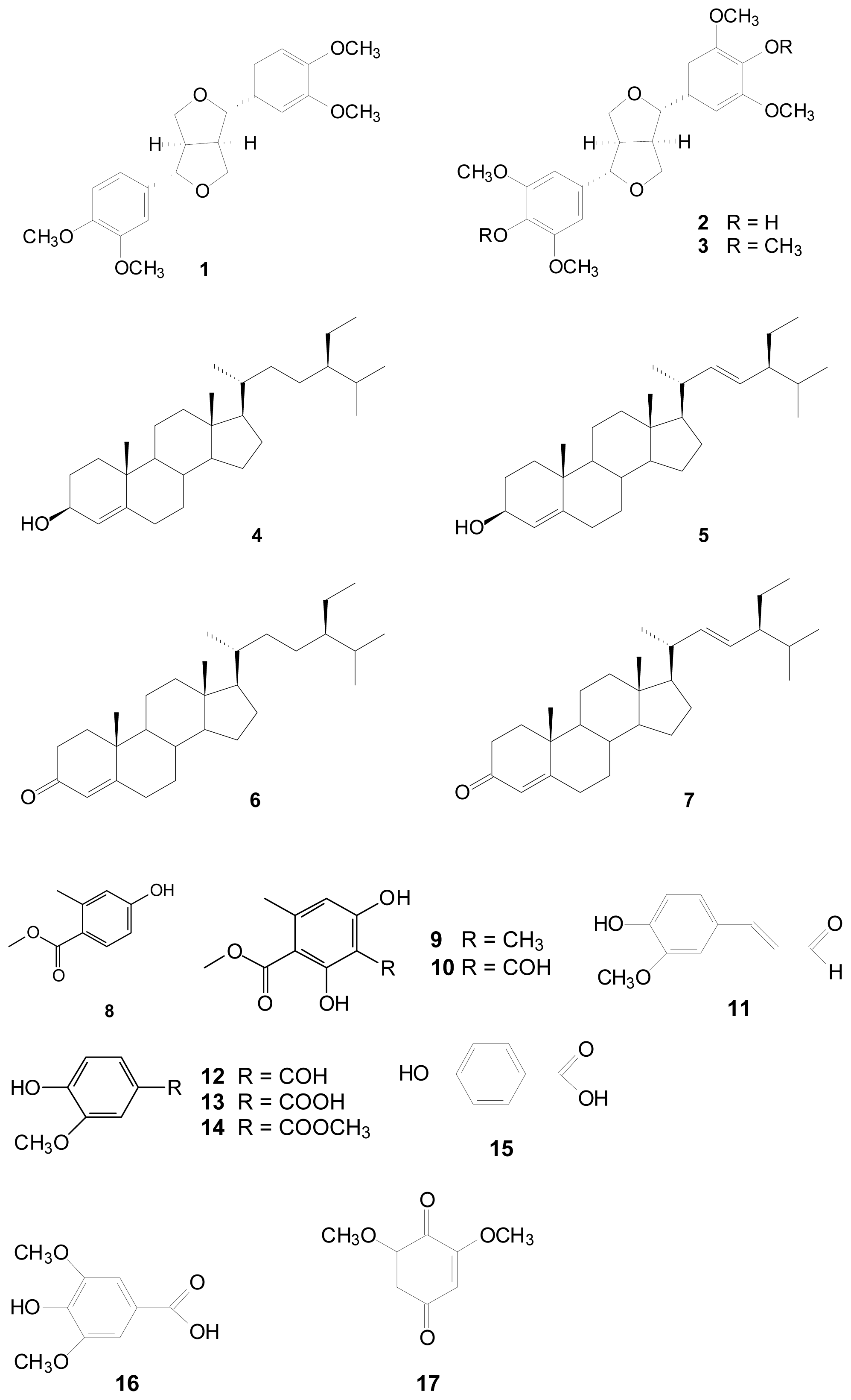
Figure 1. The chemical structures of compounds 117 from the stems of L. tulipifera.
Ijms 14 01698f1
Figure 2. The B16F10 cell viability with various doses (1, 10, 100 μM) of L. tulipifera compounds were examined using MTT assay. The control group had no additional agents. p < 0.05.
Ijms 14 01698f2
Figure 3. Tyrosinase activities (A) and melanin contents (B) of B16F10 cells treated with various concentrations (1, 10, 100 μM) of the compounds isolated from L. tulipifera for 48 h culture. The control group had no additional agents. p < 0.05.
Ijms 14 01698f3
Table 1. Antioxidative properties of compounds isolated from Liriodendron tulipifera determined by various assay. All compounds, including controls, were used at 100 μM. (-), no testing; (ns), no significance.
CompoundsDPPH (%)ABTS (%)Chelating (%)Reducing power (OD700)
Vitamin C a88.6 ± 1.876.4 ± 5.6--
EDTA b--86.9 ± 4.5-
BHA c---0.98 ± 0.1
(−)-Eudesmin (1)ns64.1 ± 5.6ns0.28 ± 0.0
(+)-Syringaresinol (2)38.5 ± 4.884.8 ± 7.3ns0.66 ± 0.3
(+)-Yangambin (3)ns28.1 ± 6.713.5 ± 0.60.24 ± 0.0
β-Sitosterol (4)nsnsnsns
Stigmasterol (5)nsnsnsns
β-Sitostenone (6)ns52.4 ± 7.4ns0.29 ± 0.0
Stigmastenone (7)nsnsnsns
Methyl 4-hydroxy-2-methylbenzoate (8)nsns17.4 ± 9.10.18 ± 0.0
β-Orcinol carboxylate (9)ns79.8 ± 2.2ns0.45 ± 0.0
Methyl haematommate (10)ns72.5 ± 6.119.8 ± 8.30.28 ± 0.0
Coniferyl aldehyde (11)nsnsns0.24 ± 0.0
Vanillin (12)ns19.4 ± 5.5ns0.24 ± 0.0
Vanillic acid (13)ns22.7 ± 5.2ns0.18 ± 0.0
Methyl vanillate (14)nsnsnsns
p-Hydroxybenzoic acid (15)nsnsnsns
Syringic acid (16)ns49.4 ± 10.8ns0.25 ± 0.0
2,6-Dimethoxy-p-quinone (17)ns31.5 ± 13.525.5 ± 6.80.29 ± 0.0
Data were expressed as a mean value of at least three independent experiments.
aVitamin C was used as a positive control on DPPH assay at 100 μM;
bEDTA was used as a positive control on metal chelating ability at 100 μM;
cBHA was used as a positive control on reducing power at 100 μM.
Table 2. Inhibitions of L. tulipifera compounds on mushroom tyrosinase at 100 μM. (ns), no significance.
L. tulipifera compoundsMushroom tyrosinase inhibition (%)
Kojic acid a89.2 ± 0.1
(−)-Eudesmin (1)ns
(+)-Syringaresinol (2)ns
(+)-Yangambin (3)ns
β-Sitosterol (4)ns
Stigmasterol (5)ns
β-Sitostenone (6)ns
Stigmastenone (7)ns
Methyl 4-hydroxy-2-methylbenzoate (8)23.2 ± 0.4
β-Orcinol carboxylate (9)ns
Methyl haematommate (10)ns
Coniferyl aldehyde (11)ns
Vanillin (12)ns
Vanillic acid (13)22.8 ± 0.4
Methyl vanillate (14)ns
p-Hydroxybenzoic acid (15)15.4 ± 0.4
Syringic acid (16)10.6 ± 0.4
2,6-Dimethoxy-p-quinone (17)ns
Data were expressed as a mean value of at least three independent experiments.
aKojic acid was used as a positive control of mushroom tyrosinase assay at 100 μM.