Article

Ochratoxin A Inhibits Mouse Embryonic Development by Activating a Mitochondrion-Dependent Apoptotic Signaling Pathway

1
Department of Life Science, National Pingtung University of Science and Technology, Pingtung 912, Taiwan
2
Department of Bioscience Technology and Center for Nanotechnology, Chung Yuan Christian University, Chung Li 320, Taiwan
3
Department of Cell and Molecular Biology, Institute of Biomedical Sciences, Medical College of Chang Gung University, Tao-Yuan 333, Taiwan
*
Author to whom correspondence should be addressed.
Int. J. Mol. Sci. 2013, 14(1), 935-953; https://doi.org/10.3390/ijms14010935
Submission received: 22 October 2012 / Revised: 10 December 2012 / Accepted: 24 December 2012 / Published: 7 January 2013
(This article belongs to the Special Issue Signalling Molecules and Signal Transduction in Cells)

Abstract

:
Ochratoxin A (OTA), a mycotoxin found in many foods worldwide, causes nephrotoxicity, hepatotoxicity, and immunotoxicity, both in vitro and in vivo. In the present study, we explored the cytotoxic effects exerted by OTA on the blastocyst stage of mouse embryos, on subsequent embryonic attachment, on outgrowth in vitro, and following in vivo implantation via embryo transfer. Mouse blastocysts were incubated with or without OTA (1, 5, or 10 μM) for 24 h. Cell proliferation and growth were investigated using dual differential staining; apoptosis was measured using the terminal deoxynucleotidyl transferase-mediated dUTP nick-end labeling (TUNEL) assay; and embryo implantation and post-implantation development were assessed by examination of in vitro growth and the outcome of in vivo embryo transfer, respectively. Blastocysts treated with 10 μM OTA displayed a significantly increased level of apoptosis and a reduction in total cell number. Interestingly, we observed no marked difference in implantation success rate between OTA-pretreated and control blastocysts either during in vitro embryonic development (following implantation in a fibronectin-coated culture dish) or after in vivo embryo transfer. However, in vitro treatment with 10 μM OTA was associated with increased resorption of post-implantation embryos by the mouse uterus, and decreased fetal weight upon embryo transfer. Our results collectively indicate that in vitro exposure to OTA triggers apoptosis and retards early post-implantation development after transfer of embryos to host mice. In addition, OTA induces apoptosis-mediated injury of mouse blastocysts, via reactive oxygen species (ROS) generation, and promotes mitochondrion-dependent apoptotic signaling processes that impair subsequent embryonic development.

1. Introduction

Ochratoxin A (OTA) is a mycotoxin produced principally by ubiquitous strains of Aspergillus and Penicillium[1]. OTA is one of the most common food-contaminating mycotoxins, and is often isolated from beans, grains, cereals and spices. In addition, OTA has contaminated coffee, grape juice, wine, beer and bread [2]. It is very difficult to completely avoid dietary exposure to OTA because the chemical occurs widely in various food stuffs [3,4]. Therefore, it is important to study the adverse effects of OTA on humans. Previous studies have found that OTA is nephrotoxic and hepatotoxic, and causes neurodegenerative disease [5]. In addition, recent work has shown that OTA creates oxidative stress in several regions of the mouse midbrain and hippocampus, compromising brain development [6]. OTA is a potent carcinogen and induces tumors in the kidney, mammary gland, and liver [79]. A recent study found that OTA triggered apoptosis via activation of a mitochondrion-dependent pathway. OTA induces apoptosis by elevating ROS generation, by causing mitochondrial transmembrane potential to be lost via opening of mitochondrial pores, by triggering the release of cytochrome c, and by activating caspase [10]. Thus, although OTA appears to exert multiple biological actions, and is cytotoxic [4,11], very few studies conducted to date have explored whether OTA negatively affects embryonic development.
During normal embryogenesis, apoptosis (a unique morphological pattern of cell death) functions to remove abnormal or redundant cells from pre-implantation embryos [12,13]. Apoptotic processes do not occur prior to the blastocyst stage during normal mouse embryonic development [14]. Induction of apoptosis during early stages of embryogenesis (i.e., following exposure to a teratogen) compromises embryonic development [1519]. Additionally, several chemical and physical triggers of apoptosis create oxidative stress via ROS generation [15,20]. This suggests that oxidative stress and apoptosis are closely linked and that ROS generators are potent inducers of apoptosis.
Apoptosis plays an important role in embryonic development [21]. Although several studies have shown that apoptosis is essential for normal embryonic development [2224], excessive apoptosis triggered in early embryos by exposure to mechanistically diverse teratogens can cause developmental injury [15,16,2527]. Previous studies found that OTA induced apoptosis in mammalian cells, including monkey and human kidney epithelial cells, porcine kidney PK15 cells, and human OK cells [2831]. In addition, developmental neurotoxicity caused by OTA was reflected in both morphological and behavioral changes in rodents [32,33]. Pretreatment of cultured post-implantation rat embryos with OTA caused dose-dependent reductions in yolk sac diameter, crown-rump length, somite number count, and protein and DNA content [34]. However, the details of the injuries caused by OTA, and the mechanisms of OTA action, during either the pre- or post-implantation stages of embryonic development, remain unclear. In the present study, we investigated whether OTA exerted cytotoxic effects on early-stage development of mouse blastocysts. We found that OTA suppressed embryonic cell proliferation during the blastocyst stage predominantly by inducing apoptosis of the inner cell mass (ICM). We also monitored subsequent blastocyst development in vitro and after embryo transfer in vivo.

2. Results

2.1. Effects of OTA on Mouse Blastocysts

To explore whether OTA was cytotoxic, we treated mouse blastocysts with 1, 5, or 10 μM OTA at 37 °C for 24 h, and measured apoptosis using the TUNEL assay. OTA at 10 μM clearly induced apoptosis (Figure 1A); the level of cell death was nine-fold higher in OTA-treated blastocysts than in untreated controls (Figure 1B).

2.2. Effects of OTA on Cell Proliferation

We used differential staining to examine cell proliferation in blastocysts treated with 1, 5, or 10 μM OTA for 24 h, and in untreated controls. The total and ICM cell numbers in blastocysts treated with 10 μM OTA were significantly lower than in controls (Figure 2A). Annexin V and PI staining were used to identify the cell death modes. Significantly higher numbers of Annexin V-positive/PI-negative (apoptotic) cells were evident in the ICM of treated blastocysts compared to controls, but no such difference was apparent in the trophectoderm (TE) (Figure 2B). Thus, OTA induces significant apoptosis in the ICM but not the TE of mouse blastocysts, impairing developmental potential.

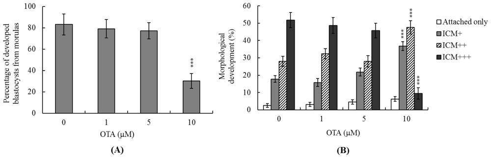
2.3. Effects of OTA on Mouse Embryonic Developmental Potential in Vitro

We next analyzed the effects of OTA on embryonic pre-implantation, implantation, and post-implantation development in vitro. Untreated control morulae developed into blastocysts at a frequency of ~83%, compared to only 30.4% of morulae treated with 10 μM OTA (Figure 3A). To explore the effects of OTA on implantation and post-implantation events in vitro, blastocysts were treated with 1, 5, or 10 μM OTA (300–320 blastocysts in each group), or remained untreated (control blastocysts), and the implantation rate and subsequent development over 8 days in culture were analyzed. The implantation rate of and the extent of attachment to fibronectin-coated dishes were similar in the OTA-treated and control groups (Figure 3B). Importantly, OTA-pretreated blastocysts were less successful in achieving post-implantation developmental milestones than were control blastocysts (Figure 3B), indicating that OTA affects the in vitro potential of blastocysts to develop features characteristic of post-implantation embryos.

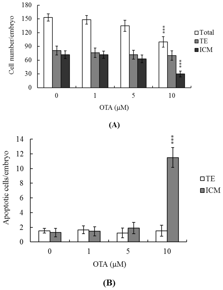
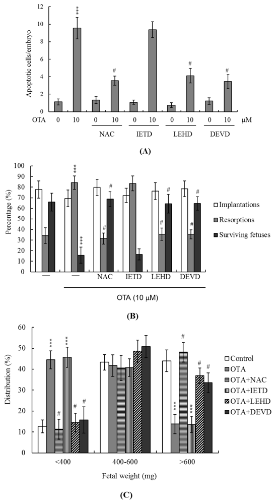
2.4. Effects of OTA on the Developmental Potential of Blastocysts in Vivo

To further explore the effects of OTA on blastocyst development in vivo, we performed embryo transfers on mouse blastocysts pretreated with OTA or not, and examined uterine contents 13 days post-transfer (day 18 fetus). The implantation ratio of the group pretreated with OTA (10 μM) was not significantly different from that of the untreated control group (Figure 4A). Embryos that implanted but failed to develop were subsequently resorbed. However, the proportion of such embryos was significantly higher in the group pretreated with 10 μM OTA compared to the control group (Figure 4A). Interestingly, no difference in placental weight was evident between the OTA-treated and untreated groups (Figure 4B), but fetal weight was lower in the OTA-treated group. Moreover, both earlier and current experiments by our group have shown that 35%–40% of normal mouse fetuses weigh more than 600 mg at day 18 of pregnancy after mouse embryo transfer, and the average weight of all surviving fetuses was ~600 ± 12 mg in the untreated control group of the present experiment [16,17,35]. Fetal weight is an important indicator of developmental status, and the average fetal weight of untreated controls thus serves as a key indicator of the developmental status of OTA-treated blastocysts. Only about 14% of fetuses in the group pretreated with 10 μM OTA weighed over 600 mg, in contrast to 45.5% of control fetuses (Figure 4C). Thus, exposure to OTA at the blastocyst stage is associated with a risk of poor post-implantation development.

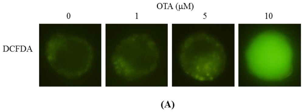
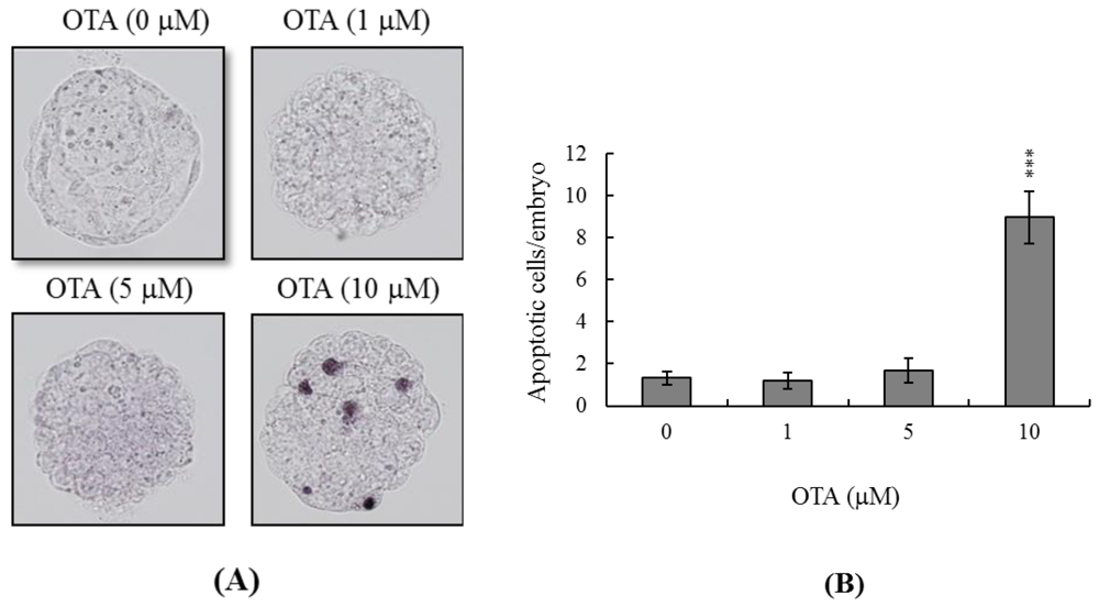
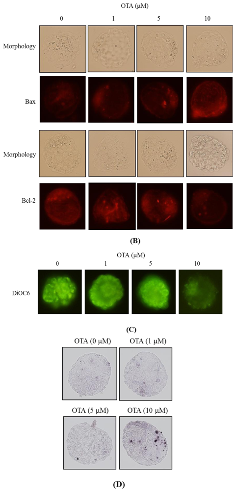
2.5. ROS Generation and Mitochondrion-Dependent Apoptotic Processes Are Involved in Blastocyst Death Induced by OTA

In light of the data of previous reports and our recent finding that ROS effectively induce apoptosis [16,36,37], we used the fluorescent dye DCF-DA to measure ROS content in OTA-treated mouse blastocyst cells. As shown in Figure 5A, OTA at 10 μM directly induced an increase in fluorescence intensity in such cells, compared with that of untreated control cells. Changes in the expression levels of Bax and Bcl-2 affect the action of the mitochondrion-dependent apoptotic pathway [38,39]; high and low Bax/Bcl-2 ratios are associated with lower and higher apoptotic thresholds, respectively. We thus explored whether OTA induced apoptosis via modulation of Bax and Bcl-2 expression. Immunostaining revealed that the Bax and Bcl-2 levels increased and decreased, respectively, upon OTA treatment of mouse blastocysts (Figure 5B). Examination of the effect of OTA on the mitochondrial membrane potential (MMP) of mouse blastocyst cells revealed that treatment with 10 μM OTA suppressed DiOC6(3) uptake into mitochondria, indicative of significant loss of MMP (Figure 5C). In addition, 10 μM OTA significantly activated caspase-3; this is an important feature of apoptosis (Figure 5D). To further explore the roles played by ROS and apoptosis-associated events in OTA-induced apoptosis, we added a recognized ROS scavenger, N-acetyl cysteine (NAC), and various caspase-specific inhibitors, to OTA-treated mouse blastocysts. Pretreatment of cells with NAC (500 μM) attenuated OTA-induced apoptosis (Figure 6A). In addition, pretreatment with inhibitors specific for caspase-9 (Z-LEHD-FMK) and caspase-3 (Z-DEVD-FMK) effectively blocked apoptosis, whereas the caspase-8-specific inhibitor Z-IETD-FMK did not (Figure 6A). Importantly, treatment with 10 μM OTA was associated with a lower implantation ratio, and the failure of further development was effectively blocked by addition of NAC and inhibitors of caspase-9 and caspase-3 at the time of embryo transfer. Animals pretreated with the caspase-8 inhibitor were similar in all respect to untreated controls (Figure 6B). In addition, the lower fetal weight in the group treated with 10 μM OTA was effectively rescued by pretreatment with NAC and specific caspase-9 and -3 inhibitors (Figure 6C). We thus suggest that OTA triggers ROS generation, in turn activating mitochondrion-dependent apoptotic processes in mouse blastocyst cells.

3. Discussion

Chemical or physical injury can affect normal progression of the complex and precisely orchestrated process of embryonic development, leading to malformation or miscarriage of the embryo. It is thus important to explore the possible teratogenic effects of various chemical agents and environmental toxins. Several previous reports have shown that OTA is a neurotoxic mycotoxin that causes oxidative stress, DNA damage, and mitochondrial dysfunction [10,40]. OTA-induced oxidative DNA damage has been found in the brain [6], and OTA increased oxidative stress in neural stem/progenitor cells [41]. A recent study found that OTA compromised hippocampal neurogenesis in vivo and that such injury might be associated with memory loss and deficits in learning and memory [41]. OTA not only induces neurotoxicity, but also kills other types of cells. Thus, exposure to OTA triggers caspase-dependent apoptosis via the mitochondrial pathway in human hepatocarcinoma cells [42] and disturbs calcium flux, causing DNA damage and apoptosis, in porcine kidney PK15 cells [30]. Thus, OTA may be a teratogen if ingested during pregnancy. Several papers have shown the teratogenic effects of OTA in mouse, rat, and pig [4346]. However, the regulatory effects and related mechanisms of OTA-induced cytotoxicity on early embryonic development have not been studied in detail. We therefore employed an in vitro assay to assess the cytotoxic effects of OTA during embryonic development, and sought to explain the mechanism of action of the chemical. Preliminary experiments revealed that OTA triggered apoptosis of mouse blastocysts only when the cells were incubated with OTA for at least 12 h; the effects endured for 24 h (data not shown). We therefore incubated blastocysts in medium containing 1–10 μM OTA for 24 h. Cell numbers fell as apoptosis developed (Figure 1). TUNEL staining revealed that treatment of mouse blastocysts with 10 μM OTA induced an 8.9-fold rise in apoptosis (Figure 1). Further, dual differential and Annexin V staining showed that the OTA-induced apoptosis occurred primarily in the ICM (Figure 2).
The TE arises from the trophoblast at the blastocyst stage and develops into a sphere of epithelial cells surrounding the ICM and the blastocoel. These cells contribute to placenta formation and are required for development of the mammalian conceptus [47]. Thus, reduction in the numbers of cells in the TE lineage may reduce both implantation and embryonic viability [48,49]. However, in our experiments, OTA induced apoptosis only in the ICM, thus not in the TE, and did not deleteriously affect embryonic attachment or outgrowth either in vitro or in vivo (Figures 24). Previous studies have shown that a reduction of ~30% or more in the number of ICM cells is associated with a high risk of fetal loss or developmental injury, even when the implantation rate and TE cell numbers are normal [50]. In addition, high numbers of ICM cells are essential to ensure viable implantation, and a reduction in cell numbers may decrease embryonic viability [48,49,51]. However, we found, in the present work, that OTA-induced embryonic cell death occurred only in the ICM, compromising post-implantation development, but exerting no effect on implantation per se (Figures 24). Although apoptosis is employed to eliminate unwanted cells during normal embryonic development, this process does not normally occur at the blastocyst stage [12,13]. Apoptotic action before or during blastocyst development is likely to result in deletion of important cell lineages, thus affecting embryonic development and potentially leading to miscarriage or embryonic malformation [14]. Our coworkers are currently using primary cell cultures derived from mouse ICM and TE to examine the negative effects of OTA on these cells, and to begin investigating the underlying mechanisms. Their preliminary results have revealed that OTA induces ICM cell apoptosis at concentrations higher than 10 μM. Low doses of OTA (<5 μM) had no injury effects in TE cell lines, whereas high concentrations (20–40 μM) triggered apoptosis. Notably, OTA appears to induce apoptosis in both mouse ICM and TE cell cultures through the mitochondria-dependent apoptotic pathway, as evident from increases in the Bax/Bcl-2 ratio and decreases in mitochondrial membrane potential. Accordingly, we conclude that OTA induces apoptosis in the ICM and TE via the same regulatory mechanism, but the treatment dose required to induce negative effects can vary by cell type. In view of our findings that OTA reduced cell number and increased apoptosis specifically in the ICM of mouse blastocysts, we determined whether OTA affected embryonic implantation and mortality, and/or caused developmental delay in post-implantation mouse embryos either in vitro or in vivo (Figure 2). We found that OTA-treated blastocysts exhibited a reduced level of embryonic development and a higher level of embryonic death both in vitro and in vivo (Figures 3 and 4).
Moreover, OTA has been shown to induce cell death with an IC50 value of 14 μM in porcine kidney PK15 cells [30]. Preliminary experiments by our group further revealed that OTA dose-dependently triggers mouse embryonic stem cell (ESC) apoptosis with an IC50 value of 12.7 μM, as determined by MTT assays performed after 24 h of exposure (data not shown). In addition, our initial HPLC results showed that serum OTA levels were about 9.6 μM in mice that had been exposed to drinking water supplemented with 20 μM OTA over 4 days (data not shown). Our embryo transfer assays showed that the 13-days-post-transfer (day 18 fetus) fetal weight of the OTA-pretreated group (10 μM) was significantly lower than that of the untreated control group (Figure 4C). However, we did not perform immunohistochemical staining of developmental markers in various organs of day 18 fetuses. To further determine the impact of OTA on early embryonic development in a stem cell assay model, our coworkers recently incubated cells with or without OTA and examined their ability to form embryoid bodies in vitro. Embryoid body formation was significantly decreased in cells pretreated with OTA. To ascertain whether the expression levels of OCT 4 and phosphorylated STAT3 (two well-known pluripotent markers) are affected by OTA, stem cells were treated with or without OTA for 24 h. Immunoblotting experiments revealed that OTA had no significant effect on the expression levels of OCT 4 or phosphorylated STAT3, compared to the untreated control group. Moreover, treatment of embryoid body cells with 50 ng/mL nerve growth factor (NGF) for 14 days induced their differentiation into nerve cells, as reflected by expression of microtubule associated protein-2 (MAP-2), a major nerve cell biomarker. Notably, pre-treatment with 10 μM OTA effectively inhibited the NGF-induced expression of MAP-2 (data not shown). Collectively, these results suggest that OTA inhibits early embryonic development and may have injury effects on neuronal organs. These experimental data showed that OTA induces apoptosis and negatively affects mouse embryonic development both in vitro and in vivo. Thus, OTA appears to compromise both pre- and post-implantation embryonic development at doses that are physiological, in the sense that they may be attained via dietary intake.
Mechanistically, our data show that OTA directly evokes intracellular oxidative stress (Figure 5A) leading to ROS-mediated apoptosis of mouse blastocyst cells (Figure 6A). This seems to involve the mitochondrion-dependent apoptotic pathway, as indicated both by the OTA-induced changes in the intracellular levels of Bcl family members (Bax and Bcl-2) and the loss of mitochondrial membrane potential (Figure 5B). Our findings are consistent with those of previous studies showing that OTA could trigger the mitochondrion-dependent apoptotic process, as revealed by loss of mitochondrial transmembrane potential, increased ROS production, mitochondrial relocalization of Bax, release of cytochrome c, and activation of caspases [10]. Given that recent studies have shown that addition of specific compounds to commonly used cell culture media triggers generation of ROS, such as hydrogen peroxide [52,53], we co-incubated OTA and culture medium, and measured ROS levels using the ferrous iron oxidation-xylenol orange method [52]. No artifactual ROS generation was detected under such conditions (data not shown). Importantly, a well-known ROS scavenger, NAC, effectively prevented OTA-induced apoptosis in mouse blastocysts (Figure 6A). We explored the precise mechanism of OTA-induced apoptosis in such blastocysts (Figures 5 and 6). NAC is not suitable for use in our animal model due to the high dose (400 μM) that is required to prevent OTA-induced embryonic development injury in vitro (Figure 6). However, we observed that pretreatment with NAC (400 μM) did not fully prevent OTA-induced cell apoptosis in mouse blastocysts (Figure 6A). This suggests that OTA may have a dual mode of action to cause cell apoptosis, such as through ROS generation and the formation of DNA adducts, potentially explaining why the antioxidant only partially inhibited OTA-induced apoptosis in mouse blastocyst-stage embryos. Although additional studies will be required to assess this possibility in detail, our present work provides important new insights into the negative impact of OTA on early embryonic development.
A recent study showed that OTA treatment of human peripheral blood mononuclear cells triggered the release of reactive oxygen species (ROS) and increased the levels of 8-hydroxydeoxyguanosine (8-OHdG), an important biomarker of oxidative DNA stress [54]. Investigation further showed that OTA treatment of human peripheral blood mononuclear cells in vitro could induce cell cycle arrest at G1 phase by down-regulating the protein expression levels of CDK4 and cyclinD1, and also trigger cell apoptosis. These results demonstrated that ROS is involved in OTA-induced DNA damage and G1 arrest in human peripheral blood mononuclear cells, suggesting that OTA may negatively affect embryonic development in mouse blastocysts via ROS-induced DNA damage. Our results collectively show that OTA triggers apoptosis of the ICM cells of blastocysts, leading to impairment of embryonic development via ROS generation, which in turn stimulates various adverse downstream processes characteristic of the mitochondrion-dependent apoptotic pathway.
A previous study further found that OTA has a high potential to initiate skin tumors in mouse skin in vivo, acting through oxidative stress, MAPK signaling and DNA damage. The same study also found that anti-oxidants may help prevent OTA-induced tumorigenesis [55]. Moreover, OTA has been shown to inhibit cell proliferation and downregulate heat shock protein 70 and 27 in cultured human hepatocellular carcinoma cells, but without inducing significant generation of reactive oxygen species [42]. DNA damage following OTA treatment of cultured human hepatocellular carcinoma cells induced apoptosis via the p53 protein, which triggers mitochondria- and caspase-dependent apoptotic processes. The p53 protein directly interacts with BCL-2 family proteins; this permits mitochondrial outer membrane permeabilization, leading to the release of apoptogenic proteins, such as cytochrome c, which activate caspases to trigger the cell death cascade. In addition, none oxidant, OTA seems to be genotoxic, triggering mitochondrial- and caspase-dependent apoptosis. From these results, we conclude that OTA may inhibit the transcriptional process. However, oxidative damage is not a major contributor to OTA toxicity in human hepatocellular carcinoma cells [42]. Based on the prior results and our present data, we speculate that OTA may have dual means to cause apoptosis, such as through ROS generation and the formation of DNA adducts. This would explain why antioxidants only partially inhibit OTA-induced apoptosis in mouse blastocyst-stage embryos (Figure 6A). Thus, although future work will be required to examine the detailed mechanisms of this process, our present study provides important new insights into the negative impacts of OTA on early embryonic development.

4. Experimental Section

4.1. Materials

Ochratoxin A (OTA), Pregnant mare’s serum gonadotropin (PMSG), Bovine serum albumin (BSA), sodium pyruvate and puerarin were purchased from Sigma (St. Louis, MO, USA). Human chorionic gonadotropin (hCG) was obtained from Serono (NV Organon Oss, The Netherlands). The TUNEL in situ cell death detection kit was obtained from Roche (Mannheim, Germany) and CMRL-1066 medium was from Gibco Life Technologies (Grand Island, NY, USA). Z-DEVD-FMK, Z-LEHD-FMK and Z-IETD-FMK were from Calbiochem (La Jolla, CA, USA).

4.2. Collection of Mouse Morulas and Blastocysts

ICR mice were from National Laboratory Animal Center (Taiwan, ROC). This research was also approved by the Animal Research Ethics Board of Chung Yuan Christian University (Taiwan, ROC). All animals received humane care, as outlined in the Guidelines for Care and Use of Experimental Animals (Canadian Council on Animal Care, Ottawa, 1984). All mice were maintained on breeder chow (Harlan Teklad chow) with food and water available ad libitum. Housing was in standard 28 cm × 16 cm × 11 cm (height) polypropylene cages with wire-grid tops and kept under a 12 h day/12 h night regimen. Nulliparous females (6–8 weeks old) were superovulated by injection of 5 IU PMSG followed 48 h later by injection of 5 IU hCG, and then mated overnight with a single fertile male of the same strain. The day a vaginal plug was found was defined as day 0 of gestation. Plug-positive females were separated for experimentation. Morulas were obtained by flushing the uterine tubes on the afternoon of gestation day 3, and blastocysts were obtained by flushing the uterine horn on day 4; in both cases the flushing solution consisted of CMRL-1066 culture medium containing 1 mM glutamine and 1 mM sodium pyruvate. Expanded blastocysts from different females were pooled and randomly selected for experiments.

4.3. OTA Treatment and TUNEL Assay

Blastocysts were incubated in medium containing with or without 1, 5, or 10 μM OTA for 24 h. For apoptosis detection, embryos were washed in OTA-free medium, fixed, permeabilized and subjected to TUNEL labeling using an in situ cell death detection kit (Roche Molecular Biochemicals, Mannheim, Germany) according to the manufacturer’s protocol. Photographic images were taken under brightfield illumination using a fluorescence microscope (Olympus BX70, Tokyo, Japan).

4.4. OTA Treatment and Cell Proliferation

Blastocysts were incubated with or without culture medium containing indicated concentrations of OTA (1–10 μM) for 24 h. Then, blastocysts were washed with OTA-free medium and dual differential staining was used to facilitate counting of cell numbers in the inner cell mass (ICM) and trophectoderm (TE) [48]. Blastocysts were incubated in 0.4% pronase in M2-BSA medium (M2 medium containing 0.1% bovine serum albumin) for removal of the zona pellucida. The denuded blastocysts were exposed to 1 mM trinitrobenzenesulphonic acid (TNBS) in BSA-free M2 medium containing 0.1% polyvinylpyrrolidone (PVP) at 4 °C for 30 min, and then washed with M2 medium (Sigma, St. Louis, MO, USA) [56]. The blastocysts were further treated with 30 μg/mL anti-dinitrophenol-BSA complex antibody in M2-BSA at 37 °C for 30 min, and then with M2 medium supplemented with 10% whole guinea-pig serum as a source of complement, along with 20 μg/mL bisbenzimide and 10 μg/Ml propidium iodide (PI), at 37 °C for 30 min. The immunolysed blastocysts were gently transferred to slides and protected from light before observation. Under UV light excitation, the ICM cells (which take up bisbenzimidine but exclude PI) appeared blue, whereas the TE cells (which take up both fluorochromes) appeared orange-red. Since multinucleated cells are not common in pre-implantation embryos [57], the number of nuclei was considered to represent an accurate measure of the cell number. Photographic images were taken under a fluorescence microscope (Olympus IX71, Tokyo, Japan).

4.5. Annexin V Staining

Blastocysts were incubated in 0, 1, 5 or 10 μM OTA for 24 h, washed with OTA-free culture medium, and then stained using an Annexin V-FLUOS staining kit (Roche, Mannheim, Germany), according to the manufacturer’s instructions. Briefly, the blastocysts were incubated in M2-BSA for removal of the zona pellucida, washed with PBS plus 0.3% BSA, and then incubated for 60 min with a mixture of 100 μL binding buffer, 1 μL fluorescein isothiocyanate (FITC)-conjugated Annexin V and 1 μL PI. After incubation, the embryos were washed and photographed using a fluorescence microscope under fluorescent illumination. Cells staining Annexin V+/PI− were considered apoptotic, while those staining Annexin V+/PI+ were considered necrotic.

4.6. Morphological Analysis of Embryonic Development

Blastocysts were cultured according to a modification of the previously reported method [58]. Briefly, embryos were cultured in 4-well multidishes at 37 °C. For group culture, four embryos were cultured per well. The basic medium consisted of CMRL-1066 supplemented with 1 mM glutamine and 1 mM sodium pyruvate plus 50 IU/mL penicillin and 50 mg/mL streptomycin (hereafter called culture medium). For treatments, the embryos were incubated with 0, 1, 5 or 10 μM OTA for 24 h. Thereafter, the embryos were cultured for 3 days in culture medium supplemented with 20% fetal calf serum, and for 4 days in culture medium supplemented with 20% heated-inactivated human placental cord serum, for a total culture time of 8 days from the onset of treatment. Embryos were inspected daily under a phase-contrast dissecting microscope, and developmental stages were classified according to established methods [59,60]. Under these culture conditions, each hatched blastocyst attached to the fibronectin and grew to form a cluster of ICM cells over the trophoblastic layer via in a process called TE outgrowth. After a total incubation period of 96 h, morphological scores for outgrowth were estimated. Growing embryos were classified as either “attached” or “outgrowth”, with the latter defined by the presence of a cluster of ICM cells over the trophoblastic layer. As described previously [61,62], ICM clusters were scored according to shape, ranging from compact and rounded ICM (+++) to a few scattered cells (+) over the trophoblastic layer.

4.7. Blastocyst Development following Embryo Transfer

To examine the ability of expanded blastocysts to implant and develop in vivo, the generated embryos were transferred to recipient mice. ICR females (white skin color) were mated with vasectomized males (C57BL/6J; black skin color; from National Laboratory Animal Center, Taiwan, ROC) to produce pseudopregnant dams as recipients for embryo transfer. To ensure that all fetuses in the pseudopregnant mice came from embryo transfer (white color) and not from fertilization by C57BL/6J (black color), we examined the skin color of the fetuses at day 18 post-coitus. To assess the impact of OTA on post-implantation growth in vivo, blastocysts were exposed to 0, 1, 5 and 10 μM OTA for 24 h, and then 8 embryos were transferred in parallel to the paired uterine horns of day 4 pseudopregnant mice. The surrogate mice were killed on day 18 post-coitus, and the frequency of implantation was calculated as the number of implantation sites per number of embryos transferred. The incidence rates of resorbed and surviving fetuses were calculated as the number of resorptions or surviving fetuses, respectively, per number of implantations. The weights of the surviving fetuses and placentae were measured immediately after dissection.

4.8. Immunofluorescent Cell Stain

Mouse blastocyst cells were fixed by formaldehyde, permeabilized by 1% Triton X-100, blocked by bovine serum albumin (5 mg/mL in PBS), and incubated with anti-Bax or anti-Bcl-2 antibodies (40 mg/mL) at room temperature for 3 h. After washing three times with PBS, embryos were incubated with second antibody conjugated with FITC or Rhodamine (TRITC) (1:100) at room temperature for 1 h and then observed under a fluorescence microscope (Olympus BX70, Tokyo, Japan).

4.9. Statistics

The data were analyzed using one-way ANOVA and t-tests and are presented as the mean ± SEM, with significance at p < 0.05.

5. Conclusions

In summary, for the first time, we have shown that OTA induces apoptosis in the ICM (only) of mouse blastocysts via ROS- and mitochondrion-dependent pathways, decreasing embryonic development and viability. OTA is thus potentially hazardous to normal embryonic development. Further studies are required to determine the effects of dietary OTA on embryonic development during pregnancy, and to define the precise regulatory mechanism(s) whereby OTA affects embryonic development, possibly acting as a teratogen during human embryogenesis.

Acknowledgment

This work was supported by grants (NSC101-2311-B-033-001-MY3, NSC101-2632-M-033-001-MY2 and NSC 99-2632-M-033-001-MY3) from the National Science Council of Taiwan, ROC.
  • Conflict of InterestThe authors declare no conflicts of interest.

References

  1. Council of Agricultural Science and Technology (CAST), Mycotoxins: Risks in Plant, Animal, and Human Systems; CAST: Ames, IA, USA, 2003.
  2. IPCS. Safety evaluation of certain mycotoxins in food. WHO Food Addit. Ser. 2001, 47, 103–415.
  3. Walker, R. Risk assessment of ochratoxin: Current views of the European Scientific Committee on Food, the JECFA and the Codex Committee on Food Additives and Contaminants. Adv. Exp. Med. Biol 2002, 504, 249–255. [Google Scholar]
  4. Pfohl-Leszkowicz, A.; Manderville, R.A. Ochratoxin A: An overview on toxicity and carcinogenicity in animals and humans. Mol. Nutr. Food Res 2007, 51, 61–99. [Google Scholar]
  5. Zhang, X.; Boesch-Saadatmandi, C.; Lou, Y.; Wolffram, S.; Huebbe, P.; Rimbach, G. Ochratoxin A induces apoptosis in neuronal cells. Genes Nutr 2009, 4, 41–48. [Google Scholar]
  6. Sava, V.; Reunova, O.; Velasquez, A.; Harbison, R.; Sanchez-Ramos, J. Acute neurotoxic effects of the fungal metabolite ochratoxin-A. Neurotoxicology 2006, 27, 82–92. [Google Scholar]
  7. Kanisawa, M.; Suzuki, S. Induction of renal and hepatic tumors in mice by ochratoxin A, a mycotoxin. Gann = Gan 1978, 69, 599–600. [Google Scholar]
  8. Kanisawa, M. Pathogenesis of human cancer development due to environmental factors. Gan No Rinsho 1984, 30, 1445–1456. [Google Scholar]
  9. Boorman, G. NTP Technical Report on the Toxicology and Carcinogenesis Studies of Ochratoxin A (CAS No. 303-47-9) in F344/N Rats (Gavage Studies), NTP TR 358, NIH Publication No. 89-2813; National Institute of Health: North Carolina, NC, USA, 1989. [Google Scholar]
  10. Bouaziz, C.; Sharaf el dein, O.; Martel, C.; Golli, E.E.; Abid-Essefi, S.; Brenner, C.; Lemaire, C.; Bacha, H. Molecular events involved in ochratoxin A induced mitochondrial pathway of apoptosis, modulation by Bcl-2 family members. Environ. Toxicol 2011, 26, 579–590. [Google Scholar]
  11. Pfohl-Leszkowicz, A.; Manderville, R.A. An update on direct genotoxicity as a molecular mechanism of ochratoxin a carcinogenicity. Chem. Res. Toxicol 2012, 25, 252–262. [Google Scholar]
  12. Hardy, K. Cell death in the mammalian blastocyst. Mol. Hum. Reprod 1997, 3, 919–925. [Google Scholar]
  13. Hardy, K.; Stark, J.; Winston, R.M. Maintenance of the inner cell mass in human blastocysts from fragmented embryos. Biol. Reprod 2003, 68, 1165–1169. [Google Scholar]
  14. Byrne, A.T.; Southgate, J.; Brison, D.R.; Leese, H.J. Analysis of apoptosis in the preimplantation bovine embryo using TUNEL. J. Reprod. Fertil 1999, 117, 97–105. [Google Scholar]
  15. Hsuuw, Y.D.; Chang, C.K.; Chan, W.H.; Yu, J.S. Curcumin prevents methylglyoxal-induced oxidative stress and apoptosis in mouse embryonic stem cells and blastocysts. J. Cell. Physiol 2005, 205, 379–386. [Google Scholar]
  16. Chan, W.H. Ginkgolide B induces apoptosis and developmental injury in mouse embryonic stem cells and blastocysts. Hum. Reprod 2006, 21, 2985–2995. [Google Scholar]
  17. Chan, W.H. Impact of genistein on maturation of mouse oocytes, fertilization, and fetal development. Reprod. Toxicol 2009, 28, 52–58. [Google Scholar]
  18. Chan, W.H. Effects of citrinin on maturation of mouse oocytes, fertilization, and fetal development in vitro and in vivo. Toxicol. Lett 2008, 180, 28–32. [Google Scholar]
  19. Chan, W.H. Ginkgolides induce apoptosis and decrease cell numbers in mouse blastocysts. Biochem. Biophys. Res. Commun 2005, 338, 1263–1267. [Google Scholar]
  20. Jacobson, M.D. Reactive oxygen species and programmed cell death. Trends Biochem. Sci 1996, 21, 83–86. [Google Scholar]
  21. Thompson, C.B. Apoptosis in the pathogenesis and treatment of disease. Science 1995, 267, 1456–1462. [Google Scholar]
  22. Brill, A.; Torchinsky, A.; Carp, H.; Toder, V. The role of apoptosis in normal and abnormal embryonic development. J. Assist. Reprod. Genet 1999, 16, 512–519. [Google Scholar]
  23. Lotz, K.; Proff, P.; Bienengraeber, V.; Fanghaenel, J.; Gedrange, T.; Weingaertner, J. Apoptosis as a creative agent of embryonic development of bucca, mentum and nasolacrimal duct. An in vivo study in rats. J. Craniomaxillofac. Surg 2006, 34, S8–S13. [Google Scholar]
  24. Weingaertner, J.; Proff, P.; Bienengraeber, V.; Gedrange, T.; Fanghaenel, J.; Lotz, K. In vivo study of apoptosis as a creative agent of embryonic development of the primary nasal duct in rats. J. Craniomaxillofac. Surg 2006, 34, S3–S7. [Google Scholar]
  25. Huang, F.J.; Shen, C.C.; Chang, S.Y.; Wu, T.C.; Hsuuw, Y.D. Retinoic acid decreases the viability of mouse blastocysts in vitro. Hum. Reprod 2003, 18, 130–136. [Google Scholar]
  26. Shang, E.H.; Wu, R.S. Aquatic hypoxia is a teratogen and affects fish embryonic development. Environ. Sci. Technol 2004, 38, 4763–4767. [Google Scholar]
  27. Detmar, J.; Rabaglino, T.; Taniuchi, Y.; Oh, J.; Acton, B.M.; Benito, A.; Nunez, G.; Jurisicova, A. Embryonic loss due to exposure to polycyclic aromatic hydrocarbons is mediated by Bax. Apoptosis 2006, 11, 1413–1425. [Google Scholar]
  28. Li, J.; Yin, S.; Dong, Y.; Fan, L.; Hu, H. p53 activation inhibits ochratoxin A-induced apoptosis in monkey and human kidney epithelial cells via suppression of JNK activation. Biochem. Biophys. Res. Commun 2011, 411, 458–463. [Google Scholar]
  29. Golli-Bennour, E.E.; Kouidhi, B.; Bouslimi, A.; Abid-Essefi, S.; Hassen, W.; Bacha, H. Cytotoxicity and genotoxicity induced by aflatoxin B1, ochratoxin A, and their combination in cultured Vero cells. J. Biochem. Mol. Toxicol 2010, 24, 42–50. [Google Scholar]
  30. Klaric, M.S.; Zeljezic, D.; Rumora, L.; Peraica, M.; Pepeljnjak, S.; Domijan, A.M. A potential role of calcium in apoptosis and aberrant chromatin forms in porcine kidney PK15 cells induced by individual and combined ochratoxin A and citrinin. Arch. Toxicol 2012, 86, 97–107. [Google Scholar]
  31. Sauvant, C.; Holzinger, H.; Gekle, M. Proximal tubular toxicity of ochratoxin A is amplified by simultaneous inhibition of the extracellular signal-regulated kinases 1/2. J. Pharmacol. Exp. Ther 2005, 313, 234–241. [Google Scholar]
  32. Hood, R.D.; Naughton, M.J.; Hayes, A.W. Prenatal effects of Ochratoxin A in hamsters. Teratology 1976, 13, 11–14. [Google Scholar]
  33. Mayura, K.; Reddy, R.V.; Hayes, A.W.; Berndt, W.O. Embryocidal, fetotoxic and teratogenic effects of ochratoxin A in rats. Toxicology 1982, 25, 175–185. [Google Scholar]
  34. Mayura, K.; Edwards, J.F.; Maull, E.A.; Phillips, T.D. The effects of ochratoxin A on postimplantation rat embryos in culture. Arch. Environ. Contam. Toxicol 1989, 18, 411–415. [Google Scholar]
  35. Chan, W.H.; Shiao, N.H. Effect of citrinin on mouse embryonic development in vitro and in vivo. Reprod. Toxicol 2007, 24, 120–125. [Google Scholar]
  36. Chan, W.H. Citrinin induces apoptosis via a mitochondria-dependent pathway and inhibition of survival signals in embryonic stem cells, and causes developmental injury in blastocysts. Biochem. J 2007, 404, 317–326. [Google Scholar]
  37. Chan, W.H. Citrinin induces apoptosis in mouse embryonic stem cells. IUBMB Life 2008, 60, 171–179. [Google Scholar]
  38. Yu, F.; Watts, R.N.; Zhang, X.D.; Borrow, J.M.; Hersey, P. Involvement of BH3-only proapoptotic proteins in mitochondrial-dependent Phenoxodiol-induced apoptosis of human melanoma cells. Anticancer Drugs 2006, 17, 1151–1161. [Google Scholar]
  39. Criollo, A.; Galluzzi, L.; Chiara Maiuri, M.; Tasdemir, E.; Lavandero, S.; Kroemer, G. Mitochondrial control of cell death induced by hyperosmotic stress. Apoptosis 2007, 12, 3–18. [Google Scholar]
  40. Yoon, S.; Cong, W.T.; Bang, Y.; Lee, S.N.; Yoon, C.S.; Kwack, S.J.; Kang, T.S.; Lee, K.Y.; Choi, J.K.; Choi, H.J. Proteome response to ochratoxin A-induced apoptotic cell death in mouse hippocampal HT22 cells. Neurotoxicology 2009, 30, 666–676. [Google Scholar]
  41. Sava, V.; Velasquez, A.; Song, S.; Sanchez-Ramos, J. Adult hippocampal neural stem/progenitor cells in vitro are vulnerable to the mycotoxin ochratoxin-A. Toxicol. Sci 2007, 98, 187–197. [Google Scholar]
  42. El Golli Bennour, E.; Rodriguez-Enfedaque, A.; Bouaziz, C.; Ladjimi, M.; Renaud, F.; Bacha, H. Toxicities induced in cultured human hepatocarcinoma cells exposed to ochratoxin A: Oxidative stress and apoptosis status. J. Biochem. Mol. Toxicol 2009, 23, 87–96. [Google Scholar]
  43. Wangikar, P.B.; Dwivedi, P.; Sinha, N. Effect in rats of simultaneous prenatal exposure to ochratoxin A and aflatoxin B1. I. Maternal toxicity and fetal malformations. Birth Defects Res. B 2004, 71, 343–351. [Google Scholar]
  44. Biro, K.; Barna-Vetro, I.; Pecsi, T.; Szabo, E.; Winkler, G.; Fink-Gremmels, J.; Solti, L. Evaluation of spermatological parameters in ochratoxin A—Challenged boars. Theriogenology 2003, 60, 199–207. [Google Scholar]
  45. Patil, R.D.; Dwivedi, P.; Sharma, A.K. Critical period and minimum single oral dose of ochratoxin A for inducing developmental toxicity in pregnant Wistar rats. Reprod. Toxicol 2006, 22, 679–687. [Google Scholar]
  46. Jennings-Gee, J.E.; Tozlovanu, M.; Manderville, R.; Miller, M.S.; Pfohl-Leszkowicz, A.; Schwartz, G.G. Ochratoxin A: In utero exposure in mice induces adducts in testicular DNA. Toxins 2010, 2, 1428–1444. [Google Scholar]
  47. Cross, J.C.; Werb, Z.; Fisher, S.J. Implantation and the placenta: key pieces of the development puzzle. Science 1994, 266, 1508–1518. [Google Scholar]
  48. Pampfer, S.; de Hertogh, R.; Vanderheyden, I.; Michiels, B.; Vercheval, M. Decreased inner cell mass proportion in blastocysts from diabetic rats. Diabetes 1990, 39, 471–476. [Google Scholar]
  49. Kelly, S.M.; Robaire, B.; Hales, B.F. Paternal cyclophosphamide treatment causes postimplantation loss via inner cell mass-specific cell death. Teratology 1992, 45, 313–318. [Google Scholar]
  50. Tam, P.P. Postimplantation development of mitomycin C-treated mouse blastocysts. Teratology 1988, 37, 205–212. [Google Scholar]
  51. Chen, C.C.; Chan, W.H. Impact effects of puerarin on mouse embryonic development. Reprod. Toxicol 2009, 28, 530–535. [Google Scholar]
  52. Long, L.H.; Clement, M.V.; Halliwell, B. Artifacts in cell culture: Rapid generation of hydrogen peroxide on addition of (−)-epigallocatechin, (−)-epigallocatechin gallate, (+)-catechin, and quercetin to commonly used cell culture media. Biochem. Biophys. Res. Commun 2000, 273, 50–53. [Google Scholar]
  53. Halliwell, B. Oxidative stress in cell culture: An under-appreciated problem? FEBS Lett 2003, 540, 3–6. [Google Scholar]
  54. Liu, J.; Wang, Y.; Cui, J.; Xing, L.; Shen, H.; Wu, S.; Lian, H.; Wang, J.; Yan, X.; Zhang, X. Ochratoxin A induces oxidative DNA damage and G1 phase arrest in human peripheral blood mononuclear cells in vitro. Toxicol. Lett 2012, 211, 164–171. [Google Scholar]
  55. Kumar, R.; Ansari, K.M.; Chaudhari, B.P.; Dhawan, A.; Dwivedi, P.D.; Jain, S.K.; Das, M. Topical application of ochratoxin a causes DNA damage and tumor initiation in mouse skin. PLoS One 2012, 7, e47280. [Google Scholar]
  56. Hardy, K.; Handyside, A.H.; Winston, R.M. The human blastocyst: Cell number, death and allocation during late preimplantation development in vitro. Development 1989, 107, 597–604. [Google Scholar]
  57. Gardner, R.L.; Davies, T.J. Lack of coupling between onset of giant transformation and genome endoreduplication in the mural trophectoderm of the mouse blastocyst. J. Exp. Zool 1993, 265, 54–60. [Google Scholar]
  58. Huang, F.J.; Wu, T.C.; Tsai, M.Y. Effect of retinoic acid on implantation and post-implantation development of mouse embryos in vitro. Hum. Reprod 2001, 16, 2171–2176. [Google Scholar]
  59. Witschi, E. Characterization of Developmental Stages. Part II. Rat. In Biology Data Book, 2nd ed; Federation of American Societies of Experimental Biologies: Washington DC, USA, 1972; pp. 178–180. [Google Scholar]
  60. Armant, D.R.; Kaplan, H.A.; Lennarz, W.J. Fibronectin and laminin promote in vitro attachment and outgrowth of mouse blastocysts. Dev. Biol 1986, 116, 519–523. [Google Scholar]
  61. Pampfer, S.; Wuu, Y.D.; Vanderheyden, I.; de Hertogh, R. In vitro study of the carry-over effect associated with early diabetic embryopathy in the rat. Diabetologia 1994, 37, 855–862. [Google Scholar]
  62. Chan, W.H. Embryonic toxicity of sanguinarine through apoptotic processes in mouse blastocysts. Toxicol. Lett 2011, 205, 285–292. [Google Scholar]
Figure 1. Ochratoxin A (OTA) induces apoptosis in mouse blastocysts. (A) Mouse blastocysts were treated with OTA (1, 5, or 10 μM) for 24 h, or left untreated, and the extent of apoptosis was determined using transferase-mediated dUTP nick-end labeling (TUNEL) staining followed by light microscopy. TUNEL-positive cells are shown in black; (B) The mean numbers of apoptotic (TUNEL-positive) cells per blastocyst was calculated as six to eight experiments. Data are based on at least 220 blastocysts from each group. Values are presented as means ± SEMs. ***p < 0.001 versus the control group.
Ijms 14 00935f1
Figure 2. Effects of OTA on blastocyst viability. Mouse blastocysts were treated with OTA (1, 5, or 10 μM) for 24 h, or left untreated. (A) The total number of cells per blastocyst, and the numbers of cells in the inner cell mass (ICM) and the trophectoderm (TE) were counted; (B) The proportions of Annexin V-positive/PI-negative cells in the blastocysts of each group were determined. Data are based on at least 230 blastocysts from each group. Values are presented as means ± SEMs of five separate experiments. ***p < 0.001 versus the control group.
Ijms 14 00935f2
Figure 3. In vitro development of mouse embryos exposed to OTA at the blastocyst stage. (A) Mouse morulae were treated with OTA (1, 5, or 10 μM) for 24 h, or left untreated, and next cultured for an additional 24 h at 37 °C. The proportions of morulae that developed into blastocysts were counted; (B) Mouse blastocysts were treated with OTA (1, 5, or 10 μM) for 24 h, or left untreated, and observed in culture for seven days post-treatment. Growing embryos were classified as either “attached” or “outgrowth,” with the latter defined by the presence of a cluster of inner cell mass (ICM) cells over the trophoblastic layer. ICM clusters were scored according to shape, ranging from compact and rounded ICM (+++) to a few scattered cells (+) over the trophoblastic layer. Blastocysts were identified as exhibiting attachment only, or as ICM (+), ICM (++), or ICM (+++), via morphological assessment, as described in Materials and Methods. Values are presented as means ± SEMs of six experiments. ***p < 0.001 versus the control group.
Ijms 14 00935f3
Figure 4. Effects of OTA on mouse blastocysts in terms of in vivo implantation, resorption, fetal survival and fetal weight. (A) Mouse blastocysts were treated with OTA (1, 5, or 10 μM) for 24 h, or left untreated. Implantation, resorption, and numbers of surviving fetuses, were analyzed, as described in Materials and Methods. The implantation proportions represent the number of implantations per transferred embryo, ×100. The proportions of resorption or fetal survival are the numbers of resorptions or surviving fetuses per-implantation, ×100; (B) Placental weights were measured in each of 40 recipient mice; (C) Weight distribution of surviving fetuses on day 18 post-coitus. Surviving fetuses were obtained via embryo transfer of control and OTA-pretreated blastocysts, as described in Materials and Methods (a total of 320 blastocysts were transferred to 40 recipients). ***p < 0.001 versus the OTA-free group. 2.5.
Ijms 14 00935f4
Figure 5. Effects of OTA on ROS generation and mitochondrion-dependent apoptotic processes in mouse blastocysts. Mouse blastocysts were treated with OTA (1, 5, or 10 μM), or left untreated, for 24 h. (A) ROS generation was detected by staining with a 20 μM solution of the DCF-DA fluorescent dye; (B) Bax and Bcl-2 expression levels were assessed via immunostaining using anti-Bax and anti-Bcl-2 antibodies, respectively. The protocol is described in Materials and Methods; (C) To determine changes in mitochondrial membrane potential, embryos were incubated with 40 nM DiOC6(3) at 37 °C for 1 h and examined under a fluorescence microscope; (D) Activation of caspase-3 was measured by immunostaining with anti-activated caspase-3 antibody for 3 h, followed by addition of a secondary antibody conjugated with peroxidase (1:100 dilution) for 1 h. Finally, 20 μL of DAB-substrate solution was added to each embryo, and incubation for 2 min at room temperature followed. Cells containing activated caspase-3 are shown in black.
Ijms 14 00935f5aIjms 14 00935f5b
Figure 6. Effects of ROS scavengers and caspase inhibitors on in vivo implantation, resorption, fetal survival, and fetal weight, after treatment of embryos with OTA. Mouse blastocysts were pretreated with 400 μM N-acetyl cysteine (NAC), 300 μM Z-IETD-FMK (IETD), 300 μM Z-LEHD-FMK (LEHD), or 300 μM Z-DEVD-FMK (DEVD) for 1 h, or left untreated. Blastocysts were incubated with 10 μM OTA for a further 24 h. (A) Apoptosis was detected via TUNEL staining, as described in the legend to Figure 1; (B) The extent of implantation and resorption, and the numbers of surviving fetuses, were analyzed via embryo transfer, as described in Materials and Methods and in the legend to Figure 4; (C) The weight distribution of surviving fetuses on day 18 post-transfer. Surviving fetuses were obtained via embryo transfer of control and OTA-pretreated blastocysts (a total of 320 blastocysts were transferred to 40 recipients). ***p < 0.001 versus the OTA-free group and # p < 0.001 versus the group treated with 10 μM OTA.
Ijms 14 00935f6

