Article

The Effect of the Aerial Part of Lindera akoensis on Lipopolysaccharides (LPS)-Induced Nitric Oxide Production in RAW264.7 Cells

1
Department of Chinese Pharmaceutical Sciences and Chinese Medicine Resources, China Medical University, Taichung 404, Taiwan
2
Department of Biological Science and Technology, National Pingtung University of Science and Technology, Pingtung 912, Taiwan
3
Department of Forestry, National Chung Hsing University, Taichung 402, Taiwan
4
Agricultural Biotechnology Research Center, Academia Sinica, Taipei 115, Taiwan
5
Graduate Institute of Natural Products, Kaohsiung Medical University, Kaohsiung 807, Taiwan
6
The Experimental Forest Management Office, National Chung-Hsing University, Taichung 402, Taiwan
7
Tsuzuki Institute for Traditional Medicine, China Medical University, Taichung 402, Taiwan
*
Authors to whom correspondence should be addressed.
Int. J. Mol. Sci. 2013, 14(5), 9168-9181; https://doi.org/10.3390/ijms14059168
Submission received: 12 March 2013 / Revised: 12 April 2013 / Accepted: 15 April 2013 / Published: 26 April 2013
(This article belongs to the Section Biochemistry)

Abstract

:
Four new secondary metabolites, 3α-((E)-Dodec-1-enyl)-4β-hydroxy-5β-methyldihydrofuran-2-one (1), linderinol (6), 4′-O-methylkaempferol 3-O-α-l-(4″-E-p-coumaroyl)rhamnoside (11) and kaempferol 3-O-α-l-(4″-Z-p-coumaroyl) rhamnoside (12) with eleven known compounds—3-epilistenolide D1 (2), 3-epilistenolide D2 (3), (3Z,4α,5β)-3-(dodec-11-ynylidene)-4-hydroxy-5-methylbutanolide (4), (3E,4β,5β)-3-(dodec-11-ynylidene)-4-hydroxy-5-methylbutanolide (5), matairesinol (7), syringaresinol (8), (+)-pinoresinol (9), salicifoliol (10), 4″-p-coumaroylafzelin (13), catechin (14) and epicatechin (15)—were first isolated from the aerial part of Lindera akoensis. Their structures were determined by detailed analysis of 1D- and 2D-NMR spectroscopic data. All of the compounds isolated from Lindera akoensis showed that in vitro anti-inflammatory activity decreases the LPS-stimulated production of nitric oxide (NO) in RAW 264.7 cell, with IC50 values of 4.1–413.8 μM.

1. Introduction

Lindera akoensis (Lauraceae) is an endemic evergreen tree that grows in broad-leaved forests in lowlands throughout Taiwan; it is often used as a fence. Aporphines [1], alkaloids [2], sesquiterpenoids [35], flavonoids [6], butanolides [6], furanoids [7], chalconoids [8] and phenolic compounds [9,10] are widely distributed in the plants of the genus of Lindera. Some isolates exhibit biological activities, including suppressed the contraction of thoracic aorta [1], anti-mycobacterial [6], anti-inflammatory [11], against human lung cancer cell (SBC-3) [12], inhibitory osteoclast differentiation [10], slowing down of the progression of diabetic nephropathy in mice [12], anti-nociceptivity [13], inhibition on human acyl-coenzyme A cholesterol acyltransferase activity and antioxidation of low density lipoprotein [9]. Only one piece of literature had reported the chemical constituents and anti-mycobacterial activity from the root of L. akoensis [6].
The folk usage of L. akoensis is in the treatment of trauma and inflammation [14]. Butanolides showed anti-inflammation in previous studies [15,16]. In a random screening for inhibitory activity of various Chinese traditional medicines toward nitric oxide (NO) production in vitro by RAW264.7 cells, the EtOH extract of the aerial parts of L. akoensis showed a significant activity. Thus, the constituents of L. akoensis were investigated. This paper deals with the structure elucidation of the new compounds, and the inhibitory activity of the isolates toward nitric oxide (NO) production towards RAW264.7 cells is also discussed.

2. Results and Discussion

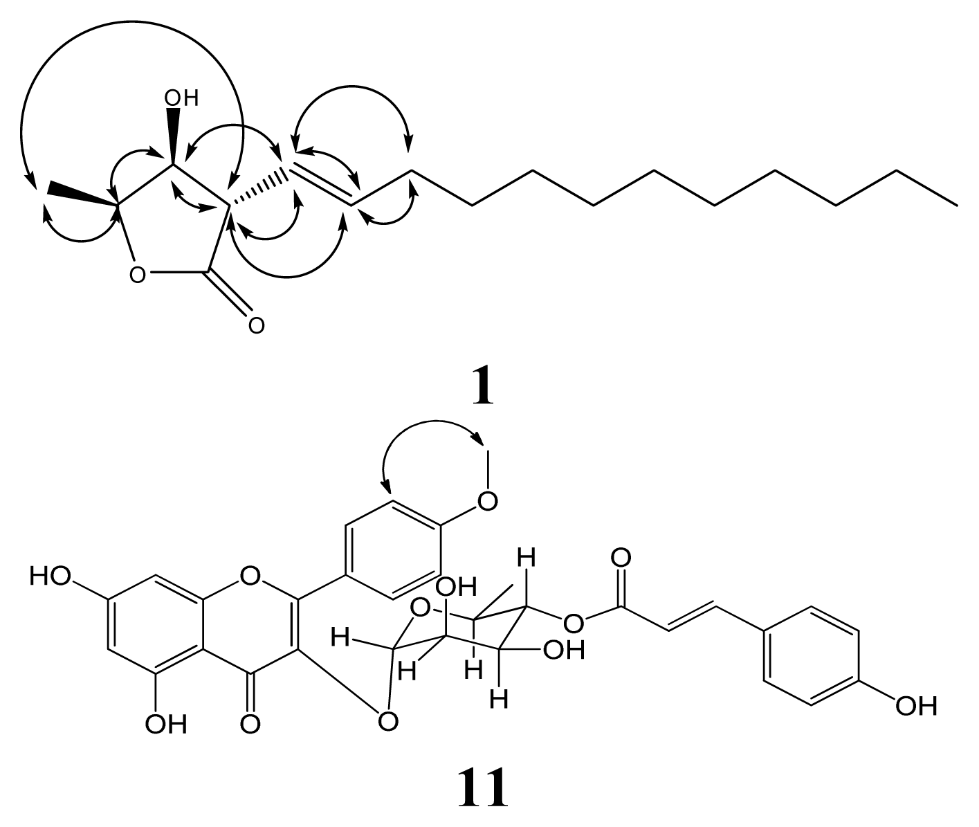
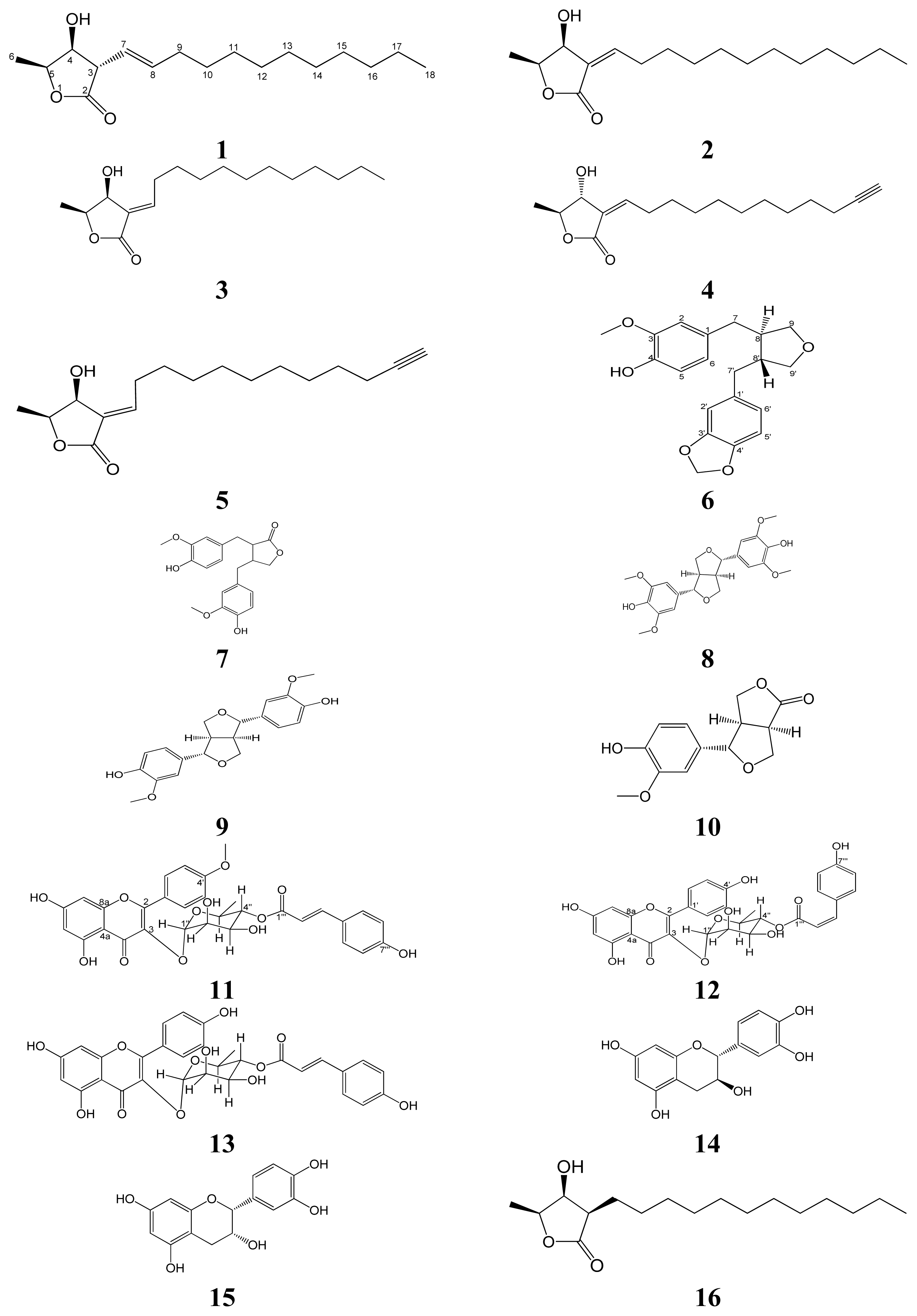
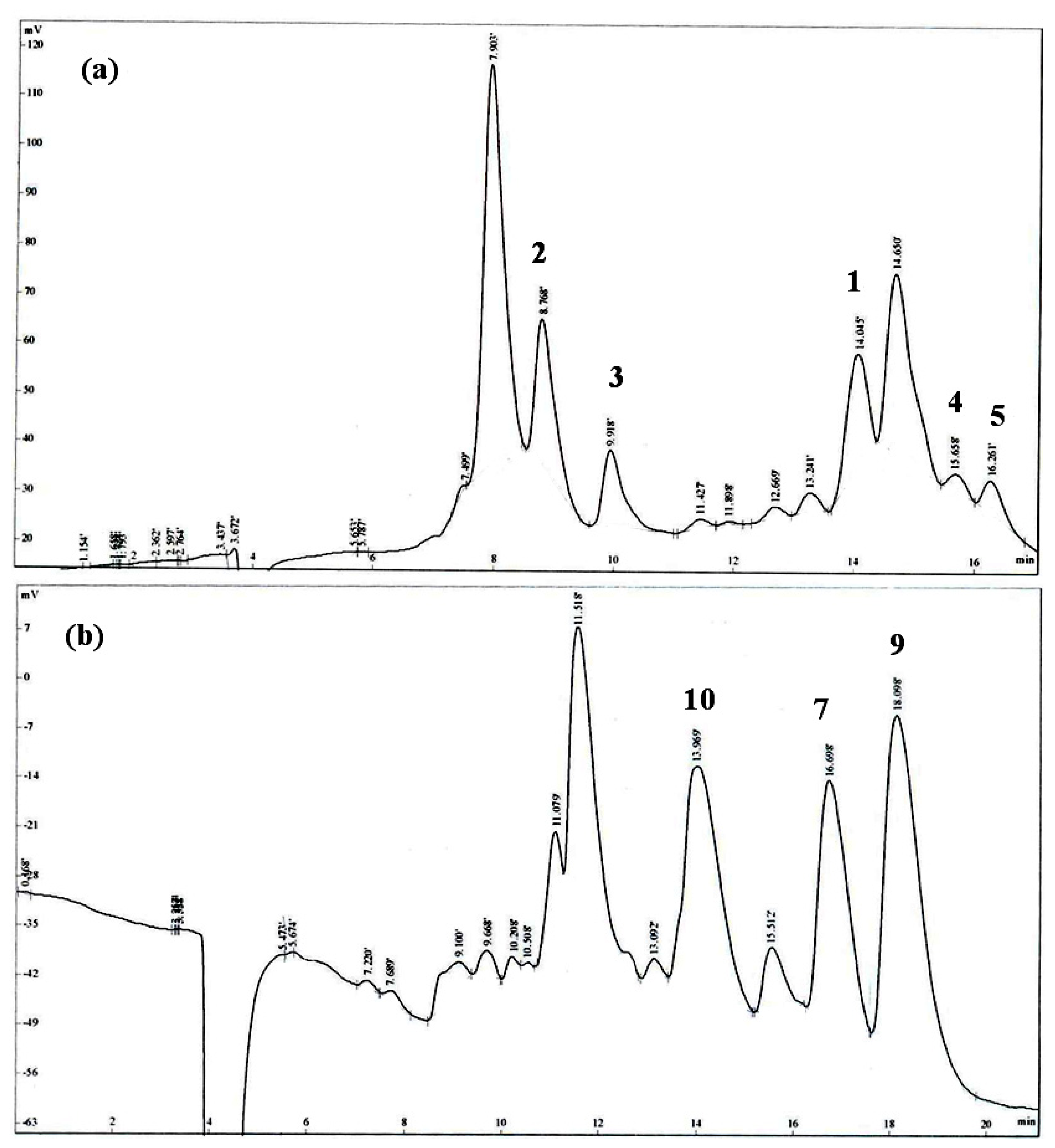
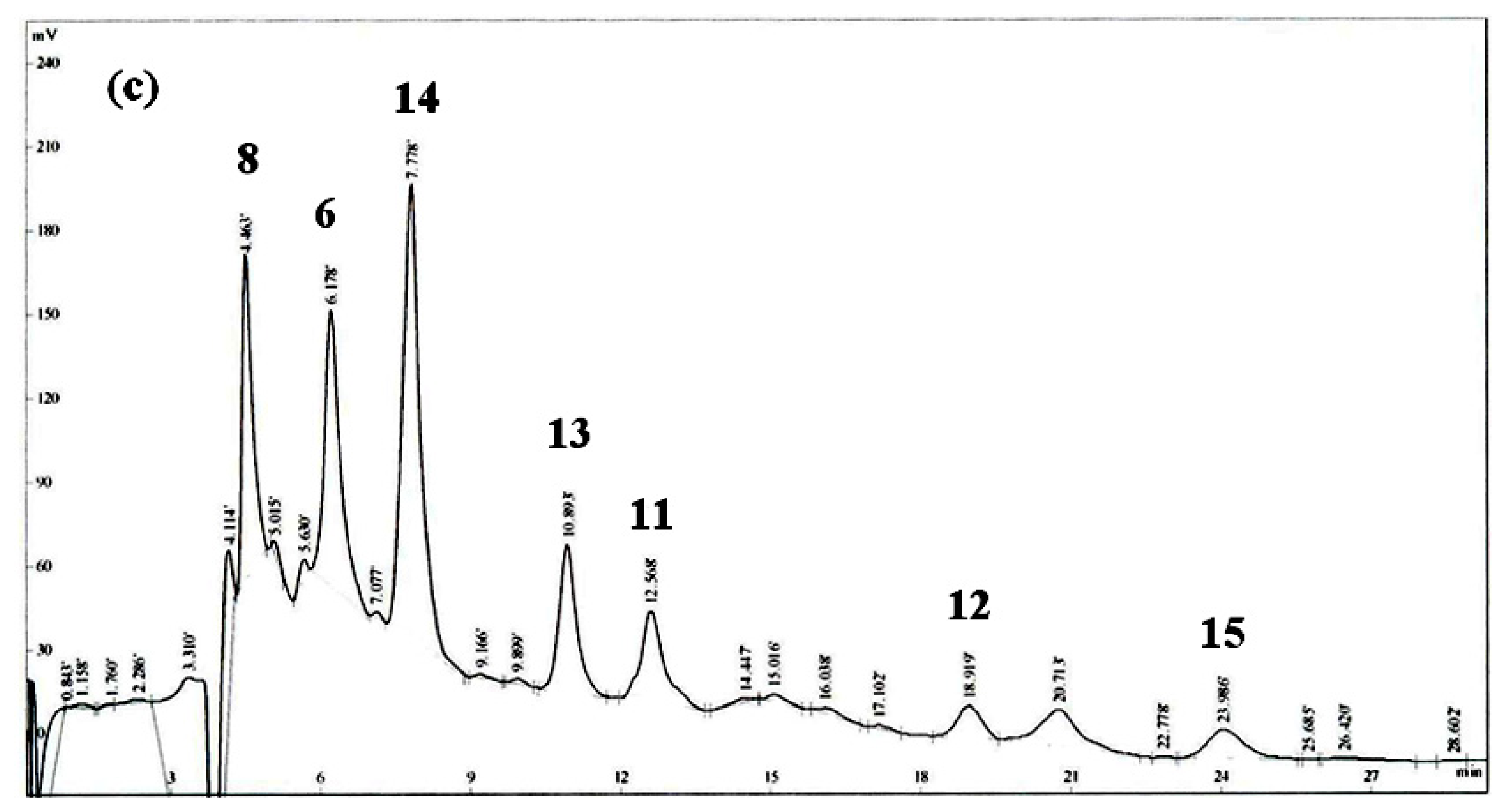
Isolation and Structural Elucidation

The aerial part of L. akoensis was air-dried and then extracted by EtOH and purified. Extensive normal phase Si gel column chromatographic purification of the EtOAc-soluble fraction afforded four new compounds, 3α-((E)-Dodec-1-enyl)-4β-hydroxy-5β-methyldihydrofuran-2-one (1), linderinol (6), 4′-O-methylkaempferol 3-O-α-l-(4″-E-p-coumaroyl)rhamnoside (11), kaempferol 3-O-α-l-(4″-Z-p-coumaroyl) rhamnoside (12), as well as eleven known compounds, 3-epilistenolide D1 (2) [17], 3-epilistenolide D2 (3) [17], (3Z,4α,5β)-3-(dodec-11-ynylidene)-4-hydroxy-5-methylbutanolide (4) [18], (3E,4β,5β)-3-(dodec-11-ynylidene)-4-hydroxy-5-methylbutanolide (5) [19], matairesinol (7) [20], syringaresinol (8) [21], (+)-pinoresinol (9) [22], salicifoliol (10) [23], 4″-p-coumaroylafzelin (13) [24], catechin (14) [25] and epicatechin (15) [25] (Figure 1).
Compound 1 was isolated as an optically inactive colorless oil ([α]2°D ± 0 (c 0.4, CHCl3)) and showed the presence of hydroxy (3401 cm−1), olefin (1682 cm−1) and γ-lactone (1759 cm−1) functionalities groups in its infrared (IR) spectrum. The high resolution electron impact mass spectrometry (HREIMS) data determined the molecular formula to be C17H30O3 (m/z 282.2198 ([M]+; calcd 282.2195)). The 1H-NMR spectrum showed signals similar to those of (3β,4β,5β)-3-dodecyl-4- hydroxy-5-methyldihydrofuran-2-one (16) (not purified in this research) [18] at δH 3.19 (1H, dd, J = 6.5, 4.7 Hz), δH 4.23 (dd, J = 4.7, 4.5), δH 4.64 (qd, J = 6.5, 4.5) were assigned to H-3, H-4 and H-5, respectively (Table 1). The chemical shift and coupling patterns of H-4 and H-5 suggested that the relative configuration of 1 was identified similar to that of 16. This conclusion was supported by comparison of the 1H and 13C NMR data of 1 with those of reported compounds having a cis-relationship between H-4 and H-5. 1H-NMR spectrum of 1 was similar to that of compounds 3 with 4β-hydroxy-5β-methyl groups. Two olefinic H-atoms were assigned the signals at δH 5.37 (1H, dd, J = 15.4, 6.5 Hz, H-7), δH 5.72(1H, dt, J = 15.4, 7.4 Hz, H-8), and nine CH2-group signals were observed (δH 2.04 (2H, q, J = 7.4 Hz, H-9), δH 1.24 (16H, m, H-10–17)). The H-7 was coupled with H-3 and H-8, with coupling constant 6.5 Hz and 15.4 Hz, respectively, establishing the trans-geometry of Δ7. Compared with 3β-((E)-dodec-1-enyl)-4β-hydroxy-5β-methyldihydrofuran-2-one in our previous study [26], the only difference was the configuration of the H-3((δH 3.19, dd, J = 6.5, 4.7 Hz), δC 52.7 (C-3)). The key correlation of NOESY spectrum, H-3, has correlation with H-6 and no correlation with H-5, moreover H-4 and H-5 having NOESY correlation, confirmed that H-4 and H-5 in the same phase and H-3 was in opposite side of H-3 (Figure 2). The zero optical rotation value indicated that there may exist in compound 1 a racemic mixture. All protons and carbons were confirmed by 1D and 2D spectra. Thus, 1 was identified as 3α-((E)-dodec-1-enyl)-4β-hydroxy-5β-methyldihydrofuran-2-one.
Compound 6 was a pale yellow amorphous solid, ([α]2°D = +20.2° (c = 0.42, CHCl3)); it has a λmax at 284.6 nm (logɛ 3.27) in the ultraviolet (UV) spectrum and shows the presence of hydroxy (3310 cm−1) and benzene (1605 and 1512 cm−1) functionalities in its IR spectrum. The HREIMS data determined the molecular formula to be C20H22O5 (m/z 342.1530 ([M]+; calcd 342.1467)). The 1H-NMR spectrum showed signals similar to those of matairesinol (7), such as the CH2 group at δH 2.62 (2H, dd, J = 12.8, 5.0 Hz, Ha-7, Ha-7′), δH 2.72 (2H, dd, J = 12.8, 8.5 Hz, Hb-7, Hb-7′), δH 3.51 (2H, dd, J = 11.2, 6.4 Hz, Ha-9, Ha-9′), δH 3.77 (2H, dd, J = 11.2, 6.4 Hz, Hb-9, Hb-9′) were assigned to H-7, H-7′, H-9 and H-9′, respectively. One set of the ABX system of aromatic protons exhibited at δH 6.61(1H,1H, s, H-2), δH 6.68 and 6.79 (each 1H, d, J = 8.0 Hz, H-6, H-5); the other set of aromatic protons showed at δH 6.61 (1H, s, H-2′), δH 6.57 and 6.62 (each 1H, d, J = 8.0 Hz, H-6′, H-5′). The proton signals assignments are elucidation by HMBC technology. In addition, three functional groups attached on the different phenyl groups were revealed from the following 1H-NMR signals: δH 5.90 (2H, s, methylene dioxide), 3.82 (3H, Ar-OMe, having a NOESY correlation to H-2) and 5.66(1H, s, Ar-OH). The positive value of optical rotation could be inferred the trans-configuration between dibenzyl substituents on C-8 and C-8′. Based on the 1H- and 13C-NMR (Table 1), COSY, NOESY, HSQC and HMBC experiments, the structure of 6 was tentatively named as linderinol.
Compound 11 was a pale yellow amorphous solid, ([α]2°D ± 0° (c = 8.3, CH3OH)). Its molecular formula was determined to be C31H28O12 by HR-ESI-MS spectrometry (m/z 592.1576 ([Na]+; calcd 592.5446). The IR spectrum exhibited bands at 3426 and 1651 cm−1 due to a hydroxyl and a conjugated carbonyl group. The NMR signals of rhamnose were easily assigned by their characteristic multiplicities, especially on the unique proton signal of the methyl, which was up-field at δH 0.78 (3H, d, J = 6.3 Hz), shielded by a C-ring, the aromatic ring of flavon [24]. An A2X2 coupling system at δH 7.49 (2H, d, J = 8.6 Hz, H-5‴, -9‴) and 6.84 (2H, d, J = 8.6 Hz, H-6‴, -8‴), as well as two olefinic proton signals at δH 6.25 and 7.53 (each 1H, d, J = 16.0 Hz) could be observed in the presence of a E-p-coumaroyl moiety. The H-4″ triplets (δH 4.91, t, J = 9.7 Hz) in this compound appeared at a relatively low field with respect to the corresponding signal of afzelin [24]. Hence, this compound is esterified at this position. The apigenin group could be observed by NMR spectra, matching the literature [27], but the proton signal at H-3 (δH 6.76, 1H, s) cannot be detected; moreover, a conspicuous difference of the carbon signal between C-3 of 11C 135.7) and C-3 of apigenin (δC 103.2) was observed. By this evidence, we speculated that rhamnose connected on apigenin with a C-3-C-1″ linkage, just like the common afzelin; this speculation was certificated by 1- and 2-D NMR. A methoxy, with a resonance at δH 3.85 (3H, s), correlated with C-4′ (δC 163.6) on the HMBC spectrum, indicating that C-4′ was the position where it linked with a methoxy; furthermore, the significant NOE correlation on position 3′ (δH 7.14, 2H, d, J = 8.8) and a methoxy (δH 3.85, 3H, s) proved this. The rhamnoside and E-p-coumaroyl configurations were decided by the 1D-, 2D-NMR and comparison of the 1H- and 13C-NMR spectrum of compound 13 [24]. Based on the above deduction, 11 was designated to be a new compound 4′-O-methylkaempferol 3-O-α-l-(4″-E-p-coumaroyl)rhamnoside.
Compound 12 was a pale yellow amorphous solid, ([α]2°D ± 0° (c = 4.5, CH3OH)). Its molecular formula was determined to be C30H26O12 by HR-ESI-MS spectrometry (m/z 578.1416 ([Na]+; calcd 578.1424). Together, a 2D technique predicted 12 as a combination with three units of p-coumaroyl, rhamnose and kaempferol derivative, such as in compound 13 [24]; the Z-configuration of C-2‴ and C-3‴ on the p-coumaroyl moiety was deduced by the smaller coupling constant (12.8 Hz), a higher shift of two olefinic proton signals (δH 5.75 and 6.87) and a lower shift of H-5‴ (δH 7.66, d, J = 8.6 Hz), comparing to the corresponding protons in 11. Based on the above deduction, 12 was designated as a new compound, kaempferol 3-O-α-l-(4″-Z-p-coumaroyl)rhamnoside.
3-epilistenolide D1 (2), 3-epilistenolide D2 (3), (3Z,4α,5β)-3-(dodec-11-ynylidene)-4-hydroxy-5- methylbutanolide (4) and (3E,4β,5β)-3-(dodec-11-ynylidene)-4-hydroxy-5-methylbutanolide (5) were isolated as light yellow oils, whereas matairesinol (7), syringaresinol (8), (+)-pinoresinol (9), salicifoliol (10), 4″-p-coumaroylafzelin (13), catechin (14) and epicatechin (15) were obtained as pale yellow solids. The 1H and 13C NMR spectra of compounds 25, 710 and 1315 were confirmed by comparison of their spectral data with the reported value from the literature.

2.2 Anti-Inflammatory Activity

NO, produced from l-arginine by NO synthase, has various biological actions, e.g., as a defense and regulatory molecule for homeostatic equilibrium [28]. However, in pathophysiologic conditions, such as inflammation, there is an increased production of NO by inducible NO synthase (iNOS) [29]. Macrophages have been expected to be an origin of inflammation, because they contain various chemical mediators that may be responsible for several inflammatory stages [30]. The inhibitory activity toward NO production, induced by lipopolysaccharides (LPS), by murine macrophage-derived RAW264.7 cells, was assayed. These compounds from L. akoensis were screened by anti-inflammatory activity in vitro with a decrease in nitrite of the LPS-stimulated production in RAW 264.7 cells with IC50 values of 4.1–413.8 μM (Table 2).

3. Experimental Section

3.1. Chemicals

LPS (endotoxin from Escherichia coli, serotype 0127:B8), indomethacin, MTT (3-[4,5-dimethylthiazol-2-yl]-2,5-diphenyltetrazolium bromide) and other chemicals were purchased from Sigma Chemical Co. (St. Louis, MO, USA).

3.2. General

UV spectra were obtained with a Shimadzu Pharmaspec-1700 UV-Visible spectrophotometer. Optical rotations were obtained with a Jasco P-1020 polarimeter. Infrared spectra were obtained with a Shimadzu IRprestige-21 Fourier transform infrared spectrophotometer. 1D- and 2D-NMR spectra were recorded with a Bruker DRX-400 FT-NMR spectrometer. Mass spectrometric (HR-EI-MS and HR-ESI-MS) data were generated at the Mass Spectrometry Laboratory of the Chung Hsing University. Column chromatography was performed using Merck Si gel (30–65 μM), and TLC analysis was carried out using aluminum pre-coated Si plates; the spots were visualized using a UV lamp at λ = 254 nm. HPLC chromatograms were obtained with a Shimadzu LC-6A and a IOTA-2 RI-detector with a Phenomenex luna silica(2) 250 × 10 column.

3.3. Plant Material

Lindera akoensis was collected and identified by Dr. Yen-Hsueh Tseng (Department of Forestry, National Chung Hsing University) at Taichung, Taiwan, in July, 2008.

3.4. Extraction and Isolation

The materials were totally dried under dark in air. The dried aerial part of L. akoensis (5.9 kg) was cut into small pieces and soaked in 95% ethanol (60 liter, 7 days × 3). After filtration, the crude extract was concentrated and stored under vacuum to yield an brown thick paste (337.8 g) that was suspended in H2O (1000 mL) and extracted with ethyl acetate (1000 mL, 3 times). The resulting ethyl acetate extract was concentrated to yield 127.8 g of a brown thick oil that was purified by 1900 g silica gel with a particle size 0.063–0.200 mm and an internal diameter of the column, 15 cm packed height, 25 cm chromatography, using a gradient of increasing polarity with n-hexane/ethyl acetate (99:1–0:100) as the mobile phase and separated into 21 fractions on the basis of TLC analysis for random isolation of compounds. Fraction 11 (5.08 g) was re-separated by chromatography and semi-preparative HPLC with 40% EtOAc in n-hexane to afford pure, butanolide 1 (6.1 mg, 0.00104‰), 2 (7.7 mg, 0.00131‰), 3 (7.8 mg, 0.00132‰) and 4 (2.1 mg, 0.00036‰) and 5 (1.6 mg, 0.00027‰). Fraction 15 (6.82 g) was re-separated by chromatography and semi-preparative HPLC with 50% EtOAc in n-hexane to afford pure lignans 7 (2.3 mg, 0.00039‰), 9 (1.5 mg, 0.00025‰) and 10 (1.2 mg, 0.00020‰). Fraction 16 (7.15 g) was re-separated by chromatography and semi-preparative HPLC with 60% EtOAc in n-hexane to afford pure lignans 6 (8.3 mg, 0.00141‰), 8 (15.8 mg, 0.00268‰), 11 (16.6 mg, 0.00281‰), 12 (8.9 mg, 0.00151‰), 13 (38.3 mg, 0.00649‰), 14 (62.3 mg, 0.01056‰) and 15 (2.2 mg, 0.00037‰). The flow of semi-preparative HPLC was 1.5 mL/min, the chromatograms of compounds showed on Figure 3.
3α-((E)-dodec-1-enyl)-4β-hydroxy-5β-methyldihydrofuran-2-one (1). Colorless oil; mp: 75.5–77.0 °C; [α]2°D ± 0° (c = 0.4, CHCl3); HR-EI-MS m/z: 282.2198 [M]+ (calcd for C17H30O3, 282.2195); IR (KBr) λmax: 3401, 1759, 1682, 1379 cm−1; 1H-NMR and 13C-NMR (400/100 MHz, in CDCl3): see Table 1.
Linderinol (6). Yellow amorphous solid; mp: 76.0–76.5°C; [α]2°D + 20.2° (c = 0.42, CHCl3); HR-EI-MS m/z: 342.1530 [M]+ (calcd for C20H22O5, 342.1467); UVmax (CH3OH): 253, 284 nm; IR (KBr) λmax: 3310, 1605, 1512 cm−1; 1H-NMR and 13C-NMR (500/125 MHz, in CDCl3): see Table 1.
4′-O-methylkaempferol 3-O-α-l-(4″-E-p-coumaroyl) rhamnoside (11). Yellow amorphous solid; [α]2°D ±0° (c = 8.3, CH3OH); HR-ESI-MS m/z: 592.1576 [Na]+ (calcd for C31H28O12, 592.5446); UVmax (CH3OH): 313, 277, 267, 247 nm; IR (KBr) λmax: 3426, 2924, 1651, 1605, 1512, 1173 cm−1; 1H-NMR and 13C-NMR (500/125 MHz, in CDCl3): see Table 3.
Kaempferol 3-O-α-l-(4″-Z-p-coumaroyl) rhamnoside (12). Yellow amorphous solid; [α]2°D ± 0° (c = 4.5, CH3OH); HR-ESI-MS m/z: 578.1416 [Na]+ (calcd for C30H26O12, 578.1424); UVmax (CH3OH): 313, 277, 266, 247 nm; IR (KBr) λmax: 3418, 2978, 1651, 1605, 1513, 1173 cm−1; 1H-NMR and 13C-NMR (500/125 MHz, in CDCl3): see Table 3.

3.5. Cell Culture

A murine macrophage cell line RAW264.7 (BCRC No. 60001) was purchased from the Bioresources Collection and Research Center (BCRC) of the Food Industry Research and Development Institute (Hsinchu, Taiwan). Cells were cultured in plastic dishes containing Dulbecco’s Modified Eagle Medium (DMEM, Sigma, St. Louis, MO, USA) supplemented with 10% fetal bovine serum (FBS, Sigma, St. Louis, MO, USA, USA) in a CO2 incubator (5% CO2 in air) at 37 °C and subcultured every 3 days at a dilution of 1:5 using 0.05% trypsin-0.02% EDTA in Ca2+-, Mg2+-free phosphate-buffered saline (DPBS).

3.6. Measurement of Nitric Oxide/Nitrite

NO production was indirectly assessed by measuring the nitrite levels in the cultured media and serum determined by a colorimetric method based on the Griess reaction. The cells were incubated with butanolides (0, 3.125, 6.25, 12.5, 25 and 50 μg/mL) in the presence of LPS (100 ng/mL) at 37 °C for 24 h. Then, cells were dispensed into 96-well plates, and 100 μL of each supernatant was mixed with the same volume of Griess reagent (1% sulfanilamide, 0.1% naphthyl ethylenediamine dihydrochloride and 5% phosphoric acid) and incubated at room temperature for 10 min; the absorbance was measured at 540 nm with a Micro-Reader (Molecular Devices Orleans Drive, Sunnyvale, CA, USA). Serum samples were diluted four times with distilled water and deproteinized by adding 1/20 volume of zinc sulfate (300 g/L) to a final concentration of 15 g/L. After centrifugation at 10,000× g for 5 min at room temperature, 100 μL supernatant was applied to a microtiter plate well, followed by 100 μL of Griess reagent. After 10 min of color development at room temperature, the absorbance was measured at 540 nm with a Micro-Reader. By using sodium nitrite to generate a standard curve, the concentration of nitrite was measured form absorbance at 540 nm.

3.7. Cell Viability

Cells (2 × 10⁵) were cultured in 96-well plate containing DMEM supplemented with 10% FBS for 1 day to become nearly confluent. Then cells were cultured with compounds 15 in the presence of 100 ng/mL LPS (lipopolysaccharide) for 24 h. After that, the cells were washed twice with DPBS and incubated with 100 μL of 0.5 mg/mL MTT for 2 h at 37 °C testing for cell viability. The medium was then discarded, and 100 μL dimethyl sulfoxide (DMSO) was added. After 30-min incubation, the absorbance at 570 nm was read using a microplate reader (Molecular Devices, Sunnyvale, CA, USA).

3.8. Statistical Analysis

IC50 values were estimated using a non-linear regression algorithm (SigmaPlot 8.0; SPSS Inc., Chicago, IL, USA, 2002). Statistical evaluation was carried out by one-way analysis of variance (ANOVA followed by Scheffe’s multiple range tests).

4. Conclusions

These fifteen compounds 115 exhibited no significant cytotoxic activity. As to anti-inflammatory activity, compounds 2 and 3 are stronger than the other three, butanolides 1, 4 and 5. According to our previous study [26], the active site may result from the conjugation between the γ-lactone and olefinic functionalities despite the E- or Z-form. Although compounds 4 and 5 also possessed conjugation of γ-lactone and olefinic functionalities, it showed no significant active, due to the terminal acetylene group retarding the activity. Therefore, the butanolides that have saturated terminal or vinyl-terminal [24] were more active in anti-inflammatory than the acetylene-terminal group ones.
Comparing compounds 610, there is no significant difference on anti-inflammatory activity of the 8-8′ linkage lignans 6 and 7, regardless of whether there is the presence of methoxy, γ-lactone or methylene dioxide groups; instead, the symmetry lignans 8 and 9 exhibited stronger anti-inflammatory activity than asymmetric ones. The number of methoxy group and the symmetry benzene ring may play an important role to affect anti-inflammatory activity.
Comparing flavonoids 1115, there is no significant difference on anti-inflammatory activity of 1113, regardless of whether there is the presence of methoxy, E- or Z-form of the p-coumaroyl group, but the flavonoids, which have a rhamnoside and p-coumaroyl group (1113), exhibited stronger anti-inflammatory activity than catechin (14) and epicatechin (15).

Acknowledgments

Financial was supported from the China Medical University (CMU99-Tsuzuki) and the Taiwan Department of Health Clinical Trial and Research Center of Excellence (DOH 102-TD-B-111-004).

Conflict of Interest

The authors declare no conflict of interest.

References

  1. Chen, C.C.; Lin, C.F.; Huang, Y.L. Bioactive constituents from the flower buds and peduncles of Lindera megaphylla. J. Nat. Prod 1995, 9, 1423–1425. [Google Scholar]
  2. Chang, Y.C.; Chen, C.Y.; Chang, F.R.; Wu, Y.C. Alkaloids from Lindera glauca. J. Chin. Chem. Soc 2001, 48, 811–815. [Google Scholar]
  3. Cheng, X.L.; Ma, S.C.; Wei, F.; Wang, G.L.; Xiao, X.Y.; Lin, R.C. A new sesquiterpene isolated from Lindera aggregata (SIMS) KOSTERM. Chem. Pharm. Bull 2007, 55, 1390–1392. [Google Scholar]
  4. Takamasa, O.; Akito, N.; Munehiro, N.; Makoto, I.; Li, Y.M.; Shinya, M.; Hajime, M.; Hisayoshi, F. New sesquiterpene lactones from water extract of the root of Lindera strychnifolia with cytotoxicity against the human small cell cancer, SBC-3. Tetrahedron. Lett 2005, 46, 8657–8660. [Google Scholar]
  5. Kouni, I.; Hirai, A.; Fukushige, A.; Jiang, Z.H.; Takashi, T. New eudesmane sesquiterpenes from the root of Lindera strychnifolia. J. Nat. Prod 2001, 64, 286–288. [Google Scholar]
  6. Chang, S.Y.; Chen, M.J.; Peng, C.F.; Chang, H.S.; Chen, I.S. Antimycobacterial butanolides from the root of Lindera akoensis. Chem. Biodivers 2008, 5, 2690–2698. [Google Scholar]
  7. Zhang, M.; Zhang, C.F.; Sun, Q.S.; Wang, Z.T. Two new compounds from Lindera chunii Merr. Chin. Chem. Lett 2006, 17, 1325–1327. [Google Scholar]
  8. Leong, Y.W.; Harrison, L.J.; Bennett, G.J.; Kadir, A.A.; Connolly, J.D. A dihydrochalcone from Lindera lucida. Phytochemistry 1998, 5, 891–894. [Google Scholar]
  9. Song, M.C.; Nigussie, F.; Jeong, T.S.; Lee, C.Y.; Regassa, F.; Markos, T.; Baek, N.I. Phenolic compounds from the roots of Lindera fruticosa. J. Nat. Prod 2006, 69, 853–855. [Google Scholar]
  10. Song, M.C.; Nigussie, F.; Yang, H.J.; Kim, H.H.; Kim, J.Y.; Chung, D.K.; Baek, N.I. Phenolic glycosides from Lindera fruticosa root and their inhibitory activity on osteoclast differentiation. Chrm. Pharm. Bull 2008, 5, 707–710. [Google Scholar]
  11. Wang, S.Y.; Lan, X.Y.; Xiao, J.H.; Yang, J.C.; Kao, Y.T.; Chang, S.T. Antiinflammatory activity of Lindera erythrocarpa fruits. Phytother. Res 2008, 22, 213–216. [Google Scholar]
  12. Ohno, T.; Takemura, G.; Murata, I.; Kagawa, T.; Akao, S.; Minatoguchi, S.; Fujiwara, T.; Fujiwara, H. Water extract of the root of Lindera strychnifolia slows down the progression of diabetic nephropathy in db/db mice. Life Sci 2005, 77, 1391–1403. [Google Scholar]
  13. Zhao, Q.; Zhao, Y.; Wang, K. Antinociceptive and free radical scavenging activities of alkaloids isolated from Lindera angustifolia Chen. J. Ethnopharmacol 2006, 106, 408–413. [Google Scholar]
  14. Department of Health, Committee on Chinese Medicine and Pharmacy, The Catologue of Medicinal Plant Resourses in Taiwan; Taipei, Taiwan, 2003.
  15. Kondo, S.; Mitsunaga, T. Anti-inflammatory Agents Containing Butanolides. JP 2008150347, 2008. [Google Scholar]
  16. Kim, N.Y.; Ryu, J.H. Butanolides from Machilus thunbergii and their inhibitory activity on nitric oxide synthesis in activated macrophages. Phytother. Res 2003, 17, 372–375. [Google Scholar]
  17. Lee, S.S.; Chang, S.M.; Chen, C.H. Chemical constituents from Alseodaphne andersonii. J. Nat. Prod 2001, 64, 1548–1551. [Google Scholar]
  18. Cheng, W.; Zhu, C.G.; Xu, W.D.; Fan, X.N.; Yang, Y.C.; Li, Y.; Cheng, X.G.; Wang, W.J.; Shi, J.G. Chemical constituents of the bark of Machilus wangchiana and their biological activities. J. Nat. Prod 2009, 72, 2145–2152. [Google Scholar]
  19. Tsai, I.L.; Hung, C.H.; Duh, C.Y.; Chen, J.H.; Lin, W.Y.; Chen, I.S. Cytotoxic butanolides from the stem bark of formosan Lindera communis. Planta Med 2001, 67, 865–866. [Google Scholar]
  20. Kaoru, U.; Ariko, S.; Masanori, K.; Akiru, U.; Takao, T. Studies on differentiation-inducers from arctium fructus. Chem. Pharm. Bull 1993, 41, 1774–1779. [Google Scholar]
  21. Chen, C.Y.; Wu, T.Y.; Chang, F.R.; Wu, Y.C. Lignans and kauranes from the stems of Annona Cherimola. J. Chin. Chem. Soc 1998, 45, 629–634. [Google Scholar]
  22. Cowan, S.; Stewart, M.; Abbiw, D.K.; Latif, Z.; Sarker, S.D.; Nash, R.J. Lignans from Strophanthus gratus. Fitoterapia 2001, 72, 80–82. [Google Scholar]
  23. Chang, H.S.; Lee, S.J.; Yang, C.W.; Chen, I.S. Cytotoxic Sesquiterpenes from Magnolia kachirachirai. Chem. Biodivers 2010, 7, 2737–2747. [Google Scholar]
  24. Walmir, S.G.; Massayoshi, Y.; Otto, R.G. Benzylisoquinoline alkaloids and flavonols from Ocotea vellosiana. Phytochemistry 1995, 39, 815–816. [Google Scholar]
  25. Zhao, J.; Zhou, X.W.; Chen, X.B.; Wang, Q.X. α-Glucosidase inhibitory constituents from Toona sinensis. Chem. Nat. Compd 2009, 45, 244–246. [Google Scholar]
  26. Yang, C.P.; Huang, G.J.; Huang, H.C.; Chen, Y.C.; Chang, C.I.; Wang, S.Y.; Chen, I.S.; Tseng, Y.H.; Chien, S.C.; Kuo, Y.H. A new anti-inflammatory butanolide from the aerial part of Lindera akoensis. Molecules 2012, 17, 6585–6592. [Google Scholar]
  27. Ha, T.J.; Lee, J.H.; Lee, M.H.; Lee, B.W.; Kwon, H.S.; Park, C.H.; Shim, K.B.; Kim, H.T.; Baek, I.Y.; Jang, D.S. Isolation and identification of phenolic compounds from the seeds of Perilla frutescens (L.) and their inhibitory activities against α-glucosidase and aldose reductase. Food Chem 2012, 135, 1397–1403. [Google Scholar]
  28. Geller, D.A.; Billiar, T.R. Molecular biology of nitric oxide synthases. Cancer Metastasis Rev 1998, 17, 7–20. [Google Scholar]
  29. Moncada, S.; Palmer, R.M.; Higgs, E.A. Nitric oxide: Physiology, pathophysiology, and pharmacology. Pharmcol. Rev 1991, 43, 109–142. [Google Scholar]
  30. Luo, Y.; Liu, M.; Dai, Y.; Yao, X.; Xia, Y.; Chou, G.; Wang, Z. Norisoboldine inhibits the production of pro-inflammatory cytokines in lipopolysaccharide-stimulated RAW 264.7 cells by down-regulating the activation of MAPKs but not NF-κB. Inflammation 2010, 33, 389–397. [Google Scholar]
Figure 1. Structures of 116.
Ijms 14 09168f1
Figure 2. Significant NOESY correlations (↔) of 1 and 11.
Ijms 14 09168f2
Figure 3. The chromatograms of 115 on semi-preparative HPLC.
Ijms 14 09168f3aIjms 14 09168f3b
Table 1. NMR data (CDCl3) of 1 and 6. δ in ppm, J in Hz.
16

No.δHaδCbδHcδCd
1132.4
2175.66.61 (s)109.3
33.19 (dd, J = 6.5, 4.7)52.7146.5
44.23 (dd, J = 4.7, 4.5)74.6143.8
54.64 (qd, J = 6.5, 4.5)78.16.79 (d, J = 8.0)114.2
61.39 (d, J = 6.5)13.86.68 (d, J = 8.0)121.7
75.37 (dd, J = 15.4, 6.5)120.92.62 (dd, J = 12.8, 5.0)35.9
2.72 (dd, J = 12.8, 8.5)
85.72 (dt, J = 15.4, 7.4)136.71.85 (m)44.1
92.04 (q, J = 7.4)32.63.51 (dd, J = 11.2, 6.4)60.6
3.77 (dd, J = 11.2, 6.4)
10–151.24 (br s)29.0–31.9
16–171.24 (br s)22.7
180.86 (t, J = 6.6)14.1
1′134.4
2′6.61 (s)111.4
3′147.6
4′145.7
5′6.62 (d, J = 8.0)108.1
6′6.57 (d, J = 8.0)121.9
7′2.62 (dd, J = 12.8, 5.0)35.9
2.72 (dd, J = 12.8, 8.5)
8′1.85 (m)44.1
9′3.51 (dd, J = 11.2, 6.4)60.5
3.77 (dd, J = 11.2, 6.4)
OCH33.82 (s)55.9
OCH2O5.90 (s)100.8
Recorded at
a400 MHz;
b100 MHz;
c500 MHz;
d125 MHz.
Table 2. Cell viability and in vitro decrease of nitrite of LPS-stimulated production in RAW 264.7 cell activities of compound 115.
CompoundCytotoxicity IC50 (μM)Inhibition of NO production IC50 (μM)
178.0 ± 5.120.1 ± 0.3
232.6 ± 0.54.1 ± 0.1
327.7 ± 1.64.5 ± 0.1
4138.8 ± 2.821.7 ± 0.4
5142.8 ± 1.933.4 ± 1.0
6>292.4196.0 ± 4.0
7>279.3178.8 ± 12.1
8>239.249.7 ± 4.5
9>279.390.4 ± 8.6
10>400.0311.6 ± 14.1
11>84.562.5 ± 2.2
12>86.567.9 ± 1.9
13>86.576.9 ± 7.3
14>517.2413.8 ± 6.9
15>517.2351.7 ± 37.4
indomethacin182.9 ± 5.5
Values are expressed as mean ± SD of three replicates.
Table 3. NMR data (Methanol-d4) of11 and 12. δ in ppm, J in Hz.
1112

No.δHaδCbδHaδCb
2159.1159.6
3135.7135.8
4179.6179.9
4a106.1106.1
5158.7158.8
66.22, d, J = 2.0100.16.21, d, J = 2.0100.1
7166.2166.1
86.38, d, J = 2.095.06.38, d, J = 2.095.0
8a163.3163.4
1′124.1122.7
2′7.84, d, J = 8.8132.07.73, d, J = 8.5132.1
3′7.14, d, J = 8.8115.46.94, d, J = 8.5116.6
4′163.6161.8
5′7.14, d, J = 8.8115.46.94, d, J = 8.5116.6
6′7.84, d, J = 8.8132.07.73, d, J = 8.5132.1
1″5.62, br s102.45.51, d, J = 1.0102.9
2″4.23, br s71.94.23, dd, J = 3.0, 1.072.0
3″3.91, dd, J = 9.7, 2.970.23.89, dd, J = 9.7, 3.070.3
4″4.91, t, J = 9.774.94.90, t, J = 9.774.6
5″3.18, m69.83.28, m69.9
6″0.78, d, J = 6.317.80.78, d, J = 6.317.8
1‴168.8167.8
2‴6.25, d, J = 16.0115.35.75, d, J = 12.8116.0
3‴7.53, d, J = 16.0146.86.87, d, J = 12.8145.8
4‴127.3127.7
5‴7.49, d, J =8.6131.47.66, d, J = 8.6134.0
6‴6.84, d, J = 8.6117.06.74, d, J = 8.6116.0
7‴161.4160.3
8‴6.84, d, J = 8.6117.06.74, d, J = 8.6116.0
9‴7.49, d, J = 8.6131.47.66, d, J = 8.6134.0
OCH33.85, s56.3
Recorded at
a500 MHz;
b125 MHz.

