Review

A Molecular and Chemical Perspective in Defining Melatonin Receptor Subtype Selectivity

Biotechnology Research Institute, State Key Laboratory of Molecular Neuroscience, Division of Life Science, The Hong Kong University of Science and Technology, Hong Kong
*
Author to whom correspondence should be addressed.
Int. J. Mol. Sci. 2013, 14(9), 18385-18406; https://doi.org/10.3390/ijms140918385
Submission received: 2 June 2013 / Revised: 16 July 2013 / Accepted: 26 August 2013 / Published: 6 September 2013
(This article belongs to the Special Issue Advances in the Research of Melatonin)

Abstract

:
Melatonin is primarily synthesized and secreted by the pineal gland during darkness in a normal diurnal cycle. In addition to its intrinsic antioxidant property, the neurohormone has renowned regulatory roles in the control of circadian rhythm and exerts its physiological actions primarily by interacting with the G protein-coupled MT1 and MT2 transmembrane receptors. The two melatonin receptor subtypes display identical ligand binding characteristics and mediate a myriad of signaling pathways, including adenylyl cyclase inhibition, phospholipase C stimulation and the regulation of other effector molecules. Both MT1 and MT2 receptors are widely expressed in the central nervous system as well as many peripheral tissues, but each receptor subtype can be linked to specific functional responses at the target tissue. Given the broad therapeutic implications of melatonin receptors in chronobiology, immunomodulation, endocrine regulation, reproductive functions and cancer development, drug discovery and development programs have been directed at identifying chemical molecules that bind to the two melatonin receptor subtypes. However, all of the melatoninergics in the market act on both subtypes of melatonin receptors without significant selectivity. To facilitate the design and development of novel therapeutic agents, it is necessary to understand the intrinsic differences between MT1 and MT2 that determine ligand binding, functional efficacy, and signaling specificity. This review summarizes our current knowledge in differentiating MT1 and MT2 receptors and their signaling capacities. The use of homology modeling in the mapping of the ligand-binding pocket will be described. Identification of conserved and distinct residues will be tremendously useful in the design of highly selective ligands.

1. Introduction

1.1. Melatonin and Melatonin Receptors

Melatonin (N-acetyl-5-methoxytryptamine) is a potent free radical scavenger and a regulator of redox-active enzymes in many plants [1], while in animals and humans, it is an important hormone with renowned regulatory roles in the mammalian circadian rhythm [2]. In both nocturnal and diurnal species, melatonin is produced by the pineal gland during the dark phase at night and its production is acutely suppressed by light. With the continuing output of the pineal gland, melatonin circulates in a physiologically active concentration at night [3] and induces the nocturnal melatonin signal that encodes time of day and length of day information to the central biological clock [4].
The primary role of melatonin in circadian phase readjustment, sleep initiation and support of sleep maintenance are largely mediated by the membrane receptors and to these effects, melatonin has been widely used in the treatment of chronobiological disorders such as seasonal affective disorders, insomnia and sleep disorders caused by blindness, shift work and jet lag [58]. A general formulation of 3 mg of melatonin is widely available as a nutritional supplement in the United States for producing pharmacological melatonin levels in the circulation [9]. Administration of 5 mg of melatonin before an imposed sleep period could advance the sleep phase in individuals with delayed sleep phase disorder without affecting total sleep duration [10,11]. Sustained melatonin treatment has also been shown to entrain rest-activity rhythms in blind people with free running disorder [12]. For travelers across multiple time zones, a daily regimen of melatonin of between 0.5 and 5 mg after arrival appears to be effective in reducing jet lag symptoms [13].
Melatonin also regulates an immense diversity of physiological and pathophysiological processes, including retinal physiology, seasonal reproductive cycles, cancer development and growth, immune modulation, antioxidation and free radical scavenging, mitochondrial respiration, cardiovascular function, bone metabolism, intermediary metabolism and gastrointestinal physiology [4,14,15]. Hence, melatonin was suggested to have a variety of clinical applications such as inhibiting the proliferation of various types of cancer cells [16] and exhibiting immunomodulatory properties [17]. As a potent antioxidant and free radical scavenger, melatonin offers neuroprotective and anti-inflammatory effects to neuronal cells from free radical and neurotoxin-induced damage and displays potential therapeutic benefits in treating neurodegenerative diseases including Parkinson’s disease, Alzheimer’s disease, muscular sclerosis, stroke and neuroendocrine disorders [1822].

1.2. G Protein-Coupled Melatonin Receptors

Besides its free radical scavenging property, melatonin exerts most of its major physiological actions by interacting with seven-pass transmembrane G protein-coupled melatonin receptors. The first melatonin receptor was cloned from Xenopus laevis immortalized melanophore mRNAs which was expressed only in non-mammalian species [23]. Subsequently, cDNAs encoding two different forms of human melatonin receptors have been cloned and denoted as types 1 and 2 melatonin receptors (MT1 and MT2, respectively) [24,25]. The binding characteristics of the two receptor subtypes are similar, exhibiting subnanomolar affinities for melatonin [26]. MT1 and MT2 are separately mapped to chromosome 4q35.1 and 11q21-22, respectively, and encoding two proteins with 350 and 365 amino acids, respectively [24,27], sharing 55% overall identity and 70% within membrane domains [24,25]. Like many other G protein-coupled receptors (GPCRs), melatonin receptors are potentially glycosylated at their extracellular N-termini, and protein kinase C (PKC), casein kinase 1 and 2, and protein kinase A (PKA) phosphorylation sites are found in the intracellular C-terminal tails [26], which are involved in the functional regulations. An orphan GPCR, GPR50, shares 45% amino acid sequence identity with MT1 and MT2 [28], but its function is poorly understood. In fact, GPR50 does not bind melatonin at all. Intriguingly, co-expression studies showed that GPR50 dimerizes with either MT1 or MT2 and inhibits agonist binding of the MT1 but not MT2 and prevents the recruitment of intracellular signaling partners such as G proteins and β-arrestins to the receptor dimers [29]. Although non-validated, melatonin has been suggested to act as a ligand of the orphan nuclear receptor of the retinoic acid receptor family, named as retinoid Z receptor (RZR) and retinoid acid receptor-related orphan receptor (ROR) [30]. The RZR/ROR subfamily consists of three subtypes (α, β, γ) and four splice variants of the α–subtype, with RORα1 and RORα2 receptors being involved in immune modulation and are suggested to regulate cytokine production by immune cells upon melatonin binding [31]. Both MT1 and MT2 are distributed in neuronal and peripheral tissues [15,32]. MT1 and MT2 are found in the suprachiasmatic nucleus (SCN) at high densities [33] and their expression levels are synchronized with the diurnal rhythm [34]. In addition to the SCN, functional melatonin receptors are mainly localized in the brain [35] and many peripheral tissues, including the ovary [36], testis [37], arteries and heart [38], liver and kidney [39], adipocytes [40] and the immune system [26,31,41]; reviewed in references [26] and [32].

2. Melatonin Receptor Signaling

As a member of the superfamily of G protein-coupled receptors, melatonin receptors are capable of influencing a number of signaling cascades through the heterotrimeric guanine nucleotide binding proteins. Activation of melatonin receptors leads to the dissociation of the heterotrimeric G proteins, and the Gα subunit and Gβγ complex thereby interact with various downstream effectors. Upon receptor activation, melatonin receptors primarily inhibit adenylyl cyclases (AC) via the pertussis toxin (PTX)-sensitive Gi proteins (Gαi2 and Gαi3 isoforms) [42]. The decline in cAMP subsequently suppresses protein kinase A activity and nuclear factor CREB (cAMP responsive element binding protein) phosphorylation. Melatonin has been shown to play a role in the rhythmic regulation of clock gene expression via the AC/cAMP pathway [43]. Phosphorylated CREB can bind to the CRE site on the mPER1 promoter and enhance the clock gene expression [44]. The melatonin-induced signaling cascade may modulate the circadian rhythm of the SCN through counteracting the effect of pituitary adenylyl cyclase activating peptide (PACAP) in the formation of phosphorylated CREB [45].
In addition to the cAMP-dependent signaling, Gi-coupled melatonin receptors can utilize PTX-insensitive Gs, Gz and G16 for signal propagation [46]. Through Gq-coupling or the dissociation of βγ subunits of Gi, melatonin stimulates the activity of phospholipase C (PLC), which in turn converts phosphatidylinositol (PIP2) into diacylglycerol (DAG) and inositol 1,4,5-triphosphatase (IP3). The elevated level of second messengers activate protein kinase C (PKC) and calcium signaling by calmodulin kinases (CaMK), thereby stimulating the mitogen-activated protein kinase (MAPK) cascade, including ERK, JNK and p38. The extracellular signal-regulated kinase (ERK) is mainly stimulated by growth factors, while c-Jun N-terminal kinase (JNK) and p38 MAPK are more responsive to cellular stress and cytokines [47]. JNK is capable of modulating the activities of several transcription factors including c-Jun, ATF-2 and Elk-1, to promote activating protein-1 (AP-1) transcriptional activity and to induce c-fos activation [48,49]. Thus, melatonin may regulate gene transcription via different pathways, through the activation of MAPK cascades, or through the suppression of CREB by inhibition of adenylyl cyclase. The role of melatonin in transcriptional events has been demonstrated in MCF-7 human breast cancer cells, which endogenously express the MT1 receptor. Treatment of MCF-7 cells with melatonin induces activation of JNK and ERK [46]. The observed phosphorylation of MAPK is postulated to upregulate the activity of c-fos, which dimerize with c-jun to promote AP-1 transcription activity in cell proliferation, differentiation, survival and apoptosis.
The presence of both MT1 receptor and G16 in hematopoietic cells suggests that the melatonin signaling plays an important role in hematopoietic development and immune regulation. In fact, melatonin can regulate cytokine production and has been reported to activate T helper cells through the induction of interleukin-2 (IL-2), as well as triggering interleukin-6 (IL-6) mediated monocyte activation [50]. The functional coupling between Gi-coupled melatonin receptor with G16 has previously been shown to induce JNK phosphorylation in vitro, which allows melatonin receptors to modulate transcriptional events. In native Jurkat T cells, activation of MT1 receptor induces biphasic phosphorylation of signal transducer and activator of transcription 3 (STAT3) at Ser727 and Tyr705 through G16. STAT Ser727 phosphorylation occurred in the early phase, whereas STAT3 Tyr705 phosphorylation appeared only after prolonged exposure to 2-iodomelatonin [51]. The MT1/G16 mediated phosphorylation of STAT3 Ser727 is accompanied by elevated productions of IL-6, GM-CSF and CXCL-8, which serves as an autocrine signal to induce the late Tyr705 phosphorylation. The release of various cytokines also suggests a role for melatonin in promoting the differentiation of naive T cells into efficient B-lymphocyte helper T cells and the induction of neutrophil chemotaxis [52,53]. As STAT3 regulates gene transcription in cell proliferation and differentiation, it raises the possibility that melatonin may modulate the immunofunctions of hematopoietic cells and participate in different immunoenhancing events by mediating distinct Ser727 and Tyr705 phosphorylations.
Functionally, MT1 and MT2 have distinctive physiological roles. For example, MT1 mediates melatonin-regulated cardiac vasoconstriction [33,38,45], whereas MT2 activation dilates cardiac vessels and modulates inflammatory as well as immune responses [45,54,55]. The use of subtype-selective antagonists and transgenic animals has further demonstrated the different pathways mediated by the two receptor subtypes. MT1 knockout mice have improper sensorimotor gating and depression, indicating that the MT1 signaling is critical for normal brain function [56]. Another behavioral effect reported in knockout mice study is a deficit in memory based on the elevated plus-maze test in MT2 knockout mice [57]. However, the two receptors have been suggested to act in a complementary way in the SCN to modulate the body’s circadian rhythms as well as the sleep-wake cycle. Studies in mice showed that the neuronal firing in the SCN is suppressed by MT1 thereby implicating MT1 in sleep promotion [33]. A more recent study showed that MT1 may also modulate biological clock-related gene expressions as the expression of most clock genes is reduced in the pituitary of MT1 knockout mice but not in MT2 knockout mice [58]. On the other hand, it has been shown that the phase advancement of rat SCN neuron firing is mediated by MT2, hence implying its role in causing phase-shifting of the circadian rhythmic activity of SCN neurons [45,59]. These effects have been confirmed for MT1 on SCN slices by pharmacological experiments [33] or by in vivo treatment with the MT2 selective antagonist 4P-PDOT for MT2 [60]. While the melatonin receptor subtypes may work in concert to regulate various chronobiotic and homeostatic responses, the distinct roles of MT1 and MT2 spur interest in developing subtype-specific pharmacological agents to pinpoint their individual roles in the regulation of circadian rhythmicity, or promoting sleep without phase-shifting the circadian clock.

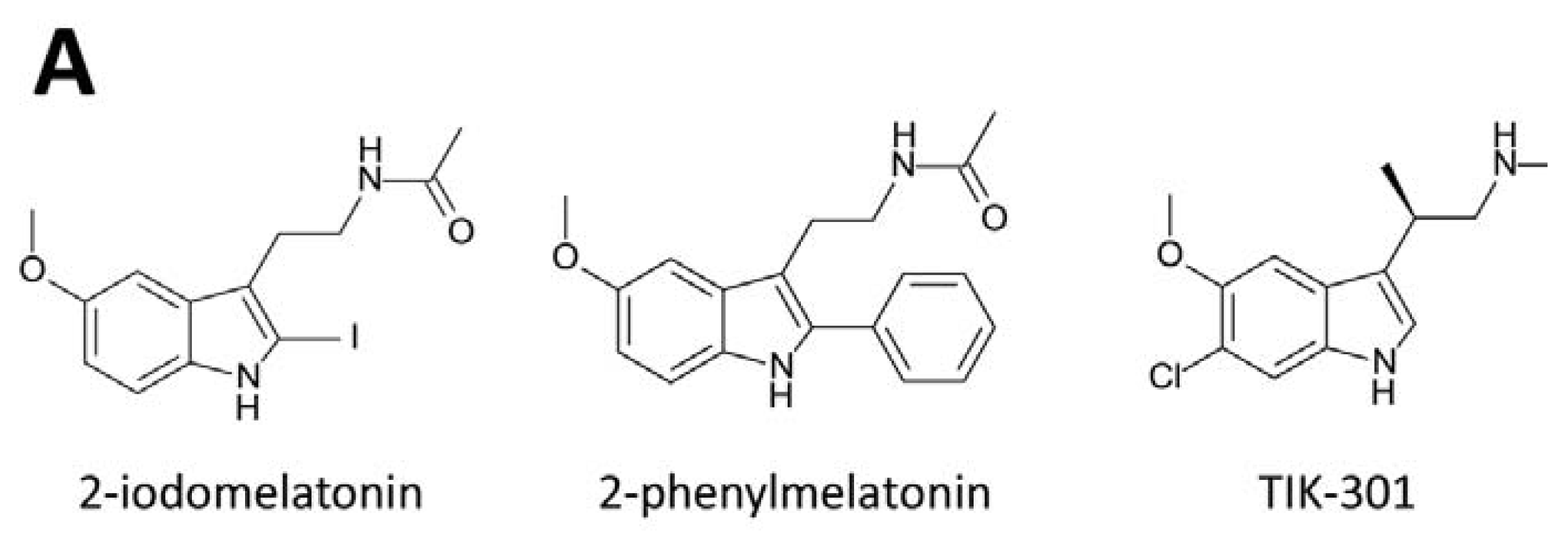
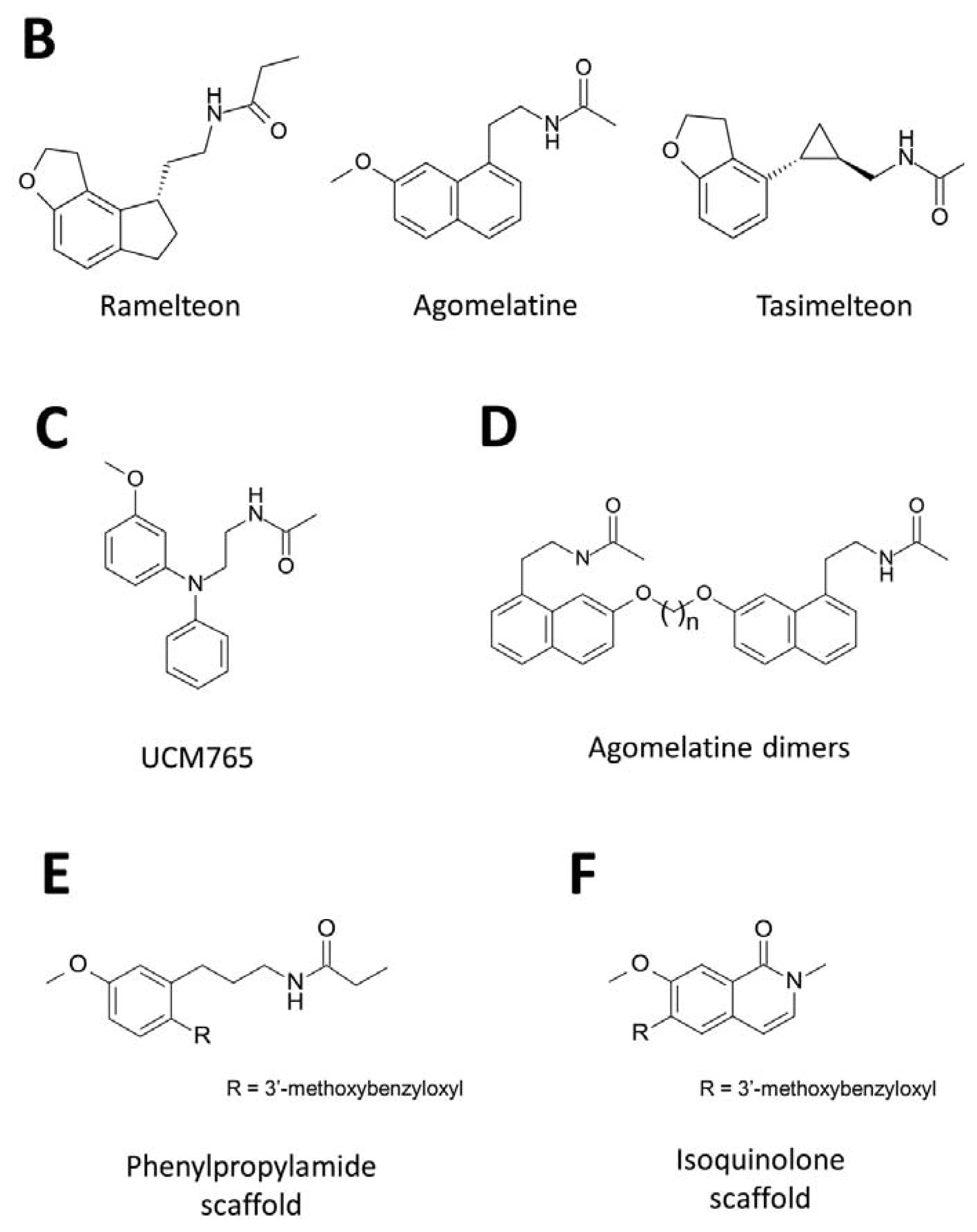
3. Development of Synthetic Melatoninergics

The therapeutic potential of melatonin is limited by its non-specific actions at multiple receptors as well as its unfavorable pharmacokinetic properties, such as high first-pass metabolism, short half-life and poor oral bioavailability [61]. Hence, much work has been undertaken to discover and develop new classes of melatoninergic ligands with improved pharmacological properties including receptor subtype selectivity and higher binding affinity. Melatoninergics agonists such as 2-iodomelatonin, 2-phenylmelatonin [62,63] and TIK-301 (β-methyl-6-chloromelatonin) [64] were therefore developed (Figure 1A), resembling the indolic core structure of melatonin with slightly higher affinity toward MT2. Bioisosteric replacements of the melatonin structure have also been widely adopted resulting in numerous non-indoleamine melatoninergics with heterocyclic scaffolds appearing in the literature. The ever-growing list includes indane, naphthalene, benzoxazole and benzofuran, fluroene, carbazole, quinoline, isoquinoline, benzopyran, benzothiazole, benzoxazine, benzothiazine, benzoxadole, azaindole, benzylpiperidine, biphenyl, pyridine, aryloxyanilide and phenylpropylamide [6568]. Ramelteon (Figure 1B) is an indane derivative with critical features for melatonin receptor binding and shows high binding affinity at both MT1 and MT2 receptors (Ki CHO-hMT1 = 14 pM; Ki CHO-hMT2 = 45 pM) [61,69]. Another widely used strategy to obtain potent melatoninergic ligands is by replacement of the indole moiety with aromatic fragments. Agomelatine (Figure 1B) is a naphthalene derivative [70,71] that retains a similar binding affinity as melatonin but possesses both melatonin agonist and serotonin antagonist properties (Ki CHO-hMT1 = 0.1 nM; Ki CHO-hMT2 = 0.12 nM; Ki CHO-h5-HT2C = 708 nM) [72,73]. By mimicking the indane structure of Ramelteon, a dihydrobenzofuran scaffold is linked to the ethylamidomethyl chain by a cyclopropyl ring. Tasimelteon (Figure 1B) displays melatoninergic activity with minimal structural requirements for receptor binding [74]. A diverse array of synthetic melatoninergic ligand from different structural scaffolds has been developed and their receptor affinity, intrinsic activity, subtype selectivity and metabolism were studied in relation to their chemical design. Despite these advances, very few subtype-selective melatoninergic ligands are available for research purposes, let alone drug development. Luzindole and 5-methoxyluzindole are among the early identified melatoninergics showing modest MT2 selectivity. The incorporation of 2-benzyl group appears to reduce binding affinity at the MT1, leading to MT2 selectivity [75]. Similarly, analogs like N0889 and DH97 are endowed with a higher affinity for MT2 receptor and Ki(MT1)/Ki(MT2) ratios ranging from 31- to 89-fold [75,76]. Further variations of the 2-benzyl group have been made by cyclization to constrain its orientation, yielding tetracyclic compounds like K185 and IIK7, which also exhibited 90- and 132-fold higher affinity for the MT2 than for the MT1 receptor, respectively [77]. The role of an aromatic group right next to the alkylamide chain in subtype selectivity is further supported by other melatoninergics with non-indole scaffolds. Binding affinity of 4P-PDOT, S24773, S24014 and S28407 display over 100-fold preferences toward the MT2 receptor [72,78,79]. Despite being receptor subtype selective, none of these melatoninergic agents had been clinically developed and marketed as a treatment for circadian rhythm related sleep disorders. The development of a subtype-selective drug candidate may result in a safer medicine with more favorable pharmacological profiles. UCM765 (Figure 1C) is a relatively simple N-phenylaniline-based compound which preferentially binds to MT2 with an affinity two orders of magnitude higher than to MT1 [80,81]. A metabolically more stable analog, UCM924, has also been developed without compromising binding affinity. Their MT2 selectivity are believed to be more favorable for re-entrainment of the circadian rhythm and alleviation of symptoms related to shifted or poorly coupled circadian oscillations. Discovery of MT1-selective compounds is unexpectedly rare. A bulky substituent replacing the 5-methoxy group of melatonin or its naphthalenic analog gives rise to a series of modestly MT1-selective compounds. A key feature is that most of them are dimeric derivatives resembling two molecules of agomelatine attached to both ends of apolymethylene chain (Figure 1D) [79]. The length of the linking chain can vary from two to eight, but three gives the highest selectivity ratio. Functional characterization of one of these dimeric melatoninergics reveals its antagonistic activity with approximately 40-fold MT1 selectivity. Another series of MT1-selective ligands are N-(anilinoalkyl)amides bearing 3-arylalkyloxy or 3-alkyloxy substituents at the aniline ring. Derivative with a phenylbutyloxy substituent was shown to be a potent partial agonist, displaying 78-fold selectivity for the MT1 receptor [82].
The emergence of melatoninergic agonist, partial agonist and antagonist not only helps to delineate melatonin receptor mediated actions, but many of the compounds were also used as pharmacological tools in rationalizing their structure activity relationships (SAR), providing new insights into the development of new classes of melatoninergic therapeutic agents. Previous studies of the structure-activity relationship have revealed that the 3-acylaminoethyl chain and 5-methoxy groups on the indole ring of melatonin are the crucial components in receptor binding; removal of either the acylaminoethyl chain or the methoxy group would lead to significant loss of receptor affinity [62,83]. The size of the amide terminal group on the acylaminoethyl chain appears to modulate ligand affinity on the melatonin receptors. Extending the acetyl group to propionyl endowed the compound with improved binding affinity and potency, but substituents higher than butyl or composed of branched structure have an adverse effect on the affinity. There have been reports that substitutions with cyclopropylcarbonyl or cyclobutylcarbonyl group would shift the intrinsic activity from agonist to antagonist [84,85]. The relative distance of the amide group and the methoxyl group is also critical for ligand-receptor interaction. Melatonin analogs with the methoxyl group moving to positions 4, 6 or 7 exhibited reduced affinity, indicating that 5-methoxyl group is optimal for receptor binding [85,86]. Replacement of the 5-methoxyl group by hydrogen, hydroxyl or alkoxy groups also decreased affinity, while halogen substituents retained both affinity and intrinsic activity [87]. Affinity of melatonin can be further enhanced by substituting a halogen, methyl or a phenyl group at the C2 position of the indole ring. A series of substituted analogs also displays higher affinity for the MT2 receptor, indicating that these substituents are better tolerated in the MT2 binding pocket and are likely interacting with an auxiliary binding subdomain in the receptor [88,89]. Furthermore, it has been subsequently revealed that the indole scaffold does not actually play a major role in the receptor binding. Either shifting the position of the nitrogen atom in the pyrrolidine ring or replacing indole with other aromatic fragments does not generally affect the receptor affinity [80,90]. With the appropriately positioned acylaminoethyl and methoxy groups, bioisosteres, such as Ramelteon and agomelatin, have demonstrated potent melatoninergic activity similar to the indole-based derivatives [85,89].
A number of synthetic melatoninergics with improved pharmacokinetic properties compared to melatonin have been developed into clinical uses, but none of them exhibits selectivity towards either receptor subtype [91]. The development of a subtype-selective drug candidate may result in a safer medicine with more favorable pharmacological profiles. A novel series of substituted N-[3-(3-methoxyphenyl)propyl]amides were identified in our laboratory displaying superb binding affinities toward human MT2, and MT1 to a lesser extent [92,93]. Similar to melatonin, a terminal simple alkyl group (up to propyl) attached on the propylamide chain progressively improves the intrinsic activity, whereas a terminal phenyl substitute is devoid of any binding. Particularly, introduction of a 3′-methoxyl group to the benzyloxyl substitute incorporated at C6 position of the phenylpropylamide scaffold (analogous to C2 position of melatonin) dramatically enhances the binding affinity toward MT2 but not MT1 (Figure 1E). Furthermore, unpublished results in our laboratory indicate that a 3′-methoxybenzyloxyl group linked with a bicyclic isoquinolone scaffold (Figure 1F) without the protruding alkylamide chain can still bind MT2 at modest to high affinity when compared with melatonin, and these compounds are basically inactive toward MT1. Substitution at the 6 position provided a potent agonist, which was endowed with high MT2 binding affinity (Ki = 26.6 nM) and 139-fold selectivity for the MT2 receptor. The physiological significance of such exclusively MT2-selective compounds awaits further studies.

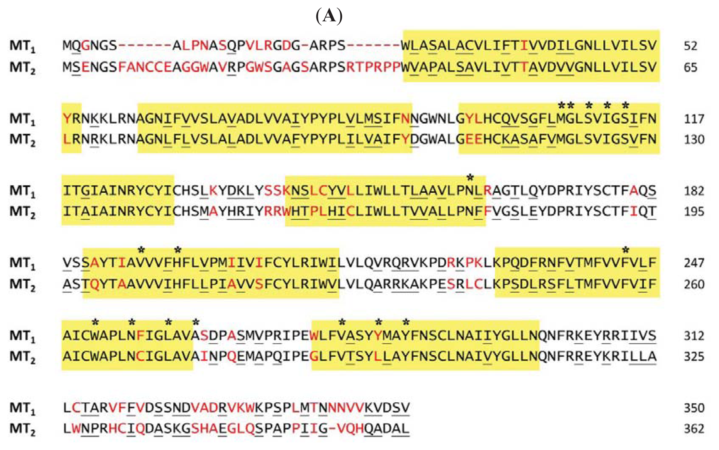
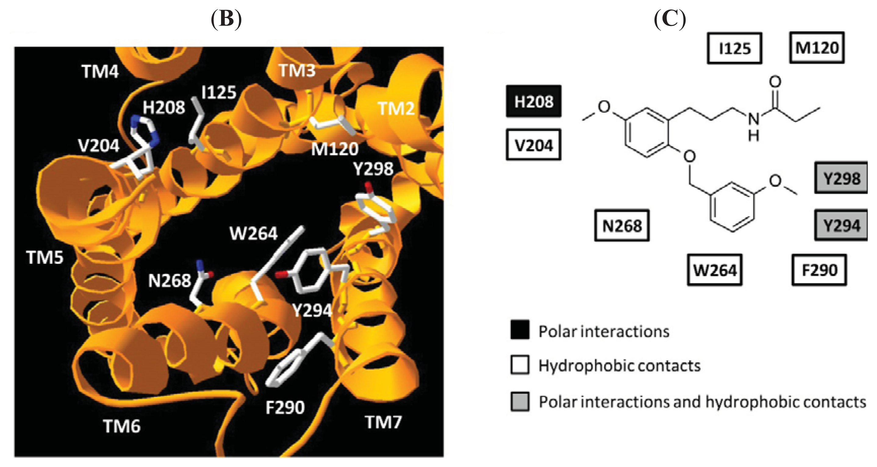
4. Homology Models of Melatonin Receptors

Similar to other rhodopsin-like GPCRs, melatonin receptors contain seven putative transmembrane helices connected by three intracellular and three extracellular loops. These helices are believed to superpose with that of rhodopsin-like receptors and have common residues at a conserved position. The structural similarity has allowed the interactions between melatonin receptors and their ligands to be predicted through a combination of site directed mutagenesis and homology modeling approaches. Sequence alignment of MT1 and MT2 showed that the position of a conserved histidine in TM5 (His195 for MT1 and His208 for MT2) is identical to that utilized in the ligand binding site of many other GPCRs, conferring an important role of the residue in ligand recognition (Figure 2A). Substitution of the histidine residue with alanine reduced the binding affinity of melatonin but did not affect the affinity for the antagonist luzindole [94,95]. Previous studies of structure-activity relationship have revealed that the 5-methoxy group of melatonin or equivalent methoxy group of other melatoninergic agonists is a crucial component in receptor binding [87]. The oxygen of the methoxy group has been postulated to form a hydrogen bond with the histidine residue on TM5, which is essential for receptor activation.
Residues located within TM3, 5, 6 and 7 have also been extensively studied by site-directed mutagenesis, but it appeared that the conserved histidine is the only common residue correlated to the binding site of the two subtypes. TM3 of rhodopsin-like GPCRs contains a relatively large number of cysteine, serine and threonine residues than the other TM domains. These residues can form hydrogen bonds with the peptide backbone and facilitate receptor conformational change at different functional states. Ser110 and Ser114 in TM3 of MT1 seem to be involved in agonist binding through interacting with the acetamidoethyl chain of melatonin [96]. Mutations of serine residues to alanine reduced melatonin affinity, while the binding of luzindole was unaffected, inferring the subset of residues for binding agonist and antagonist may differ. In contrast, the equivalent TM3 serine residues (Ser123 and Ser127) on MT2 are not important for ligand binding [94]. The distinct tolerance to conserved serine mutations hinted of structural divergence between the two subtypes.
Ligand binding interaction was relatively less evident in MT2 receptor and different hypotheses have been proposed to predict the putative binding site. Gerdin et al. suggested that both the asparagine in the TM4 (Asn175) and the conserved His208 participate in the interaction with the 5-methoxy group of melatonin, while two residues in the TM6 domains (Phe257 and Trp264) facilitate the binding of aromatic compounds such as luzindole and 4P-PDOT through π-π interaction [94]. On the other hand, Mazna and colleagues have identified several residues in TM3 (Met120, Gly121 and Ile125), TM5 (Val204), TM6 (Leu272 and Ala275) and TM7 (Val291 and Leu295) that are crucial for ligand binding as mutations at these positions reduced the binding affinity for melatonin. The subset of residues identified was found to be involved in agonist binding possibly via hydrophobic interactions with the indole group and N-acetyl group of 2-iodomelatonin [97,98]. The authors also hypothesized a potential role for Asn268 and Tyr298 in facilitating the specific interaction of His208 with the 5-methoxy substituent of melatonin.
Based on the hypothesis proposed by Gerdin et al. [94], Farce et al. re-orientated the helix bundles in their proposed receptor models to represent an active form of melatonin receptor upon binding melatonin [99]. To place the critical residues (Ser110, Ser114 and His195 in MT1; Asn175 and His208 in MT2, respectively) in proper position to bind melatonin, the authors have to rotate TM3 and TM5 helices of MT1 receptor in a clockwise manner, while rotating TM4 of MT2 counter-clockwise. The distinct conformations adopted by the two receptor subtypes in response to endogenous melatonin binding may confer functional selectivity in signaling upon receptor activation.
Mor et al. have published SAR data showing that the incorporation of a distant 2-benzyl substituent on the indole scaffold of melatonin specifically enhances MT2 binding [100]. The data gathered suggest that an additional binding cavity formed by a large group of hydrophobic amino acid side chains may exist in the MT2 receptor binding site to accommodate the aromatic out-of-plane substituent. To identify the putative hydrophobic pocket, homology models of MT1 and MT2 receptors have been built using the crystal structure of bovine rhodopsin as a template. In the MT2 model proposed by Rivara et al., the aromatic ring of the reference MT2 selective antagonist N-[1-(4-chloro-benzyl)-4-methoxy-1H-indol-2-ylmethyl]-propionamide lied in a putative hydrophobic pocket located between TM3, TM5 and TM6, and in close vicinity to the side chain of Trp264 upon ligand docking [101]. Molecular dynamics simulations confirmed that the distance between Trp264 and the aromatic substituent of the antagonist is observed to be closer in MT2 than that of the MT1 receptor complex. The two interacting groups form a stable π-π interaction when in close proximity and enhance MT2 selectivity. The observed interaction, however, limits the conformational freedom of the CWXP motif in TM6 and prevents receptor activation, thus providing an explanation for the antagonist activity.
Among the family of rhodopsin-like GPCRs, melatonin receptors and opsins are phylogentically close and may share a common ancestor. Despite their sequence similarity, the partially active opsin structure is suggested to be conformationally different from a ligand binding activated receptor, inferring diverse mechanisms underlying GPCR activation throughout the family. The homology modeling of small molecule binding aminergic receptor therefore may provide a reference set of amino acids for determining the ligand binding residues in the melatonin receptor [102,103]. Protein sequence alignment of a diverse array of vertebrate receptor orthologues revealed that amino residue 6.48 (equivalent to Trp264 in MT2) is conserved in most rhodopsin-like GPCRs and is likely to play a general role in receptor activation by stabilizing conformational alternation of side chains upon agonist binding [102,104]. Base on the aminergic binding site defined by evolutionary trace analysis, Gloriam et al. predicted 12 positions on TM3 (3.32, 3.33, 3.37 and 3.40), TM5 (5.42, 5.43, 5.46 and 5.47), TM6 (6.51 and 6.52) and TM7 (7.42 and 7.43) are involved in the generic binding site (corresponding residues in MT1 and MT2 receptors are listed in Tables 1 and 2) [105]. The subset of residues identified is in agreement with the MT2 receptor homology model proposed by Mazna and colleagues [97,98].
Agomelatine is an antidepressant which binds specifically to melatonin and type 2C serotonin (5-HT2C) receptors [70,71]. This cross reactivity suggests that both receptors may share a similar structure-activity relationships profile upon binding agomelatine. The modeling of the agomelatine-bound state of the serotonin receptor therefore may allow us to identify important structural features that are may be similarly present in the melatonin receptors. An extensive screening of antagonist-bound 5-HT2C models suggested that antagonists are caged between TM3, TM5, TM6 and TM7, and agomelatine adopts a conformation wherein its protonated amine group interacts with Asp134 (3.32) and Tyr358 (7.43) through hydrogen bonding [107]. As observed in the respective crystal structures, the naphthalene ring of agomelatine also participates in π-π stacking binding interactions with the hydrophobic cluster comprising Trp324 (6.48), Phe327 (6.51) and Phe328 (6.52) [108]. These two identified structural features are not only valuable to the design of new antidepressants, they also help to further our understanding of the nature of residues that are required for receptor-ligand interaction.
Homology modeling approaches have identified a subset of residues and potential structural determinants that appear to be critical for receptor ligand binding [82,109]. However, the reliability of the prediction is susceptible to the degree of similarity between the target sequence and the template of crystallized receptors. Comparative analyses of prospective modeled ligand-receptor complexes generated from distinct classes of GPCR templates suggest that 35%–40% sequence identity boundaries are required for reliable homology modeling in general [110]. Although modeling between closely related receptor subtypes, and especially, among the same class of GPCRs is generally successful, distant homology modeling based on a phylogenetically remote receptor template often fails to produce accurate receptor models due to significant deviation in structures [109]. Melatonin shares a low sequence identity with the available crystallized receptor, and hence, receptor modeling is usually accompanied by experimental data from mutagenesis studies. Recent resolution of crystal structures of aminergic receptors allows melatonin receptor homology models to be built by using a structurally similar receptor template and this approach may facilitate our understanding of the melatonin receptor structure.
On the basis of mutagenesis studies demonstrating that the conserved histidine residue (His195 for MT1 and His208 for MT2) is important for melatonin binding and activation [94,95], possible interaction of His208 in MT2 with the 3-methoxy group of phenylpropylamide was probed by assessing the potency difference between the wild-type and mutant receptors. When the histidine residue in the MT2 receptor was mutated to alanine (His208Ala), MT2-selective phenylpropylamide compounds exhibited a significant loss in potency and efficacy (unpublished data). Despite their difference in subtype selectivity, the data suggests that the interaction between the methoxy group and the histidine is critical for both melatonin and phenylpropylamide binding, and the two agonists may stabilize the receptor binding pocket conformation in a similar manner. In the presence of the selectivity determinant 3′-methoxybenzyloxyl group at C6 position of the phenylpropylamide scaffold, compounds preferentially activate MT2 over MT1. According to receptor models proposed by Mor et al. [100] and Rivara et al. [101], an additional binding cavity present in MT2 is able to accommodate the aromatic substituent, thereby increasing the MT2 affinity of phenylpropylamide compounds bearing a 3′-methoxybenzyloxyl moiety. Base on the homology modeling of melatonin and serotonin receptors, such a cavity is most likely formed by a large group of hydrophobic amino acid side chains located at TM6 (Trp264, Asn268) and TM7 (Phe290, Tyr294, Tyr298) (Figure 2B,C). Molecular interactions between the 3′-methoxyl substituent on the benzyl ring and the MT2 receptor, however, are less understood. To characterize structural features responsible for subtype selective ligand binding at the MT2 receptor, additional mutagenesis studies will need to include selective residues that participate in the formation of the putative hydrophobic binding pocket.
Lastly, a novel series of MT2-selective isoquinolone derivatives (Figure 1F) can bind to and activate the His208Ala MT2 receptor mutant. These compounds possess a substituted 3-methoxylbenzyloxyl substituent at their bicyclic scaffold but have little structural resemblance to melatonin. They exhibit an EC50 comparable to that of melatonin towards MT2 and are totally inactive at MT1. Functional assays also demonstrated that the compound-induced response in His208Ala was essentially indistinguishable from that observed with the wild-type receptor, leading to the speculation that agonists can bind to the MT2 receptor in at least two configurations as His208 may not be required for receptor activation. Identification of the residues of MT2 receptor that participate in MT2-selective ligand docking awaits further mutagenesis studies

5. Conclusions

Melatonin exerts its physiological actions through mediating multiple signaling pathways via transmembrane melatonin receptors. Parallel signaling mechanisms of melatonin also promote transcriptional activities by recruiting various kinases and transcription factors, acting on the downstream of signaling pathways in a cell type specific manner. Hence, melatonin displays a pleiotropic functional profile in addition to its primary role in regulating circadian rhythm and sleep promotion. With regard to the distinct role of melatonin receptor subtypes in chronobiology and immunomodulation, subtype-selective melatoninergics are of emerging interest since they may result in a safer medicine with more promising therapeutic effects. Further clarifications of receptor ligand molecular interactions will guide the development of melatonin receptor subtype-specific therapeutic agents while structural data remains relatively limited.

Acknowledgments

Studies by the authors are supported in part by the National Key Basic Research Program of China (2013CB530900), the Hong Kong Jockey Club, Research Grant Council (HKUST 660108) and University Grant Council of Hong Kong (T13-607/12R).

Conflicts of Interest

The authors declare no conflict of interest.

References

  1. Poeggeler, B.; Reiter, R.J.; Tan, D.X.; Chen, L.D.; Manchester, L.C. Melatonin, hydroxyl radical-mediated oxidative damage, and aging: A hypothesis. J. Pineal Res 1993, 14, 151–168. [Google Scholar]
  2. Kennaway, D.; Wright, H. Melatonin and circadian rhythms. Curr. Top. Med. Chem 2002, 2, 199–209. [Google Scholar]
  3. Geoffriau, M.; Claustrat, B.; Veldhuis, J. Estimation of frequently sampled nocturnal melatonin production in humans by deconvolution analysis: Evidence for episodic or ultradian secretion. J. Pineal Res 1999, 27, 139–144. [Google Scholar]
  4. Pandi-Perumal, S.; Srinivasan, V.; Maestroni, G.; Cardinali, D.; Poeggeler, B.; Hardeland, R. Melatonin: Nature’s most versatile biological signal? FEBS J 2006, 273, 2813–2838. [Google Scholar]
  5. Cardinali, D.; Furio, A.; Reyes, M.; Brusco, L. The use of chronobiotics in the resynchronization of the sleep-wake cycle. Cancer Causes Control 2006, 17, 601–609. [Google Scholar]
  6. Dawson, D.; Armstrong, S. Chronobiotics—Drugs that shift rhythms. Pharmacol. Ther 1996, 69, 15–36. [Google Scholar]
  7. Arendt, J.; Skene, D. Melatonin as a chronobiotic. Sleep Med. Rev 2005, 9, 25–39. [Google Scholar]
  8. Touitou, Y.; Bogdan, A. Promoting adjustment of the sleep-wake cycle by chronobiotics. Physiol. Behav 2007, 90, 294–300. [Google Scholar]
  9. Sack, R.L.; Auckley, D.; Auger, R.R.; Carskadon, M.A.; Wright, K.P.; Vitiello, M.V.; Zhdanova, I.V.; Medicine, A.A.O.S. Circadian rhythm sleep disorders: part I, basic principles, shift work and jet lag disorders. An American Academy of Sleep Medicine review. Sleep 2007, 30, 1460–1483. [Google Scholar]
  10. Kayumov, L.; Brown, G.; Jindal, R.; Buttoo, K.; Shapiro, C.M. A randomized, double-blind, placebo-controlled crossover study of the effect of exogenous melatonin on delayed sleep phase syndrome. Psychosom. Med 2001, 63, 40–48. [Google Scholar]
  11. Sack, R.L.; Auckley, D.; Auger, R.R.; Carskadon, M.A.; Wright, K.P.; Vitiello, M.V.; Zhdanova, I.V.; Medicine, A.A.O.S. Circadian rhythm sleep disorders: part II, advanced sleep phase disorder, delayed sleep phase disorder, free-running disorder, and irregular sleep-wake rhythm. An American Academy of Sleep Medicine review. Sleep 2007, 30, 1484–1501. [Google Scholar]
  12. Skene, D.J.; Arendt, J. Circadian rhythm sleep disorders in the blind and their treatment with melatonin. Sleep Med 2007, 8, 651–655. [Google Scholar]
  13. Herxheimer, A.; Petrie, K.J. Melatonin for the prevention and treatment of jet lag. Cochrane Database Syst. Rev. 2002. [Google Scholar] [CrossRef]
  14. Arendt, J. Melatonin and human rhythms. Chronobiol. Int 2006, 23, 21–37. [Google Scholar]
  15. Ekmekcioglu, C. Melatonin receptors in humans: biological role and clinical relevance. Biomed. Pharmacother 2006, 60, 97–108. [Google Scholar]
  16. Ram, P.; Dai, J.; Yuan, L.; Dong, C.; Kiefer, T.; Lai, L.; Hill, S. Involvement of the mt1 melatonin receptor in human breast cancer. Cancer Lett 2002, 179, 141–150. [Google Scholar]
  17. Carrillo-Vico, A.; Reiter, R.; Lardone, P.; Herrera, J.; Fernández-Montesinos, R.; Guerrero, J.; Pozo, D. The modulatory role of melatonin on immune responsiveness. Curr. Opin. Investig. Drugs 2006, 7, 423–431. [Google Scholar]
  18. Bondy, S.C.; Sharman, E.H. Melatonin and the aging brain. Neurochem. Int 2007, 50, 571–580. [Google Scholar]
  19. De Jonghe, A.; Korevaar, J.C.; van Munster, B.C.; de Rooij, S.E. Effectiveness of melatonin treatment on circadian rhythm disturbances in dementia. Are there implications for delirium? A systematic review. Int. J. Geriatr. Psychiatry 2010, 25, 1201–1208. [Google Scholar]
  20. Jan, J.E.; Reiter, R.J.; Wong, P.K.; Bax, M.C.; Ribary, U.; Wasdell, M.B. Melatonin has membrane receptor-independent hypnotic action on neurons: An hypothesis. J. Pineal Res 2011, 50, 233–240. [Google Scholar]
  21. Larson, E.B.; Zollman, F.S. The effect of sleep medications on cognitive recovery from traumatic brain injury. J. Head Trauma Rehabil 2010, 25, 61–67. [Google Scholar]
  22. Reiter, R.J.; Tan, D.X.; Manchester, L.C.; Tamura, H. Melatonin defeats neurally-derived free radicals and reduces the associated neuromorphological and neurobehavioral damage. J. Physiol. Pharmacol 2007, 58, 5–22. [Google Scholar]
  23. Ebisawa, T.; Karne, S.; Lerner, M.; Reppert, S. Expression cloning of a high-affinity melatonin receptor from Xenopus dermal melanophores. Proc. Natl. Acad. Sci. USA 1994, 91, 6133–6137. [Google Scholar]
  24. Reppert, S.; Godson, C.; Mahle, C.; Weaver, D.; Slaugenhaupt, S.; Gusella, J. Molecular characterization of a second melatonin receptor expressed in human retina and brain: The Mel1b melatonin receptor. Proc. Natl. Acad. Sci. USA 1995, 92, 8734–8738. [Google Scholar]
  25. Reppert, S.; Weaver, D.; Ebisawa, T. Cloning and characterization of a mammalian melatonin receptor that mediates reproductive and circadian responses. Neuron 1994, 13, 1177–1185. [Google Scholar]
  26. Dubocovich, M.; Markowska, M. Functional MT1 and MT2 melatonin receptors in mammals. Endocrine 2005, 27, 101–110. [Google Scholar]
  27. Slaugenhaupt, S.; Roca, A.; Liebert, C.; Altherr, M.; Gusella, J.; Reppert, S. Mapping of the gene for the Mel1a-melatonin receptor to human chromosome 4 (MTNR1A) and mouse chromosome 8 (Mtnr1a). Genomics 1995, 27, 355–357. [Google Scholar]
  28. Reppert, S.; Weaver, D.; Ebisawa, T.; Mahle, C.; Kolakowski, L.J. Cloning of a melatonin-related receptor from human pituitary. FEBS Lett 1996, 386, 219–224. [Google Scholar]
  29. Levoye, A.; Dam, J.; Ayoub, M.; Guillaume, J.; Couturier, C.; Delagrange, P.; Jockers, R. The orphan GPR50 receptor specifically inhibits MT1 melatonin receptor function through heterodimerization. EMBO J 2006, 25, 3012–3023. [Google Scholar]
  30. Becker-André, M.; Wiesenberg, I.; Schaeren-Wiemers, N.; André, E.; Missbach, M.; Saurat, J.; Carlberg, C. Pineal gland hormone melatonin binds and activates an orphan of the nuclear receptor superfamily. J. Biol. Chem 1994, 269, 28531–28534. [Google Scholar]
  31. Skwarlo-Sonta, K.; Majewski, P.; Markowska, M.; Oblap, R.; Olszanska, B. Bidirectional communication between the pineal gland and the immune system. Can. J. Physiol. Pharmacol 2003, 81, 342–349. [Google Scholar]
  32. Pandi-Perumal, S.; Trakht, I.; Srinivasan, V.; Spence, D.; Maestroni, G.; Zisapel, N.; Cardinali, D. Physiological effects of melatonin: Role of melatonin receptors and signal transduction pathways. Prog. Neurobiol 2008, 85, 335–353. [Google Scholar]
  33. Liu, C.; Weaver, D.; Jin, X.; Shearman, L.; Pieschl, R.; Gribkoff, V.; Reppert, S. Molecular dissection of two distinct actions of melatonin on the suprachiasmatic circadian clock. Neuron 1997, 19, 91–102. [Google Scholar]
  34. Reiter, R. Pineal melatonin: cell biology of its synthesis and of its physiological interactions. Endocr. Rev 1991, 12, 151–180. [Google Scholar]
  35. Dubocovich, M.; Rivera-Bermudez, M.; Gerdin, M.; Masana, M. Molecular pharmacology, regulation and function of mammalian melatonin receptors. Front. Biosci 2003, 8, d1093–d1108. [Google Scholar]
  36. Clemens, J.; Jarzynka, M.; Witt-Enderby, P. Down-regulation of mt1 melatonin receptors in rat ovary following estrogen exposure. Life Sci 2001, 69, 27–35. [Google Scholar]
  37. Frungieri, M.; Mayerhofer, A.; Zitta, K.; Pignataro, O.; Calandra, R.; Gonzalez-Calvar, S. Direct effect of melatonin on Syrian hamster testes: Melatonin subtype 1a receptors, inhibition of androgen production, and interaction with the local corticotropin-releasing hormone system. Endocrinology 2005, 146, 1541–1552. [Google Scholar]
  38. Masana, M.; Doolen, S.; Ersahin, C.; Al-Ghoul, W.; Duckles, S.; Dubocovich, M.; Krause, D. MT(2) melatonin receptors are present and functional in rat caudal artery. J. Pharmacol. Exp. Ther 2002, 302, 1295–1302. [Google Scholar]
  39. Naji, L.; Carrillo-Vico, A.; Guerrero, J.; Calvo, J. Expression of membrane and nuclear melatonin receptors in mouse peripheral organs. Life Sci 2004, 74, 2227–2236. [Google Scholar]
  40. Zalatan, F.; Krause, J.; Blask, D. Inhibition of isoproterenol-induced lipolysis in rat inguinal adipocytes in vitro by physiological melatonin via a receptor-mediated mechanism. Endocrinology 2001, 142, 3783–3790. [Google Scholar]
  41. Guerrero, J.; Reiter, R. Melatonin-immune system relationships. Curr. Top. Med. Chem 2002, 2, 167–179. [Google Scholar]
  42. Brydon, L.; Roka, F.; Petit, L.; de Coppet, P.; Tissot, M.; Barrett, P.; Morgan, P.; Nanoff, C.; Strosberg, A.; Jockers, R. Dual signaling of human Mel1a melatonin receptors via G(i2), G(i3), and G(q/11) proteins. Mol. Endocrinol 1999, 13, 2025–2038. [Google Scholar]
  43. Masana, M.I.; Dubocovich, M.L. Melatonin receptor signaling: finding the path through the dark. Sci. STKE 2001, 2001. [Google Scholar] [CrossRef]
  44. Travnickova-Bendova, Z.; Cermakian, N.; Reppert, S.M.; Sassone-Corsi, P. Bimodal regulation of mPeriod promoters by CREB-dependent signaling and CLOCK/BMAL1 activity. Proc. Natl. Acad. Sci. USA 2002, 99, 7728–7733. [Google Scholar]
  45. Jin, X.; von Gall, C.; Pieschl, R.; Gribkoff, V.; Stehle, J.; Reppert, S.; Weaver, D. Targeted disruption of the mouse Mel(1b) melatonin receptor. Mol. Cell. Biol 2003, 23, 1054–1060. [Google Scholar]
  46. Chan, A.; Lai, F.; Lo, R.; Voyno-Yasenetskaya, T.; Stanbridge, E.; Wong, Y. Melatonin mt1 and MT2 receptors stimulate c-Jun N-terminal kinase via pertussis toxin-sensitive and -insensitive G proteins. Cell Signal 2002, 14, 249–257. [Google Scholar]
  47. Kyriakis, J.M.; Banerjee, P.; Nikolakaki, E.; Dai, T.; Rubie, E.A.; Ahmad, M.F.; Avruch, J.; Woodgett, J.R. The stress-activated protein kinase subfamily of c-Jun kinases. Nature 1994, 369, 156–160. [Google Scholar]
  48. van Dam, H.; Duyndam, M.; Rottier, R.; Bosch, A.; de Vries-Smits, L.; Herrlich, P.; Zantema, A.; Angel, P.; van der Eb, A.J. Heterodimer formation of cJun and ATF-2 is responsible for induction of c-jun by the 243 amino acid adenovirus E1A protein. EMBO J 1993, 12, 479–487. [Google Scholar]
  49. Zinck, R.; Cahill, M.A.; Kracht, M.; Sachsenmaier, C.; Hipskind, R.A.; Nordheim, A. Protein synthesis inhibitors reveal differential regulation of mitogen-activated protein kinase and stress-activated protein kinase pathways that converge on Elk-1. Mol. Cell. Biol 1995, 15, 4930–4938. [Google Scholar]
  50. Garcia-Mauriño, S.; Gonzalez-Haba, M.G.; Calvo, J.R.; Rafii-El-Idrissi, M.; Sanchez-Margalet, V.; Goberna, R.; Guerrero, J.M. Melatonin enhances IL-2, IL-6, and IFN-gamma production by human circulating CD4+ cells: A possible nuclear receptor-mediated mechanism involving T helper type 1 lymphocytes and monocytes. J. Immunol 1997, 159, 574–581. [Google Scholar]
  51. Lau, W.W.; Ng, J.K.; Lee, M.M.; Chan, A.S.; Wong, Y.H. Interleukin-6 autocrine signaling mediates melatonin MT(1/2) receptor-induced STAT3 Tyr(705) phosphorylation. J. Pineal Res 2012, 52, 477–489. [Google Scholar]
  52. Eddahri, F.; Denanglaire, S.; Bureau, F.; Spolski, R.; Leonard, W.J.; Leo, O.; Andris, F. Interleukin-6/STAT3 signaling regulates the ability of naive T cells to acquire B-cell help capacities. Blood 2009, 113, 2426–2433. [Google Scholar]
  53. Shen, L.; Fahey, J.V.; Hussey, S.B.; Asin, S.N.; Wira, C.R.; Fanger, M.W. Synergy between IL-8 and GM-CSF in reproductive tract epithelial cell secretions promotes enhanced neutrophil chemotaxis. Cell. Immunol 2004, 230, 23–32. [Google Scholar]
  54. Drazen, D.; Nelson, R. Melatonin receptor subtype MT2 (Mel 1b) and not mt1 (Mel 1a) is associated with melatonin-induced enhancement of cell-mediated and humoral immunity. Neuroendocrinology 2001, 74, 178–184. [Google Scholar]
  55. Drazen, D.; Bilu, D.; Bilbo, S.; Nelson, R. Melatonin enhancement of splenocyte proliferation is attenuated by luzindole, a melatonin receptor antagonist. Am. J. Physiol. Regul. Integr. Comp. Physiol 2001, 280, R1476–R1482. [Google Scholar]
  56. Weil, Z.; Hotchkiss, A.; Gatien, M.; Pieke-Dahl, S.; Nelson, R. Melatonin receptor (MT1) knockout mice display depression-like behaviors and deficits in sensorimotor gating. Brain Res. Bull 2006, 68, 425–429. [Google Scholar]
  57. Larson, J.; Jessen, R.; Uz, T.; Arslan, A.; Kurtuncu, M.; Imbesi, M.; Manev, H. Impaired hippocampal long-term potentiation in melatonin MT2 receptor-deficient mice. Neurosci. Lett 2006, 393, 23–26. [Google Scholar]
  58. Von Gall, C.; Weaver, D.; Moek, J.; Jilg, A.; Stehle, J.; Korf, H. Melatonin plays a crucial role in the regulation of rhythmic clock gene expression in the mouse pars tuberalis. Ann. N Y Acad. Sci 2005, 1040, 508–511. [Google Scholar]
  59. Dubocovich, M.; Hudson, R.; Sumaya, I.; Masana, M.; Manna, E. Effect of MT1 melatonin receptor deletion on melatonin-mediated phase shift of circadian rhythms in the C57BL/6 mouse. J. Pineal Res 2005, 39, 113–120. [Google Scholar]
  60. Gerdin, M.; Masana, M.; Rivera-Bermúdez, M.; Hudson, R.; Earnest, D.; Gillette, M.; Dubocovich, M. Melatonin desensitizes endogenous MT2 melatonin receptors in the rat suprachiasmatic nucleus: Relevance for defining the periods of sensitivity of the mammalian circadian clock to melatonin. FASEB J 2004, 18, 1646–1656. [Google Scholar]
  61. Uchikawa, O.; Fukatsu, K.; Tokunoh, R.; Kawada, M.; Matsumoto, K.; Imai, Y.; Hinuma, S.; Kato, K.; Nishikawa, H.; Hirai, K.; et al. Synthesis of a novel series of tricyclic indan derivatives as melatonin receptor agonists. J. Med. Chem 2002, 45, 4222–4239. [Google Scholar]
  62. Dubocovich, M.; Takahashi, J. Use of 2-[125I]iodomelatonin to characterize melatonin binding sites in chicken retina. Proc. Natl. Acad. Sci. USA 1987, 84, 3916–3920. [Google Scholar]
  63. Nonno, R.; Lucini, V.; Pannacci, M.; Mazzucchelli, C.; Angeloni, D.; Fraschini, F.; Stankov, B. Pharmacological characterization of the human melatonin Mel1a receptor following stable transfection into NIH3T3 cells. Br. J. Pharmacol 1998, 124, 485–492. [Google Scholar]
  64. Mulchahey, J.; Goldwater, D.; Zemlan, F. A single blind, placebo controlled, across groups dose escalation study of the safety, tolerability, pharmacokinetics and pharmacodynamics of the melatonin analog beta-methyl-6-chloromelatonin. Life Sci 2004, 75, 1843–1856. [Google Scholar]
  65. Hardeland, R. Investigational melatonin receptor agonists. Expert Opin. Investig. Drugs 2010, 19, 747–764. [Google Scholar]
  66. Zlotos, D.P. Recent advances in melatonin receptor ligands. Arch. Pharm. (Weinheim) 2005, 338, 229–247. [Google Scholar]
  67. Mor, M.; Rivara, S.; Pala, D.; Bedini, A.; Spadoni, G.; Tarzia, G. Recent advances in the development of melatonin MT(1) and MT(2) receptor agonists. Expert Opin. Ther. Pat 2010, 20, 1059–1077. [Google Scholar]
  68. Zlotos, D.P. Recent progress in the development of agonists and antagonists for melatonin receptors. Curr. Med. Chem 2012, 19, 3532–3549. [Google Scholar]
  69. Kato, K.; Hirai, K.; Nishiyama, K.; Uchikawa, O.; Fukatsu, K.; Ohkawa, S.; Kawamata, Y.; Hinuma, S.; Miyamoto, M. Neurochemical properties of ramelteon (TAK-375), a selective MT1/MT2 receptor agonist. Neuropharmacology 2005, 48, 301–310. [Google Scholar]
  70. Bourin, M.; Mocaër, E.; Porsolt, R. Antidepressant-like activity of S 20098 (agomelatine) in the forced swimming test in rodents: involvement of melatonin and serotonin receptors. J. Psychiatry Neurosci 2004, 29, 126–133. [Google Scholar]
  71. Millan, M.; Gobert, A.; Lejeune, F.; Dekeyne, A.; Newman-Tancredi, A.; Pasteau, V.; Rivet, J.; Cussac, D. The novel melatonin agonist agomelatine (S20098) is an antagonist at 5-hydroxytryptamine2C receptors, blockade of which enhances the activity of frontocortical dopaminergic and adrenergic pathways. J. Pharmacol. Exp. Ther 2003, 306, 954–964. [Google Scholar]
  72. Audinot, V.; Mailliet, F.; Lahaye-Brasseur, C.; Bonnaud, A.; Le Gall, A.; Amossé, C.; Dromaint, S.; Rodriguez, M.; Nagel, N.; Galizzi, J.; et al. New selective ligands of human cloned melatonin MT1 and MT2 receptors. Naunyn. Schmiedeberg Arch. Pharmacol 2003, 367, 553–561. [Google Scholar]
  73. Ettaoussi, M.; Sabaouni, A.; Rami, M.; Boutin, J.A.; Delagrange, P.; Renard, P.; Spedding, M.; Caignard, D.H.; Berthelot, P.; Yous, S. Design, synthesis and pharmacological evaluation of new series of naphthalenic analogues as melatoninergic (MT1/MT2) and serotoninergic 5-HT2C dual ligands (I). Eur. J. Med. Chem 2012, 49, 310–323. [Google Scholar]
  74. Hardeland, R. Tasimelteon, a melatonin agonist for the treatment of insomnia and circadian rhythm sleep disorders. Curr. Opin. Investig. Drugs 2009, 10, 691–701. [Google Scholar]
  75. Dubocovich, M.; Masana, M.; Iacob, S.; Sauri, D. Melatonin receptor antagonists that differentiate between the human Mel1a and Mel1b recombinant subtypes are used to assess the pharmacological profile of the rabbit retina ML1 presynaptic heteroreceptor. Naunyn. Schmiedeberg Arch. Pharmacol 1997, 355, 365–375. [Google Scholar]
  76. Teh, M.; Sugden, D. Comparison of the structure-activity relationships of melatonin receptor agonists and antagonists: Lengthening the N-acyl side-chain has differing effects on potency on Xenopus melanophores. Naunyn. Schmiedeberg Arch. Pharmacol 1998, 358, 522–528. [Google Scholar]
  77. Faust, R.; Garratt, P.J.; Jones, R.; Yeh, L.K.; Tsotinis, A.; Panoussopoulou, M.; Calogeropoulou, T.; Teh, M.T.; Sugden, D. Mapping the melatonin receptor. 6. Melatonin agonists and antagonists derived from 6H-isoindolo[2,1-a]indoles, 5,6-dihydroindolo[2,1-a]isoquinolines, and 6,7-dihydro-5H-benzo[c]azepino[2,1-a]indoles. J. Med. Chem 2000, 43, 1050–1061. [Google Scholar]
  78. Wallez, V.; Durieux-Poissonnier, S.; Chavatte, P.; Boutin, J.; Audinot, V.; Nicolas, J.; Bennejean, C.; Delagrange, P.; Renard, P.; Lesieur, D. Synthesis and structure-affinity-activity relationships of novel benzofuran derivatives as MT(2) melatonin receptor selective ligands. J. Med. Chem 2002, 45, 2788–2800. [Google Scholar]
  79. Descamps-François, C.; Yous, S.; Chavatte, P.; Audinot, V.; Bonnaud, A.; Boutin, J.; Delagrange, P.; Bennejean, C.; Renard, P.; Lesieur, D. Design and synthesis of naphthalenic dimers as selective MT1 melatoninergic ligands. J. Med. Chem 2003, 46, 1127–1129. [Google Scholar]
  80. Rivara, S.; Mor, M.; Bedini, A.; Spadoni, G.; Tarzia, G. Melatonin receptor agonists: SAR and applications to the treatment of sleep-wake disorders. Curr. Top. Med. Chem 2008, 8, 954–968. [Google Scholar]
  81. Rivara, S.; Lodola, A.; Mor, M.; Bedini, A.; Spadoni, G.; Lucini, V.; Pannacci, M.; Fraschini, F.; Scaglione, F.; Sanchez, R.O.; et al. N-(substituted-anilinoethyl)amides: design, synthesis, and pharmacological characterization of a new class of melatonin receptor ligands. J. Med. Chem 2007, 50, 6618–6626. [Google Scholar]
  82. Rivara, S.; Pala, D.; Lodola, A.; Mor, M.; Lucini, V.; Dugnani, S.; Scaglione, F.; Bedini, A.; Lucarini, S.; Tarzia, G.; et al. MT(1)-Selective Melatonin Receptor Ligands: Synthesis, Pharmacological Evaluation, and Molecular Dynamics Investigation of N-{[(3-O-Substituted) anilino]alkyl}amides. ChemMedChem 2012, 7, 1954–1964. [Google Scholar]
  83. Sugden, D.; Pickering, H.; Teh, M.T.; Garratt, P.J. Melatonin receptor pharmacology: toward subtype specificity. Biol. Cell 1997, 89, 531–537. [Google Scholar]
  84. Navajas, C.; Kokkola, T.; Poso, A.; Honka, N.; Gynther, J.; Laitinen, J.T. A rhodopsin-based model for melatonin recognition at its G protein-coupled receptor. Eur. J. Pharmacol 1996, 304, 173–183. [Google Scholar]
  85. Garratt, P.; Travard, S.; Vonhoff, S.; Tsotinis, A.; Sugden, D. Mapping the melatonin receptor. 4. Comparison of the binding affinities of a series of substituted phenylalkyl amides. J. Med. Chem 1996, 39, 1797–1805. [Google Scholar]
  86. Spadoni, G.; Mor, M.; Tarzia, G. Structure-affinity relationships of indole-based melatonin analogs. Biol. Signals Recept 1999, 8, 15–23. [Google Scholar]
  87. Mor, M.; Rivara, S.; Silva, C.; Bordi, F.; Plazzi, P.V.; Spadoni, G.; Diamantini, G.; Balsamini, C.; Tarzia, G.; Fraschini, F.; et al. Melatonin receptor ligands: synthesis of new melatonin derivatives and comprehensive comparative molecular field analysis (CoMFA) study. J. Med. Chem 1998, 41, 3831–3844. [Google Scholar]
  88. Sugden, D.; Chong, N.; Lewis, D. Structural requirements at the melatonin receptor. Br. J. Pharmacol 1995, 114, 618–623. [Google Scholar]
  89. Spadoni, G.; Stankov, B.; Duranti, A.; Biella, G.; Lucini, V.; Salvatori, A.; Fraschini, F. 2-Substituted 5-methoxy-N-acyltryptamines: synthesis, binding affinity for the melatonin receptor, and evaluation of the biological activity. J. Med. Chem 1993, 36, 4069–4074. [Google Scholar]
  90. Beresford, I.; Browning, C.; Starkey, S.; Brown, J.; Foord, S.; Coughlan, J.; North, P.; Dubocovich, M.; Hagan, R. GR196429: A nonindolic agonist at high-affinity melatonin receptors. J. Pharmacol. Exp. Ther 1998, 285, 1239–1245. [Google Scholar]
  91. Mody, S.; Hu, Y.; Ho, M.; Wong, Y. In search of novel and therapeutically significant melatoninergic ligands. Recent Pat. CNS Drug Discov 2007, 2, 241–245. [Google Scholar]
  92. Hu, Y.; Ho, M.; Chan, K.; New, D.; Wong, Y. Synthesis of substituted N-[3-(3-methoxyphenyl)propyl] amides as highly potent MT(2)-selective melatonin ligands. Bioorg. Med. Chem. Lett 2010, 20, 2582–2585. [Google Scholar]
  93. Chan, K.H.; Hu, Y.; Ho, M.K.; Wong, Y.H. Characterization of Substituted Phenylpropylamides as Highly Selective Agonists at the Melatonin MT(2) Receptor. Curr. Med. Chem 2013, 20, 289–300. [Google Scholar]
  94. Gerdin, M.; Mseeh, F.; Dubocovich, M. Mutagenesis studies of the human MT2 melatonin receptor. Biochem. Pharmacol 2003, 66, 315–320. [Google Scholar]
  95. Kokkola, T.; Watson, M.A.; White, J.; Dowell, S.; Foord, S.M.; Laitinen, J.T. Mutagenesis of human Mel1a melatonin receptor expressed in yeast reveals domains important for receptor function. Biochem. Biophys. Res. Commun 1998, 249, 531–536. [Google Scholar]
  96. Conway, S.; Mowat, E.; Drew, J.; Barrett, P.; Delagrange, P.; Morgan, P. Serine residues 110 and 114 are required for agonist binding but not antagonist binding to the melatonin MT(1) receptor. Biochem. Biophys. Res. Commun 2001, 282, 1229–1236. [Google Scholar]
  97. Mazna, P.; Obsilova, V.; Jelinkova, I.; Balik, A.; Berka, K.; Sovova, Z.; Ettrich, R.; Svoboda, P.; Obsil, T.; Teisinger, J. Molecular modeling of human MT2 melatonin receptor: the role of Val204, Leu272 and Tyr298 in ligand binding. J. Neurochem 2004, 91, 836–842. [Google Scholar]
  98. Mazna, P.; Berka, K.; Jelinkova, I.; Balik, A.; Svoboda, P.; Obsilova, V.; Obsil, T.; Teisinger, J. Ligand binding to the human MT2 melatonin receptor: the role of residues in transmembrane domains 3, 6, and 7. BioChem. Biophys Res. Commun 2005, 332, 726–734. [Google Scholar]
  99. Farce, A.; Chugunov, A.O.; Logé, C.; Sabaouni, A.; Yous, S.; Dilly, S.; Renault, N.; Vergoten, G.; Efremov, R.G.; Lesieur, D.; et al. Homology modeling of MT1 and MT2 receptors. Eur. J. Med. Chem 2008, 43, 1926–1944. [Google Scholar]
  100. Mor, M.; Spadoni, G.; Di Giacomo, B.; Diamantini, G.; Bedini, A.; Tarzia, G.; Plazzi, P.V.; Rivara, S.; Nonno, R.; Lucini, V.; et al. Synthesis, pharmacological characterization and QSAR studies on 2-substituted indole melatonin receptor ligands. Bioorg. Med. Chem 2001, 9, 1045–1057. [Google Scholar]
  101. Rivara, S.; Lorenzi, S.; Mor, M.; Plazzi, P.V.; Spadoni, G.; Bedini, A.; Tarzia, G. Analysis of structure-activity relationships for MT2 selective antagonists by melatonin MT1 and MT2 receptor models. J. Med. Chem 2005, 48, 4049–4060. [Google Scholar]
  102. Shi, L.; Javitch, J.A. The binding site of aminergic G protein-coupled receptors: the transmembrane segments and second extracellular loop. Annu. Rev. Pharmacol. Toxicol 2002, 42, 437–467. [Google Scholar]
  103. Kristiansen, K. Molecular mechanisms of ligand binding, signaling, and regulation within the superfamily of G-protein-coupled receptors: molecular modeling and mutagenesis approaches to receptor structure and function. Pharmacol. Ther 2004, 103, 21–80. [Google Scholar]
  104. Shi, L.; Liapakis, G.; Xu, R.; Guarnieri, F.; Ballesteros, J.A.; Javitch, J.A. Beta2 adrenergic receptor activation. Modulation of the proline kink in transmembrane 6 by a rotamer toggle switch. J. Biol. Chem 2002, 277, 40989–40996. [Google Scholar]
  105. Gloriam, D.E.; Foord, S.M.; Blaney, F.E.; Garland, S.L. Definition of the G protein-coupled receptor transmembrane bundle binding pocket and calculation of receptor similarities for drug design. J. Med. Chem 2009, 52, 4429–4442. [Google Scholar]
  106. Dubocovich, M.; Delagrange, P.; Krause, D.; Sugden, D.; Cardinali, D.; Olcese, J. International Union of Basic and Clinical Pharmacology. LXXV. Nomenclature, Classification, and Pharmacology of G Protein-Coupled Melatonin Receptors. Pharmacol. Rev 2010, 62, 343–380. [Google Scholar]
  107. Renault, N.; Gohier, A.; Chavatte, P.; Farce, A. Novel structural insights for drug design of selective 5-HT(2C) inverse agonists from a ligand-biased receptor model. Eur. J. Med. Chem 2010, 45, 5086–5099. [Google Scholar]
  108. Farce, A.; Dilly, S.; Yous, S.; Berthelot, P.; Chavatte, P. Homology modelling of the serotoninergic 5-HT2c receptor. J. Enzyme Inhib. Med. Chem 2006, 21, 285–292. [Google Scholar]
  109. Pala, D.; Beuming, T.; Sherman, W.; Lodola, A.; Rivara, S.; Mor, M. Structure-based virtual screening of MT2 melatonin receptor: influence of template choice and structural refinement. J. Chem. Inf. Model 2013, 53, 821–835. [Google Scholar]
  110. Kufareva, I.; Rueda, M.; Katritch, V.; Stevens, R.C.; Abagyan, R.; participants, G.D. Status of GPCR modeling and docking as reflected by community-wide GPCR Dock 2010 assessment. Structure 2011, 19, 1108–1126. [Google Scholar]
  111. Cherezov, V.; Rosenbaum, D.; Hanson, M.; Rasmussen, S.; Thian, F.; Kobilka, T.; Choi, H.; Kuhn, P.; Weis, W.; Kobilka, B.; et al. High-resolution crystal structure of an engineered human beta2-adrenergic G protein-coupled receptor. Science 2007, 318, 1258–1265. [Google Scholar]
Figure 1. Structures of melatonin ligands. (A) 2-iodomelatonin, 2-phenylmelatonin and TIK-301; (B) Ramelateon, Agomelatine and Tasimelteon; (C) MT2 selective ligands, UCM765; (D) MT1 selective ligands, agomelatine dimmers; (E) Substituted N-[3-(3-methoxyphenyl) propyl] amide (N-(3-{5-Methoxy-2-[2-(3-methoxy-phenyl)-methyleneoxy]-phenyl}-propyl)-propionamide); (F) Substituted isoquinolone (7-Methoxy-6-(3-methoxy-benzyloxy)-2-methylisoquinolin- 1(2H)-one).
Ijms 14 18385f1aIjms 14 18385f1b
Figure 2. Ligand binding pocket of phenylpropylamide-bound MT2 receptor structure. (A) Superposition of human MT1 and MT2 receptor sequences. Residues corresponding to the transmembrane regions are overtyped in yellow. Identical or conserved residues are in black, conserved substitutions are underlined. Residues differing from one subtype to the other are in red. An asterisk (*) indicates a previously characterized single point mutation to the hMT2 melatonin receptor; (B) Extracellular view of the model of the MT2 receptor. Only the proposed amino acid residues participated in the binding of phenylpropylamide (Met120, Ile125, Val204, His208, Trp264, Asn268, Phe290, Tyr294 and Tyr298) are shown. The model of the MT2 receptor was created by using homology modeling from the published crystal structure of the human β2 adrenergic receptor solved at 2.4 Å resolution (PDB access code 2RH1) [111]. Figure was generated using DeepView/Swiss-PdbViewer v3.7; (C) Schematic representation of the interactions between MT2 receptor and the ligand phenylpropylamide. Mutation of amino acid in black box indicates potential polar interactions. Mutations of amino acids in white boxes indicate potential hydrophobic interactions, and grey boxes indicate both potential polar interactions and hydrophobic interactions.
Ijms 14 18385f2aIjms 14 18385f2b
Table 1. Characterizations of single point mutations to the hMT1 melatonin receptor.
MutationGeneral TM numberingBinding/functional characterizatics
S110A3.35Kd increased by 8-fold, Bmax reduced by 10-fold. Ki increased by 9-fold. No change in Ki of luzindole [96].
S114A3.39Kd increased by 9-fold, Bmax reduced by 4-fold. Ki increased by 4-fold. No change in Ki of luzindole [96].
H195A5.46EC50 of melatonin or 2-iodomelatonin reduced by 3–6 fold in yeast CPRG assay [95].
The equilibrium dissociation constant (Kd) and the maximum binding capacity (Bmax) were determined in saturation studies using 2-[125I]-iodomelatonin. Unless otherwise specified, Ki represents the equilibrium dissociation constant of melatonin. Please refer to reference [106] for more details on mutagenesis studies of melatonin receptors.
Table 2. Characterizations of single point mutations to the hMT2 melatonin receptor
MutationGeneral TM numberingBinding/functional characterizatics
M120A3.32No change in Kd, Bmax reduced by 3-fold. Ki increased by 2-fold.
Ki of 4P-PDOT reduced by 6-fold [98].
G121A3.33No change in Kd or Bmax. Ki increased by 3-fold [98].
G121I3.33No change in Kd or Bmax. Ki increased by 2-fold [98].
S123A3.35No change in Kd or Ki. Bmax reduced by 5-fold [94].
S127A3.39No change in Kd or Ki. Bmax reduced by 3-fold [94].
I125A3.37No change in Kd or Bmax. No change in Ki [98].
N175A4.60No change in Kd or Bmax. Ki increased by 4-fold [94].
V204A5.42NSB [97]
H208A5.46Kd increased by 2-fold, Ki increased by 4-fold. No change in Bmax [94].
F257A6.41No change in Kd or Bmax. No change in Ki [94].
W264A6.48Kd reduced by 2-fold, no change in Ki. Bmax reduced by 23-fold [94].
N268A6.52NSB [98]
N268D6.52NSB [98]
N268L6.52NSB [98]
N268Q6.52No change in Kd or Bmax. No change in Ki. [98]
L272A6.56NSB [97]
A275I6.59NSB [98]
A275V6.59No change in Kd or Bmax. No change in Ki. [98]
V291A7.36NSB [98]
V291I7.36NSB [98]
L295A7.40NSB [98]
L295I7.40NSB [98]
L295V7.40NSB [98]
Y298A7.43NSB [97]
NSB, no specific binding. The equilibrium dissociation constant (Kd) and the maximum binding capacity (Bmax) were determined in saturation studies using 2-[125I]-iodomelatonin. Unless otherwise specified, Ki represents the equilibrium dissociation constant of melatonin. Please refer to reference [106] for more details on mutagenesis studies of melatonin receptors.

